Prevalence of haemoparasites of cattle in four districts of Assam, India and Bordering Bhutan
Bạn đang xem bản rút gọn của tài liệu. Xem và tải ngay bản đầy đủ của tài liệu tại đây (546.14 KB, 11 trang )
Int.J.Curr.Microbiol.App.Sci (2020) 9(11): 1166-1176
International Journal of Current Microbiology and Applied Sciences
ISSN: 2319-7706 Volume 9 Number 11 (2020)
Journal homepage:
Original Research Article
/>
Prevalence of Haemoparasites of Cattle in Four Districts of Assam, India
and Bordering Bhutan
Dipanjali Mushahary, Kanta Bhattacharjee*,
Prabhat Chandra Sarmah and Dilip Kumar Deka
Department of Parasitology, College of Veterinary Science, Assam Agricultural University,
Khanapara, Guwahati, India
*Corresponding author
ABSTRACT
Keywords
Cattle,
Haemoprotozoan,
Theileria orientalis,
Babesia bigemina,
Anaplasma
marginale, Bhutan,
Blood smear
Article Info
Accepted:
10 October 2020
Available Online:
10 November 2020
The present investigation was done for one year to record the prevalence of haemoparasite
species in crossbred and domestic cattle along the four Indo-Bhutan border districts of
Assam. Microscopic examination of blood smears from 533 cattle resulted in overall
prevalence of 67.35% and detection of three species, viz. Theileria orientalis (62.85%),
Babesia bigemina (2.62%) and Anaplasma marginale (1.87%). Infection was found higher
in crossbred (75.32%) than in indigenous cattle (66.00%), which was statistically highly
significant (P<0.01). Age wise, prevalence of haemoparasites were found highest in adult
cattle >3 years (80.42%) followed by young (68.11%) and lowest in calves (54.85%).
Breed wise, higher prevalence was recorded in crossbred (75.32%) than in indigenous
cattle (66.00%). Sex wise, females revealed higher prevalence of haemoparasites (74.02%)
than the male counterpart (58.22%). Cattle and other animals are being regularly traded
across the porous Indo-Bhutan border areas. However, there is no record on the
haemoparasitic diseases prevailing in the border districts. The various factors related to
prevalence of haemoparasite species of cattle in these areas are discussed.
Introduction
Livestock is an important subsector of Indian
Agriculture economy with cattle (199.10
million) and buffalo (105.30 million)
accounting for 16.24% and 56.90%
respectively of world bovine population
(Livestock census 2007, Govt. of India).
Assam situated in India’s Northeast region is
characterized by low milk producing cattle
with average productivity of 1.34 L/ day
against the all-India average of 2.77 L/day.
There still exists a huge gap in the production
and consumption of milk and other livestock
products. Apart from the indigenous cattle,
crossbreds are being produced by upgradation of indigenous animals with Jersey
and Holstein Friesian exotic germ plasms.
Arthropod borne haemoprotozoan and
haemorickettsial
diseases
like
trypanosomosis,
theileriosis,
babesiosis,
anaplasmosis and ehrlichiosis cause huge
economic loss to the livestock industry
1166
Int.J.Curr.Microbiol.App.Sci (2020) 9(11): 1166-1176
throughout the world, responsible for high
degree of morbidity and leading to mortality
or long term debilitating effects causing
anaemia, emaciation and reduction in milk
production in both exotic and crossbred cattle
(PD-ADMAS, 2005-06). Further, recovered
animals become carriers, and are a potential
source of infection to healthy susceptible
population (Callow, 1984). In India, the cost
of tick and tick borne disease (TTBD) control
in animals has been estimated to be US $
498.7 million per annum (Minjauw and
McLeod, 2003).
Theileriosis caused by Theileria annulata is
an important disease and in India, the annual
loss reported is approximately US$ 800
million. Theileria orientalis, another parasite
under the genus and previously considered to
be non-pathogenic or mildly pathogenic,
occur in all continents, are transmitted mainly
by
Amblyomma,
Rhipicephalus
and
Haemaphysalis ticks and cause benign
theileriosis (Uilenberg, 1981). There was no
local
knowledge
on
prevalence
of
haemoparasite other than Babesia and
Anaplasma in cattle of Assam till the report of
Kakati (2013) who recorded predominance of
Boophilus microplus tick vector and incidence
of Theileria orientalis besides B. bigemina
and A. marginale in indigenous and crossbred
cattle and record of mortality among
crossbreds but no T. annulata. Bovine
anaplasmosis, caused by A. marginale, is an
important
haemorickettsial
disease
transmitted biologically by ticks and
mechanically by biting flies and blood
contaminated fomites causing significant
economic losses in tropical and subtropical
areas of world (Ristic, 1981).
The Northeast India represents the transition
between India, Myanmar, Bangladesh, China
and Bhutan and is the geographical gateway
for much of flora and fauna (Rai, 2008).
Animal diseases often transcend international
boundaries (Trans Border Animal DiseasesTADs) through unabated movement of
animals and can become the cause of national
emergencies so far the animal and human
health is concerned. Bhutan, known as the
“Thunder Dragon Country” is a tiny
independent kingdom bordered in the east,
west and south by the Indian states of
Arunachal Pradesh, Sikkim, Assam and West
Bengal, while in the north by China. Among
diseases of cattle, tick borne diseases as
babesiosis, theileriosis and anaplasmosis are
the major recognized problem in Bhutan
(Phanchung et al., 2012; Tshering and Dorji,
2013). The border trade between the India and
Bhutan takes place through several
recognized passes or duars. Assam is the
major state of which six districts such as
Kokrajhr, Bongaigaon, Chirang, Baksa,
Udalguri
and
Sonitpur
covering
approximately 1000 square miles area share
boundary with Bhutan.
Livestock for milk production and draught
purpose are being regularly traded through the
porous border, which are considered to be the
risk factors for transmission of various
diseases. Therefore studies on these
organisms in these porous borders are of great
importance in the monitoring and surveillance
of trans-boundary animal diseases.
Materials and Methods
Study area
The present study was carried out in four
districts of Assam (26.24°-26.6897°N
Latitude and 90.16°-91.9099°E Longitude)
namely, Kokrajhar, Chirang, Baksa and
Udalguri along the Indo-Bhutan border areas
for one year w.e.f. April 2016 to March 2017.
The environmental temperature ranged from
8° to15°C during winter and 35° to 38° C
during the summer.
1167
Int.J.Curr.Microbiol.App.Sci (2020) 9(11): 1166-1176
Study design
Statistical analysis
A total of 533 cattle (456 indigenous and 77
crossbred) were considered for the prevalence
of haemoprotozoan and haemorickettsial
diseases in the study area. Collection of
animal level data such as age, sex, breed, and
husbandry practices were collected by
interviewing the owners/farmers. According
to age, animals were categorized into calves
(<1 year), young (1-3 years) and adult (>3
years). Indigenous (Bos indicus) and
crossbred (Holstein Friesian, Jersey, Bos
taurus X Bos indicus) cattle of either sex were
selected randomly on the basis of their
availability. The crossbred animals were stallfed, kept on concrete/ semi concrete floors
whereas the indigenous animals were of open
grazed type and raised on muddy floor of the
sheds.
SAS Enterprise Guide 4.3 software program
was employed for the data analysis using Chisquare (χ2) test. The results were expressed in
percentage with p-value and the significance
was determined with p value of <0.05. Odds
Ratio was calculated according to the formula
given by Schlesselman (1982).
Microscopic detection of haemoparasites
Anticoagulated blood was collected from 128
animals in Kokrajhar district, 139 from
Chirang, 146 from Baksa and 120 from
Udalguri district, thin blood smears prepared
and stained with Giemsa stain. Smears were
then examined under high power (40X) and
oil immersion objective (100X) of a
compound microscope for detection of
parasite within the red blood cells and
lymphocytes. The parasites were identified on
the basis of their characteristic morphology
(Levine, 1978; Soulsby, 1982). Percent
parasitaemia (No. of parasitized cell /Total
no. of respective cell x 100 = % parasitaemia)
in positive cases was estimated by counting at
least 10 randomly chosen microscopic fields
containing a single layer of non overlapping
cells under oil immersion objective. Failure to
detect parasite in a blood smear even after
examination of at least 500 oil immersion
fields over a time period of 20-30 minutes
was recorded as negative blood sample.
Results and Discussion
Prevalence of haemoparasites in crossbred
and indigenous cattle
In the present study, the prevalence of
haemoparasites in cattle by blood smear
examination was found to be 67.35%, either
in single or mixed infections (Table 1). Three
species of blood parasites were identified as
Theileria orientalis (62.85%) followed by
Anaplasma marginale (2.62%) and Babesia
bigemina (1.87%). No case of Theileria
annulata, Babesia bovis and Trypanosoma
evansi was recorded. Statistically, the
difference in species wise prevalence of
haemoparasites was highly significant
(P<0.01). Overall prevalence was recorded
highest in Chirang district (71.22%) followed
by Kokrajhar (70.31%), Baksa (67.12%) and
Udalguri (60.00%), the prevalence was
statistically highly significant (P<0.01).
Varying prevalence of haemoprotozoan
parasites was reported by workers from India
and abroad (3.9% by Sebele et al., 2015 in
Ethiopia; 22.9% by Singh et al., 2012 in
Ludhiana; 27.2% by Kohli et al., 2014 in
Dehradun; 43.18% by Ananda et al., 2009 in
Bangalore; 56.89% by Kakati, 2013 in Assam;
76.85% by Reetha et al., 2012 in Tamil Nadu).
There are very scanty reports on T. orientalis
in India which might be due to the fact that this
parasite responsible for Oriental theileriosis
was earlier considered as a mild one and had
usually no ill effects in cattle.
1168
Int.J.Curr.Microbiol.App.Sci (2020) 9(11): 1166-1176
Table.1 Prevalence of haemoparasites in crossbred and indigenous cattle from Indo-Bhutan border districts of Assam
Haemoparasite
species recorded
Kokrajhar
CrossIndigebred
nous
(n=18)
(n=110)
No.
No.
positive
positive
(%)
(%)
12
72
(66.67)
(65.45)
2
2
(11.11)
(1.81)
0
2
(0.00)
(1.81)
14
76
(77.77)
(69.09)
90
(70.31)
Theileria
orientalis
Babesia bigemina
Anaplasma
marginale
Total
Overall
prevalence
Highly significant
Chirang
CrossIndigebred
nous
(n=25)
(n=114)
No.
No.
positive
positive
(%)
(%)
20*
70
(80.00)
(61.40)
0
1
(0.00)
(0.87)
0
8
(0.00)
(7.01)
20
79
(80.00)
(69.29)
99
(71.22)
Baksa
CrossIndigebred
nous
(n=19)
(127)
No.
No.
positive
positive
(%)
(%)
10
84
(52.63)
(66.14)
0
2
(0.00)
(1.57)
1
1
(5.26)
(0.78)
11
87
(57.89)
(68.50)
98
(67.12)
Udalguri
CrossIndigebred
nous
(n=15)
(n=105)
No.
No.
positive
positive
(%)
(%)
10
57
(66.67)
(54.28)
1
2
(6.67)
(1.90)
2
0
(3.33)
(0.00)
13
59
(86.66)
(56.19)
72
(60.00)
Total
CrossIndigebred
nous
(n=77)
(n=456)
No.
No.
positive
positive
(%)
(%)
52
283
(67.53)
(62.85)
3
7
(3.89)
(1.53)
3
11
(3.89)
(2.41)
58
301
(75.32)
(66.00)
359
(67.35)
Odds Ratio
Significance
level
(χ2)
Cross-bred
Vs
Indigenous
=1.57
P
< 0.001
Table.2 Prevalence of haemoparasites in relation to sex of cattle in Indo-Bhutan border districts of Assam
Haemoparasite
species recorded
Theileria orientalis
Babesia bigemina
Anaplasma marginale
Total
Overall prevalence
Kokrajhar
Male
Female
(n=54)
(n=74)
No.
No.
positive
positive
(%)
(%)
25
59
(46.29)
(79.72)
0
4
(0.00)
(5.40)
0
2
(0.00)
(2.70)
25
65
(46.29)
(87.82)
90
(70.31)
Chirang
Male
Female
((n=61)
(n=78)
No.
No.
positive
positive
(%)
(%)
41
49
(67.21)
(62.82)
0
1
(0.00)
(1.28)
1
7
(1.63)
(8.97)
42
57
(68.85)
(73.07)
99
(71.22)
Baksa
Male
Female
(n=59)
(n=87)
No.
No.
positive
positive
(%)
(%)
39
55
(66.10)
(63.21)
0
2
(0.00)
(2.29)
0
2
(0.00)
(2.29)
39
59
(66.10)
(67.81)
98
(67.12)
Highly significant
1169
Udalguri
Male
Female
(n=51)
(n=69)
No.
No.
positive
positive
(%)
(%)
23
44
(45.09)
(63.76)
1
2
(1.96)
(2.89)
1
1
(1.96)
(1.44)
25
47
(49.01)
(68.11)
72
(60.00)
Total
Male
Female
(n=225)
(n=308)
No.
No.
positive
positive
(%)
(%)
128
207
(56.88)
(67.20)
1
9
(0.44)
(2.92)
2
12
(0.88)
(3.89)
131
228
(58.22)
(74.02)
359
(67.35)
Significance level
(χ2)
P<0.0001
Int.J.Curr.Microbiol.App.Sci (2020) 9(11): 1166-1176
Table.3 Prevalence of haemoparasitic infection in relation to age of cattle
District
Kokrajhar
Age group
n=No. examined
Calf
(n=50)
Young (n=31)
Adult (n=47)
Chirang
Calf
(n=51)
Young (n=35)
Adult (n=53)
Baksa
Calf (n=54)
Young
(n=40)
Adult (n=52)
Udalguri
Calf (n=51)
Young (n=32)
Adult (n=37)
Total
Calf (n=206)
Young
(n=138)
Adult (n=189)
Theileria
orientalis
No.positive
(%)
17
(34.00)
25
(70.96)
42
(89.36)
33
(64.70)
21
(60.00)
36
(67.92)
33
(61.11)
27
(67.50)
34
(65.38)
30
(58.82)
15
(46.87)
23
(62.16)
113
(54.85)
88
(63.76)
135
(71.42)
Haemoparasites recorded
Babesia
Anaplasma
bigemina
marginale
No.positive
No. positive
(%)
(%)
0
0
(0.00)
(0.00)
2
0
(6.45)
(0.00)
2
2
(4.25)
(4.25)
0
0
(0.00)
(0.00)
0
3
(0.00)
(8.57)
1
5
(1.88)
(9.43)
0
0
(0.00)
(0.00)
0
0
(0.00)
(0.00)
2
2
(3.84)
(3.84)
0
0
(0.00)
(0.00)
1
0
(3.12)
(0.00)
1
2
(2.70)
(5.40)
0
0
(0.00)
(0.00)
3
3
(2.17)
(2.17)
6
11
(3.17)
(5.82)
Highly significant
1170
Total
No. positive
(%)
17
(34.00)
27
(77.41)
46
(97.87)
33
(64.70)
24
(68.57)
42
(79.24)
33
(61.11)
27
(67.50)
38
(73.07)
30
(58.82)
16
(50.00)
26
(70.27)
113
(54.85)
94
(68.11)
152
(80.42)
Odds Ratio
Significance level
(χ2)
Calf Vs Young
=1.22
Adult Vs Calf
=3.58
P<0.001
Int.J.Curr.Microbiol.App.Sci (2020) 9(11): 1166-1176
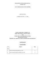
Fig.1 (a-h): Erythrocytic parasites of Theileria orientalis (1000 X magnification)
1171
Int.J.Curr.Microbiol.App.Sci (2020) 9(11): 1166-1176
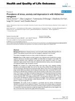
Fig.1 (i-j) Giemsa stained blood smear of cattle showing intralymphocytic parasites of Theileria
orientalis, KBb (1000 X magnification)
Fig.2 (a-d): Giemsa stained blood smear of cattle showing Babesia bigemina (1000 X)
Fig.2 (e): Anaplasma marginale inside RBC (1000X)
1172
Int.J.Curr.Microbiol.App.Sci (2020) 9(11): 1166-1176
However, Kakati (2013) observed outbreaks
of theileriosis with mortality in cattle of
Assam due to T. orientalis and confirmed its
virulence similar to recent reports made by
Aparna et al., (2011) from India and many
other countries like Australia (Eamens et al.,
2013), Michigan, USA (Bayugar et al., 2002)
and Japan (Yokoyama et al., 2010). Several
workers from different parts of India recorded
in addition to the above three species, the
prevalence of T. annulata and T. evansi in
cattle and buffaloes (Das and Sharma, 1991;
Ananda et al., 2009; Sahoo et al., 2012).
T. orientalis inside the erythrocytes were
found in different forms such as rod, bar,
comma, ring (annular), dot, crescent shaped,
finger like, tail like with trailing cytoplasm
and tetrad (dividing form). Merozoites were
also found extracellularly (Fig.1 a-h).
Schizont of T. orientalis (Koch Blue Body)
was also detected in the lymphocytes in a few
cases (Fig.1 i-j). Piroplasms of B. bigemina
varied in shape from ovoid or vacuolar forms,
amoeboid to typically paired pryiforms with
acute angle inside the erythrocytes and also in
extracellular location (Fig.2 a-d).
Inclusion bodies of A. marginale were dot
shaped appearing in the margin or periphery
of erythrocytes (Fig.2 e). Aparna et al.,
(2011), Kakati (2013) and Anupama et al
(2015) also reported similar forms of T.
orientalis. Babesia organisms with different
morphology were similar to the description
given by Soulsby (1982), Singh et al., (2011)
and Jyothisree et al (2013). Inclusion bodies
of A. marginale conformed to those of
workers (Atif et al., 2012; Kakati, 2013).
According to the type of cattle, prevalence
recorded was higher in crossbred (75.32%)
compared to indigenous cattle (66.00%), the
difference being highly significant (P<0.01).
Crossbred animals were 1.57 times more
susceptible
to
acquire
tick-borne
haemoparasites than the indigenous cattle.
These findings are in congruent with the
results of several workers from India and
abroad (Khan et al., 2004; Nair et al., 2013;
Naik et al., 2016). Atif et al., (2012) in their
work in Bangladesh reported significantly
higher
prevalence
of
tick-transmitted
haemoparasitic
diseases
(Babesiosis,
Anaplasmosis and Theileriosis) in crossbred
as compared to indigenous cattle.
Findings of several workers concluded that
indigenous breeds exhibit a high level of
resistance to ticks and tick-transmitted
diseases. According to Radostits et al.,
(2000), crossbred cattle populations were
more predisposed than indigenous cattle
because of natural resistance and endemic
stability between host-parasite relationships.
Chaudhri et al., (2013) also reported higher
prevalence of B. bigemina (3.89%) in
crossbred cows than in indigenous ones
(1.53%), thus agreeing to our findings.
Sex wise prevalence of haemoparasites in
cattle
The female animals were found to have
overall higher (74.02%) infection rate of
haemoparasites than the male (58.22%)
counterparts, the difference was found to be
highly significant (P<0.01), as shown in
Table-2. Prevalence of haemoparasite
infection was recorded highest in females of
Kokrajhar district (87.82%) and least in Baksa
(67.81%),
The findings of the present work are in
congruent with that of several workers
(Rajput et al., 2005; Kakati, 2013; Naik et al.,
2016). The possible reason for higher
prevalence in female animals might be due to
examination of more number of female cattle,
hormonal
disturbances
and
immunosuppression in advanced pregnancy
and or lactation in high producing females.
1173
Int.J.Curr.Microbiol.App.Sci (2020) 9(11): 1166-1176
Age wise prevalence of haemoparasites in
cattle
In the present study, the adult cattle (> 3
years) recorded highest infection of
haemoparasites (80.42%) followed by young
(68.11%) and calves (54.85%) which was
highly significant (P<0.01), (Table-3) thus
agreeing to reports of Ruprah (1985), Ananda
et al (2009), Mohanta et al., (2011), Kakati
(2013) and Naik et al., (2016). Adult cattle
were 3.58 times more susceptible than calves
to haemoparasitic infection. The lower
prevalence in young animals compared to
adults can be attributed to the restricted
grazing of young animals which tends to
reduce their chance of contact with the
vectors of these diseases. It is assumed that
aged animals are more susceptible to blood
protozoan diseases than the younger animals
due to inverse age resistance (Urquhart et. al.,
1996).
In conclusion, the present study conducted for
the first time in Indo- Bhutan border districts
of Assam showed haemoparasite infection
due to T. orientalis, B. bigemina and A.
marginale in the cattle population and are
considered to be endemic for the
haemoparasites.
Acknowledgement
The authors are grateful to the Head, Deptt. of
Parasitology and the Dean, College of
Veterinary Science, AAU, Khanapara for
providing necessary facilities to carry out the
research work.
References
Ananda, K.J., D’Souza, P. E., and
Puttalakshmamma,
G.C.2009.
Prevalence
of
Haemoprotozoan
diseases in crossbred cattle in
Banglore North. Vet. World. 2 (1):15-
16.
Anupama, R., Srinivasan, S.R., and Parthiban,
M. 2015. Molecular studies on
Theileriosis and Identification of
Theileria orientalis in India using
PCR. Indian Vet. J. 92 (2): 9-11.
Aparna, M., Ravindran, R., Vimalkumar,
M.B., Lakshmanan, B., Rameshkumar,
P., Kumar, K.G.A., Promod, K.,
Ajithkumar, S., Ravishankar, C.,
Devada, K., Subramanian, H., George,
A.J., and Ghosh, S. 2011.Molecular
characterization of Theileria orientalis
causing fatal infection in crossbred
adult bovines of South India. Parasitol.
Int. 60: 524-529.
Atif, F.A., Khan, M.S., Iqbal, H.J., Arshad,
G.M., Ashraf, E., and Ullah, S. 2012.
Prevalence of Anaplasma marginale,
Babesia bigemina and Theileria
annulata infections among cattle in
Sargodha District, Pakistan. Afr. J.
Agri. Res. 7(22): 3302-07.
Bayugar, R.C., Pillars, R., Schlater, J., and
Holmana, P.J. 2002. Theileria buffeli
infection of a Michigan cow
confirmed by small subunit ribosomal
RNA gene analysis. Vet. Parasitol.105,
105-110.
Callow, L. L., 1984. Protozoan and rickettsial
diseases. In: Australian bureau of
animal health, Animal Health in
Australia.
Vol
5.
Australian
Government Publishing Services,
Canberra. 121-216.
Chaudhri, S.S., Bisla, R.S., Bhanot, V., and
Singh, H. 2013. Prevalence of
Haemoprotozoan infections in pyretic
Dairy animals of eastern Haryana.
Indian J. Anim. Res. 47 (4): 344-347.
Das,S.S., and Sharma, N.N.1991. Prevalence
of Theileria infection in Hyaloma
anatolicum anatolicum in north
districts of Tripura (India). J. Vet.
Parasitol. 5, 25-27.
Eamens,
G.J., Bailey,
G., Jenkins
C.,
1174
Int.J.Curr.Microbiol.App.Sci (2020) 9(11): 1166-1176
and Gonsalves, J.R. 2013. Significance
of Theileria orientalis types in
individual affected beef herds in New
South Wales based on clinical, smear
and PCR findings. Vet Parasitol. 196,
96-105.
Jyothisree, Ch., Naik, S., and Samatha, V.
2013. A study on Prevalence and
Clinico- Therapeutic management of
Babesiosis in H.F. crossbred cattle in
Anantapur district of Andhra Pradesh.
Int. J. Food, Agri.Vet. Sci. 3(2): 88-91.
Kakati, P. 2013. Studies on ticks and tick
borne haemoparasitic infection of
cattle in Assam. M.V.Sc. Thesis,
Assam Agricultural University, Jorhat,
Khanapara. Pp: 107
Khan, M.Q., Zahoor, A., Jahangir, M., and
Mirza, M.A.2004. Prevalence of blood
parasites in cattle and buffaloes. Pak.
Vet. J. 24(4):193-194.
Kohli, S., Atheya, U.K., and Thapliyal, A.
2014. Prevalence of theileriosis in
cross bred cattle: its detection through
blood
smear
examination
and
polymerase
chain
reaction
in
Dehradun district, Uttarakhand, India.
Int. J. Livest. Prod. 7(3): 168- 171.
Livestock census 2007. Ministry of Animal
Husbandry, dairying and Fisheries,
Ministry of Agricultre, Govt. of India.
Levine, N.D. 1978. Text Book of Veterinary
Parasitology. Minneapolis: Burgees.
Pp: 406.
Minijauw, B., and McLeod, A. 2003. The
impact of ticks and tick-borne diseases
on livelihood of small scale and
marginal livestock owners in India and
eastern and southern Africa. Tickborne diseases and poverty research
report,
DFID
Animal
Health
Programme Centre for Tropical
Veterinary Medicine, University of
Edinburgh, U.K., pp: 24-57
Mohanta, U.K., Anisuzzaman., and Mondal,
M.M.H. 2011. Tick and tick borne
protozoan diseases of livestock in the
selected hilly areas of Bangladesh, Int.
J. Agril. Res. Innov. & Tech. 1 (1&2):
60-63.
Naik, B.S., Maiti, S.K., and Raghuvanshi,
P.D.S.2016. Prevalence of Tropical
Theileriosis in Cattle in Chhattisgarh
State. J. Anim. Res. 6 (6): 1043-45.
Nair, A.S., Ravindran, R., Lakshmanan, B.,
Sreekumar, C., Kumar, S.S., Raju, R.,
Tresamol, P.V., Vimalkumar, M.B.,
and Saseendranath, M.R. 2013. Bovine
carriers of Anaplasma marginale and
Anaplasma bovis in South India. Trop.
Biomed. 30 (1): 105–112.
Phanchung et al. 2012. Small holder dairy
farming in Bhutan: Characteristics,
constraints
and
development
opportunities. Chapter 2., pp. 18-25.
PD-ADMAS, 2005-06. Annual report of
Project Directorate of Animal disease
Monitoring and Surveillance.
Radostits, O.M., Gay, C.C., Blood, D.C., and
Hinchcliff, K.W. 2000. Veterinary
Medicine. A Textbook of the Diseases
of Cattle, Sheep, Pigs, Goats and
Horses, 9th ed. W.B. Saunders,
London.
Rai, M. 2008. The fauna of Northeast India.
gspot.i
n/2008/10/fauna-of-northeastindia.html
Rajput, Z. I., Song-hua, H.U., Arijo, A.G.,
Habib, M., and Khalid, M. 2005.
Comparative study of Anaplasma
parasites in tick carrying buffaloes and
cattle. J. Zhejiang Univ. Sci. B. 6 (11):
1057-1062.
/>57
Reetha, T.L., Thomas, K.S., and Babu, M.
2012. Occurrence of Haemoprotozoan
Infection in Bovine. Int. J. Applied
BioRes. 13, 1-2.
Ristic, M. 1981. Anaplasmosis. In: M.Ristic
and I. McIntyre (ed), Diseases of cattle
1175
Int.J.Curr.Microbiol.App.Sci (2020) 9(11): 1166-1176
in the tropics. Martinus Nijhoff,
Boston, MA. pp 443–468.
Ruprah, M.S. 1985. A Text Book of Clinical
Protozoology. Oxonian Press Pvt. Ltd,
New Delhi.
Sahoo, B.M., Panda, M.R., Dehuri, M.,
Bishnoi, P.C., Panda, S.K., and
Mohanty, B.N. 2012. Epidemiology of
haemoprotozoan infection in cattle of
coastal districts of Odisha. XXIII
National Congress of Veterinary
Parasitology, 12-14 December, 2012,
CVSc., AAU, Khanapara, Guwahati22; S-I; pp :28.
Schlesselman, J.J. 1982. Case-Control
Studies. 1st edition, Oxford University
Press, New York, Pp. 174-177.
Sebele,T., Zewedu, F., and Getachew, A.G.
2015. A study of the prevalence of
haemoparasites of ruminants in and
around Debre-zeit, Central Ethiopia.
Afr. J. Parasitol. Res.2 (3): 66-71.
Singh, H., Singh, N.K., Chand, N.,
Deshmukh, S., Singh, J.R.S., and Rath,
S.S. 2011. Concurrent infection of
Babesia bigemina and Theileria
annulata in Holstein Friesian cow: A
case report. J. Vet. Parasitol. 25 (1) :
82-83.
Singh, N. K., Singh, H., Jyoti, Haque, M., and
Rath, S.S. 2012. Prevalence of
parasitic infections in cattle of
Ludhiana district, Punjab. J. Parasit.
Dis. 36 (2):256–259.
Soulsby, E.J.L.1982. Helminths, arthropods
and protozoa of domesticated animals.
7th ed. London: Bailliere Tindall. pp:
809.
Tshering, G., and Dorji, N. 2013. Prevalence
of gastrointestinal parasites in free
range cattle:a case study in haa
district, Bhutan. J. Anim. Hlth. Prod.
1, 36-37.
Uilenberg, G. 1981. Theilerial species of
domestic livestock. In: Irvin, A.D.,
Cunningham, M.P. and Young, A.S.
(eds.), Advances in the control of
Theileriosis.
Martinus
Nijhoff
Publishers, The Hague. 137p.
Urquhart, G.M., Armour, A., Duncan, J.L.,
Dunn, A.M., and Jennings, F.W. 1996.
Veterinary Parasitology. 2nd edn.
Black Well Sciences Ltd., p: 231.
Yokoyama, N., Ueno, A., Izuno, D., Kuboki,
N.,
Khukhuu, A., Igarashi,
T.,Miyahara, T., Shiraishi, R., Kudo,
M., Oshiro, S., Zakimi, C., Sugimoto,
K., and Inokuma, H. 2010. Genotypic
Diversity of Theileria orientalis
Detected from Cattle Grazing in
Kumamoto and Okinawa Prefectures
of Japan. J. Vet. Med. Sci. 73(3): 305312.
How to cite this article:
Dipanjali Mushahary, Kanta Bhattacharjee, Prabhat Chandra Sarmah and Dilip Kumar Deka.
2020. Prevalence of Haemoparasites of Cattle in Four Districts of Assam, India and Bordering
Bhutan. Int.J.Curr.Microbiol.App.Sci. 9(11): 1166-1176.
doi: />
1176