A case report of aspergillosis accompanied by saccular bronchodilation after bronchial thermoplasty in a 19-year-old woman
Bạn đang xem bản rút gọn của tài liệu. Xem và tải ngay bản đầy đủ của tài liệu tại đây (962.94 KB, 4 trang )
(2020) 20:312
Sasada et al. BMC Pulm Med
/>
Open Access
CASE REPORT
A case report of aspergillosis accompanied
by saccular bronchodilation after bronchial
thermoplasty in a 19‑year‑old woman
Shinji Sasada1* , Kenshiro Ohmura2, Tomoyo Oguri1,3, Yutaro Fujimoto1, Saori Murata1, Yumi Tsuchiya1,
Kota Ishioka1, Saeko Takahashi1, Morio Nakamura1 and Masahiro Kaji2
Abstract
Background: Fungal infections are rarely reported as a complication of bronchial thermoplasty (BT) in patients without immunosuppressive comorbidity.
Case presentation: A 19-year-old woman college student was admitted to our hospital owing to uncontrolled
severe asthma despite using the maximum dose of steroid inhalation. She experienced asthmatic attacks more
frequently while cheerleading, which is an extracurricular activity. She received BT because she wanted to continue
cheerleading. After the second BT session, she developed more sputum and cough. During the third session, white
secretion and saccular bronchodilation appeared in the left lower bronchus. Aspergillus fumigatus was detected in
the culture of the bronchial lavage sample, and saccular bronchodilation in the affected bronchus was observed on
computed tomography (CT). Five months after the start of oral itraconazole, her subjective symptoms as well as her
CT findings improved. Her asthma condition improved enough for the patient to continue cheerleading without
exacerbation.
Conclusions: It is necessary to consider the possibility of respiratory tract infections including fungal infections after
BT. Detailed observations of the entire bronchus and sample collection for microbial culture are highly recommended.
Keywords: Aspergillosis, Saccular bronchodilation, Bronchial thermoplasty, Severe asthma, Case report
Background
Bronchial thermoplasty (BT) is a medical procedure
in which bronchoscopy is used and controlled thermal
energy is applied to the bronchial wall to decrease the
smooth muscles. This procedure is also effective for the
treatment of severe asthma. Bronchial edema and radiological changes are commonly known major complications of BT, but infections are rarely reported. The AIR2
trial reported that only one patient with lower respiratory
tract infection required hospitalization in the BT group
*Correspondence:
1
Department of Respiratory Medicine, Tokyo Saiseikai Central Hospital,
1‑4‑17 Mita, Minato‑ku, Tokyo 108‑0073, Japan
Full list of author information is available at the end of the article
[1]. Here, we present a case of aspergillosis after BT in a
young asthmatic patient.
Case presentation
The patient was a 19-year-old woman with no comorbidities other than asthma. She was a non-smoker. She had
been treated for severe asthma with high doses of inhaled
corticosteroids plus long-acting β2-agonists, antileukotriene, theophylline, and antihistamine. The patient participated in cheerleading as an extracurricular activity at
her college. During cheerleading, she experienced asthmatic attacks, which became increasingly frequent, and
she therefore sought medical consultation. The patient
wanted to continue cheerleading and enquired about
strengthening her treatment. However, she did not want
© The Author(s) 2020. Open Access This article is licensed under a Creative Commons Attribution 4.0 International License, which
permits use, sharing, adaptation, distribution and reproduction in any medium or format, as long as you give appropriate credit to the
original author(s) and the source, provide a link to the Creative Commons licence, and indicate if changes were made. The images or
other third party material in this article are included in the article’s Creative Commons licence, unless indicated otherwise in a credit line
to the material. If material is not included in the article’s Creative Commons licence and your intended use is not permitted by statutory
regulation or exceeds the permitted use, you will need to obtain permission directly from the copyright holder. To view a copy of this
licence, visit http://creativecommons.org/licenses/by/4.0/. The Creative Commons Public Domain Dedication waiver (http://creativeco
mmons.org/publicdomain/zero/1.0/) applies to the data made available in this article, unless otherwise stated in a credit line to the data.
Sasada et al. BMC Pulm Med
(2020) 20:312
to take regular corticosteroids or antibody agents. She
refused biological treatment owing to the cost associated
with long-term continuation and the desire to have children. Therefore, the patient decided to undergo BT.
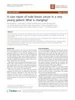
Chest computed tomography (CT) showed a thickened
bronchial wall (Fig. 1a); the percentage forced expiratory
volume (%FEV) was 86.2%, and exhaled nitric oxide was
45 ppm. Blood test results revealed the following: 515
U/mL, IgE; 8900/μL, white blood cell count; and 143/
μL, eosinophils. The patient received 30 mg/day prednisolone for 3 days before BT treatment and continued it
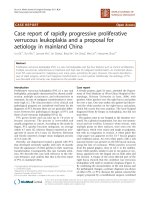
for 1 day post-BT treatment. The sessions for the right
and left lower lobe were properly completed at 3-week
intervals, and the BT activation numbers were 31 and 24
times, respectively (Fig. 2a).
After the second BT session, she developed more sputum and cough. During the third session, white secretion
and saccular bronchodilation appeared in the left lower
bronchus (B9 and B10) (Fig. 2b). At the same time, bronchial washing was performed. Her CT also showed saccular bronchodilation in the affected bronchus (Fig. 1b).
Aspergillus fumigatus was detected in the bronchial lavage culture. Oral itraconazole administration was started,
Page 2 of 4
and serum β-d-glucan and aspergillus antigen were
found to be negative. Five months later, the subjective
symptoms and CT findings improved (Fig. 1c). Although
respiratory function remained unchanged (%FEV, 88%),
exhaled nitric oxide reduced to 15 ppm. After BT, the
inhaled steroid dose was reduced owing to a remarkable improvement in asthma symptoms. The patient
was able to continue her cheerleading activity without
exacerbations.
Discussion and conclusions
Previous studies have evaluated the efficacy and safety
of BT in patients with severe asthma. The AIR2 study
showed that BT improved asthma-related quality of
life (QOL) and significantly controlled the frequency
of severe exacerbations [1]. In addition, the efficacy of
BT can last for at least 5 years [2]. Moreover, Japanese
patients with asthma had improved QOL, less exacerbations, less symptoms, and less obstructive pulmonary
function after BT [3]. However, some complications associated with BT have been reported. Burn et al. reported
that their BT patients experienced adverse events more
frequently than those described in previous clinical trials
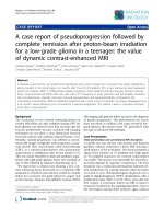
Fig. 1 Chest computed tomography images. a Left lower lobe with thickened bronchial wall before bronchial thermoplasty (BT). b Saccular
bronchodilation of left lower bronchus (B9, B10) (arrows) and nodular consolidation (triangle) are seen 3 weeks after BT. c Bronchodilation and
consolidation are improved 5 months after the start of oral itraconazole administration
Sasada et al. BMC Pulm Med
(2020) 20:312
Page 3 of 4
Fig. 2 Bronchoscopic images. a Bronchial thermoplasty (BT) activation to left B9. b White sputum (arrows) and saccular bronchodilation (triangles)
are seen 3 weeks after BT (B)
[4]. Iikura et al. reported a case of aspergillus and nocardia infection after BT [5]: the patient was a 35-year-old
man who regularly received systemic corticosteroids,
which resulted in chronic immunodeficiency. In our case,
the patient did not receive regular systemic corticosteroids, and the prophylactic steroid dose was 30 mg/body
weight, which was less than the recommended dose of
50 mg/body weight in Japan [6]. In our experience, highdose inhaled corticosteroids and minimal prophylactic
systemic corticosteroids may cause fungal infections even
in the absence of immunosuppressive comorbidities.
In addition, heat energy from BT may cause tissue fragility that predisposes the patient to respiratory tract
infections [7]. However, in this case, the activation number for the left lower lobe was 24 times, which was much
less than the average [6], and it developed while the
damage to the respiratory tract was kept to a minimum.
Potential respiratory aspergillosis has been previously
reported in asthmatic patients who received inhaled corticosteroids [8, 9]. Hypothetically, it is considered that
the mucosal defense is reduced following BT; thus, the
pre-existing aspergillus molds become engrafted. In this
case, aspergillosis was not tested for before performing BT. From this experience, pre-screening of chronic
infections of the respiratory tract should be carried out.
On the other hand, serum β-D-glucan and the aspergillus antigen were negative after occurring aspergillosis,
the reason for the negative may be that the infection
remained localized and the medication was started early.
If the aspergillosis was not diagnosed and treated early,
the bronchial deformity might have become irreversible.
We report a case of aspergillosis infection accompanied by saccular bronchodilation after BT. Regardless
of the strength of asthma treatment and the absence of
an immunosuppressive comorbidity, it is necessary to
consider the possibility of respiratory tract infections,
including fungal infections. Detailed observations of
the entire bronchus and sample collection for microbial
culture are highly recommended.
Abbreviations
BT: Bronchial thermoplasty; CT: Chest computed tomography; QOL: Quality of
life; FEV: Forced expiratory volume.
Acknowledgements
We would like to thank Masami Yoshihara, the medical interpreter and the
medical coordinator at Tokyo Saiseikai Central Hospital for English language
editing.
Authors’ contributions
All authors have read and approved the manuscript, and significantly contributed to this paper. SS, KO, TO, YF, SM, YT, KI, ST, MN, MK: Conception and
design, literature review, manuscript writing and correction, final approval of
manuscript. All authors read and approved the final manuscript.
Funding
No funding sources were used.
Availability of data and materials
All data and material are available for sharing if needed.
Ethics approval and consent to participate
Appropriate written informed consent was obtained for the publication of
this case report and accompanying images. It was approved by the Clinical
Research Ethics Committee of Saiseikai Central Hospital and was implemented
(Approval Number 2020-010-1).
Sasada et al. BMC Pulm Med
(2020) 20:312
Consent for publication
Written informed consent was obtained from the patient for publication of
this case report and any accompanying images.
Competing interests
The authors declare that they have no competing interests. This manuscript
has not been published and is not under consideration for publication
elsewhere. Additionally, all of the authors have approved the contents of this
paper and have agreed to the journal´s submission policies.
Author details
1
Department of Respiratory Medicine, Tokyo Saiseikai Central Hospital, 1‑4‑17
Mita, Minato‑ku, Tokyo 108‑0073, Japan. 2 Department of Thoracic Surgery,
Tokyo Saiseikai Central Hospital, Tokyo, Japan. 3 Department of Clinical Oncology, St. Marianna University School of Medicine, Kawasaki, Japan.
Received: 23 September 2020 Accepted: 18 November 2020
Page 4 of 4
3. Iikura M, Hojo M, Nagano N, et al. Bronchial thermoplasty for severe
uncontrolled asthma in Japan. Allergol Int. 2018;67:273–5.
4. Burn J, Sims AJ, Keltie K, et al. Procedural and shorttermsafety of bronchial
thermoplasty in clinical practice: evidence from a national registry and
hospital episode statistics. J Asthma. 2016;1:1–8.
5. Matsubayashi S, Iikura M, Numata M, et al. A case of Aspergillus and
Nocardia infections after bronchial thermoplasty. Respirol Case Rep.
2019;7:e00392.
6. Yamamoto S, Iikura M, Kakuwa T, et al. Can the number of radiofrequency
activations predict serious adverse events after bronchial thermoplasty?
A retrospective case-control study Pulm Ther. 2019;5:221–33.
7. Goorsenberg AWM, d’Hooghe JNS, de Bruin DM, et al. Bronchial thermoplasty-induced acute airway effects assessed with optical coherence
tomography in severe asthma. Respiration. 2018;15:1–7.
8. Fairfax AJ, David V, Douce G, et al. Laryngeal Aspergillosis following highdose inhaled fluticasone therapy for asthma. Thorax. 1999;54:860–1.
9. Leav BA, Fanburg B, Hadley S, et al. Invasive pulmonary Aspergillosis associated with high-dose inhaled fluticasone. N Engl J Med. 2000;343:586.
Publisher’s Note
References
1. Castro M, Rubin AS, Laviolette M, et al. Effectiveness and safety of bronchial thermoplasty in the treatment of severe asthma. Am J Respir Crit
Care Med. 2010;181:116–24.
2. Wechsler ME, Laviolette M, Rubin AS, et al. Bronchial thermoplasty—long
term safety and effectiveness in severe persistent asthma. J Allergy Clin
Immunol. 2013;132:1295–302.
Springer Nature remains neutral with regard to jurisdictional claims in published maps and institutional affiliations.
Ready to submit your research ? Choose BMC and benefit from:
• fast, convenient online submission
• thorough peer review by experienced researchers in your field
• rapid publication on acceptance
• support for research data, including large and complex data types
• gold Open Access which fosters wider collaboration and increased citations
• maximum visibility for your research: over 100M website views per year
At BMC, research is always in progress.
Learn more biomedcentral.com/submissions