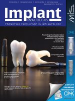
Tạp chí implant IPUS tháng 3& 4/2013 Vol 6 No2
Bạn đang xem bản rút gọn của tài liệu. Xem và tải ngay bản đầy đủ của tài liệu tại đây (15.43 MB, 65 trang )
clinical articles • management advice • practice profiles • technology reviews
March/April 2013 – Vol 6 No 2
PROMOTING EXCELLENCE IN IMPLANTOLOGY
Preventing the dreaded black
triangle during implant
placement in the esthetic
zone: part 1
Dr. Scott M. Blyer
Practice profile
Dr. Coury Staadecker
Corporate profile
Straumann
Trabecular Metal™ implants
from orthopedics to
dental implantology
Dr. Suheil M. Boutros
PAYING SUBSCRIBERS EARN 24
CONTINUING EDUCATION CREDITS
PER YEAR!
Uncovering peri-implantitis
Dr. Nikos Donos
March/April 2013 - Volume 6 Number 2
EDITORIAL ADVISORS
Steve Barter BDS, MSurgDent RCS
Anthony Bendkowski BDS, LDS RCS, MFGDP, DipDSed, DPDS,
MsurgDent
Philip Bennett BDS, LDS RCS, FICOI
Stephen Byfield BDS, MFGDP, FICD
Sanjay Chopra BDS
Andrew Dawood BDS, MSc, MRD RCS
Professor Nikolaos Donos DDS, MS, PhD
Abid Faqir BDS, MFDS RCS, MSc (MedSci)
Koray Feran BDS, MSC, LDS RCS, FDS RCS
Philip Freiburger BDS, MFGDP (UK)
Jeffrey Ganeles, DMD, FACD
Mark Hamburger BDS, BChD
Mark Haswell BDS, MSc
Gareth Jenkins BDS, FDS RCS, MScD
Stephen Jones BDS, MSc, MGDS RCS, MRD RCS
Gregori M. Kurtzman, DDS
Jonathan Lack DDS, CertPerio, FCDS
Samuel Lee, DDS
David Little DDS
Andrew Moore BDS, Dip Imp Dent RCS
Ara Nazarian DDS
Ken Nicholson BDS, MSc
Michael R. Norton BDS, FDS RCS(ed)
Rob Oretti BDS, MGDS RCS
Christopher Orr BDS, BSc
Fazeela Khan-Osborne BDS, LDS RCS, BSc, MSc
Jay B. Reznick DMD, MD
Nigel Saynor BDS
Malcolm Schaller BDS
Ashok Sethi BDS, DGDP, MGDS RCS, DUI
Harry Shiers BDS, MSc, MGDS, MFDS
Harris Sidelsky BDS, LDS RCS, MSc
Paul Tipton BDS, MSc, DGDP(UK)
Clive Waterman BDS, MDc, DGDP (UK)
Peter Young BDS, PhD
Brian T. Young DDS, MS
PUBLISHER
Lisa Moler
Email:
Tel: (480) 403-1505
MANAGING EDITOR
Mali Schantz-Feld
Email:
Tel: (727) 515-5118
ASSISTANT EDITOR
Kay Harwell Fernández
Email:
PRODUCTION MANAGER/CLIENT RELATIONS
Kim Murphy
Email:
NATIONAL SALES/MARKETING MANAGER
Drew Thornley
Email:
Tel: (619) 459-9595
NATIONAL SALES REPRESENTATIVE
Sharon Conti
Email:
Tel: (724) 496-6820
E-MEDIA MANAGER/GRAPHIC DESIGN
Email:
Greg McGuire
PRODUCTION ASST./SUBSCRIPTION COORDINATOR
Email:
Lauren Peyton
MedMark, LLC
15720 N. Greenway-Hayden Loop #9
Scottsdale, AZ 85260
Fax: (480) 629-4002
Tel: (480) 621-8955
Toll-free: (866) 579-9496 Web: www.endopracticeus.com
SUBSCRIPTION RATES
Individual subscription
1 year
(6 issues)
3 years
(18 issues)
$99
$239
© FMC 2013. All rights
reserved. FMC is part of the
specialist publishing group
Springer Science+Business Media. The publisher’s written
consent must be obtained before any part of this publication may
be reproduced in any form whatsoever, including photocopies
and information retrieval systems. While every care has been
taken in the preparation of this magazine, the publisher cannot
be held responsible for the accuracy of the information printed
herein, or in any consequence arising from it. The views
expressed herein are those of the author(s) and not necessarily
the opinion of either Implant Practice or the publisher.
Volume 6 Number 2
H
aving recently celebrated my 27th year in private practice as a periodontist, I have
been reflecting on the changes that have occurred in the profession. It is hard to
believe that at the beginning of my career I was a “full-time” specialist limiting my practice
to the prevention, diagnosis, and treatment of periodontal disease. As a resident, implant
dentistry was not a part of our curriculum, and discussions involving this topic were
relegated to lunch hour debates in the cafeteria. At that time, it was performed by a select
few who later became known as pioneers in the field.
In the mid- to late 1980s, many clinicians, including myself, were taking the courses
necessary to place dental implants and recognized the fact that one can change people’s
lives by simply restoring form and function. However, at that time, patients with hopelessly
involved dentitions often had treatment plans that were in excess of 18 months. Patient
acceptance was often difficult to obtain, as they did not necessarily understand the
advantages of implant dentistry.
With time, several innovations, some of which include an internal hex connection
and a second-generation roughened surface technology (micro and macro roughness),
improved the predictability of patient care and addressed some of the patient’s resistance
to time-intensive treatment plans. This led to wider acceptance of implant dentistry and
a paradigm shift in the 1990s, making this a treatment of choice in clinical situations that
would require sophisticated, less predictable procedures to salvage failing dentitions.
In response to market demands, esthetics became the focus of our profession.
It was no longer enough to simply restore form and function. Our endpoint had to be
an esthetically pleasing restoration. As a result, the last 10 to 15 years found clinicians
changing their mantra from surgically driven implant placement to restoratively driven
implant placement. Often, this would require one- and two-stage hard and soft tissue
grafting procedures to satisfy the esthetic demands of a consumer-educated patient
population. There was, and always will be, a percentage of the population who is
comfortable with an “at any cost” treatment approach. However, due to motivation,
time, and financial constraints, many patients would seek treatment alternatives that
also resulted in an esthetic restoration. Implant companies responded with a number of
innovations centering on surface technology and the introduction of new implant materials
(alloys) developed specifically for narrow interdental spaces, expanding our treatment
options.
More recently, another surface technology was introduced that enhanced
osseointegration through its hydrophilic and chemically active properties, resulting in
an improved surface chemistry. This is noteworthy, as these properties enable faster
osseointegration, reducing the overall loss of implant stability, which is typical after
mechanical stability due to an osteopenia. This technology is designed to give clinicians
the confidence to proceed with immediate placement in extraction sites. A byproduct
of the improved surface chemistry is the ability to load the fixture sooner, increasing the
appeal and patient acceptance of implant treatment.
Another technology that is allowing for more and more implant candidates is the
advent of new implant materials. There is a titanium-zirconium alloy that has shown higher
strengths when compared to implants made of grade 4 titanium manufactured by the
same company. Smaller diameter implants can now be placed with confidence, as fixture
fracture is less of a concern. This is clinically relevant, as often patients will not accept
treatment recommendations if large grafting procedures are necessary to create an
environment for successful implant placement.
When I graduated from my residency, I had no idea that the profession would change
as much as it has. I feel blessed to be practicing in a time when dentistry continues
to evolve where we now have the ability to meet and exceed patient expectations
with respect to restoring form and function — as well as replacing teeth that are
indistinguishable from those lost. I can only hope that the innovations that will occur in the
next 27 years will be as noteworthy as those in the past.
Dr. Robert Miller
Miami and Boca Raton, Florida
Implant practice 1
INTRODUCTION
Reflections on an ever-evolving profession
TABLE OF CONTENTS
Clinical
Practice profile
6
Dr. Coury Staadecker: The art of harnessing synergy
Dr. Staadecker discusses the many facets of his practice that set the stage for
guiding and maintaining true patient wellness.
Uncovering peri-implantitis
Dr. Nikos Donos talks about the
growing importance of peri-implant
disease and explains how the latest
research is shaping treatment...... 14
Guided surgery – understanding
the risks
Dr. Peter Sanders explains the
importance of gaining experience in
conventional implant placement prior
to using CT guided surgery.......... 18
Continuing
education
Preventing the dreaded
black triangle during implant
placement in the esthetic zone:
part 1
Dr. Scott M. Blyer examines ways
to avoid a frustrating complication of
dental implant therapy................. 22
Treatment planning of implants
Corporate profile
10
Straumann: Shaping the future together
Straumann® – a global leader in implant dentistry offering surgical, restorative,
regenerative, and digital solutions for the dental and lab business – is a pioneer
of innovative technologies.
2 Implant practice
in the esthetic zone: part 1
In the first part of a series of
articles, Drs. Sajid Jivraj, Mamaly
Reshad, and Winston Chee look
at the diagnostic factors that affect
the predictability of peri-implant
esthetics ..................................... 28
Volume 6 Number 2
ORTHOPHOS XG 3D
The right solution for
your diagnostic needs.
Implantologists
Endodontists
Orthodontists
will benefit from highquality pan and ceph
images for optimized
therapy planning.
General Practitioners
will achieve greater
diagnostic accuracy
for routine cases.
ORTHOPHOS XG 3D
will enjoy instantly
viewable 3D volumetric
images for revealing
and measuring canal
shapes, depths
and anatomies.
will appreciate the
seamless clinical
workflow from initial
diagnostics, to treatment
planning, to ordering
surgical guides and final
implant placement.
The advantages of 2D & 3D in one comprehensive unit
ORTHOPHOS XG 3D is a hybrid system that provides clinical
workflow advantages, along with the lowest possible effective
dose for the patient. Its 3D function provides diagnostic accuracy
when you need it most: for implants, surgical procedures and
volumetric imaging of the jaws, sinuses and other dental anatomy.
For standard 2D images, it offers the most comprehensive selection
of pan and ceph programs to meet virtually all needs, from standard
panoramic programs for adults and children, to extraoral bitewing,
sinus, TMJ options and many more.
Automatic patient positioning
The new Auto-Positioner measures the exact tilt of the patient’s
occlusal plane and automatically adjusts the height for an optimal
panoramic image within the sharp layer, thereby preventing incorrect
positioning and reducing re-takes.
For more information, visit www.Sirona3D.com
or call Sirona at: 800.659.5977
www.facebook.com/Sirona3D
TABLE OF CONTENTS
34
Small-diameter
implant
treatment
Step-By-Step
Product profile
Event preview
Fast, profitable, and patient-
CPK – Complete Prosthetic Kit
friendly denture stabilization
3M™ ESPE™ MDI Mini Dental Implants
.....................................................34
from MIS Implants Technologies
Simplifies the restorative component
of Implant Dentistry .......................46
4th annual NYU College of
Dentistry Global Implantology
Week ...........................................52
Technology
Trabecular Metal implants
from orthopedics to dental
™
implantology
Dr. Suheil M. Boutros focuses on the
applications for a new type of implant
.....................................................38
4 Implant practice
i-CAT® FLX — the latest
advancement in Cone Beam 3D
For greater flexibility in scanning,
planning, and treatment ................48
Introducing a new implant
designed exclusively for
overdentures - the LOCATOR®
Overdenture Implant system ....50
Diary.......................................56
Materials &
equipment .....................62
Volume 6 Number 2
79459-US-1208 © 2012 DENTSPLY International, Inc.
Abutments as individual
as your patients
Available for all major implant systems and in your
choice of titanium, gold-shaded titanium and four
shades of zirconia, ATLANTIS™ patient-specific
ATLANTIS BioDesign Matrix™
The four features of the ATLANTIS BioDesign Matrix™
work together to support soft tissue management
for ideal functional and esthetic result. This is the
true value of ATLANTIS™ for you and
your patients.
CAD/CAM abutments help to eliminate the need
for inventory management of stock components and
simplify the restorative procedure.
ATLANTIS VAD™
Designed from the
final tooth shape.
Natural Shape™
Shape and
emergence profile
based on individual
patient anatomy.
Soft-tissue Adapt™
Optimal support for
soft tissue sculpturing
and adaptation to the
finished crown.
Find out how ATLANTIS™ can
bring simplicity and esthetics
to your practice. Just take an
implant-level impression, send it to your
laboratory and ask for ATLANTIS today.
Custom Connect™
Strong and stable fit –
customized connection for
all major implant systems.
800-531-3481 • www.dentsplyimplants.com
PRACTICE PROFILE
What can you tell us about your
background?
I earned my dental degree from Ohio State
University in 1997 and my Periodontics
Certificate from the Naval Postgraduate
Dental School in Bethesda, Maryland.
While pursuing my periodontics certificate,
I also earned a Master of Science degree
from George Washington University. When
on staff at the Naval Medical Center San
Diego, I mentored numerous general
practice residents and lectured extensively.
While in private practice in Seattle,
Washington, I continued my involvement
with academics as a Clinical Instructor
and Affiliate Professor at the University
of Washington, Department of Graduate
Periodontics. Additionally, I am the former
Senior Clinical Editor of the Seattle Study
Club Journal, reaching over 8,000 dentists
worldwide. I am a Diplomate of the
American Board of Periodontology and an
Accredited Fellow of the American Society
of Dental Anesthesiology.
Is your practice
implants?
limited
to
As a periodontist, there are three distinct
facets of my practice that include (1)
treatment of periodontal disease, (2) implant
therapy, and (3) periodontal plastic surgery.
Being well versed in all three areas sets the
stage to guide and maintain true patient
wellness. Additionally, these facets blend
seamlessly not only to establish health,
but also to maintain optimal function and
esthetics.
Why did you decide to focus on
implantology?
While I was attending dental school during
the mid-1990s, it was the birthplace of
modern day implantology. Implant design
and technology have continued to evolve,
but with all the different manufacturers,
implants have become similar. We can
now provide our patients with a tooth
replacement that predictably makes them
“whole” again. I can emotionally identify
with the innate and powerful sense of selfpreservation. With prosthetic treatment
other than implant therapy, the treatment
is either collaterally destructive or foreign
to the patient. Patients simply perceive
implants as being a part of themselves
6 Implant practice
and therefore self-preserving.
Having
the opportunity to return a sense of selfesteem and confidence is just as joyful for
me as it is for my patients.
How long have you been
practicing, and what systems do
you use?
I have been practicing dentistry for
more than 15 years. I exclusively use
Straumann® and Nobel Biocare® dental
implant products.
What
training
undertaken?
have
you
Following graduation from Dental School
at The Ohio State University, I continued
my training in an Advanced Education
General Dentistry (AEGD) Residency in the
U.S. Navy. The AEGD Residency piqued
my interest in periodontics and implants.
Shortly thereafter, I applied and graduated
from a 3-year residency in periodontics
from the Naval Postgraduate Dental
School. Over the course of the following
8 years, I was a didactic instructor and
Affiliate Professor at the Naval Medical
Center, San Diego and the University of
Washington, respectively. I also had the
good fortune of becoming part of the
Seattle Study Club “university without
walls” continuing education organization
as a co-director and Senior Clinical Editor.
Who has inspired you?
After I had reached my goals within the
military and was ready to pursue private
practice, I was introduced to Dr. Michael
Cohen, founder of the Seattle Study Club.
Dr. Cohen invited me to become partner
Volume 6 Number 2
PRACTICE PROFILE
Pairing sound
clinical knowhow
with new technology
and materials is an
art form. Critically
evaluating and
reevaluating yourself
and each other
Humanitarian operation while Dr. Staadecker was in the Navy in Mombasa, Kenya
allows us to grow in a
positive direction from
which our patients
Dr. Staadecker and his
partner Dr. Donald C. Dornan
benefit most.
in his practice, co-director in the Seattle
Study Club and Senior Clinical Editor in the
SSC Journal.
The Seattle Study Club is recognized
as one of the most advanced and exciting
dental continuing education groups today.
Dr. Cohen is one of the few practitioners
in the country to have constructed a
successful bridge between didactic and
clinical programming. Building on the
traditional study club model, he has added
original and more powerful programming to
maximize member interest.
I then had the good fortune to return
to California in Newport Beach and partner
with Dr. Donald C. Dornan in private
practice. Dr. Dornan is the most skilled,
humble, and accomplished periodontist
I know. The proof of Dr. Dornan’s deft
clinical abilities resides in our hygiene
maintenance program for over 40 years.
What is the most satisfying aspect
of your practice?
The interpersonal relationships that I have
forged with my patients over the years
is daily motivation. This energy is like
oxygen in my blood. There is a symbiotic
relationship in caring for my patients who I
consider friends for life.
Professionally, what are you most
proud of?
I have been blessed often with being in
the right place at the right time. In my
professional training and in life, I have had
the opportunity to be guided by gifted
mentors that have molded the way I think
and approach patients. As an Affiliate
Professor at the University of Washington
in the Graduate Periodontics Department,
I had the chance to give back to the
dental community. The residents at UW
were intelligent, eager, and passionate to
learn. Passing along the techniques that
I have developed throughout my career is
like opening my heart. Years later, I have
continued to stay in touch with many of my
former residents.
What do you think is unique about
your practice?
There is a great deal of diversity,
innovation, and experience within our
practice. Pairing sound clinical knowhow
with new technology and materials is an art
form. Critically evaluating and reevaluating
yourself and each other allows us to grow
in a positive direction from which our
patients benefit most.
Volume 6 Number 2 Implant practice 7
PRACTICE PROFILE
During a Half Ironman—swimming, biking, and running
What has been your biggest
challenge?
The future of dentistry resides in molecular
biology and the capability to harvest cells.
Stem cell research has come a long way
but has not made it to our practices yet.
Influencing stem cells to down or up
regulate in the presence of disease is also
becoming more noteworthy. Clinicians
and the general population are becoming
more aware of the periodontal-systemic
relationship.
clinicians is a joy! Now, I have started a
study club, Apres Continuum, based upon
interdisciplinary treatment planning.
The doctors involved in Apres
Continuum are dedicated to the
advancement of team treatment planning
and total case management as the ultimate
tool for achieving ideal comprehensive care.
They have also committed themselves to
excellence in their profession and in the
management of their practices.
As we settle into the 21st century,
technological advances continue to shape
a challenging and innovative future for the
dental health care profession. How can the
demands of this rapidly changing field be
met? What skills and knowledge will be
necessary to move comfortably into the
future? How can all aspects of dentistry,
whether periodontics, oral surgery, or
endodontics, be incorporated into one’s
practice, thereby “bridging the disciplines?”
The answers to these questions are crucial
to comprehending the role that continuing
education will play in the future of our
profession.
What are your top tips for maintaining a successful practice?
What advice would you give to
budding implantologists?
My top tip for maintaining a successful
practice is to find what makes you
passionate, and leverage off of that
passion. I found myself involved in many
cases that required a comprehensive
approach, which led me to becoming
involved with interdisciplinary study clubs.
The challenging nature of these cases and
the opportunity to work closely with astute
First, know your strengths, work within
your strengths, and pass those gifts along
to your patients.
Secondly, develop a strong level of
communication between the restorative
dentist and implant surgeon. Working
together as a team will benefit your patients
and practice immensely.
Finally, work with an interdisciplinary
I believe that dentists are often
perfectionists. I am no exception to this,
which is both a blessing and a curse. Even
with all of the advances in technology
that we have available to us, there are still
limitations in our biology. Accepting these
limitations can be challenging.
What would you have become if
you had not become a dentist?
An architect.
What is the future of implants and
dentistry?
8 Implant practice
team that values treatment planning.
What are your hobbies, and what
do you do in your spare time?
I am an avid outdoorsman and former
triathlete. Ski trips with my friends and
family are always the highlight of the year. IP
TOP FAVORITES
1.Periolase® by Millennium
2. Acellular Dermal Matrix
3. Tunneling Instrument (KMIS1) by G. Hatzell &
Son
4. DASK Lateral Wall Sinus Bur by Dentium
USA
5. SonicWeld by KLS Martin
6.Straumann® immediate temporary abutment
7. Molly Moon’s Salted Caramel Ice Cream,
Seattle, WA
8. Paseo’s Caribbean Roast Plato, Seattle, WA
9. Thurman Café’s Thurman Burger, Columbus,
OH
10.Ikko’s Sweet Shrimp in Miso Soup, Costa
Mesa, CA
11.Juliette Kitchen & Bar’s Pork Cheek small
plate, Newport Beach, CA
12.W Hotel, South Beach (Miami Beach), FL
13.Earl Grey at Uva’s in Vancouver, BC
14.Backcountry at Whistler Blackcomb, BC
15.Ohmi Filet at The Met, Seattle, WA
16.Osso Bucco at Caffé dei Poeti in Madrid,
Spain
17.Portola Coffee in Costa Mesa, CA
Volume 6 Number 2
Roxolid® for All featuring
the Loxim™ Transfer Piece
Designed to give you confidence in all cases through the
combination of advanced material and surface technology
Roxolid implants with Loxim can increase your treatment
options, expand your prosthetic options and make implant
insertion and restoration as easy as 1–2–3.
www.straumann.us
800/488 8168
CORPORATE PROFILE
Straumann
Who we are
Straumann® – a global leader in implant
dentistry offering surgical, restorative,
regenerative, and digital solutions for the
dental and lab business – is a pioneer
of innovative technologies. We are
SM
committed to Simply Doing More for
dental professionals and patients. With
world-class customer service, highly
skilled technical support, and a team of
experienced professionals readily available
to you, our vision is to be the commercial
partner of choice in implant, restorative,
and regenerative dentistry.
With its corporate headquarters in
Basel, Switzerland, and North American
headquarters in Andover, Massachusetts,
Straumann’s products and services are
available in more than 70 countries. Having
pioneered many influential technologies
and techniques in dentistry, the company’s
mission is to enable dental professionals to
restore their patients’ dental function and
overall oral health.
What drives us – our core beliefs
Reliability is our trademark
We deliver peace of mind. Our customers
and patients trust us for consistent quality
and service excellence.
Simplicity is our strength
In an increasingly complex world, we
seek solutions that make life simpler for
customers and patients.
Customers are our inspiration
We are dedicated to the success of all our
10 Implant practice
Shaping the future together
customers. We always seek to understand
their perspective and to deliver what we
promise.
People are our success
Our success depends on skilled, caring,
trustworthy, and diverse individuals who
work as a team and share our passion for
innovative solutions and service excellence.
Achieving more is our future
We strive relentlessly for better solutions
and to create value for our stakeholders.
We must always believe in our ability to
achieve more.
Why dental professionals trust in
our products
Straumann has won the confidence
of its customers with this promise: a
strong foundation of scientific and clinical
evidence supporting the specialization,
reliability, and simplicity that define every
Straumann solution. With more than
3,000 published peer-reviewed studies,
along with what has been learned in
more than 50 years of research in various
scientific fields, Straumann products have
demonstrated their long-term effectiveness
through research studies following good
clinical practice. This reliability made the
Straumann® Dental Implant System one of
the most widely used systems in the world
with more than 9 million implants sold.
Straumann’s 30-year relationship with
the International Team for Implantology
(ITI®) unites more than 11,000 dental
professionals from all fields of implant
dentistry and dental tissue regeneration.
Straumann has won
the confidence of
its customers with
this promise: a
strong foundation of
scientific and clinical
evidence supporting
the specialization,
reliability, and simplicity
that define every
Straumann solution.
Volume 6 Number 2
Our tradition of innovation
The number of innovations Straumann
has produced continues to grow, from
the SLA® implant surface in 1998 to the
hydrophilic SLActive® implant surface in
2006, the Roxolid® material in 2009 to a
new generation of small diameter implant
– the Narrow Neck CrossFit® – in 2012.
Beginning April 2013, Straumann makes
Roxolid available in all implant diameters
with the introduction of Roxolid® for All –
Straumann strength, simplified. Roxolid for
All with the new Loxim™ transfer piece is
designed to provide you with confidence
in all cases through the advanced material
and surface combination with the flexibility
of more treatment options and efficient
implant placement through simplified
handling.
Straumann’s dedication to innovation
provides clinicians the products they need
to meet the clinicial demands in daily
practice.
The Straumann® Dental Implant
System – surgical and restorative
solutions
What does simplicity mean?
One
system. One kit. A variety of indications.
Straumann offers a complete line of both
Soft Tissue Level and Bone Level implants
for maximum flexibility and efficiency with
SLA and SLActive surface technologies
designed for treatment predictability and
your choice of titanium grade 4 or Roxolid
material, which is designed to provide more
confidence when placing small diameter
implants.
With characteristics such as double
roughness treatment for greater bone-toimplant contact, the SLA implant surface
is designed to allow loading in just 6
weeks after implant placement in healthy
patients with sufficient bone quality and
quantity. The SLActive surface takes the
topography of the SLA surface to the next
level. Through its surface chemistry, it is
designed to deliver faster osseointegration1
to enhance confidence in all treatments,
reduce healing times from 6-8 weeks to
3-4 weeks,2 and increase predictability in
stability-critical treatment protocols.
The Roxolid material enabled the
design of the Narrow Neck CrossFit
Implant. Roxolid – the first Titanium
Zirconium alloy developed specifically
for the needs of dental implantology
— features higher tensile3 and fatigue4
strengths and osseointegration when
compared to Straumann SLActive titanium
implants5. The CrossFit Connection is
designed to provide a secure and precise
fit between the Straumann implant and
authentic Straumann abutments.
This year, Roxolid for All offers
you the advanced material of Roxolid
and the surface technology of SLActive
combined with simplified handling with the
development of the Loxim transfer piece.
Loxim is pre-mounted to the implant,
self-retained and designed for clockwise
and counter-clockwise rotations with
one-step implant insertion. The additional
treatment options offered by Roxolid for
All may result in a less invasive procedure
or fewer procedures, helping to increase
the acceptance of implant treatment to
patients.
Excellent restorative outcomes –
authenticity
As the company that
pioneered single-stage
tissue-level
implants,
Straumann
has
a
strong track record
in, and vision for,
dental
implantology.
Precision is the hallmark
of
the
Straumann
product
portfolio.
From Bone Control
Design® to the implantabutment connections,
Straumann products are
manufactured to exacting specifications.
Look-alike implant and abutment
systems attempt to copy the original
manufacturer’s design, but cannot give
assurance of equal precision or material
quality. Compromises, such as a poor
connection between the implant and
abutment, can lead to complications.
When it comes to long-term stability and
excellent restorative outcomes, providing
genuine Straumann components from our
complete prosthetic portfolio is important.
Now you can eliminate all doubt with
the Straumann Online Verification Tool and
NEW Laser Etched Titanium Abutments
that enable you to confirm that you have
purchased and received an original
Straumann component.*
Straumann
Implants.
Straumann
Abutments.
Straumann Authenticity.
Straumann regeneration solutions
Straumann offers a complete portfolio of
oral tissue regeneration solutions for various
treatment situations. Some of the most
exciting research and development within
the dental market is being conducted on
regeneration, showing the body’s potential
to rebuild lost structures. Straumann is on
the forefront of this research with the use
of the polyethylene glycol (PEG) technology
in dental applications and more expansive
research on enamel matrix derivative
(EMD).
With over 400 scientifically supported
clinical publications, including results over
10 years, Straumann® Emdogain™ is a
protein-based gel designed to promote
predictable regeneration of lost periodontal
hard and soft tissue, helping to save and
stabilize teeth. Clinicians have learned that
treating gingival recession cases may be
an important strategy in practice growth,
and the use of Emdogain6 may decrease
Volume 6 Number 2 Implant practice 11
CORPORATE PROFILE
An independent academic association,
ITI actively promotes networking and
exchange among its members at meetings,
courses, and congresses with the objective
of improving treatment methods and
outcomes for the benefit of their patients.
CORPORATE PROFILE
tooth sensitivity to hot and cold, support
the regeneration of lost bone and tissue,7
and boost confidence by providing a more
natural-looking appearance.8 Emdogain
was recently featured on Lifetime TV’s The
Balancing Act as a treatment of choice to
fight the effects of gum disease.
Straumann® Bone Graft Solutions
provide a choice of quality products
designed to support the regeneration of
the patient’s own vital bone. Straumann®
AlloGraft is processed with LifeNet
Health®‘s proprietary and patented
Allowash XG® technology, designed to
remove and inactivate viruses and bacteria
with a Sterility Assurance Level (SAL) of
10-6, and maintain the biomechanical and/
or biochemical properties of the tissue.
Straumann delivers several AlloGraft
products, each designed to meet a specific
clinical and patient need.
Straumann®
MembraGel®,
an
advanced technology hydrogel membrane
used in treatment with Guided Bone
Regeneration (GBR), is a precise, simple
and quick application – a next generation
membrane. With its gel-like consistency
and its formation in situ, MembraGel is
adaptable to various types and sizes of
bone defects and can be precisely applied
to the surgical site. MembraGel is designed
to function as a barrier to prevent ingrowth
of soft tissue into the defect region
and stabilize the underlying bone graft
material, confining it to the site of bone
augmentation. Straumann MembraGel
was launched in conjunction with a wellreceived, specialized education program
that includes hands-on product trainings
and covers all aspects of the application.
On the cutting edge of digital
dentistry
What will shape the future of dentistry?
Digitalization.
Straumann’s
complete
digital package is designed for seamless
connectivity to simplify workflows and
offer interdisciplinary care amongst the
treatment team.
Straumann® CARES® Digital Solutions
delivers a full prosthetic digital workflow
across guided surgery, intraoral scanning,
and CADCAM technology that is reliable,
precise, and dedicated to the needs of
clinicians and laboratory technicians.
Straumann®
solutions
CARES®
digital
Guided Surgery offers a clear view of
patient bone structure, nerve position,
12 Implant practice
vascular structures, and the final implant
location to simplify the planning and
execution of complex procedures with the
goal of reducing surgical and prosthetic
complications. Guided Surgery, based
on computerized 3D treatment planning
software, is designed to offer the surgeon
more predictable outcomes and more
accurate financial estimates for the patient.
Guided Surgery and 3D treatment planning
has expanded the ability to communicate
with referrals and patients. This can lead
to improved case acceptance and practice
growth.
Straumann® CARES® CADCAM is
an integrated prosthetic design system,
including a state-of-the-art scanner,
software, and a leading material offering an
applications range. Through alliances with
industry leaders such as Ivoclar Vivadent
AG®, 3M ESPE, and VITA, Straumann
offers high-performance ceramic materials
for first-class esthetic restorations. From
customized abutments to screw-retained
bar and bridge solutions, applications
are available for a multitude of patient
situations.
Intraoral scanning can replace
conventional impression taking and
enables the lab to digitally design
CADCAM crowns, bridges, or customized
abutment restorations without the need for
a stone model. Straumann’s goal is to help
you reduce time to the final restoration,
eliminate manual processes, and decrease
remakes via a CADCAM production
process by employing a digital workflow.
Simply Doing MoreSM
Straumann is not only a commercial
partner for premium products. Even more
importantly, we strive to help you grow
your practice. From a wide range of patient
education materials to practice growth
tools that are developed based on your
needs, we will work with you every day
to differentiate your practice. When you
work with Straumann, you have a network
of dental professionals who are by your
side every day. We are committed to your
success – and the esthetic results your
patients demand.
Today. Tomorrow. Together.
Straumann invites you to grow with us.
We are working on multiple initiatives
that will help shape the future of
dentistry. Dedication to research has
allowed Straumann to deliver meaningful
innovations that help clinicians improve
the quality of care and life for patients.9
Straumann values the longstanding trust
of customers, working with clinicians
to help grow their practices through a
variety of channels. From comprehensive
continuing education courses designed to
deliver the latest technologies and clinically
relevant scientific information for surgical
and restorative clinicians, office staff, and
dental labs to customer loyalty programs,
Straumann stands behind more than just
their products – Straumann stands behind
their customers.
With a full pipeline of innovative
technologies, products, services, and
solutions to address the changing trends in
dentistry, clinicians should want to choose
Straumann as their commercial partner of
choice. At Straumann, the future is today.
IP
This information
Straumann.
was
provided
by
References
*Straumann recommends that you use only
original Straumann prosthetic components to
restore Straumann implants.
1. Compared to SLA® in an animal model.
2. Compared to SLA.
3. Norm ASTM F67 (states min. tensile strength
of annealed titanium).
4. Data on file.
5. Gottlow J, et al. Evaluation of a new titaniumzirconium dental implant: a biomechanical and
histological comparative study in the mini pig.
Clin Imp Dent Relat Res. 2012;14(4);538-545.
6. In combination with coronally advanced flap.
7. McGuire MK, Nunn M. Evaluation of human
recession defects treated with coronally
advanced flaps and either enamel matrix
derivative or connective tissue. Part 2: histologic
evaluation. J Periodontol. 2003;74:1126-1135.
8. McGuire MK, Nunn M. Evaluation of
human recession defects treated with
coronally advanced flaps and either enamel
matrix derivative or connective tissue. Part 1:
comparison of clinical parameters. J Periodontol
2003;74:1110-1125.
9. Academy of Osseointegration. What are the
benefits of dental implants? Retrieved February
7, 2013. />Accessed February 7, 2013.
Volume 6 Number 2
Dental Implant ComplICatIons:
Providing SolutionS for your Practice
friday, May 17, 2013
■
Mark your calendars! Back by popular demand, this year’s event
will take place friday, May 17, 2013 in San francisco, ca. our
group of seven speakers will come together at the Westin St. francis
to provide you information from their experiences on this topic that
is coming to the forefront of the dental world.
receive $20 off your
tuition by entering
discount code “ImplantUS”
the Westin St. francis
■
San francisco, ca
program date:
Friday, May 17, 2013
speakers:
Sang-Choon Cho, DDS
Stuart J. Froum, DDS
Ronald E. Jung, DMD, PhD
Dean Morton, BDS, MS
Kirk L. Pasquinelli, DDS
Paul S. Rosen, DMD, MS
Ray C. Williams, DMD
agenda:
7:00am – 8:00am
8:00am – 5:00pm
5:00pm – 6:00pm
Registration
Program
Cocktail Reception
“Your program was terrific! The speakers were knowledgeable and their material
was outstanding! You even arranged great weather! Please let me know when I
can sign up for next year’s program.”
–Dr. Kenneth R. Levine
Location:
“Course was amazing. Engaging speakers and was able to apply things I learned
the next day I was in my office! As a restorative dentist who works in the same office
as my surgical team, I have always enjoyed learning the surgical end so that it can
enhance my ability to communicate the complete treatment to patients during case
presentations.”
–Dr. Jay Freedman
Straumann will provide 7.0 Continuing Education
Credits for this program
Visit to learn more and register
*$20 off cannot be combined with other available discounts.
Please see website for complete program details and pricing.
The Westin St. Francis
335 Powell Street
San Francisco, CA 94102
Straumann would like to thank the following
sponsors:
CLINICAL
Uncovering peri-implantitis
Dr. Nikos Donos talks about the growing importance of peri-implant disease and explains how the latest
research is shaping treatment
What is peri-implantitis – and how
does it differ from periodontitis?
Peri-implantitis is a disease affecting the
tissues around a dental implant, whereas
periodontitis is a disease affecting the
tissues surrounding a natural tooth. They
share a lot of common clinical features
in terms of pocket formation, bleeding
upon probing, inflammation, and bone
loss. However, at a recent consensus
conference of the European Federation
of Periodontology (EFP), it has been
shown that despite similarities in terms
of clinical features and etiology between
peri-implantitis and periodontitis, critical
histopathological differences exist between
the lesions created by these diseases.
How can dentists diagnose it?
It is usually by a combined clinical and
radiographic diagnosis. During clinical
examination, pockets and bleeding upon
probing might be seen. In this case, a
radiographic evaluation is needed – you
can compare the bone loss in association
with the clinical signs that have occurred
during the intervals between X-rays.
It is recommended by the EFP that in
order to establish baseline, a radiograph
should be taken to determine alveolar
bone loss after physiologic remodeling
has been completed. In the same report,
it is suggested that time of prosthesis
installation is the point to establish baseline
criteria.
Should it be treated differently
than periodontitis?
There is usually a two-step procedure: a
nonsurgical treatment initially, and finally a
surgical treatment.
While it has been shown that
nonsurgical treatment might be adequate
Nikos Donos, DDS, MS, FHEA, FRCSEng
PhD, has held the positions of Head and Chair
of Periodontology, as well as the Director
of Research, and Chair of Department of
Clinical Research, and Director of Eastman
Clinical Investigation Centre, UCL-Eastman Dental
Institute in London, England.
14 Implant practice
to treat the clinical symptoms for periimplant mucositis, this is often not the
case with peri-implantitis. Furthermore,
today we are not in a position to claim that
we have a predictable surgical approach
that will eliminate or resolve the disease.
Unfortunately, there are studies indicating
that even after a surgical procedure, a
number of peri-implantitis cases continue
to progress with the loss of implant as a
result.
Nevertheless, there are two surgical
approaches: the resective and the
regenerative approach.
The resective approach aims to
eliminate the pockets around the implants
and expose the contaminated implant
surface, in order for the patient to perform
oral hygiene procedures and control the
plaque formation.
The regenerative approach, when
the defect configuration allows it, leads
to bone regeneration around the implant
(with a significant variability, if any, of
Volume 6 Number 2
CLINICAL
reosseointegration) through the use of
membranes and bone grafts, according
to the treatment principle of guided bone
regeneration.
Again, there is no long-term data
discussing/evaluating the efficacy of these
two surgical approaches.
It is important to add that we often
need to use antibiotics for both surgical
and nonsurgical techniques.
Is peri-implantitis
problem?
a
“When we discuss
implant failure, we are
usually talking about
the complete loss of
the implant.”
growing
Peri-implant disease could well become a
bigger issue in the future, given that many
patients wish to be treated with dental
implants. As they become more accessible
to more people, there is the possibility that
we’ll see more cases of peri-implantitis in
the future. But there is also the possibility
that dentists are becoming more aware of
the disease and case selection, whereas
peri-implantitis was not previously thought
of as a common problem.
IMPLANT PRACTICE AD_2PR.pdf
1
prior to the placement of implants; and, at
the end, the regular maintenance of these
patients. There is a lot of discussion about
identifying these susceptible patients, but
unfortunately, there is still no easy way
to define who, within the periodontallycompromised population, will be more
susceptible to further complications than
others.
Can improperly managing the soft
tissues lead to implant failure?
Soft tissue management is a very important
element to consider within implant
treatment.
But when we discuss implant failure,
we are usually talking about the complete
loss of the implant.
Therefore, as far as complete failure, I
do not think that solely managing the soft
tissues in a non-optimal manner will always
lead directly to failure. However, if we talk
about failure in terms of making cosmetic
compromises, then this can, of course,
result directly from improper management
Does it only affect those with a
previous diagnosis of periodontitis?
There is a significant amount of literature
indicating that patients with periodontitis are
definitely more susceptible to developing
biologic complications (peri-implantitis).
The important element in these cases is
appropriate case selection by the dentist;
the complete resolution of the periodontal
disease by the specialist in periodontics
2/18/13
10:52 PM
CERTIFIED PRE-OWNED CONE BEAM
C
M
Y
CM
SUPERSTORE!
STARTING AT
49,900
$
MY
CY
CMY
Financing Available
K
Delivery Training
Installation Manufacturer's
Warranty
Call 888.246.5611 or visit renewdigital.com.
© Renew Digital, LLC 2013.
Volume 6 Number 2 Implant practice 15
CLINICAL
of the soft tissues.
The dentist needs to be very well
trained in managing the soft tissues
because the esthetic demands these days
are very high – patients being treated with
dental implants often expect to have the
same smiles as “the models in the implant
brochures.” There is a very high level of
expectation in this field on the part of
patients.
the specialty of periodontology, and it is
regarded as a complex level of treatment.
The ADEE (Association for Dental Education
in Europe) held a workshop in Prague in
2008 that decided the treatment of periimplantitis is a major competency where
a significant level of training is required
(specialist level).
Is this a mistake inexperienced
implant dentists are more likely to
make?
I think that an understanding of periimplantitis and periodontitis will become
very important in the future.
A significant number of dental implants
will continue to be placed on a global level,
and the data so far shows that a proportion
of these patients will present biologic
complications with their implants, and
they will require treatment with predictable
outcomes. The demand for very good
esthetic results, and the fact that patients
wish to have faster treatments, will, most
probably, lead to further exciting research
in terms of implant surfaces. I also think
we will see exciting developments in the
restorative components, too, where new
materials will appear that allow better
esthetics and better resistance to fracture.
I think that any inexperienced dentist in any
type of dental discipline is more likely to
make mistakes, but experienced dentists
can make mistakes, too. As with all
disciplines, it’s important to have the right
training for the safety of your patients.
Is peri-implantitis only an issue for
dentists treating complex cases?
All dentists who do implant dentistry – but
also those who do not – should be mindful
of peri-implantitis, and be able to advise
their patients on how to avoid it. Treatment
of peri-implantitis, though, is a condition
that forms part of the official curriculum for
What does the future hold for
implant dentistry?
References
Claffey N, Clarke E, Polyzois I, Renvert S.
Surgical treatment of peri-implantitis. J Clin
Periodontol. 2008;35(suppl):316-332.
Dereka X, Mardas N, Chin S, Petrie A, Donos N.
A systematic review on the association between
genetic predisposition and dental implant
biological complications. Clin Oral Implants Res.
2012;23(7):775-788.
Donos N. Summary of: Specialists’ management
decisions and attitudes towards mucositis and
peri-implantitis. Br Dent J. 2012;212(1):30-31.
Donos N, Laurell L, Mardas N. Hierarchical
decisions on teeth vs. implants in the
periodontitis-susceptible patient: the modern
dilemma. Periodontol 2000. 2012;59(1):89-110.
Donos N, Mardas N, Buser D. An outline of
competencies and the appropriate postgraduate
educational pathways in implant dentistry. Eur J
Dent Educ. 2009;13(suppl 1):45-54.
Lang NP, Berglundh T. Periimplant diseases:
where are we now? Consensus of the Seventh
European Workshop on Periodontology. J Clin
Periodontol. 2011;38(suppl 11):178-181.
Lindhe J, Meyle J. Peri-implant diseases:
Consensus report of the Sixth European
Workshop on Periodontology. J Clin Periodontal.
2008;35(suppl 8):282-285.
Nibali L, Donos N. Radiographic bone fill of
peri-implantitis defects following nonsurgical
therapy: report of three cases. Quintessence Int.
2011;42(5):393-397.
IP
Address the implant complexities you face everday with...
3 EASY WAYS TO SUBSCRIBE
VISIT
www.implantpracticeus.com
CALL
1.866.579.9496
16 Implant practice
$99 1 year
$239 3 years
Volume 6 Number 2
You’re saving smiles.
They’ll smile at the savings!
Springstone makes implant dentistry more affordable
No-Interest* Plans PLUS Extended Plans as low as 3.99%*
Lowest Payments Help Patients Say YES
Case Size
$5,000
$10,000
$20,000
$40,000
Our Extended Plan
LOWEST Payment
$102
$181
$334
$667
“The Other Guy’s”
Lowest Payment (@ 14.9%)**
$119
$237
$475
n/a
60 mo. @ 7.99% APR
72 mo. @ 8.99% APR
84 mo. @ 9.99% APR
84 mo. @ 9.99% APR
* For plan details, please visit springstoneplan.com. ** Based on publicly available as of 2/7/13.
Let’s Talk!
800-630-1663
Learn more at hellospringstone.com
12-Month
No-Interest Payment
$417
$834
$1,667
n/a
CLINICAL
Guided surgery – understanding the risks
Dr. Peter Sanders explains the importance of gaining experience in conventional implant placement prior to
using CT guided surgery
I
t is commonly known that cone beam
computed tomography (CBCT) and
computed tomography (CT) guided surgery
can improve the placement of implants, with
great precision and accuracy. Technologies
such as cone beam scanning, 3D imaging
software, and surgical guides can achieve
a level of precision that up until recent years
was unheard of.
When planned and executed well,
the use of CT guided surgery improves
comfort in treatment, lowers the volume of
local anesthetic required, reduces surgical
trauma, and cuts down on chair time for
the patient. But while the potential for
complication decreases and higher levels
of precision are achieved, there are certain
risks associated with surgical guidance
that remain. These risks are related to the
tactile feedback we sacrifice when relying
on such technology.
Earning experience
To successfully place a dental implant
using CT guided surgery, a strong
knowledge of the anatomy is critical. But
as more and more dental surgeons are
using guided surgery, many (especially
those new to implants) are foregoing the
vital foundation of experience gained
through manual or conventional implant
procedures. Consequently, the anatomical
knowledge and experience gained through
such procedures may be missing.
CT guided surgery has relieved the
surgeon of making many decisions that
are commonly experienced in conventional
placement during surgery. However
Dr. Peter Sanders is the clinical director and
lead implant dentist at Dental Confidence.
He was recently awarded the Fellowship of
the Faculty of General Dental Practice by the
Royal College of Surgeons (RCS) in London
and regularly attends implant conferences and training
events across the globe.
Dr. Sanders is also responsible for delivery of the
FGDP(UK) Implant Diploma program at Leeds Dental
School and examining at the Royal College of Surgeons
of England.
For more information visit www.dentalconfidence.com.
18 Implant practice
Figure 1: Planning in SimPlant
without this experience, the risk relating
to anatomical hazards can increase and
therefore precision can be compromised –
potentially leading to iatrogenic damage or
implant failure.
In actual fact, the use of guided surgery
requires the same skills, experience,
and anatomical knowledge as manual
placement, to ensure any potential risks
are recognized and avoided.
For example, if during the osteotomy
protocol a higher or lower level of resistance
occurs while drilling the bone, previous
experience of manual implant placement
will alert you to the unpredicted resistance,
indicating bone density or alignment
issues. Conversely, little prior experience of
conventional surgical placement may leave
this warning sign unheeded.
Guiding principles
Other potential risks lie in using
stereolithographic
surgical
guides.
Stereolithographic guides provide superior
accuracy in the placement of implants,
especially tooth- and bone-supported
guides. However, this accuracy needs to
be supported with experience to achieve
the precision the technology is capable of.
Mucosa-supported guides are often
misconstrued as the easiest to use, but
they can be the least accurate in terms
of positioning. If a guide is positioned,
but there is a slight misalignment in the
initial stages, or the guide is moved from
its original position, this could potentially
make the implant’s proximity to adjacent
nerves, teeth, or blood vessels a hazard.
Additional tissue pressure on seating
the guide may also result in implants being
placed more deeply than planned. The use
of a tissue punch approach during mucosaguided surgery may also lead to the
permanent loss of useful or critical attached
mucosa, potentially compromising the final
outcome.
Bone-supported guides are very
stable, and therefore accurate, but require
a much larger flap to be raised than may
otherwise be desirable, with the inherent
consequences of increased trauma and
risks.
When using a guide, the surgeon is
compelled to follow the path of the drilling
sleeves, without being able to visually verify
the accuracy of the guide, which can also
obscure the view of the implant position.
This may mean that errors such as
Volume 6 Number 2
e
O
r Metal M
at
ss
eoi
n co r p ora
tio
Tr
abe
cular bon
l
Tr a
ula
n
ec
ria
b
OR TION
T F RA WTH
AN PO INGRO
PL OR NE
E IM INC TH + BO
TH EO ROW
SBSNE ONG
O O
On
gro
wth + Ingrow
th
*
Artistic Rendering
I am the Zimmer® Trabecular Metal™ Dental Implant, the first dental implant
to offer a mid-section with up to 80% porosity—designed to enable bone INGROWTH as well as bone
ONGROWTH. Through osseoincorporation, I harness the tried-and-true technology of Trabecular Metal
Material, used by Zimmer for over fifteen years in orthopedics. My material adds a high volume of
ingrowth designed to enhance secondary stability.... and I am Zimmer.
Visit TrabecularMetal.zimmerdental.com
to view a special bone ingrowth animation and
request a Trabecular Metal Technology demo.
www.zimmerdental.com
©2012 Zimmer Dental Inc. All rights reserved. * Data on file with Zimmer Dental.
Please check with a Zimmer Dental representative for availability and additional information.
e
CLINICAL
Figures 2A-2D: Screenshots showing the planning stages. This ensures that not only are the implants placed in the ideal position from surgical and prosthetic perspectives, but also that
the abutment angles and collar heights can be preselected
Figures 3A-3B: Stereolithographic drill guides with metal collars to control drill angle and
depth
Figures 4A-4C: Bone level drill guide in position and in use
control over the depth of the drilling may
be more difficult to assess as visual access
is impaired. In such cases, conventional
implant experience is critical.
A lack of knowledge may lead one to
accept a misaligned guide position that
an experienced surgeon (with a history
of manual implant placement) would
recognize as incorrect.
Stereolithographic surgical guides can
improve accuracy and precision, but they
can sometimes present certain limitations.
For example, a guide may only be used
when there is sufficient ridge width. This
means that some conservative techniques
such as ridge expansion, ridge splitting, or
bone condensing are not possible.
Guided surgery is also incompatible
with techniques such as internal sinus lifts
and deep implant placements. Without the
experience of manually placing implants, a
good knowledge of these techniques may
not have been acquired. If primary stability
is an issue, a surgeon may need to be
able to improvise using such techniques, if
20 Implant practice
Figure 5: Pre-selected abutments aligned
as planned
treatment is to be successful.
Hot under the collar
Overheating is also a risk. Inadequate
cooling from irrigation of the surgical area
can cause necrosis of the bone, leading
to implant failure. By using a guide, the
likelihood of overheating the bone can
often be increased.
Most systems use external irrigation,
whereby coolant saline is used to reduce
the temperature of the external area of the
drill, but with this, there is an increased risk
of the bur and bone overheating.
The most effective way of cooling the
drill is through internal irrigation, where the
possibility of overheating is significantly
reduced. The alternative is to continually
remove the drill completely in order to
cool it, but this will lead to a less accurate
osteotomy, as the hole gets larger and less
precise with each reinsertion.
Without a background of conventional
methods of placement, a surgeon
increases his/her chances of overheating
bone as his/her frame of reference – in
terms of understanding when irrigation has
not been effective is limited.
Acknowledging experience
Overall, CT guidance can greatly enhance
precision and accuracy, but in order to
eliminate possibilities of risk, hazards, or
potential failure, first-hand experience of
manual implant procedure should always
be gained prior to its use.
While the advances of technology
should be embraced, it is important that
we do not forget the value of first-hand
knowledge and experience.
Using CT/CBCT guided surgery is
often perceived as the “easier” way to place
implants, which in many cases may be
true. However, as with any form of surgery,
it is vital to have a thorough understanding
of all of the potential risks and hazards so
they can be both recognized and avoided
before damage or failure occurs. IP
Volume 6 Number 2
Let’s redefine experTise
The art of flexible fields of view
Workflow integration | Humanized technology | diagnostic excellence
CS 9300 / CS 9300 Select
Solutions that give you more
confidence at every angle
the Cs 9300 extraoral imaging system combines outstanding image quality, low dose
exposure and high flexibility through selectable fields of view in one compact and
versatile solution. now with every angle, you get a better, more accurate view of your
patients’ dental anatomy, allowing you to diagnose with confidence and ease.
• 5 x 5 to 17 x 13.5 cm fields of view—
including a new 10 x 10 cm for the Cs 9300 select
• Panoramic, 3d and optional cephalometric imaging
• Up to 90 μm image resolution
• intelligent dose management
Call 800.944.6365 or visit www.carestreamdental.com/cs9300ip
© Carestream Health, inc. 2013
8697 Pe Ad 0213
n ow
avail able
in
two
versions
CONTINUING EDUCATION
Preventing the dreaded black triangle during implant
placement in the esthetic zone: part 1
Dr. Scott M. Blyer examines ways to avoid a frustrating complication of dental implant therapy
A
s the evolution of dental implant therapy
in terms of technology and technique
marches on, still one of the greatest
challenges remains — the achieving of
predictable esthetic results. The implant
practitioner must have a strong grasp
conceptually, surgically, and prosthetically
to deliver not only what patients want,
but what they expect in the esthetic zone.
As a specialist, barriers to an esthetically
pleasing result should be identified and
related to the patient and the restoring
dentist. When they are explained before
they occur, they are a warning; when
explained after they occur, they become
an excuse.
The focus of this article is the
avoidance of a frustrating complication, the
open gingival embrasure, also referred to as
the black triangle. Preventive interventions
for the black triangle should be considered
(1) preoperatively, (2) surgically, and (3)
postoperative prosthetically (Figure 1).
1. Preoperative assessment/interventions:
Always when planning on implantation in
the esthetic zone, gingival biotype should
be identified before continuing further.
Patients with a thin biotype often have long
narrow maxillary central incisors. This type
of gingival support is more susceptible
to recession and open embrasures.
When a thin biotyped patient requiring a
tooth extraction and implant placement
is identified, there should be meticulous
Dr. Blyer is a dual-degree, double boardcertified oral and maxillofacial surgeon with
offices in Islandia and New York City. He
has teaching positions in multiple hospitals,
authored textbook chapters and books,
written numerous research papers, and lectured around
the globe. He is a reviewer for two well-respected
medical publications, and his resume reflects numerous
awards in leadership, research, patient care, and
compassion. He was voted one of America’s 80 top
oral and maxillofacial surgeons by his peers in 2010. He
is a certified speaker for Straumann® Dental Implants.
22 Implant practice
Educational aims and objectives
This article aims to identify preventative interventions for the black triangle
preoperatively, surgically, and postoperative prosthetically.
Expected outcomes
Correctly answering the questions on page 26, worth 2 hours of CE, will
demonstrate the reader can:
•Identify the importance of gingival biotypes when planning implantation in the
esthetic zone.
•Learn about surgical considerations such as flap design, supporting bone,
angulation, and mesio-distal relationships.
•Realize the various aspects of prosthetic planning such as platform switching,
contact points, temporization, tooth shape, and final crown position.
Figure 1: To avoid creating an open gingival embrasure
during implant placement and restoration, many considerations must be in place from before one starts until final
restoration
attention to performing an “atraumatic
extraction.”
Considering a hard and/
or soft tissue graft and a 3- to 6-month
consolidation period should be considered
prior to implant placement.1
Patients with a thicker biotype typically
have short and wide central incisors.2 They
have thicker osseous structure with thick
and wide papilla. These thicker biotype
patients have less recession, better
vasculature to the papilla, and better tissue
resilience.
If a tooth with poor gingival
support is planned for an extraction and
implant placement, this tooth can be
orthodontically extruded over a period of 4
weeks. The extrusion should be parallel to
the long axis of the tooth to advance the
buccal and interdental bone supporting
the papilla coronally (Figures 2A and 2B).
This should be considered in those areas
with an apically positioned gingival margin,
and flat gingival scallop.3 A 2 mm coronal
overcorrection is ideal anticipating some
recession over time.4 Cochran, et al.,
(2002) reported that soft-tissue changes
(e.g., recession) of approximately 1
mm take place in the first year after the
restorative therapy is performed on a
one-stage implant.5 Anticipating this with
overcorrection is ideal.
Volume 6 Number 2
CONTINUING EDUCATION
Figure 2A: Orthodontic extrusion (before)
Figure 2B: Orthodontic extrusion (after)
To quote Dr. D. Garber, “Soft tissue is the
issue, but bone sets the tone.” Bone loss
around an implant will increase the distance
from the contact point to the bone, resulting
in an inadequate papilla.
Figure 3: Papilla-sparing incision
2. Surgical considerations:
Fabricating a surgical guide prior to implant
placement can help determine the need for
site development and help assist proper
angulation and positioning of an implant in
the esthetic zone.
A) Flap design: Numerous interdental
papilla-preserving incisions have been
described. Most of these incisions restrict
the vertical release component in the
papilla area (Figure 3). Restricting flap
elevation can minimize the amount of bone
resorption,6 thus helping in the preservation
of the interdental papilla. Different variations
of techniques for papilla preservation have
been described, most of them emphasizing
limiting the vertical releasing incisions in the
papillary area.7
B) Supporting bone: To quote Dr.
D. Garber, “Soft tissue is the issue, but
bone sets the tone.” Bone loss around an
implant will increase the distance from the
contact point to the bone, resulting in an
inadequate papilla. For a two-piece implant,
changes occur after placement of the
abutment. Biological width is reestablished
as 1.5 to 2 mm of bone resorption occurs
circumferentially. If proper spacing were
not respected, the resultant interproximal
bone resorption will not support the papilla,
and it will be evident in papilla loss.
Bone resorption along the gingival
margins has been most pronounced when
the facial thickness was less than 1.4 mm,
while the possibility of bone gain has been
seen at a 2 mm thickness. This is why the
authors concluded that 2 mm is a critical
thickness for the integrity of facial plate
after stage 2.8
C)Angulation: Proper angulation
of implant placement will preserve bonesupporting gingival tissue around the
implant. This angulation should resemble
the angulation of the long access of the
adjacent teeth. In the maxilla, a slight
palatal inclination can help preserve thin
buccal bone.2
D) Buccolingual position is critical for
a proper esthetic result (Figure 1). This can
best be estimated on a model. The center
of the implant should be 4 mm from an
imaginary line connecting the incisal edges
of adjacent teeth. The buccal aspect of the
implant should touch that imaginary incisal
edge line. There are times when slight
variations of this can be favored (Figure 4).
E) Mesio-distal relationship: Implants
Volume 6 Number 2 Implant practice 23
CONTINUING EDUCATION
Figure 4: The center of the implant should be 4 mm from
an imaginary line drawn from the incisal edges of the
adjacent teeth
Figure 5: Implant placement should be 3 to 5 mm apical
to the gingival margin of adjacent teeth
Figure 6: The platform matched side (A) of the implant shows an abutment with the same diameter
implant. This implant/abutment interface will result in bone loss around the microgap. The platform
switched side (B) shows a smaller diameter abutment which shifts the microgap medially, preserving
crestal bone attachment and papilla support
should be placed no closer than 1.5 mm
to a natural tooth. Two adjacent implants
should be placed no closer than 3 mm.9
F)Apically: The implant should be
placed 3 to 5 mm below the gingival margin
of the adjacent teeth10 (Figure 5).
3. Prosthetic planning
A)Platform
switching:
Platform
switching is another technique employed
to preserve crestal bone height around the
implant which helps support the papilla.
At the implant abutment interface, also
referred to as the microgap, 1.2 to 1.3 mm
of horizontal and vertical bone loss can be
anticipated. Platform switching has been
a proven method for minimizing or even
eliminating this unwanted loss11 (Figure 6).
B) Contact point: Tarnow, et al.,12
examined the existence of interdental
papillae in humans, and this study has
been duplicated multiple times since.13
24 Implant practice
The authors found that when the distance
from the contact point to the alveolar bone
was less or equal to 5 mm, the papilla
was present 98% of the time, while at 6
mm, it dropped to 56%, and at 7 mm it
was only present 27% of the time between
natural teeth and implants. In two adjacent
implants, the distance between contact
point to alveolar bone was <3.5 mm to
maintain papilla formation9 (Figure 7).
The mean papilla length for an implanttooth relationship was found to be 6.5
mm; for an implant-implant relationship,
the mean papilla length was 4.5 mm.14
In another study, the papilla length was
determined to be 3.4 mm between
adjacent implants.15
The importance of the alveolar bone
to contact point distance is of colossal
importance in maintaining or creating
proper interdental papilla formation.
C)Temporization: The provisional
restoration placed in the edentulous space
will also impact the gingival architecture.
If a pontic should be placed, it should be
ovoid and not overbulked on the facial.
The pontic should extend initially 2.5 mm
below the free gingival margin. This will
allow the pontic to be situated within 1
mm of the facial and interproximal bone
and will give support to the surrounding
facial gingiva and the interdental papilla.
After a 4-week healing period, the height
of the pontic should be adjusted to extend
approximately 1.5 mm below the tissue.16 If
a removable appliance should be placed, it
should lay passive and not impinge on the
tissues.
Placement of a provisional restoration
at the time of stage 2 can also reshape the
interdental papilla tissue favorably. When
possible, a provisional restoration is helpful
in prosthetically guiding the soft tissue into
its final position for a 4- to 6-week period.17
Volume 6 Number 2

