Tạp chí implant IPUS tháng 8 &9/ 2013 Vol 6 No4
Bạn đang xem bản rút gọn của tài liệu. Xem và tải ngay bản đầy đủ của tài liệu tại đây (18.08 MB, 68 trang )
clinical articles • management advice • practice profiles • technology reviews
August/September 2013 – Vol 6 No 4
PROMOTING EXCELLENCE IN IMPLANTOLOGY
Corporate profile
Henry Schein Dental
Surgical Solutions
Minimally invasive crestal
approach technique for
sinus elevation
Drs. Ziv Mazor, Andreas Ioannou,
Narayan Venkataraman,
George Kotsakis, and Udatta Kher
Practice profile
Dr. David Feinerman
PAYING SUBSCRIBERS EARN 24
CONTINUING EDUCATION CREDITS
PER YEAR!
The evolution and
advancement of
dental implants
Drs. Robert J. Miller
and Randi J. Korn
Treatment planning of implants
in the esthetic zone: part three
Drs. Sajid Jivraj, Mamaly Reshad,
and Winston Chee
WHEN THE OSTEOTOMY MUST BE NARROW -
SO MUST YOUR IMPLANT CHOICE
Choose the LOCATOR® Overdenture Implant System
2.5mm Cuff Heights 4mm
2.4mm
Diameters
2.9mm
included with each Implant
It’s a fact – denture patients commonly have narrow ridges and will
require bone grafting before standard implants can be placed. Many
of these patients will decline grafting due to the additional treatment
time or cost. For these patients, the new narrow diameter LOCATOR
Overdenture Implant System (LODI) may be the perfect fit. Make LODI
your new go-to implant for overdenture patients with narrow ridges
or limited finances and stop turning away patients who decline
grafting. Your referrals will love that LODI features all the benefits of
the LOCATOR Attachment system that they prefer, and that all of the
restorative components are included.
Discover the benefits that LODI can bring to your practice today
by visiting www.zestanchors.com/LODI/31 or calling
855.868.LODI (5634).
©2013 ZEST Anchors LLC. All rights reserved. ZEST and LOCATOR
are registered trademarks of ZEST IP Holdings, LLC.
EDITORIAL ADVISORS
Steve Barter BDS, MSurgDent RCS
Anthony Bendkowski BDS, LDS RCS, MFGDP, DipDSed, DPDS,
MsurgDent
Philip Bennett BDS, LDS RCS, FICOI
Stephen Byfield BDS, MFGDP, FICD
Sanjay Chopra BDS
Andrew Dawood BDS, MSc, MRD RCS
Professor Nikolaos Donos DDS, MS, PhD
Abid Faqir BDS, MFDS RCS, MSc (MedSci)
Koray Feran BDS, MSC, LDS RCS, FDS RCS
Philip Freiburger BDS, MFGDP (UK)
Jeffrey Ganeles, DMD, FACD
Mark Hamburger BDS, BChD
Mark Haswell BDS, MSc
Gareth Jenkins BDS, FDS RCS, MScD
Stephen Jones BDS, MSc, MGDS RCS, MRD RCS
Gregori M. Kurtzman, DDS
Jonathan Lack DDS, CertPerio, FCDS
Samuel Lee, DDS
David Little DDS
Andrew Moore BDS, Dip Imp Dent RCS
Ara Nazarian DDS
Ken Nicholson BDS, MSc
Michael R. Norton BDS, FDS RCS(ed)
Rob Oretti BDS, MGDS RCS
Christopher Orr BDS, BSc
Fazeela Khan-Osborne BDS, LDS RCS, BSc, MSc
Jay B. Reznick DMD, MD
Nigel Saynor BDS
Malcolm Schaller BDS
Ashok Sethi BDS, DGDP, MGDS RCS, DUI
Harry Shiers BDS, MSc, MGDS, MFDS
Harris Sidelsky BDS, LDS RCS, MSc
Paul Tipton BDS, MSc, DGDP(UK)
Clive Waterman BDS, MDc, DGDP (UK)
Peter Young BDS, PhD
Brian T. Young DDS, MS
CE QUALITY ASSURANCE ADVISORY BOARD
Dr. Alexandra Day BDS, VT
Julian English BA (Hons), editorial director FMC
Dr. Paul Langmaid CBE, BDS, ex chief dental officer to the Government
for Wales
Dr. Ellis Paul BDS, LDS, FFGDP (UK), FICD, editor-in-chief Private
Dentistry
Dr. Chris Potts BDS, DGDP (UK), business advisor and ex-head of
Boots Dental, BUPA Dentalcover, Virgin
Dr. Harry Shiers BDS, MSc (implant surgery), MGDS, MFDS, Harley St
referral implant surgeon
PUBLISHER | Lisa Moler
Email:
Tel: (480) 403-1505
MANAGING EDITOR | Mali Schantz-Feld
Email:
Tel: (727) 515-5118
ASSISTANT EDITOR | Kay Harwell Fernández
Email:
Tel: (386) 212-0413
EDITORIAL ASSISTANT | Mandi Gross
Email:
Tel: (727) 393-3394
DIRECTOR OF SALES | Michelle Manning
Email:
Tel: (480) 621-8955
NATIONAL SALES/MARKETING MANAGER
Drew Thornley
Email:
Tel: (619) 459-9595
NATIONAL SALES REPRESENTATIVE
Sharon Conti
Email:
Tel: (724) 496-6820
PRODUCTION/DIGITAL MARKETING MANAGER
Greg McGuire
Tel: (480) 621-8955
Email:
PRODUCTION ASST./SUBSCRIPTION COORD.
Lauren Peyton
Email:
Tel: (480) 621-8955
MedMark, LLC
15720 N. Greenway-Hayden Loop #9
Scottsdale, AZ 85260
Tel: (480) 621-8955
Fax: (480) 629-4002
Toll-free: (866) 579-9496 Web: www.implantpracticeus.com
SUBSCRIPTION RATES
1 year
(6 issues)
3 years
(18 issues)
$99
$239
© FMC 2013. All rights reserved.
FMC is part of the specialist
publishing group Springer
Science+Business Media. The publisher’s written consent must be
obtained before any part of this publication may be reproduced in
any form whatsoever, including photocopies and information retrieval
systems. While every care has been taken in the preparation of this
magazine, the publisher cannot be held responsible for the accuracy
of the information printed herein, or in any consequence arising from
it. The views expressed herein are those of the author(s) and not
necessarily the opinion of either Implant Practice or the publisher.
Volume 6 Number 4
This is no longer your father’s
implant dentistry!
T
he axiom “I placed the implant where the bone was” is a dated concept in implant
dentistry today and no longer accepted as the “norm.” Osseous grafting has become
an integral part of implant treatment, allowing ideal implant placement without the
compromises we accepted in the past related to where the residual bone remained.
Practitioners who have been involved with implant treatment, both surgically and
restoratively for 20 or more years have witnessed the evolution afforded by advances
in creating bone where is it needed so that the fixtures can be placed where restorative
demands dictate. It has been long preached that implant dentistry is a restorative
treatment with a surgical component. In the past due to resorptive patterns, restoratively
we had to compromise in some patients where the fixtures could be placed. This often
forced compromises in the esthetic results or created challenges to home hygiene care for
the patient. Advances in grafting materials and techniques permit a true restorative-driven
treatment resulting in ideal placement of the fixtures regardless of where the bone lies
prior to treatment.
Predictability was not always the word associated with oral osseous grafting. Early
endeavors using rib, tibia, hip, and other areas distant from the oral cavity resulted
in mixed results, often demonstrating resorption of the host graft over time and
postoperative issues (i.e., discomfort) at the donor site.
Yet, what “goes around comes around.” Philip Boyne, one of the early pioneers
(1970s) in the use of titanium mesh as a cage to contain graft materials at the host site,
has seen his concepts generally embraced with the advances in grafting materials.
Titanium mesh is available from multiple manufacturers, pre-shaped to the different
regions of the arch that can be placed either with or without simultaneous fixture
placement, allowing the graft to be undisturbed until integration has occurred to the
underlaying bed. The sinus augmentation techniques of Hilt Tatum, also from the
1970s, have seen new light with embracing of his pioneering approach of crestal-driven
augmentation. Simpler, easier, more predictable crestal sinus augmentation has opened
the door to more practitioners being able to provide this service and allow implant
placement in the deficient posterior maxilla, as well as providing the patient with a less
traumatic approach to improving bone height in this region of the mouth.
We have also witnessed remarkable improvements in the osseous graft materials
themselves. The demineralized bottled bone allograft materials that were the standard
years ago have been replaced by materials that are better processed and engineered to
direct bone growth (osseoconductive) and stimulate bone growth (osseoinductive), and
provide improved handling.
Bovine osseous products continue to be utilized, but synthetic osseous grafting
materials have evolved to provide grafts that are completely replaced by native host bone
leaving no remnants behind following healing of the site. Bone morphogenic proteins
(BMP) provided from select companies, along with factors derived from the patient’s own
plasma, are helping us better engineer our grafts providing better quality results in less
time. Additionally, “putty” forms of osseous graft materials available, both alloplastic and
synthetic, allow improved ease of placement without unwanted distribution of the graft
material beyond the site, and shaping of the graft to the dimensions of the desired ridge at
placement. This circumvents the issues associated with granular graft materials that had
been accepted yet undesired.
CBCT has opened new frontiers permitting better evaluation of osseous structure
and related anatomical features. The CAD/CAM-derived surgical stents from the 3D
planning allow the restorative team to determine where the coronal portion of the
restoration needs to be placed and where bone may need to be created to accomplish
those restorative goals.
Today, implant dentistry is truly a restoratively-driven treatment modality allowing us
to replicate what Mother Nature had originally provided the patient.
Gregori M. Kurtzman
DDS, MAGD, FACD, FPFA, FADI, DICOI, DADIA
Implant practice 1
INTRODUCTION
August/September 2013 - Volume 6 Number 4
TABLE OF CONTENTS
Case study
An advanced mini dental
implant case: 25 extractions
and insertion of 15 MDIs for a
Practice profile
6
Dr. David Feinerman: Communication, attention to detail, and
hard work
This clinician strives to balance a full-scope oral and maxillofacial surgery practice
and family fun.
quadriplegic patient
Dr. M. Dean Wright uses MDIs to
treat a challenging case............... 14
Adjunctive laser treatment in
extraction/immediate implant
placement
Dr. Robert J. Miller discusses
technology that is changing the face
of implants at the speed of light... 18
Clinical
Minimally invasive crestal
approach technique for sinus
elevation utilizing a cartridge
Corporate profile
12
Henry Schein Dental Surgical Solutions
From cotton rolls to cone beams, this new division is a one-stop shop for the
specialty practice.
2 Implant practice
delivery system
Drs. Ziv Mazor, Andreas Ioannou,
Narayan Venkataraman, George
Kotsakis, and Udatta Kher delve
into ways to overcome insufficient
vertical bone height in the posterior
maxilla in conjunction with maxillary
sinus lift....................................... 20
An affordable overdenture option
for an edentulous ridge
Dr. Ara Nazarian discusses the
benefits of a small diameter implant
................................................... 26
Volume 6 Number 4
TABLE OF CONTENTS
Best of class
Implant Practice US congratulates
the 18 winners of Pride Institute’s
“Best of Class” Technology
Awards ........................................30
Continuing
education
Treatment planning of implants in
the esthetic zone: part 3
In the final part of the series, Drs. Sajid
Jivraj, Mamaly Reshad, and Winston
Chee look at the considerations for
multiple implant placement............32
36
Monitoring, diagnosis, and
treatment of peri-implant diseases
treatment of peri-implant diseases
Drs. Cemal Ucer, David Speechley,
Simon Wright, and Eddie Scher look
at the clinical headlines from the
Association of Dental Implantology
UK’s consensus meeting...............36
Step-by-step
Osstell ISQ
As easy as 1, 2... ..........................42
Technology
The evolution and advancement of
dental implants
Drs. Robert J. Miller and Randi J.
Korn discuss some history behind
new implant technology ................44
4 Implant practice
Product profile
Innovative practices and
Monitoring, diagnosis, and
On the horizon
LAPIP protocol from Millennium
Dental Technologies, Inc. offers
a patient-friendly, predictable
solution for ailing implants .......54
innovations in technology
Dr. Justin Moody introduces his
technology column with insights to
improve the implant planning and
placement process........................48
Industry news
Straumann® introduces
Emdogain™ 015 – designed to
provide versatility in patient
treatment
New smaller size syringes will
help clinicians provide Emdogain
regenerative therapy to more patients
.....................................................50
Zimmer Dental Implant receives
Southern Anesthesia & Surgical
Inc. adds synthetics to the
Osteo-i® line of regenerative
products .....................................56
Luster® kits by MEISINGER.......58
Diary.......................................60
Materials &
equipment .....................64
2013 MDEA Silver Medal ...........52
Volume 6 Number 4
DENTSPLY Implants offers a
comprehensive line of implants,
including ASTRA TECH Implant
System™, ANKYLOS® and XiVE®,
digital technologies such as
ATLANTIS™ patient-specific
abutments, regenerative bone
products and professional
development programs.
We are dedicated to continuing the
tradition of DENTSPLY International,
the world leader in dentistry with
110 years of industry experience,
by providing high quality and
groundbreaking oral healthcare
solutions that create value for
dental professionals, and allows
for predictable and lasting implant
treatment outcomes, resulting in
enhanced quality of life for patients.
We invite you to join us on our journey to redefine implant dentistry.
For more information, visit www.dentsplyimplants.com.
Facilitate™
www.dentsplyimplants.com
79570-US-1212 © 2012 DENTSPLY International, Inc.
DENTSPLY Implants is the union of two successful
and innovative dental implant businesses:
DENTSPLY Friadent and Astra Tech Dental.
PRACTICE PROFILE
Dr. David Feinerman
Communication, attention to detail, and hard work
What can you tell us about your
background?
I am Board Certified as an Oral and
Maxillofacial Surgeon and have been
practicing oral surgery since 1995.
Originally from Queens, New York, I moved
to South Florida in 1997 and opened
Boynton Oral and Maxillofacial Surgery and
Implant Center, PA. I graduated Summa
Cum Laude from the State University of
New York at Albany (SUNY), and received
my DMD (Cum Laude) from Harvard
School of Dental Medicine and my MD
degree from The University of Connecticut.
Following completion of a 1-year General
Surgery and 4-year Oral and Maxillofacial
Surgery internship and residency at The
University of Connecticut, I went on to do a
1-year hospital-based maxillofacial surgery
fellowship at St. Francis Hospital and
Medical Center. During this time, I received
post-graduate training in advanced aspects
of oral and maxillofacial surgery, dental
implantology, head and neck oncologic
surgery, maxillofacial reconstruction, and
cosmetic facial surgery. From 1995–1997,
I was an associate with Connecticut
Maxillofacial Surgeons, LLC in Hartford,
Connecticut, as well as a clinical instructor
in oral and maxillofacial surgery at The
University of Connecticut School of Dental
Medicine.
In addition to private practice, I am
an Adjunct Clinical Professor at Nova
Southeastern University College of
Dental Medicine, co-chairman of the Oral
Implantology Course at the Atlantic Coast
Dental Research Clinic, and I lecture
nationally at oral and maxillofacial surgery
and oral implantology conferences. I have
published several articles in peer reviewed
journals on various oral surgery topics and
currently serve as a reviewer for several
journals including the International Journal
of Oral and Maxillofacial Surgery, the
Journal of Oral and Maxillofacial Surgery
and the Oral Surgery, Oral Pathology, Oral
Medicine, Oral Radiology and Endodontics
Journal. I have served on the South Palm
Beach County Dental Association Board
for the past 6 years and am currently on
staff at Delray Medical Center and Boca
6 Implant practice
David M. Feinerman, DMD, MD
Raton Outpatient Laser and Surgery
Center. I am a Diplomate of the American
Board of Oral and Maxillofacial Surgery,
fellow of the American Association of Oral
and Maxillofacial Surgeons, a member of
the Florida Society of Oral and Maxillofacial
Surgeons,
the
American
Dental
Association, Florida Dental Association,
American Medical Association, Florida
Medical Association, Atlantic Coast Dental
Association, South Palm Beach County
Dental Association, the Academy of
Osseointegration, the International Team of
Implantology, and the American Academy
of Implant Dentistry.
Is your practice
implants?
limited
to
My practice is a full-scope oral and
maxillofacial surgery practice consisting of
dental implantology, dentoalveolar surgery,
oral pathology, facial trauma, orthognathic
surgery, orofacial reconstruction, and
ambulatory anesthesia.
Why did you decide to focus on
implantology?
When I practiced in Connecticut, I worked
in a hospital-based oral and maxillofacial
surgery practice with a heavy emphasis
on orthognathic surgery, TMJ surgery,
and cancer reconstruction. When I moved
to Florida, the demographics of the
surrounding population leant itself to a
more office-based practice. Many patients
were being sent 15 miles north (to Palm
Beach) and 15 miles south (to Boca Raton)
for their implant surgery. There seemed to
be a void in my area (Boynton Beach), and
I decided to focus my practice in the area
of implantology.
How long have you been
practicing, and what systems do
you use?
I have been in private practice since 1995.
The Straumann® Dental Implant System is
the one I use most, but I occasionally place
Zimmer®, Nobel Biocare®, Astra, Ankylos®
and Biomet 3i™. We have all the systems
in the office.
What
training
undertaken?
have
you
As an oral and maxillofacial surgeon, I did
5 years of dental school (with one extra
Volume 6 Number 4
ROXOLID FOR ALL
®
THREE INNOVATIONS
■
■
■
■
ALL DIAMETERS
■
AWARD WINNING TECHNOLOGIES
STRENGTH - The Advanced Roxolid Material
®
SURFACE - The SLActive Technology
SIMPLICITY - The Loxim™ Transfer Piece
®
Designed to increase your treatment options and help
to increase patient acceptance of implant therapy.
www.straumann.us
800/448 8168
PRACTICE PROFILE
year of research at Harvard) and a 5-year
oral and maxillofacial surgery residency.
This included a 1-year internship in general
surgery that afforded me the time to
complete my medical degree. When the
residency concluded, I completed a 1-year
hospital-based fellowship in advanced
maxillofacial reconstruction, which included
many aspects of dental implantology and
bone grafting.
My training began at the Harvard
School of Dental Medicine. Harvard had a
very strong pre-doctoral implant program
because of the pioneering work being
I am proud that we have an established reputation and that
dentists from all over the country feel comfortable to call me if
one of their patients is vacationing in Florida and experiences
an issue that requires attention.
The staff at Boynton Oral and Maxillofacial Surgery and Implant Center
Feinerman family in Beaver Creek, Colorado
done there by Dr. Paul Schnitman. As an
oral and maxillofacial surgery resident at
the University of CT, I had the benefit of
additional instruction and clinical training
because of Dr. Tom Taylor and Dr. Leon
Assael (who were both heavily involved
early on with the ITI). At that time, only
oral surgeons were allowed to take
surgical implant training courses and, as a
resident, I took the ITI, Branemark, and IMZ
implant courses. Today, I pursue as much
continuing education as my schedule will
allow for, and I am involved with the ITI.
refer patients to the practice. Some of
them have become very close personal
friends, and it makes it easy and enjoyable
to discuss cases while working together
daily to provide comprehensive patient
care.
Who has inspired you?
When I was a first-year resident in oral
surgery at the University of CT, Drs. Belzer
and Buser visited from Switzerland and
gave a lecture to the oral surgeons. It was
a “private” lecture with only 20-30 of us in
the room, and they presented the most
unbelievable, cutting-edge, implant-related
8 Implant practice
treatment. We were all amazed at what
they were doing.
Also, at the University of CT, I was
fortunate to be taught by great surgeons
and terrific people. Many of them have
been mentors and role models not only
professionally, but personally as well.
Lastly, having a loving wife and family
is extremely motivating; it pushes me to be
the very best that I can be.
What is the most satisfying aspect
of your practice?
Our goal in the practice is to deliver
superior oral surgical care. Providing great
service to our patients is not only satisfying
to the patients, but to the entire practice.
We become very close with some patients,
and it is rewarding to help someone who is
in need of your expertise. Equal to this are
the professional and personal relationships
I have developed with the dentists who
Professionally, what are you most
proud of?
Professionally, I am proud of a few things. I
am proud that our practice has become one
of the largest implant practices in Florida as
well as nationally. I am proud that we have
an established reputation and that dentists
from all over the country feel comfortable to
call me if one of their patients is vacationing
in Florida and experiences an issue that
requires attention. I am proud that many of
my staff members have been with me since
the day I started my practice in Florida. My
two surgical assistants have been with me
for 15 and 16 years, my office manager for
Volume 6 Number 4
OSTEOGENICS
2014 GLOBAL
BONE GRAFTING SYMPOSIUM
April 4-5, 2014 | Scottsdale, AZ
Hyatt Regency Scottsdale Resort
& Spa at Gainey Ranch
Symposium Registration | $895
Optional Hands-on Workshops
Thurs, April 3
Speakers
Massimo Simion, DDS, MD
Marco Ronda, DDS
Michael Pikos, DDS
Thomas Wilson, Jr., DDS
Brian Mealey, DDS
Istvan Urban, DMD, MD, PhD
Daniel Cullum, DDS
Gustavo Avila-Ortiz, DDS, MS, PhD
Sascha Jovanovic, DDS, MS
Kirk Pasquinelli, DDS
Hom-Lay Wang, DDS, MSD, PhD
To register, call Jeni Coy at 1.888.796.1923
or visit osteogenics.com/courses.
FOR MORE INFO
osteogenics.com/courses | 888.796.1923
PRACTICE PROFILE
14 years and other staff for about 10 years
now. I am proud of the loyalty and bond I
have developed with them.
What do you think is unique about
your practice?
Our practice was one of the first oral
and maxillofacial surgery practices in the
country to go digital. We have been leaders
in developing a digital workflow that allows
computer-guided placement of dental
implants with immediate provisionalization.
I have lectured around the country on
this topic, and we have received national
recognition for our work in this field. We
have always tried to be “trendsetters” in the
field of dental implants. We were one of the
first practices to start immediately loading
implants, and most recently, we were the
first practice in South Florida to become
totally Roxolid® for All Straumann.
What has been your biggest
challenge?
My biggest challenge is probably not unique
to me, but it would be balance. It is hard to
balance a busy practice, facial trauma call
at the hospital, coaching my sons’ baseball
and basketball teams, making it to all the
school events, and being a great dad and
a devoted husband.
What would you have become if
you had not become a dentist?
In my dreams, a professional tennis player
(I played college tennis). In reality, probably
an ophthalmologist!
What is the future of implants and
dentistry?
The future is very bright for implant dentistry.
The majority of dentists in the U.S. are
still treatment planning three-unit bridges
over single implants. As the education
for implants improves (especially at the
pre-doctoral level), implants will become
more mainstream and will become more
accepted and therefore, more popular.
The U.S. lags behind many European
countries as far as implants placed per
capita. In addition, advancing technologies
and honing the digital workflow will make
implant surgery and restorations easier,
faster, and even more predictable.
What are your top tips for maintaining a successful practice?
There are a number of factors that are
10 Implant practice
Kathy and Jake Feinerman
Drew Feinerman (with brother Jake in the background)
necessary to maintain a successful practice.
If I had to choose the top three, I would say
communication, attention to detail, and hard
work. Good communication is paramount,
whether it is with the referring dentists, the
staff, or the patients. We pride ourselves
on sending prompt, detailed letters to our
referring dentists immediately after seeing
their patients. We also have monthly staff
meetings as well as a separate monthly
meeting with our office manager in order
to keep the lines of communication open.
Patients are encouraged to call the office
with any questions or concerns. Patients
also receive a detailed, written treatment
plan for implant procedures.
We stress the “attention to detail”
aspect of practice to our staff. We
frequently say that almost any practice can
get things 90-95% correct, but it is that last
5% that will differentiate us from the other
specialty practices in the area.
Hard work is a given. There are no
“silver platters,” and it takes work to be
successful at anything. Fortunately for me,
it is a “labor of love.” I arrive at the office by
6:30 a.m. each day, and I usually get home
around 7 p.m. I have dinner meetings with
referring dentists, study club meetings,
“lunch and learns,” and many other
activities to help promote the practice.
What advice would you give to
budding implantologists?
I would suggest that you know both
the surgical and restorative aspects of
implantology, regardless of whether
you are a surgeon or restorative dentist.
Knowing both aspects makes treatment
planning and execution markedly easier.
Also, choose one or two implant systems,
and become an expert on those systems.
Lastly, do not “cut corners.” Look at the big
picture, and do not risk early failures just
to “get a case.” This is a sure way to give
implants (and yourself) a bad reputation.
Take your time, do it right, and treat the
patients as if they were family members.
What are your hobbies, and what
do you do in your spare time?
Golf, ski, travel, fine wine, fine dining,
coaching my kids’ sports teams, and
spending time with family. IP
Top 10 Favorites
(in and out of the office)
1. Anytime my family is all together
2. Having a patient say “thank you”
after treatment
3. Going to the Miami Heat, Miami
Dolphins, Miami Marlins, or
Florida Panthers games with my
kids
4. Straumann® Guided Surgery
5. Watching each of my sons
perform with their jazz band
6. The Roxolid® implant
7. Watching my sons’ varsity
basketball or baseball games
8. The SLActive® surface
technology
9. Playing golf with my sons
10. The Loxim™ transfer piece
Volume 6 Number 4
YOU TAKE CARE OF PATIENTS.
WE’LL TAKE CARE OF THE REST.
Surgical Solutions, a new division of Henry Schein Dental,
is focused exclusively on the evolving needs of surgical
specialists. We redefine the customer experience by bringing
you a team of experts that combine a complete product offering
with exceptional service and proven practice-building solutions
specifically designed for the Surgical Specialist.
To learn about exclusive promotions for surgical specialists,
visit our AAP booth 639 or AAOMS booth 707.
CORPORATE PROFILE
Henry Schein Dental Surgical Solutions
From cotton rolls to cone beams, this new division is a one-stop shop for the specialty practice
I
n an efficient and fast-paced specialty
office, choosing appropriate supplies and
equipment and finding quality products
and services in one place is essential.
This year, Henry Schein Dental, the
largest worldwide distributor of dental
products, took a step towards its goal of
serving the very specialized needs of oral
surgeons and periodontists by creating a
new division, Henry Schein Dental Surgical
Solutions. From cotton rolls to cone beam
scanners, specialists can rely on Surgical
Solutions as a one-stop shop for materials,
technology, and services for oral surgeons
and periodontists.
Surgical Solutions is a result of
Henry Schein Dental’s increased focus
on bringing more comprehensive services
to oral and maxillofacial surgeons and
periodontists. For nearly 80 years, Henry
Schein Inc. has been North America’s
most reliable resource for dental supplies,
dental equipment, and dental financing
services. Neil Park, DMD, general manager
of Surgical Solutions, says, “Henry Schein
Dental is already a proven partner for
general dentists, but specialists have
specific practice requirements. As a result,
we created Surgical Solutions, with a whole
new team and a specialized focus, and
with a growing cadre of representatives
concentrated only on serving the entire
spectrum of specialists’ needs.” Dr. Park
continues, “Besides the 15,000 SKUs
in our database, Henry Schein Dental
Surgical Solutions also provides our
specialist customers with pharmaceuticals,
equipment and technology, as well as
financing options for doctors and patients,
consulting services, office design, and
architectural services.” The American
College of Oral and Maxillofacial Surgeons
has already endorsed Henry Schein’s
exclusive purchasing program for oral
surgery products.
As implant procedures evolve and
improve, specialists seek new implant
options for their armamentarium. According
to a recent report by iData Research (www.
idataresearch.net), a medical device, dental, and pharmaceutical market research
firm, the U.S. market for dental implants
12 Implant practice
is expected to regain double-digit growth
by 2013 and will help drive the dental
prosthetic market to reach over 82
million prosthetic placements by 2016.
Surgical Solutions offers its oral surgeon
and periodontist customers the tools and
materials for a successful and less stressful
implant experience.
Productive products
As an example, Surgical Solutions is the
U.S. distributor for the Camlog implant
system. As the market leader in Germany,
Camlog systems are known for their
extremely high precision, surgical simplicity,
and excellent restorative flexibility. Camlog®
Screw-Line implants are tapered, and
suitable for immediate, late, and delayed
implantation. The self-tapping thread
provides a continuous grip on the bone and
high primary stability. A new system, called
Conelog®, has exactly the same outer
geometry as Camlog, except for the height
of the Promote® surface that reaches up to
the implant shoulder. The conical internal
configuration of the implant in conjunction
with the Conelog® abutments allows
integrated platform switching. For more
convenience, both systems use the same
surgical instrument kit.
In a separate category, where a smaller
diameter implant is indicated, Surgical
Solutions offers the miniMark™ Dental
Implant System, precision engineered by
ACE Surgical Supply, a company serving
the dental specialty market for more than
40 years. This implant features the popular
Locator® Attachment by Zest Anchors— a
trusted name in securing implant-retained
dentures. This small diameter implant can
restore dental function with a standardized,
minimally invasive procedure. ACE Surgical
also offers a high quality, value priced, fullline of bone and regenerative materials,
membranes, allografts, xenografts, and
other materials needed to prepare implant
sites.
With Surgical Solutions’ CAD/CAM
options, specialists can explore the
advantages of intraoral scanners from E4D
(D4D Technologies), 3M™ ESPE,™ and
3Shape. Digitally recording the position
Neil Park, DMD
of the implant during placement greatly
simplifies the restorative procedure. “We
will be offering the scanning equipment,
the scan bodies, and everything else
needed to incorporate the technology
into the surgeon’s implant practice,” says
Dr. Park. In the fall, Surgical Solutions
will be launching a nationwide program
to introduce this technology to surgeons
through a series of courses to help bring
the equipment, concepts, and training into
the practice.
Surgical Solutions also offers a full
line of imaging products, including the
DEXIS digital X-ray system, with its stateof-the-art DEXIS® Platinum sensor and
intuitive, easy-to-use imaging software.
The single-sensor system has remarkable
image quality, is direct USB portable, and
automatically saves, dates, and tooth
numbers, and correctly orients the image
when the sensor detects radiation. For
a busy office, the One-Click-Full-Mouth
series makes it possible to reduce a
25-minute FMX procedure to 5 minutes.
The DEXIS go, a companion app to the
DEXIS Imaging Suite software, functions
as an imaging hub, displaying all images
within the patient’s record, and allowing
the clinician to communicate with patients
using an iPad®.
Volume 6 Number 4
CORPORATE PROFILE
Surgical Solutions was
created by a team of
dedicated, experienced
professionals who bring
their individual expertise to
the new division.
For those specialists who want to add
an additional dimension to their imaging and
obtain three-dimensional data and greater
precision for surgical procedures, Surgical
Solutions offers many brands of CBCT
units. Henry Schein Dental is the exclusive
distributor in the U.S. of the award-winning
i-CAT® (Imaging Sciences International)
brand of cone beam 3D imaging. The
company recently debuted the i-CAT® FLX,
to help clinicians quickly diagnose complex
problems with less radiation* (i-CAT has
data on file) and develop treatment plans
more easily and accurately. The i-CAT FLX
offers 3D planning and treatment tools for
implants, restorations, oral and maxillofacial
surgery, orthodontics, TMD, and airway
disorders. The SmartScan STUDIO™
touchscreen interface promotes ease-ofuse and flexibility, and Visual iQuity™ image
technology provides i-CAT’s clearest 2D
and 3D images. The most compelling part
of this system is that specialists can gain all
of the benefits of CBCT imaging, and with
the QuickScan+ feature can capture a fulldentition 3D scan at a lower radiation dose
than a panoramic image. Tx STUDIO™
optimized treatment planning software
provides immediate access to integrated
treatment tools for implant planning,
surgical guides, and other applications.
All of these quality products
demonstrate
that
state-of-the-art
technology is a priority at Surgical
Solutions. Dr. Park describes, “The
firm sells more X-ray equipment, CBCT
scanners, and intraoral CAD/CAM units
than everyone else, so we understand how
they work for the specialty practice.” He
adds, “For instruments, we offer the full line
of Hu-Friedy and other quality instrument
The management team at Henry Schein Dental Surgical Solutions (Left to right) Todd Colvin, Neil Park, DMD, Donald Boyd,
Maritza Alford, Kerri Leslie, Robert Riley
makers, and we also have the Henry
Schein brand of value-priced instruments.
Our representatives are a veteran group
who are committed to this industry.”
Meet the team
Surgical Solutions was created by a team
of dedicated, experienced professionals
who bring their individual expertise to the
new division. Dr. Park is a dentist with 19
years of experience with Nobel Biocare,
a global leader and pioneer in implant
systems. Dr. Park notes, “The importance
of offering focused services to oral and
maxillofacial surgeons and periodontists is
a strategy that has received tremendous
support from the very top of Henry Schein’s
executive team. George Guttroff, president
of the Dental Specialties Group, and I have
worked together very closely to bring this
new division to fruition.”
Kerri Leslie, the new head of marketing,
brings her 8 years of experience in the
medical field to spread the news of the
expanding endeavor. The knowledgeable
and enthusiastic sales team, which has
already grown to 34 reps and managers
with more expected, brings expertise
across a gamut of categories. National
Director of Sales, Maritza Alford brings
her extensive management experience
from within the Henry Schein group. Todd
Colvin, who directs sales in the Northeast
region, spent many years with the implant
giant, Zimmer, before joining Camlog/
Henry Schein 6 years ago. Donald Boyd,
regional manager for the Southeast, spent
16 years with Nobel Biocare. Robert Riley,
CDT, will serve as Director of Training and
Technical services, from a new technical
resource center in San Antonio, Texas that
answers technical questions related to any
product offered by the group. Riley has
extensive experience that includes several
key positions in the implant and orthodontic
industries.
The entire Surgical Solutions’ team is
dedicated to bringing quality technology
and products to the specialty office in a
convenient and efficient way. Dr. Park
sums up, “We will prove that we can meet
the needs of oral and maxillofacial surgeons
and periodontists. These professionals
typically purchase their products from
a variety of vendors — drugs from one
company, implants from another, bonerelated products from yet another. We can
streamline that process while providing
additional value to the practice. Our
surgical sales consultants will become a
part of the practice family in that targeted
field and help to bring our customers’
practices to higher levels of clinical and
business success.” Customers are already
sharing positive feedback on how Surgical
Solutions brings targeted and professional
service to surgical specialists. IP
This information was provided by Henry
Schein Dental Surgical Solutions.
Volume 6 Number 4 Implant practice 13
CASE STUDY
An advanced mini dental implant case: 25 extractions
and insertion of 15 MDIs for a quadriplegic patient
Dr. M. Dean Wright uses MDIs to treat a challenging case
Abstract: A previously published article
by the author reviewed the current data
on mini dental implants and their use in
denture stabilization. The case showed the
insertion of six mini implants in the maxilla to
stabilize a full upper denture, as well as four
mini implants in the mandible to support a
partial. Such a case may be categorized
as a “classic” and straightforward MDI
denture stabilization treatment. In contrast,
the case illustrated in this article — a
medical first — demonstrates the more
advanced treatments made possible
by MDIs. The patient in this case was a
quadriplegic who underwent extraction of
25 teeth, followed by placement of eight
MDIs in the maxilla and seven MDIs in the
mandible. The procedure was performed in
less than 9 hours under general anesthesia
in a hospital.
I
n an article previously published in the
May/June issue of this magazine, I
outlined my decades of experience with
dental implants, along with my belief in
the practicality and utility of mini dental
implants (MDIs) as a more affordable and
accessible alternative to traditional implants
for many patients. As stated in that article,
MDIs require less bone to place, are less
invasive, and treatment can be completed
much faster than with traditional implants.
MDIs have been used for more than 10
years, and a recent prospective clinical
study showed a 98.3% success rate after a
1-year observation period.1 A 5-year study
following 2,500 mini dental implants found
a success rate of 94.2%.2
I estimate that I place approximately
100 MDIs each month, and have
seen many times over the enthusiastic
responses of patients for whom they make
M. Dean Wright, DDS, is a 1972 graduate of Wichita
State University in Wichita, Kansas, with a BS in
Chemistry and a 1976 graduate of the Kansas City
School of Dentistry. Dr. Wright has been placing
implants since 1977, and has to date personally placed
and restored over 12,000 implants – both traditional and
small-diameter. Dr. Wright is the owner and director of
Cambridge Family Dentistry, a 20-operatory general
practice and implant center located in Wichita, Kansas.
14 Implant practice
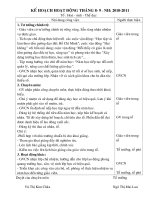
Figure 1: Panoramic X-ray showing 25 severely
abscessed and decayed teeth. Initial measurements for
implant locations and sizes were drawn on during the
consultation
Figure 2: X-ray following placement of eight maxillary and
seven mandibular MDIs. Divergence of the implants is of
no consequence
a life-changing difference. While these
implants can be used to support crowns
and bridges, they are primarily utilized
for the stabilization of dentures. Patients
experience an immediate and dramatic
boost in retention with these implants,
making it a very rewarding treatment to
offer.
The simplicity of the basic MDI
denture stabilization treatment makes it an
attractive procedure for many dentists, but
MDIs can also be utilized in complex cases
such as the one shown in this article. While
the individual techniques used in the case
illustrated here were not new to the team
involved in the procedure, I believe that the
case itself may be a medical first.
placing MDIs, combined with the fact that I
have hospital privileges at the facility where
he would be treated, presented a strong
opportunity.
An initial panoramic X-ray was taken,
which showed 25 severely abscessed and
decayed teeth (Figure 1). (A CT scanner
could not be used during treatment
planning due to the patient’s condition
and mobility restrictions.) A treatment plan
to extract the decayed teeth and place
eight MDIs in the maxilla and seven in the
mandible was presented to the patient and
accepted.
The panoramic image was used to
determine initial implant locations and
sizes. On the day prior to the surgery,
slots were cut into the immediate denture
to accommodate the future sites of the
implants, and a bite registration was taken
outside of the mouth.
On the day of the procedure, after
nasal intubation and general anesthesia,
a 4 x 4 throat pack was placed, and the
25 teeth were extracted. Any bone loss
due to breakage or tooth attachment
was harvested and used for autogenous
grafting where needed later.
Alveoplasty was then performed as
needed, and the 15 3M™ ESPE™ MDI Mini
Dental Implants were placed. The MDIs
ranged from 10 mm to 18 mm in length
and 1.8 mm to 2.4 mm in diameter. Space
limitations prohibit the inclusion of details
on the advanced technique of threading an
implant between two opposing extraction
sites, but it should be noted that varying
Case presentation
The patient in this case was a 52-yearold male who had become quadriplegic
in a tree-trimming accident some 20
years prior to this treatment. The patient’s
medical condition was a C4, C5 complete,
meaning he was paralyzed from the lower
neck down. The injury prevented proper
oral care and rapidly led to the destruction
of the patient’s teeth. The patient’s benefits
from the state of Kansas entitled him to a
single hospital treatment for the condition.
He had seen a number of local specialists
prior to visiting my office, none of whom
could come up with a satisfactory solution
given the constraints of the case.
When I met with the patient, however, I
was able to propose a realistic — although
ambitious — treatment plan. My experience
Volume 6 Number 4
Space is limited.
Sign up today!
August 2013
Code
u
u
u
u
u
u
u
u
Date
3
9
16
16
17
18
23
24
Location
Indianapolis, IN
Shreveport, LA
Chicago, IL
Virginia Beach/Richmond, VA
Chicago, IL
San Francisco/Oakland, CA
LA Area, CA
LA Area, CA
September 2013
Changing patients’ lives.
Building doctors’ practices.
Coming to a City Near You!
MDI Introductory Certification Course
Learn how 3M™ ESPE™ MDI Mini Dental Implants can help offer a
solution to patients who may be contra-indicated for conventional
implant treatment.
Already placing Mini’s? Register for an advanced course today!
Code
●
u
u
u
u
u
u
u
u
u
u
u
u
u
Date
7
13
13
13
14
20
20
20
20
21
21
27
27
28
Location
Buffalo, NY
Dallas, TX
Davenport/Iowa City, IA
Ft. Lauderdale, FL
Ft. Lauderdale, FL
Charlotte, NC
Omaha, NE
Philadelphia, PA
Scottsdale, AZ
Charlotte, NC
Cincinnati, OH
St. Louis, MO
San Jose, CA
San Jose, CA
u = Introductory Training Program
u = Advanced Training Program
● = Mini Dental Residencies
$200 OFF Tuition
Register Today by Visiting: 3MESPE.com/ImplantSeminars
Enter Promo Code* “IP200”
Buy any 12 Implants and 12 Metal Housings, Get 4 Implants
and 4 Metal Housings FREE!** Call 800-634-2249 to order.
**Promo Code “DT200” only available for 2013 3M ESPE MDI Introductory Courses
**Applies to Implants of equal or lesser value and MH-1, MH-2 and MH-3 Metal Housings
For more information or to enroll today visit
3MESPE.com/ImplantSeminars
3M ESPE Customer Care: 1-800-634-2249
3M, ESPE and Espertise are trademarks of 3M or 3M Deutschland GmbH. Used under license in Canada.
© 3M 2013. All rights reserved.
MDI
Mini Dental Implants
CASE STUDY
Figure 3: Immediate state following placement of implants
and suturing of extraction sites
densities, widths, and depths of bone
were encountered. Multiples of every size
and diameter of MDI were on hand for the
procedure in order to be prepared for any
necessary adjustments.
Experienced readers reviewing the
radiographs may note that one more
implant could have been placed in the No.
31 area above the inferior alveolar nerve;
however, without having the 3D scan and
not knowing the precise length of bone, I
did not want to risk any chance of a nerve
parasthesia, especially with this patient.
The radiographs also show how some
of the lower implants are slanted away
from the nerve areas (Figure 2). 3M ESPE
MDIs can withstand up to 30 degrees of
divergence, and this slight angle actually
adds to the final denture retention. This is
done regularly, and the visual slanting of the
MDIs on the X-rays is of no consequence.
Following placement of the implants,
the autogenous grafts were placed where
necessary and into extraction sites along
with collagen plugs, and the sites were
closed with 4-0 Vicryl™ suture (Figure
3). These steps help to preserve bone
and minimize bleeding. Practitioners are
encouraged to do a thorough job of this, as
it greatly helps in the final product.
Metal housings were snapped onto
the O-ball heads of the implants, and
rubber base reline impressions were taken
using the bite registration as a guide.
Analogs were placed in the impressions,
and the case was sent to Kaylor Dental
Lab in Wichita, Kansas, which processed
the snaps and relined the denture within
a few hours. The laboratory’s assistance
was greatly appreciated, as insertion of
the dentures on the same day helps to
minimize swelling and bleeding, and to
lessen the patient’s discomfort.
Before the conclusion of surgery, the
patient was given 10 carpules of Marcaine
so that he would be numb all day and when
the dentures were placed. Antibiotics were
16 Implant practice
Figure 4: The implants at 1 month post-op
Figure 5: Final result with dentures
Figure 6: The author and patient
given before and after surgery, as well
as an anti-inflammatory and a narcotic
painkiller. By 5 p.m., the patient returned
to the dental office, and the new dentures
were seated.
At a post-op visit 3 days later, the
patient stated that the procedure wasn’t
as bad as he had anticipated. Examination
revealed the implants held the dentures
tightly and kept them from compressing
the ridge. Our observation was that the
patient had less pain than if he had no
implants and just the immediate dentures.
A visit 1 month later showed satisfactory
healing of the tissue and a very satisfied
patient (Figures 4-6).
Their affordability, small size, and minimally
invasive nature give them capabilities that
traditional implants simply can’t match.
Eleven years ago, skeptics of MDI
treatments were numerous and vocal.
I continue to know doctors who do not
believe in MDIs, and that is, of course, their
choice. However, I believe that in the nottoo-distant future, MDIs will be as common
as amalgams and offered routinely by most
dentists. The benefits for patients are too
great to overlook, and I believe that MDIs
are one of the finest solutions you can offer
to patients who have lost or are losing their
natural teeth. IP
References
Conclusion
The two articles presented in this series
represent both the basic and advanced
capabilities of MDI treatment. As both
cases illustrate, MDIs provide dentists with
a valuable tool for denture stabilization,
proving versatile enough to be used in
everyday cases or in very challenging
treatments such as the one shown here.
1. Todorovic A, Markovic A, Šcepanovic M.
Stability and peri-implant bone resorption of
the mini implants as complete lower denture
retainers [Espertise Scientific Facts brochure]. St.
Paul, MN: 3M ESPE; 2012.
2. Shatkin TE, Shatkin S, Oppenheimer BD,
Oppenheimer AJ. Mini dental implants for
long-term fixed and removable prosthetics: a
retrospective analysis of 2514 implants placed
over a five-year period. Compend Contin Educ
Dent. 2007;28(2):92-101.
Volume 6 Number 4
CASE STUDY
Adjunctive laser treatment in extraction/immediate
implant placement
Dr. Robert J. Miller discusses technology that is changing the face of implants at the speed of light
T
hroughout the history of oral
implantology, strategies have been
based on the paradigm of placing
endosseous dental implants in healed sites.
With diminishing numbers of completely
edentulous patients being treated, there
is an increasing need to place implants
at the time of tooth removal. Additionally,
over the past decade, our discipline has
seen a dramatic change with either earlier
loading times or immediate loading. Unlike
the healed site with balanced bone density
and soft tissue coverage, extraction sites
present additional challenges with respect
to implant stability and potential presence of
infection. Therefore, if our paradigm is going
to change from placement of implants in
healed sites to one of immediate placement
in extraction sites, new modalities must
be developed. These changes, known as
“biologically-driven” surgical strategies,
reflect our understanding of the interaction
of implanted materials and living tissue.
However, they also reflect our new respect
for the consequences of placing implants
in compromised osteotomies.
Extraction site defects bring increasing
complexity with respect to initial healing
of implants. In most cases, periodontally
involved teeth or failed endodontically
treated teeth are removed, and the site is
prepared to accept an implant. Unlike the
healed site in which pathology has been
resolved, extraction sites may contain
pathogenic bacteria or granulomatous
Robert J. Miller, MA, DDS, FACD, received
both a Bachelor of Arts and Master of Arts
in Biology and then continued his education
at New York University College of Dentistry
where he received his Doctor of Dental
Surgery degree (DDS) in 1981. Upon graduation,
Dr. Miller was honored to be chosen as one of 200
applicants to complete a residency program at Flushing
Hospital and Medical Center. He is one of the few
Dentists in the United States to be Board Certified by
the American Board of Oral Implantology (ABOI). Dr.
Miller is also a Diplomate of the International Congress
of Oral Implantologists (DICOI) and holds current
memberships in the The American College of Dentists,
The American Dental Association (ADA), The Florida
Dental Association (FDA), and the South Palm Beach
County Dental Association (SPBCDA). He has been
practicing dentistry in Delray Beach, Florida for 30
years.
18 Implant practice
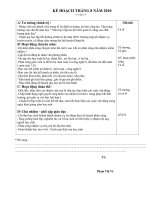
Figure 1: Fracture of an endodontically treated maxillary
cuspid with recurrent decay
Figure 2: Remnants of an apical granuloma still attached
to the root apex
Figure 3: Introduction of a 14 mm zirconium tip into the
extraction site
Figure 4: Completion of laser debridement of the apical
granuloma and de-epithelialization of the gingival sulcus
lesions that can cause infection or implant
failure. The key components of a strategy
to reduce potential complications following
implant placement in this type of site is
complete debridement of the hard tissue
and removal of epithelium in the gingival
sulcus. Sulcular epithelium harbors
periodontal pathogens that may cause
inflammation following implant placement.
These pathogens can migrate to the walls
of the portion of the implant not covered
by bone. They can delay or even prevent
integration of these exposed portions of
the implant, predisposing the implant body
to future infection and bone loss. Apical
granulomas have a different type of biologic
response. Granulomas that have formed
as a result of incomplete endodontic
debridement may harbor vegetative forms
of pathogenic bacteria. However, they may
also result in an untoward immunologic
response different from that of bacterial
origin. This may result in a cyclical biologic
process that perpetuates production
of inflammatory tissue that results in a
retrograde peri-implantitis, starting at the
implant apex and moving coronally.
The following case illustrates how
an Erbium, Chromium;YSGG laser
Figure 5: Placement of the dental implant and healing
abutment
(Biolase Technologies) can be used as
an effective means of debridement and
de-epithelialization prior to immediate
implant placement.This patient presented
with fracture of an endodontically treated
maxillary left cuspid as a result of recurrent
decay (Figure 1). The decay reached
the osseous crest making the tooth
unrestorable without a crown extension.
However, with a high smile line, the patient
opted for tooth removal and immediate
implant placement to maintain the position
of tissue architecture. Following nondestructive tooth removal and maintenance
of the facial plate, the retained root was
evaluated for depth and length. Remnants
Volume 6 Number 4
CASE STUDY
BONE GRAFTING SOLUTIONS
GUIDOR® AlloGraft
Figure 6: Removal of the healing abutment at 2 months
demonstrating regeneration of the dentogingival complex
Figure 7: One-year post-op photograph reflecting stable
gingival architecture and a healthy tissue response
of a portion of the apical granuloma can be seen still attached to
the root apex (Figure 2).
Following extraction, an erbium laser with a 14-mm zirconium
tip is introduced into the osteotomy (Figure 3). Careful debridement
of the entire extraction site is carried out until all remnants of
granulomatous tissue is removed. Additionally, the inner lining of
the sulcus up to the free gingival margin is ablated to reduce the
bacterial load and to create a bleeding interface to accelerate soft
tissue attachment to the healing abutment (Figure 4).
Following implant placement, a healing abutment is placed
and the facial defect grafted (Figure 5). In some cases, if there
is adequate initial stability, a temporary abutment and provisional
may be placed. The implant is allowed to heal for at least 2 months.
When the healing abutment is removed, we can demonstrate the
formation of a new gingival sulcus coronal to the top of the implant
and a bleeding interface apical to that zone which indicates
the presence of a hemidesmosomal attachment to the healing
abutment (Figure 6). This represents regeneration of biologic width
at the coronal aspect of the implant.
The prosthetic phase is completed, and final crown placed
on a milled titanium abutment. The final photograph (Figure 7) was
taken at 1-year post-op. This demonstrates a stable and healthy
dentogingival complex, even in a tooth position with highly parabolic
architecture and long papillae. The use of an ablative erbium laser
is ideal in implant cases when dealing with potentially infected sites
and to enhance initial healing of soft tissue architecture.
Erbium, Chromium;YSGG lasers can also be used for many
other procedures in oral implantology. These include gingival
recontouring, removal of hyperplastic tissue, flap incisions,
osseous recontouring, bone harvesting, lateral wall sinus grafts,
ridge splitting, preparation of the implant osteotomy, implant
debridement, treatment of peri-implantitis, and removal of failed
implants. IP
by LifeNet Healthđ
ã Sunstar, in partnership with LifeNet Healthđ,
is now oering GUIDORđ Allograft.
ã An osteoconductive graft material that promotes rapid healing.
ã Helps maintain space and volume with a strong matrix structure.
• Sterilized using LifeNet Allowash XG® technology
(Sterility Assurance Level of 10-6).
GUIDOR® Bioresorbable
Matrix Barrier
• Double sided bioresorbable material.
• Unique two-layer matrix design
stabilizes the wound site.
• Aids in the regeneration and
augmentation of jaw bone in
conjunction with dental implant surgery.
GUIDOR® Matrix has not been clinically tested in pregnant women,
immuno-compromised patients (diabetes, chemotherapy,
irradiation, infection with HIV) or in patients with extra large
defects or for extensive bone augmentation.
Possible complications following any oral surgery include thermal
sensitivity, flap sloughing, some loss of crestal bone height,
abscess formation, infection, pain and complications associated
with the use of anesthesia.
Complementary products provide
an easy and predictable grafting solution
For more information and exclusive specials, visit us at:
AAP Annual Meeting in Philadelphia, Booth #149
AAOMS Annual Meeting in Orlando, Booth #634
ORDER TODAY! 1-877-GUIDOR1 (1-877-484-3671)
www.GUIDOR.com
©2012 Sunstar Americas, Inc. GDR12036 80812 v1
Volume 6 Number 4 Implant practice 19
CLINICAL
Minimally invasive crestal approach technique for
sinus elevation utilizing a cartridge delivery system
Drs. Ziv Mazor, Andreas Ioannou, Narayan Venkataraman, George Kotsakis, and Udatta Kher delve into ways
to overcome insufficient vertical bone height in the posterior maxilla in conjunction with maxillary sinus lift
Introduction
Dental implants are successfully used to
replace both the form and the function of
missing teeth. The main prerequisite for
implant placement is sufficient volume of
bone in the edentulous ridge to support
the body of the implant. In the maxilla,
when severe atrophy of the edentulous
ridge exists in combination with maxillary
sinus pneumatization, maxillary sinus
augmentation surgery is frequently
employed to provide adequate vertical
bony dimensions for the placement of an
implant.
A variety of surgical techniques and
materials have been used to overcome the
problem of insufficient vertical bone height
in the posterior maxilla in conjunction with
maxillary sinus lift. This procedure aims to
increase the dimensions of the available
bone in the area by placement of bone-graft
material in the space created following the
elevation of the maxillary sinus, performed
Ziv Mazor, DMD, is a leading Israeli periodontist. He
graduated the periodontal department of Hadassah
School for Dental Medicine-Jerusalem, Israel, where
he served as clinical instructor and lecturer for
undergraduate and postgraduate dental students. Dr.
Mazor maintains private practice limited to periodontal
and implant dentistry in Raanana, Israel. Since 1993,
Dr. Mazor has been engaged in clinical research in the
field of bone augmentation and sinus floor elevation.
Dr. Mazor is the past president of the Israeli Periodontal
Society and is currently the president elect of the Israeli
Association of Oral Implants.
George Kotsakis, DDS, is a Resident in the Advanced
Education Program in Periodontology at the University
of Minnesota. Dr. Kotsakis graduated from the University
of Athens, Greece and spent 3 years in private practice
where he focused in implant treatment and complex
restorative cases. During that time he got involved in
practice-based clinical research that led him to pursue
specialty training. Dr. Kotsakis has published numerous
scientific publications in peer-reviewed journals with a
main interest in clinical and histological outcomes of
bone augmentation with different types of grafts.
Andreas Ioannou, DDS, is a Resident, Advanced
Education in Periodontology at the University of
Minnesota.
Narayan Venkataraman, MDS, is an Implantologist in
Bangalore, India.
Udatta Kher, MDS, is an Oral Surgeon in Mumbai, India.
20 Implant practice
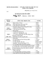
Figure 1: In contrast to the original osteotome technique, before the in-fracture of the sinus floor with the osteotome, a
small quantity of CPS is inserted in the osteotomy to function as a protective “cushion’’ during percussion
in two distinct ways: the direct sinus lift
procedure using a lateral approach and
the indirect sinus lift procedure through a
crestal approach which was introduced by
Summers in 1994.1
When the treatment of choice is
the direct sinus elevation technique,
complications can occur, including a
possibility of sinus membrane perforation.
The indirect sinus elevation technique is
less invasive, less time-consuming, and
reduces the postoperative discomfort for
the patient. The lack of direct visualization
of the membrane and the use for the
osteotomes for the fracture of the sinus
floor may lead to a risk of Schneiderian
membrane perforation as high as 26%.2
The limit of bone volume gained with the
Summers technique is approximately up to
5 mm.3
Technique-related risks such as
reports of benign paroxysmal positional
vertigo following sinus elevation utilizing
the osteotomes technique have led to the
innovation of more atraumatic modifications
of the original technique. Such one is the
minimally invasive antral membrane balloon
elevation (MIAMBE).4 In this technique, a
transalveolar approach is utilized, and the
endosteal implant osteotomy is prepared
1-2 mm below the floor of the antrum.
This surgical approach includes causing a
small fracture in the antral floor and slowly
elevating the sinus membrane with the aid
of hydraulic pressure utilizing a balloon that
inflates and ‘‘pushes’’ the Schneiderian
membrane. The gap present between
the initial position of the sinus floor and
Figure 2: The putty absorbs part of the forces that are
applied to the bone and evenly distributes the remaining
force while minimizing the risk of membrane perforation
the elevated membrane is filled with graft
materials, and an implant is placed.
In another technique, novel atraumatic
drills and reamers that can rotate in
proximity to the sinus membrane and
without perforating the Schneiderian
membrane have been utilized to make
the use of osteotomes redundant. In this
technique, an atraumatic drill is advanced
to the floor of the sinus, and then a reamer
is employed to drill any bone left at the floor
of the sinus and elevate the membrane.
Following slight elevation of the membrane
with the reamer, a carrier is used to deliver
bone graft through the osteotomy and
further advance the membrane.5
Various bone grafting materials are
frequently used in sinus lift procedures,
such as autogenous bone, freeze-dried
bone, demineralized freeze-dried bone,
xenogeneic bone, and alloplastic bone
substitutes.6-7 Recent data have shown
that bone substitutes displaying a putty
consistency can present a valuable
alternative in bone-grafting procedures.8-9
The handling characteristics of putty bone
substitutes have expanded the available
Volume 6 Number 4
CLINICAL
treatment options for bone grafting
in narrow spaces, and their physical
properties can be exploited to increase
the safety and predictability of sinus lift
procedures.
In this improvisation, viscoelastic
calcium phosphosilicate alloplastic putty
(CPS), available in a unique cartridge
delivery system, is utilized. CPS is a
completely synthetic graft substitute that is
approved for bone repair and regeneration
in dental and orthopedic osseous defects.
It is a premixed composite of 70% calcium
phosphosilicate particulate and 30%
synthetic absorbable binder. Bioactivity
of CPS results from the chemical release
of ionic dissolution products: silicon,
sodium, calcium, and phosphate, and has
shown to stimulate multiple generations
of undifferentiated cells into osteoblasts.10
CPS has been successfully used in various
osseous defects with no reported adverse
events.11,12
CPS not only acts as a “protective
cushion” but also provides hydraulic
pressure to lift the Schneiderian membrane.
This approach minimizes risks of benign
paroxysmal positional vertigo or mechanical
perforations of the Schneiderian membrane
associated with the traditional osteotome
technique. In the first case example, a
modification of the MIAMBE technique
with the use of CPS instead of an inflatable
balloon will be presented. In the second
case example, a series of atraumatic drills
will be utilized in conjunction with CPS to
perform an indirect sinus lift without the use
of osteotomes.
Illustration of the minimally invasive technique using hydraulic
pressure
The technique illustrated aims to describe
a modification of MIAMBE technique that
employs hydraulic pressure for sinus
membrane elevation. This improvisation is
made possible by the unique consistency
and delivery mechanism of the CPS graft.
The technique also helps to minimize
complications associated with the use of
osteotomes.
A Transalveolar Sinus Floor Elevation
(TSFE) technique is utilized, and the
osteotomy site is prepared to the size of
the final implant diameter and stopped 0.51 mm short of the sinus floor (Figures 1A
and 1B).
A small quantity (~0.25 cc) of the
putty graft is inserted in the implant bed to
function as a “cushion,’’ thus preventing
22 Implant practice
Figure 3:The narrow tip of the delivery system allows it to enter the narrow osteotomy and reach the floor of the sinus
Figure 4: The viscosity of the CPS that surrounds the apex of the implant aids in achieving increased primary stability
perforation of the membrane before
the osteotome is used to tap firmly and
produce a green-stick fracture (Figure 2).
A putty cartridge is snapped into the
dispensing gun, and the bent cannula of
the cartridge is placed in the osteotomy
site. The width of the cannula is narrow
enough to allow it to be inserted into the
osteotomy following the use of a 2.0 mm
pilot drill. While applying pressure against
the bone, CPS is injected into the site.
The hydraulic pressure from delivery of the
graft material elevates the sinus membrane
(Figures 3A and 3B). For every 0.5cc
injected into the sinus, the floor is elevated
approximately by 2 mm.
Following adequate elevation of the
sinus floor, an implant is placed in the
socket (Figures 4A and 4B). Approximately
85% of the graft gets remodeled into vital
bone in 5-7 months with approximately
15% residual graft after 6 months in the
site.13
Representative case of the modified reamer technique
A 50-year-old, healthy female (nonsmoker) presented for implant placement
in the edentulous upper left premolar area.
The subantral bone height was measured
at 9.3 mm in the 24 area and 5.3 mm
in the 25 area (Figure 5A). The patient
was premedicated with 2g amoxicillin 1
hour before the surgery. Following local
anesthesia, initial drilling with a 2 mm twist
drill, followed by a 2.9 mm drill to widen
the osteotome to approximately 1.0 mm
short of the sinus floor was performed
utilizing a crestal approach (Neobiotech
SCA™ kit). Subsequently, an appropriately
sized (2.8 mm in 24 area and 3.2 mm in
25 area) S-reamer was utilized until the
sinus floor was breached, while leaving
the membrane intact owing to the design
of the reamer. Separation of the sinus
floor was performed using a round-ended
depth gauge. Approximately 0.5 cc CPS
was injected into the No. 24 area and 1.5
cc into the No. 25 area (Figures 5B and
5C) using the cartridge delivery system
and continued until the hydraulic pressure
caused elevation of the sinus membrane.
Once the membrane was adequately
elevated as evidenced by the tactile
sensation of resistance to additional bone
grafting, the grafted material was laterally
spread using a paddle-shaped bone
spreader with a stopper running at 70 rpm.
4 mm x 10 mm and 5 mm x 8.5 mm CMI
IS II implants were placed in No. 24/25
areas, respectively. Implants were inserted
with a primary stability greater than 35N/
cm2 in both sites, and a healing abutment
was placed for non-submerged healing.
A 7-month postoperative radiograph
demonstrated trabecular pattern in the
grafted area indicative of the graft turnover
and bone regeneration.
Discussion
In cases where adequate amount of bone is
not available for the placement of implants
Volume 6 Number 4