Essentials of dental caries 3rd ed
Bạn đang xem bản rút gọn của tài liệu. Xem và tải ngay bản đầy đủ của tài liệu tại đây (3.66 MB, 191 trang )
www.pdflobby.com
www.pdflobby.com
Essentials of dental caries
www.pdflobby.com
Oxford University Press makes no representation, express or implied, that the
drug dosages in this book are correct. Readers must therefore always check
the product information and clinical procedures with the most up to date
published product information and data sheets provided by the manufacturers and the most recent codes of conduct and safety regulations. The authors
and the publishers do not accept responsibility or legal liability for any errors
in the text or for the misuse or misapplication of material in this work.
www.pdflobby.com
Essentials of dental caries
The disease and its management
Third edition
Edwina Kidd
Emeritus Professor of Cariology
Guy’s, King’s and St Thomas’ Dental Institute
King’s College
University of London
1
www.pdflobby.com
1
Great Clarendon Street, Oxford OX2 6DP
Oxford University Press is a department of the University of Oxford.
It furthers the University’s objective of excellence in research, scholarship,
and education by publishing worldwide in
Oxford New York
Auckland Cape Town Dar es Salaam Hong Kong Karachi
Kuala Lumpur Madrid Melbourne Mexico City Nairobi
New Delhi Shanghai Taipei Toronto
With offices in
Argentina Austria Brazil Chile Czech Republic France Greece
Guatemala Hungary Italy Japan South Korea Poland Portugal
Singapore Switzerland Thailand Turkey Ukraine Vietnam
Oxford is a registered trade mark of Oxford University Press
in the UK and in certain other countries
Published in the United States
by Oxford University Press Inc., New York
© Edwina A. M. Kidd, 2005
The moral rights of the author have been asserted
Database right Oxford University Press (maker)
First edition published by IOP Publishing Limited 1987
Second edition published by Oxford University Press 1997
This edition published 2005
All rights reserved. No part of this publication may be reproduced,
stored in a retrieval system, or transmitted, in any form or by any means,
without the prior permission in writing of Oxford University Press,
or as expressly permitted by law, or under terms agreed with the appropriate
reprographics rights organization. Enquiries concerning reproduction
outside the scope of the above should be sent to the Rights Department,
Oxford University Press, at the address above
You must not circulate this book in any other binding or cover
and you must impose this same condition on any acquirer
A catalogue record for this title is available from the British Library
Library of Congress Cataloguing in Publication Data
Kidd, Edwina A. M.
Essentials of dental caries / Edwina Kidd.–3rd ed.
Includes bibliographical references and index.
1. Dental caries.
[DNLM: 1. Dental Caries. WU 270 K47ea 2005] I. Title.
RK331.K43 2005
617,6Ј7–dc22
2004019794
ISBN 0 19 852978 3 (Pbk. : alk. paper)
10 9 8 7 6 5 4 3 2 1
Typeset by EXPO Holdings Sdn. Bhd., Malaysia
Printed in Italy
on acid-free paper by Grafiche Industriali
www.pdflobby.com
Preface
The first edition of this little book was published by John Wright in 1987,
having been commissioned over a postprandial brandy at the George Inn,
Southwark, London. The idea was to produce an easy-to-read, clinically relevant text for the junior undergraduate. The authors were frustrated by the
complexity of the cariology texts available at that time which, they felt,
lacked the clinical dimension which would take the biology to the chairside.
The book has also been used by dental nurses, dental health educators,
hygienists, and therapists. In addition scientists working in the dental field
have found this a useful introduction to clinical cariology. This title has now
found its way all over the world and is produced in CD-ROM form for some
universities.
The second, and now this third edition have been published by Oxford
University Press. The aim is still to produce a simple text to serve as a springboard for further study. This books seeks to complement more comprehensive
texts which are referenced. Other references include relevant systematic
reviews, review articles, and some original papers. The latter must be regarded
as the idiosyncratic choice of the author, but this does not devalue them in
any way.
E.A.M. KIDD
London
August 2004
www.pdflobby.com
Acknowledgements
The manuscript was word processed by Miss Audrey Fernandes, and I am
grateful for her patience and care.
This edition has a single author (EAMK) because Sally Joyston-Bechal is
now retired. However, Sally has criticized this new edition. Her logic and
attention to detail, as well as to deadlines, are irreplaceable.
E.A.M. KIDD
www.pdflobby.com
Contents
CHAPTER 1: INTRODUCTION
1.1
What is caries? 2
1.2
The carious process and the carious lesion 3
1.3
Dental plaque 3
1.4
The role of dietary carbohydrate 7
1.5
Environment of the tooth: saliva and fluoride 8
1.6
Classification of dental caries 8
1.7
Epidemiology of dental caries 12
1.8
Modifying the carious process 18
C H A P T E R 2 : C L I N I C A L A N D H I S TO L O G I C A L F E AT U R E S
OF CARIOUS LESIONS
2.1
Introduction 22
2.2
Basic enamel and dentine structure 22
2.3
The first visible sign of caries on an enamel surface 22
2.4
Dentine reactions 30
2.5
Cavitation—an important moment clinically 31
2.6
Dentine changes in the cavitated lesion: destruction and
defence 32
2.7
Inflammation of the pulp 33
2.8
The microbiology of dentine caries 36
2.9
Active and arrested lesions in dentine 36
2.10 Root caries 37
2.11 Secondary or recurrent caries 38
2.12 Residual caries 38
2.13 Why is dentine caries brown? 39
www.pdflobby.com
VIII
CONTENTS
CHAPTER 3: CARIES DIAGNOSIS
3.1
Introduction 42
3.2
Why is diagnosis important? 42
3.3
Levels of disease and diagnosis 43
3.4
Prerequisites for detection and diagnosis 44
3.5
Detection and diagnosis on individual surfaces 46
3.6
Diagnosis of caries risk 60
3.7
Explaining an individual’s caries experience 60
3.8
Categorizing caries activity status 64
C H A P T E R 4 : P R E V E N T I O N O F C A R I E S BY P L A Q U E
CONTROL
4.1
Introduction 68
4.2
Evidence of the importance of tooth cleaning 68
4.3
Mechanical removal of plaque 71
4.4
Chlorhexidine: a chemical agent for plaque control 82
CHAPTER 5: DIET AND CARIES
5.1
Acid production in dental plaque 88
5.2
Some evidence linking diet and caries 88
5.3
Frequency or amount of sugars 91
5.4
Has fluoride influenced the relationship between sugar and
caries? 91
5.5
Classification of sugars for dental health purposes 93
5.6
Recommended and current levels of sugar intake 93
5.7
Starch, fruit, and fruit sugars 93
5.8
Cultural and social pressures 93
5.9
Groups at particular risk of caries in relation to diet 94
5.10 Diet analysis 94
5.11 Dietary advice 98
www.pdflobby.com
CONTENTS
5.12 Dietary misconceptions 106
5.13 Does dietary advice work? 107
C H A P T E R 6 : F L U O R I D E S U P P L E M E N TAT I O N
6.1
Introduction 110
6.2
Crystalline structure of ename1 110
6.3
Demineralization and remineralization of dental hard
tissues 111
6.4
Fluorosis 112
6.5
Which fluoride supplement? 116
6.6
Toxicity 123
CHAPTER 7: SALIVA AND CARIES
7.1
Introduction 128
7.2
Saliva and dental health 128
7.3
Clinical management of ‘dry mouth’ 132
7.4
Saliva and caries 135
C H A P T E R 8 : PAT I E N T C O M M U N I C AT I O N A N D
M OT I V AT I O N
8.1
The essential role of the patient 142
8.2
Definition of motivation 143
8.3
Communication 143
8.4
Factors that enhance learning 148
8.5
Factors affecting motivation 150
8.6
Planning behaviour change 155
8.7
Reviewing progress and rectifying problems 156
8.8
Failure 156
IX
www.pdflobby.com
X
CONTENTS
C H A P T E R 9 : T H E O P E R AT I V E M A N A G E M E N T O F C A R I E S
Index 177
9.1
The role of operative treatment in caries management 160
9.2
Fissure sealing 163
9.3
Caries removal 171
9.4
Stablization of active disease with temporary dressings 174
www.pdflobby.com
1
Introduction
1.1 What is caries? 2
1.2 The carious process and the carious lesion 3
1.3 Dental plaque 3
1.3.1 Pathogenic properties of cariogenic bacteria 4
1.3.2 Which plaque bacteria cause caries? 4
1.3.3 Where does caries occur? 4
1.3.4 Is dental caries an infectious, preventable, disease?
1.4 The role of dietary carbohydrate 7
1.5 Environment of the tooth: saliva and fluoride 8
1.6 Classification of dental caries 8
1.7 Epidemiology of dental caries 12
1.7.1 Measuring caries activity 12
1.7.2 Practical problems with DMF and def indices 12
1.7.3 The relevance of diagnostic thresholds 13
1.7.4 Caries prevalence 14
1.7.5 The position in the UK 15
1.8 Modifying the carious process 18
7
www.pdflobby.com
2
ESSENTIALS OF DENTAL CARIES
1.1
W H AT I S C A R I E S ?
Dental caries is a process that may take place on any tooth surface in the oral
cavity where dental plaque is allowed to develop over a period of time.
Plaque formation is a natural, physiological process which will be described
in more detail in the next section. Plaque is an example of a biofilm, which
means it is not a haphazard collection of bacteria but a community of microorganisms attached to a surface. This community works together, having a collective physiology. The bacteria in the biofilm are always metabolically active.
Some of the bacteria are capable of fermenting a suitable dietary carbohydrate
substrate (such as the sugars sucrose and glucose), to produce acid, causing the
plaque pH to fall to below 5 within 1–3 minutes. Repeated falls in pH may in
time result in demineralization of the tooth surface. However, the acid produced is neutralized by saliva, so the pH increases and mineral may be
regained. This is called remineralization. The cumulative results of the deand remineralization processes may be a net loss of mineral and a carious
lesion that can be seen. Alternatively, the changes may be so slight that a
carious lesion never becomes apparent (Figure 1.1).1
From this description it becomes obvious that the carious process is an
ubiquitous, natural process. The formation of the biofilm and its metabolic
activity cannot be prevented, but disease progression can be controlled so
that a clinically visible lesion never forms: alternatively, the process can be
arrested and even advanced carious lesions may become inactive. However,
the other side of the coin is that progression of the lesion into dentine can
ultimately result in bacterial invasion and death of the pulp and spread of
infection into the periapical tissues, causing pain.
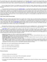
Figure 1.1. The upper anterior teeth of a young adult. In the upper picture, a
disclosing agent reveals the plaque, while in the lower picture the plaque has been
removed. White spot lesions are visible on the canines, but not on other tooth
surfaces, although plaque is present.
www.pdflobby.com
INTRODUCTION
1.2
THE CARIOUS PROCESS AND THE CARIOUS LESION
It is probably unfortunate that the word ‘caries’ is used to denote both the
carious process and the carious lesion which forms as a result of that process.
The process occurs in the biofilm at the tooth or cavity surface; the interaction of
the biofilm with the dental tissues results in the lesion in the tooth. The metabolic activity in the biofilm cannot be seen, but the lesion, which is its reflection
or consequence, can be seen. Thus the dentist is working on a reflection, and
there is a danger that the dentist might forget that the ‘action’ is in the biofilm.
Please stand in front of a mirror and look at your reflection. Do you like
what you see, or could it be improved by some makeup, a shave, a new
haircut, new clothes? You are of course concentrating on the real you and it
probably would not occur to you to pick up a brick and smash the mirror! But
if you now go into the clinic you will see dentists filling holes in teeth, and in a
way they are smashing the mirror unless they have also concentrated on
teaching the patient to modify the metabolic activity in the biofilm.
1.3
D E N TA L P L A Q U E 2
It is thought-provoking that the human body is composed of some 1014 cells,
but only about 10% of these are mammalian; the remainder are resident
microflora. Although a newborn baby’s mouth is sterile, it soon acquires
microbes, usually from the mother via saliva. More than 300 species of
microorganisms have been identified in the mouth.
Dental plaque is an adherent deposit of bacteria and their products, which
forms on all tooth surfaces and is the cause of caries. As already mentioned,
plaque is a biofilm—a community of microorganisms attached to a surface.
The populations of bacteria interact and the properties of the community
are more than the sum of the constituent species. The organisms are organized into a three-dimensional structure enclosed in a matrix of extracellular
material derived from the cells themselves and their environment.
Dental plaque formation can be described in sequential stages:
• Formation of pellicle: an acellular, proteinaceous film, derived from
saliva, which forms on a ‘naked’ tooth surface.
• Within 0–4 hours, single bacterial cells colonize the pellicle. A large proportion of these are streptococci (S. sanguis, S. oralis, S. mitis). There are
also Acintomyces species and Gram-negative bacteria. Only about 2% of
the initial streptococci are mutans streptococci, and this is of interest
because these organisms are particularly associated with the initiation of
the carious process.
• Over the next 4–24 hours the attached bacteria grow, leading to the formation of distinct microcolonies.
• In 1–14 days the Streptococcus-dominated plaque changes to a plaque
dominated by Actinomyces. Thus the population shifts; this is called
3
www.pdflobby.com
4
ESSENTIALS OF DENTAL CARIES
microbial succession. The bacterial species become more diverse and
the microcolonies continue to grow.
• In 2 weeks the plaque is mature but there are considerable site-to-site variations in its composition. Each site can be considered as unique and these
local variations may explain why lesions progress in some sites but not
others in the same mouth.
1.3.1 Pathogenic properties of cariogenic bacteria
There are a number of organisms, normally present in plaque, which can
cause caries. These cariogenic bacteria can:
• transport sugars and convert them to acid (acidogenic)
• produce extracellular and intracellular polysaccharides which contribute
to the plaque matrix; intracellular polysaccharides can be used for energy
production and converted to acid when sugars are not available
• thrive at low pH (aciduric).
1.3.2 Which plaque bacteria cause caries?
There are a number of possibilities, each of which has consequences:
• The specific plaque hypothesis proposed that only a few organisms out
of the diverse collection in the plaque flora were actively involved in the
disease. Preventive measures targeting specific bacteria (e.g. immunization) would be a logical consequence of this hypothesis.
• The non-specific plaque hypothesis considered the carious process to
be caused by the overall activity of the total plaque microflora. A consequence of this approach is that all plaque should be disturbed by mechanical plaque control (toothbrushing).
• The ecological plaque hypothesis proposes that the organisms associated with disease may be present at sound sites. Demineralization will
result from a shift in the balance of these resident microflora driven by a
change in the local environment. Frequent sugar intake (or decreased
sugar clearance if salivary secretion is low) encourages the growth of acidogenic and aciduric species, thus predisposing a site to caries. The consequence of this hypothesis is that both mechanical cleaning and some
restriction of sugar intake are important in controlling caries progression.
1.3.3 Where does caries occur?
Bacterial plaque is the essential precusor of caries and for this reason sites on
the tooth surface which encourage plaque retention and stagnation are particularly prone to progression of lesions. These sites are:
• enamel in pits and fissures on occlusal surfaces of molars and premolars
(Figure 1.2), buccal pits of molars, and palatal pits of maxillary incisors
• approximal enamel smooth surfaces just cervical to the contact point
(Figure 1.3)
www.pdflobby.com
INTRODUCTION
Figure 1.2. Occlusal caries in molars
showing stained fissures. Cavities were
present.
Figure 1.3. A carious
lesion is present on the
distal aspect of the
upper first premolar.
The lesion is shining
up through the
marginal ridge which
shows a pinkish-grey
discolouration.
• the enamel of the cervical margin of the tooth just coronal to the gingival
margin (Figure 1.4a–c)
• in patients where periodontal disease has resulted in gingival recession,
the area of plaque stagnation is on the exposed root surface (Figure 1.5)
• the margins of restorations, particularly those that are deficient or overhanging
• tooth surfaces adjacent to dentures (Figure 1.5) and bridges which make
cleaning more difficult, thus encouraging plaque stagnation.
5
www.pdflobby.com
6
ESSENTIALS OF DENTAL CARIES
a
b
c
Figure 1.4. Caries of the enamel at the cervical margin of the lower molars:
(a) The white spot lesions covered with plaque. (b) A red dye has been used to
stain the plaque so that the patient can see the plaque clearly. (c) The patient has
now removed the stained plaque with a toothbrush: the white spot lesions are
now very obvious. Note they have formed in an area of plaque stagnation and
this can been shown to the patient to demonstrate the importance of plaque
removal.
Figure 1.5. Caries on the
exposed root surface of
the mesial aspect of the
upper premolar. Note the
lesion is in an area of
plaque stagnation
adjacent to a removable
denture. Dentine is also
exposed buccally, but this
has been cleaned and
abraded by the
toothbrush and is cariesfree.
www.pdflobby.com
INTRODUCTION
1.3.4 Is dental caries an infectious, preventable, disease?
No, perhaps it is neither. Although it is caused by bacteria, these are commensal organisms, not extraneous infecting invaders. The carious process
cannot be prevented, because the activity in the biofilm is an ubiquitous,
natural process. However, the progression of lesions can be controlled. These
statements are contentious and may provoke strong reaction and interesting
discussion from your teachers!
1.4
T H E R O L E O F D I E TA R Y C A R B O H Y D R AT E
It is necessary for fermentable carbohydrates and plaque to be present on
the tooth surface for a minimum length of time for acid to form and cause
demineralization of dental enamel. These carbohydrates provide the plaque bacteria with the substrate for acid production and the synthesis of extracelluar
polysaccharides. However, carbohydrates are not all equally cariogenic. Complex carbohydrates such as starch are relatively harmless because they are not
completely digested in the mouth, but carbohydrates of low molecular weight
(sugars) diffuse readily into plaque and are metabolized quickly by the bacteria.
Thus, many sugar-containing foods and drinks cause a rapid drop in plaque pH
to a level which can cause demineralization of dental enamel. The plaque
remains acid for some time, taking 30–60 minutes to return to its normal pH (in
the region of 7). The gradual return of pH to baseline values is a result of acids
diffusing out of the plaque and buffers in the plaque and salivary film overlying
it, exerting a neutralizing effect. Repeated and frequent consumption of sugar
will keep plaque pH depressed and cause demineralization of the teeth.
The change in plaque pH may be represented graphically over a period of
time following a glucose rinse (Figure 1.6). Such a graph is called a ‘Stephan
7
Sound
6.5
pH
6
Inactive
Figure 1.6. Stephan response curves
5.5
5
Active
4.5
20
40
Time (min)
60
obtained from sound occlusal
surfaces, inactive occlusal carious
lesions and deep, active occlusal
carious cavities following a sucrose
rinse in a group of 14-year-olds. Bars
indicate standard errors.3
(Reproduced by kind permission of
Professor Fejerskov).
7
www.pdflobby.com
8
ESSENTIALS OF DENTAL CARIES
curve’ after the person who first described it in 1944. Once a cavity, or hole,
forms in the tooth, the plaque within it becomes even more efficient at producing acid. Lower pH values are recorded in plaque within cavities than in
plaque on inactive lesions or sound surfaces in the same individuals.3
The synthesis of extracellular polysaccharides from sucrose is more rapid
than from glucose, fructose, or lactose. Consequently, sucrose is the most
cariogenic sugar, although the other sugars are also harmful. Since sucrose
is also the sugar most commonly eaten, it is a very important cause of dental
caries.
1.5
E N V I R O N M E N T O F T H E TO OT H : S A L I V A A N D
FLUORIDE
Under normal conditions the tooth is continually bathed in saliva. Saliva is
supersaturated with calcium and phosphate ions and capable of remineralizing the very early stages of lesion formation, particularly when the fluoride
ion is present. Fluoride slows down the progression of lesions.
When salivary flow is diminished or absent, there is increased food
retention. Since salivary buffering capacity has been lost, an acid environment is encouraged and persists longer. This in turn encourages aciduric
bacteria which relish the acid conditions and continue to metabolize carbohydrate in the low-pH environment. The stage is set for uncontrolled
carious attack.
1.6
C L A S S I F I C AT I O N O F D E N TA L C A R I E S
Carious lesions can be classified in different ways; this section introduces and
defines this terminology.
Lesions can be classified according to their anatomical site. Thus lesions
may be found in pits and fissures or on smooth surfaces. Lesions may
start on enamel (enamel caries) or on exposed root cementum and dentine
(root caries).
Primary caries denotes lesions on unrestored surfaces. Lesions developing adjacent to fillings are referred to as either recurrent or secondary
caries. Residual caries is demineralized tissue left in place before a filling is
placed.
Carious lesions may also be classified according to their activity. A progressive lesion is described as an active carious lesion (Figure 1.1) whereas
a lesion that may have formed earlier and then stopped is referred to as
an arrested or inactive carious lesion (Figure 1.7, 1.8). This concept of
activity is very important as it impinges directly on management because
active lesions require active management. However, the distinction between
active and arrested may not be straightforward. There will be a continuum of
www.pdflobby.com
INTRODUCTION
Figure 1.7. Arrested caries on the mesial aspect of the lower second molar. The
lesion probably stopped progressing after extraction of the lower first molar.
Figure 1.8. An arrested carious
lesion in the lower first premolar.
The lesion was well into dentine, but
the tissue was hard and shiny. Note
it is plaque-free. The tooth had been
in this state for at least 10 years.
changes between active and arrested, and part of a lesion may be active while
another part is arrested. This concept is totally logical because the lesion
merely reflects the ecological balance in the overlying biofilm.
Different teeth and surfaces are involved, depending on the area of plaque
stagnation and the severity of the carious challenge. Thus, with a very mild
challenge only the most vulnerable teeth and surfaces are attacked, such
as the cervical margin of the teeth or the occlusal pits and fissures of
permanent molars. A moderate challenge may also involve the approximal
surfaces of posterior teeth. A severe challenge will cause the anterior teeth,
which normally remain caries-free, also to become carious.
Rampant caries is the name given to multiple active carious lesions
occurring in the same patient, frequently involving surfaces of teeth that are
usually caries-free. It may be seen in the permanent dentition of teenagers
and is usually due to poor oral hygiene and taking frequent cariogenic snacks
and sweet drinks between meals (Figure 1.9a–c). It is also seen in mouths
9
www.pdflobby.com
10
ESSENTIALS OF DENTAL CARIES
a
b
c
Figure 1.9. Rampant caries in young men: (a) Note these teeth look clean. This
patient is now making strenuous attempts to remove plaque with a toothbrush.
These lesions are on their way to arrest. Compare this with Figure 1.8.
(b) Despite help with oral hygiene, this patient is not keeping these teeth clean.
(c) The teeth are now disclosed and the plaque deposits are obvious. In addition,
all this man’s drinks are fizzy and sweet. This shows the devastating result of a
combination of poor oral hygiene and a high-sugar diet.
www.pdflobby.com
INTRODUCTION
Figure 1.10. Radiation caries. This patient has been irradiated in the region of
the salivary glands for the treatment of a malignant tumour. Heavy plaque
deposits are obvious over the lesions.
where there is a sudden marked reduction in salivary flow (hyposalivation)
(Figure 1.10). Radiation in the region of the salivary glands, used in the
treatment of malignant tumours, is the most common cause of an acute
reduction in salivary flow.
Early childhood caries is a term used to describe dental caries presenting in the primary dentition of young children.
Bottle caries or nursing caries are names used to describe a particular
form of rampant caries in the primary dentition of infants and young children. The problem is found in an infant or toddler who falls asleep sucking a
bottle (called a nursing bottle) which has been filled with sweetened fluids
(including milk). Alternatively, nursing caries may be found in infants using
a pacifier dipped in sweetener or in children who have a prolonged demand
breast-feeding habit. The frequency of sugar intake combined with a low
salivary flow at night are important in the development of this form of rampant caries. The clinical pattern is characteristic, with the four maxillary
deciduous incisors most severely affected (Figure 1.11).
Figure 1.11. Rampant caries of deciduous teeth. The child continually sucked a
dummy filled with rosehip syrup.
11
www.pdflobby.com
12
ESSENTIALS OF DENTAL CARIES
1.7
E P I D E M I O L O G Y O F D E N TA L C A R I E S
Epidemiology is the study of health and disease states in populations rather
than individuals. The epidemiologist defines the frequency and severity of
health problems in relation to such factors as age, sex, geography, race, economic status, nutrition, and diet. It is a bird’s-eye view of a problem which
attempts to delineate its magnitude, study its cause, and assess the efficacy of
preventive and management strategies. Epidemiological surveys are of great
importance to politicians because they should indicate areas of need where
public money may be spent appropriately.
1.7.1 Measuring caries activity
Epidemiologists are interested in both the prevalence and the incidence of a
disease. Prevalence is the proportion of a population affected by a disease or
condition at a particular time. Incidence is a measurement of the rate at which
a disease progresses. In order to measure incidence, therefore, two examinations
are required—one at the beginning and one at the end of a given time period.
The incidence of the condition is then the increase or decrease in the number of
new cases occurring in a population within that time period.
Before incidence and prevalence can be recorded, a quantitative measurement is required that will reflect accurately the extent of the disease in a
population. In the case of dental caries, the measurements of disease that are
used are:
• the number of decayed teeth with untreated carious lesions (D)
• the number of teeth which have been extracted and are therefore missing
(M)
• the number of filled teeth (F).
This measurement is known as the DMF index and is an arithmetic index
of the cumulative caries attack in a population. DMF(T) is used to denote
decayed, missing, and filled teeth; DMF(S) denotes decayed, missing, and
filled surfaces in permanent teeth and therefore takes into account the
number of surfaces attacked on each tooth. The similar indices for the
primary dentition are def(t) and def(s) where e denotes extracted teeth (to differentiate from loss due to natural exfoliation) and f denotes filled teeth or
surfaces.
1.7.2 Practical problems with DMF and def indices
There are some potential problems in the use of these indices. In young
children missing deciduous teeth may have been lost as a result of natural
exfoliation, and these must be differentiated from teeth lost due to caries. Permanent teeth are lost for reasons other than caries, such as trauma, extrac-
www.pdflobby.com
INTRODUCTION
tion for orthodontic purposes and periodontal disease, or to facilitate the construction of dentures. For this reason missing teeth may be omitted from the
indices and only decayed and filled surfaces included.
Epidemiologists take enormous trouble to achieve standardization of
examination and recording techniques. They will practice and check their
diagnoses during a clinical trial to try to ensure reproducibility. Despite this,
even a trained and experienced worker will not be completely consistent on
the same day, let alone consistent with others in studies spanning years.
In many populations there is a large filled component to the indices, and
the dentists who have done the fillings are not standardized in their diagnosis
of disease. Dentists do not practice and check their diagnostic reproducibility
in the same way as epidemiologists. In addition, there is likely to be variation
between dentists in their recording of disease. Epidemiologists carrying out
national surveys may be limited in their access to clinical facilities because
these surveys are not necessarily carried out in a dental surgery. Thus, access
to good lighting, the ability to clean and dry teeth and the opportunity to
examine radiographs may not be available. Unless radiographs are required
for clinical care, it would be unethical to use ionizing radiation.
1.7.3 The relevance of diagnostic thresholds4
The recording of caries in epidemiological surveys is usually carried out at
the ‘caries into dentine’ level of diagnosis. Enamel lesions are not recorded,
which means that epidemiological surveys inevitably underestimate the
caries problem. This may be very important because the earlier stages of
lesion formation, which are not recorded, should be managed by nonoperative preventive treatments so that the progression of lesions is controlled. The later stages (cavities) may also require restorations, in addition
to preventive treatments. However, if only these are recorded, and those
without cavities are described as ‘caries-free’, the politicians who commission the surveys in the first place may get a false impression of the dental care
needed by the population.
This has indeed happened. In the early 1990s politicians (including dental
politicians) in some developed countries gained the impression that because
many children were described as ‘caries-free’, there was a danger of overproducing dentists. As a consequence of this unfortunate terminology and a
lack of understanding of the carious process, some dental schools closed.
However, it is now realized that in many people the carious process is delayed
and thus lesions may present as cavities as the person grows older. In addition,
the improvement in the caries status means there will be fewer extractions and
thus many more teeth requiring dental care. For these reasons, more dental personnel are now needed. It must also be remembered that the arithmetic means
of DMF(T) are meaningless at the level of the individual patient.
13
www.pdflobby.com
14
ESSENTIALS OF DENTAL CARIES
1.7.4 Caries prevalence5
Dental caries is ubiquitous in modern humans, and is the main cause of
tooth loss in people of all ages. For most of the twentieth century caries was
seen as a disease of economically developed countries, with a low prevalence
in the developing world. By the late twentieth century this pattern was
changing in two ways:
• There was evidence of a rise in caries experience in some developing countries. To give an example, studies in the 1990s show dental caries as a
major problem in the former socialist countries of eastern Europe. These
countries can be considered ‘developing’ in the economic sense, and the
use of fluoride toothpastes and toothbrushes there is still low.
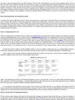
• By the late 1970s a marked reduction in caries experience among children and young adults was obvious in developed countries (Figure 1.12)
although in 1983 there were considerable differences between countries.6
DMFT
10
10.1
Australia
Denmark
Finland
Netherlands
New Zealand
Norway
Sweden
United Kingdom
USA
9.0
9
8
7.5
7.2
7
6.3
6
5
4.8
4.7
4.8
4
4.7
3.8
4.4
4.1
3.9
3.4
3
2.6
1967
71
73
74
75
76
77 78
Years
79
80
81
82
3.3
3.0
2.8
83
Figure 1.12, DMFT data from 12-year-old children of many countries
demonstrating a decline in caries prevalence between 1967 and 1983. Note the
considerable inter-country differences (data from the WHO Global Oral Data Bank
(Renson et al., 1986)6