Characteristics of Fecal Incontinence Using High Resolution Manometric assessment in Viet Duc hospital
Bạn đang xem bản rút gọn của tài liệu. Xem và tải ngay bản đầy đủ của tài liệu tại đây (715.62 KB, 73 trang )
MINISTRY OF EDUCATION AND TRAINING
MINISTRY OF HEALTH
HANOI MEDICAL UNIV ERSITY
...***.„
NGUYEN HUU TRI
CHARACTERISTICS OF FECAL INCONTINENCE USING
HIGH RESOLUTION MANOMETRIC ASSESSMENT IN
VIETDƯC HOSPITAL
Major: General Doctor
Code : 52720201
GRADUATE THESIS
2015-2021
Supervisors: M.S. Nguyen Ngoc Anil
Hanoi - 2021
r-u -ÍM CỊỈ ugc V
Hl
ACKNOWLEDGEMENT
First of all. I gratefully acknowledge the Board of President, the University Training and
Management Department of Hanoi Medical University and the Colorectal surgery center of
Viet Due University Hospital for giving me the precious opportunity to do this study.
Throughout the diesis. I wish to express my deepest gratitude to my supervisors M.S.
Nguyen Ngoc Anil for her excellent guidance, inspiration, encouragement, extremely helpful
comments and supporting me during the process of the study.
I also want to send my special thanks to all the participants. My research would not have
been possible without their cooperation.
Finally. I would also like to thank my friends and family who always beside me. support
and encourage me.
Hanoi. May lOtli. 2020
Student
Nguyen Huu Tri
r-u -ÍM Qỉ ugc V
Hl
DECLARATION
I hereby declare Thar the work entitled is my original work and have not been published
in any other dissertation, thesis for qualifications or any medical literature. I did not copy from
any other's work or from any other sources except the attached references which were listed
clearly in the text. I also pledge the data collected in the thesis is completely honest.
If there is anything wrong. I would bear all responsibilities.
Hanoi. May 10th .2021
Student
Nguyen Huu Tri
r-u -ÍM CỊỈ ugc V
Hl
TABLE OF CONTENTS
INTRODUCTION.........................................................................................................................1
CHAPTER 1: LITERATURE REVIEW......................................................................................3
1.1. Anatomy and physiology of the anorectal area.............................................................3
1.1.1. General anorectal anatomy.....................................................................................3
1.12. Anorectal physiology and incontinence mechanism.............................................5
1.2. General introduction of fecal incontinence....................................................................7
1.2.1. Definition and classification..................................................................................7
1.22. Etiology........................................................................................................................8
1.23 Pathophysiology...........................................................................................................9
1.2.4. Risk factors...........................................................................................................10
1.3. Diagnosis of fecal incontinence...................................................................................10
1.3.1. Clinical manifestation..........................................................................................10
1.3.2. Fecal incontinence’s investigation tests...............................................................11
1.4. Treatment of FI.............................................................................................................21
1.4.1. Nonoperative management...................................................................................21
1.42.
Surgery7.................................................................................................................24
1.5. Previous researches on FI.............................................................................................25
1.5.1. World....................................................................................................................25
1.52. Vietnam......................................................................................................................27
CHAPTER 2: SUBJECTS AND METHOD..............................................................................28
2.1. Subjects........................................................................................................................28
2.1.1. Sampling method.................................................................................................28
2.12.
Inclusion criteria..................................................................................................28
2.13.
Exclusion criteria.................................................................................................28
2.2. Study settings...............................................................................................................28
2.3. Study method...............................................................................................................29
2.4. Research variables and indicators...............................................................................30
2.5. Data analysis................................................................................................................31
2.6. Ethical consideration...................................................................................................31
CHAPTER 3: RESULTS...................... ................................... ............................................ 32
3.1.
General features of study population...........................................................................32
3.2.
Risk factors...................................................................................................................33
3.3.
Etiology of fecal incontinence.....................................................................................34
3.4.
HR AM in study population.......................................................................................36
3.4.1. Comparing men and women................................................................................36
3.42.
Comparing men and women................................................................................38
3.5.
3.6.
Severity of FI hi research population...................................................39
Influence of severity of FI on HRAM........................................................................39
CHAPTER 4. DISCUSSION...............................~.............................................................
41
4.1. General features of study population..........................................................................................41
4.1.1. Age.......................................................................................................................41
4.12.
Gender..................................................................................................................41
4.13.
BMI......................................................................................................................42
4.2. Risk factors................................................................................................................................42
4.2.1. Vaginal deliver’.............................................................................42
4.2.2. Physical limitation................................................................................................43
4.2.3. Urinary’ incontinence...........................................................................................43
4.3. Etiology’ of fecal incontinence..................................................................................................43
4.4. HR AM in FI patients..................................................................................................................45
4.4.1. Pressure values.....................................................................................................45
4.42.
HPZ andRAIR......................................................................................................47
4.43.
Rectal balloon volume........................................................................................48
4.4.4. HR AM results comparing urge and passive incontinence...............................48
4.5. Severity of FI.............................................................................................................................49
4.4.1. Severity of FI comparing urge and passive incontinence...................................49
4.42. The correlation between FI severity and HRAM......................................................49
CHAPTER 5: CONCLUSION-----------------------------------------------------------------------51
REFERENCES
APPENDIX 1
r-u -ÍM Qỉ ugc V
Hl
LIST OF ABBREVIATIONS
FI:
Fecal incontinence
ARM:
Anorectal manometry
HRAM:
High-resolution anorectal manometry
IAS:
Internal anal sphincter
EAS:
External anal sphincter
EAƯS:
Endoanal ultrasound
MARP:
Maximum anal resting pressure
MASP:
Maximum anal squeeze pr essure
HPZ:
High pressure zone
AƯC:
Area under the curve
EAUS:
Endoanal ultrasound
r-u -ÍM Qỉ ugc V
Hl
LIST OF ABBREVIATIONS
r-u -ÍM Qỉ ugc V
Hl
LIST OF ABBREVIATIONS
r-u -ÍM Qỉ ugc V
Hl
LIST OF FIGURES
Figure 1.1. Anatomical Structure of the anorectum.................................................................3
Figure 1.2: Anatomy of anorectal area......................................................................................4
Figure 1.3: Nerves of the anorectal area...................................................................................5
Figure 1.4: High-resolution anorectal manometry.................................................................12
Figure 1.5: Filling the balloon during rectal sensibilty test....................................................15
Figure 1.6: Anterior defect in external anal sphincter visualized on endoanal
ultrasonography..................................................................................................18
Figure 1.7: MRI defecography................................................................................................19
Figure 1.8: Stimulating the pudendal nerve............................................................................20
Figure 1.9: The nerves stimulator is implanted beneath the buttocks skin. 24 Figure 3.1:
HRA.M results comparing men and women..........................................................................37
Fig 3.2: Bivariate distribution between Wexner score and maximum anal
resting pressure(a). maximum anal squeeze pressure (b).............39
Fig 3.3: Bivariate disttibution between Wexner score and first sensation
volume (a), max tolerable volume(b)................................................................40
r-u -ÍM CỊỈ ugc V
Hl
LIST OF TABLES
Table 1.1: Normal Values of anorectal manometry...............................................................16
Table 3.1: General features of study population.....................................................................32
Table 3.2: Risk factors comparing uige and passive Fecal Incontinence .. 33
Table 3.3: Causes of Fecal incontinence comparing urge and passive incontinence............34
Table 3.4: Causes of Fecal Incontinence comparing women and men..........35
Table 3.5: HR AM results comparing men and women.........................................................36
Table 3.6. Values of HRAM comparing passive and urge incontinence.... 38
Table 3.7: Wexner score in 2 different types of FI.................................................................39
Table 4.1. Values of HRAM in FI and asymptomatic people................................................45
Table 4.2. MARP and MASP comparing urge and pasive incontinence.... 48
r-u -ÍM Qỉ Hgc V
Hl
TÓM TÁT LUẬN VÃN
A) Tiêu đề luận vân: Đánh giá các đậc diem cúa đại tiện không tự chu (ĐTKTC) dựa
trên do áp lực hậu mòn trực tràng (ALIIMTT) độ phân giái cao tại bệnh viện Việt Đửc
B) Thõng tin luận vãn
Lý do lựa chọn đề tài: DTKTC ánh hưởng nhiều đến chất lượng cuộc sống. Tuy nhiên
chưa có nhiều nghiên cứu về đặc diêm cua bệnh ờ người Việt Nam.
Mục tiêu nghiên cứu: Đánh giá các đặc điếm làm sàng và ket quá do ALHMTT ở bệnh
nhản ĐTKTC tại bệnh viện Việt Đức.
Phương pháp nghiên cứu: Nghiên cứu loạt ca bệnh trẽn 1S bệnh nhân DTKTC dền khám
tụi Bệnh viện Việt Đức vả do ALHMTT giai đoạn 2020- 2021. Sư dụng thống kê phàn tích đe
đánh giá nguyên nhân, yếu tổ nguy cơ, triệu chửng làm sàng, kcl qua do ALHMTT.
Kết quà nghiên cứu: Sa tạng chậu lã nguyên nhãn phố biến nhất ờ nữ 5/9 (55.6%) trong
khi ờ nam là phẫu thuật hậu mòn trực trảng 7/9 (77.8%). ĐTKTC cẩp ki: 8/18 (44.4%) và thụ
động: 10/18 (55.6%). Khõng có sự khác biệt VC ket qua đo ALHMTT giừa 2 nhóm trừ the tích
bom bóng tối da thắp lum ớ nhõm cấp ki (90.0 ± 28.3 so vói 123.0 X 26.7 ml; p<0.05). Áp lực
trung binh cũa hậu môn khi nghi. nhíu, rặn, ho lần lượt là 47,7 mmHg, 100,9 mmHg. 61,1
mmHg và 63.0 mmHg. Ngường nhận cam cua trực tràng và khả nâng chịu đựng tối đa cua trục
tràng lần lượt là 27.2 ml vã 108.3 ml. Có sự tương quan rõ ràng giìra sự giam ãp lực khi nghi và
nhíu cua hậu mơn và the tích chịu dựng tối da cùa trực tràng vởi mức độ nặng cua bộnh (r =
-0,48, - 0,50,-0,55; p<0.05).
Kết luận vã đề xuất: Đo áp lực hậu môn trực tràng độ phân giái cao cỏ giã trị trong chân
đoán, đánh giã mửc độ tôn thương, lẽn kế hoạch diêu trị vã tiên lượng với bệnh nhân dại tiện
khơng tự chu.
c) Từ khóa: đại tiện không tự chu. do áp lực hậu môn trực tràng, cơ thắt hậu món
a) Title: Characteristics of fecal incontinence (FI) using High- resolution manometric
assessment (HRAM) in Viet Due hospital
b) Abstract:
Reason for willing: Fecal incontinence lias a considerable impact on patients' quality of
life in both mental and physical well-being. However. in Vietnam FI has been considered as a
-W.- -ÍM Qỉ ugc V
Hl
neglected disease and received meager attention from scientists.
Problem: The aim of this study was to evaluate clinical characteristics and HR AM in
patients with fecal incontinence.
Methods: Case series study, IS patients diagnosed with fecal incontinence completed
questionnaire assessing clinical history’, symptoms and HRAM. Statistical analysis was used to
evaluate causes, risk factors, symptoms and HR AM results.
Results: Pelvic organ prolapse was the main cause of FI in women: 5/9 (55.6%) while
colorectal/anorectal surgery’ was the main cause of FI in men: 7/9 (77.8%). Urge incontinence:
8/18 and passive incontinence: 10/18. In terns of anorectal manometry, no statistical difference
found between two groups, except max tolerable volume was lower in urge incontinence (90.0
= 28.3 vs 123.0 ± 26.7 mL: p<0.05). Anal pressures during resting, squeezing, straining,
coughing were 47.7 mmHg. 100.9 mmHg. 61.1 mmHg and 63.0 mmHg. First sensation volume
and max tolerable volume were 27.2 and 108.3 mL. Maximum anal resting pressure, maximum
anal squeeze pressure, max tolerable volume decreased significantly with increasing FI severity
(r = -0.48. -0.50. -0.55; p<0.05).
Conclusion: High-resolution anorectal manometiy is valuable in diagnosis, assessment,
treatment and prognosis in patients with fecal incontinence.
c) Keywords: Fecal incontinence, high-resolution anorectal inanom?try. anal
sphincters
r-u -ÍM Qỉ ugc V
Hl
1
INTRODUCTION
Fecal incontinence (FI) is defined as the unintentional leakage of solid or liquid stool.
FI is a common disease with a prevalence ranging from 7 to 15% in the community,
depending on the definition and the survey methods used [1]. In elderly population, the
prevalence is higher, up to 47% [2].
Fl has a considerable impact on patients' quality of life. Fear of public humiliation can
lead to social isolation, loss of employment. as well as ruining relationships and lower selfesteem. Physical complications include pain, infection and skin-ulcer which is more severe in
older patients
The incidence of FI is elevated in recent years associated with risk factors such as
diabetes mellitus. lower gastrointestinal surgery and stroke. Because FI is a significant issue,
many studies are conducted annually. However, in Vietnam. Fl has been perceived as a nonserious problem by both healthcare providers and scientists. Therefore. FI patients often seek
health care in severe leakage conditions.
A thorough assessment of fecal incontinence is critical for the evaluation and
management of the anorectal disorder. Anorectal manometry is an important diagnostic tool to
evaluate anorectal motor and sensory function. In patients with FI. anal resting and squeeze
pressures help determine the presence of internal anal sphincter (IAS) and external anal
sphincter (EAS) dysfunction. Measurements of anorectal function can help to establish a proper
diagnosis for effective management. Initial treatments include patient education for diet
changes, bowel training and exercises to sttengthen pelvic floor muscles. Some researches show
those treatments can improve the symptoms about 60 percent [3]. Conservative treatment with
biofeedback therapy, electrical stimulation were also recommended. Surgery' like sacral nen e
stimulation may be an option for patients that fails with conservative treatments, or for fecal
incontinence caused by pelvic floor muscles or anal sphincter injuries.
In the world, there have been many studies on fecal incontinence because of
significant effect on quality of life. In Vietnam due to rhe lack of data on diagnosis and
r-u -ÍM CỊỈ ugc V
Hl
2
management of fecal incontinence, it requires more studies to evaluate different characteristics
of patients with FI for an optimal individualized treatment plan. Thus, we conducted this study
"Characteristics of Fecal Incontinence Using High-Resolution Manometric assessment in Viet
Due hospital" with two aims:
1. Evaluate clinical characteristics of patients with fecal incontinence.
2. Evaluate characteristics of high-resolution anorectal manometry assessment in
patients with fecal incontinence.
-■c -ÍM Qỉ ugc V
Hl
3
CHAPTER 1: LITERATURE REVIEW
1.1.
Anatomy and physiology of the anorectal area.
The rectum and anal canal are tile terminal parts of the large bowel. The anorectal canal
has the important function of regulating defecation and the role of controlling fecal continence.
1.1.1.
General anorectal anatomy
The rectum is a hollow muscular tube, 12 to 15 cm long, composed of a continuous laser
of longitudinal muscle that internsine ssith the underlying circular muscle. The anus is a tube of
muscle ssith a length of 2 to 4 cm. At rest, it forms an angle of roughly 90 degrees with the axis
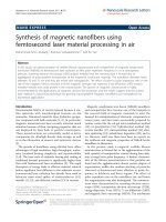
of the rectunt.[4] During defecation, the anorectal angle becomes more obtuse, whereas during
voluntary squeeze, this angle becomes more acute (Figure 1.1)
Resting
Normal
defecation
Figure 1.1. Anatomical Struct toe of the anorectum
Source: Satish S.C.Rao- 2016 [5]
• The Anal Sphincter
The anal sphincter is formed from 2 muscles: the internal anal sphincter
(IAS) and tlie external anal sphincter (EAS) (Figure 1.2). The IAS is a mostly
r-u Qỉ Hgc V Hl HỈỈ
4
fatigue-resistant
smooth
muscle
[6].[7].
The
IAS
contributes approximately 70% to 85% of the resting
sphincter pressure [8]. In this way. the IAS is primarfly
responsible for maintaining anal continence at rest.
Normally. the anus is closed by tile contraction of the
IAS. This barrier is strengthened during voluntary'
squeeze by the EAS. During defecation the IAS is relaxed
and become vertical, the lumen is opened which allows the
stool to pass distally.
The EAS is a cylindrical striated muscle under voluntary control and comprises
predominantly slow-twitch muscle fibers which enables it to have prolonged contraction [9].
Sex-related differences include a fundamentally shorter external sphincter in women than in
men both laterally and anteriorly [10]. The EAS lias a resting contraction that conưibutes about
20% of anal resting pressure (8). EAS activity is innervated by somatic nenes, the right and left
inferior rectal nerves, each derived directly from the corresponding pudendal nene (S2-S4).
Internal StAexter
Mwtcte
External sptuncter
HuKle
Ọạndi and
Haws in Penanal Skin
F/gi/re 1.2: Anatomy of anorectal area
Source: Frank H. Nett er. Atlas of Human Anatomy [11]
-c .tín CỊỈ Hgc V
Hl
5
• Nen e Structure and Sensation
The anorecrum is richly innervated by the sensory; motor, and autonomic nerves and by
the enteric nervous system. The principal nerve is the pudendal nerve, which arses from the
second, third, and fourth sacral nerves (S2, S3, S4) and innervates the EAS (Figure 1.3).
Pudendal nerve block can lead to a loss of sensation in the perianal and genital skin and
weakness of the anal sphincter muscle but rectal sensation is not affected [8Ị.
Figure 1.3: Nenes of the anorectal area
Source: Frank H. Neuer. Atlas of Human Anatomy[11]
The sensation of rectal distention is most likely transmitted along the S2. S3, and S4
parasympathetic nerves which are independent of the pudendal nerve [4]. If parasympathetic
innervation is impaired, rectal filling is only- perceived as a vague sensation of discomfort [4].
Thus, the sacral nerves play an important role in the maintenance of fecal continence.
1.1.2.
Anorectal physiology and incontinence mechanism
The recto-anal canal serves two important functions, the fecal continence and defecation.
•
Anal continence
Normal anal continence allows emission voluntarily conuolled. periodic, and selective of
the various contents: gas or liquid and solid stool. In normal conditions and during the filling of
the rectal ampulla, continence is achieved by the conttaction of the internal anal sphincter. The
distension of the rectum activates the recto-anal inhibitory reflex with consequent relaxation of
-c -ÍM Qỉ ugc V
Hl
6
the internal spliincter. and this causes a small amount of fecal material to come in contact with
the mucous membrane. This is rich in nerve endings and can differentiate the feces from the gas
and decide whether or not to defecate [12]. If defecation should be delated, the voluntary
contraction of the external anal sphincter sends back fecal material, postponing the stimulus.
Continence is based on two main elements: the ability of the rectum to host feces and anal lock
mechanism (the ability of closing anal canal), which, together with the ability of sensor)'
discrimination of the anal canal, prevents the involuntary leakage of stool.
•
Defecation
The defecation reflex is an act that takes place under the control of will. When the feces
reach the rectum this is stretched. When the person decides to defecate: Illis decision implies a
sitting position with hip flexion that results in the disappearance of the angle between the anal
canal and rectum and a complex defecator)' mechanism begins. The first occurs when the rectum
is distended by feces; a peristaltic wave is generated to push the stool from the sigmoid colon
and rectum to the anus. This reflex is. however, weak and to cause defecation must be reinforced
by the reflection of the parasympathetic that amplifies peristaltic waves and transform the reflex
of defecation in a powerful process to allow emptying of the rectum and anus. The afferent
impulses arriving at the spinal cord give rise to other effects: deep breathing.
r-u -ÍM CỊỈ ugc V
Hl
7
closure of the glottis, and contraction of the abdominal muscles, to increase the abdominal
pressure, which in turn increases the rectal pressure to push out the feces. For defecation to
occur, the voluntary' mechanism is indispensable as it inhibits the external anal sphincter because
Illis normally contracts with the arrival of stool.
• Incontinence mechanism
The ability to control evacuation is guaranteed by many factors. These include intact anal
sphincter mechanism, stool volume and consistency, intestinal motility, pelvic floor structural
integrity, cortical awareness, cognitive function, mobility' and access to facilities. Incontinence
occurs when one or more of these mechanisms are impaired and the remaining mechanisms are
unable to compensate. Although the integrity of the sphincteric mechanism plays a major part,
tliere a re other important aspects, such as stool volume and consistency, colonic transit, rectal
compliance and sensation, anorectal sensation and anorectal reflexes.
1.2. General introduction of fecal incontinence
1.2.1. Definition and classification
Fecal incontinence is the inability' to control bowel movements causing stool (feces) to
leak unexpectedly from the rectum.
There is no widely accepted approach for classifying FI. Currently. FI is classified by
separated systems based on etiology', pathophysiology (i.e., bowel disturbances, anorectal
dysfunctions), type of leakage (urge, passive, or combined), or symptom severity' scales.
Commonly, the classification based on the type of leakage is used:
-r Urge incontinence: tire patient has a sudden urge to use the bathroom but is unable to
get there in time.
* Passive incontinence: Because of the loss of sensations in anorectal area, patients can't
consciously control their defecation and stool is passed without their knowledge [13].
+ Combined incontinence: Patients have both urge and passive incontinence.
1.2.2. Etiology
There are many different causes of FI. in a patient. FI is often the result of a combinations
-c -ÍM CỊỈ ugc V
Hl
8
of factors but a single one. Common causes of FI include:
•
Fecal impaction
Chronic constipation can lead to fecal impaction. This happens when a hard stool gets
stuck in the rectum. Recurrent congestion can lead to sưetch and weakness of the sphincter,
which makes the muscles unable to stopping normal passage. Another complication of fecal
impaction is leakage of liquid fecal matter through die anus.
•
Hemorrhoids
External hemorrhoids can deter the sphincter from closing completely. Tills allows loose
stool and mucus to pass involuntarily.
•
Muscle damage
The IAS or/and EAS can be deterred from keeping the anus tightly closed when they are
damaged. Surgery in or adjacent to the anorectal region, trauma, and constipation can impair the
sphincter muscles.
•
Nen e damage
If the nerves that innervate sphincter motor are injured, the sphincter muscles won’t close
properly. Moreover, patients may lose the sensation of the urge to go to the bathroom. Some
causes of nene damage are trauma from giving birth, chronic constipation, stroke, diabetes
mellitus. multiple sclerosis.
-■c -ÍM CỊỈ ugc V
Hl
9
• Pelvic floor dysfunction
Women can experience damage to the muscles and nenes in their pelvis while giving
birth, but symptoms of peine floor dysfunction may not be immediately obvious. They may
occur after years. Besides fecal incontinence, two common complications of this condition are
rectal prolapse or rectocele.
I.2J Pathophysiology
A majority of patients with FI have reduced anal resting and/or squeeze pressures,
reflecting the weakness of the internal and/or external anal sphincters respectively [14]. Anal
sphincter damage, which is most frequently caused by obstetric or iatrogenic trauma, or
neurogenic injury can cause anal weakness. Neurogenic lesions can occur at any level of the axis
extending from the central nervous system to the external anal sphincter. In addition to anal
sphincter injury. FI is also associated with atrophy, denervation. and impaired function of the
puborectalis muscle. Excessive straining may cause increased perineal descent, which can stretch
and thereby damage the pudendal nen e and also make the anorectal angle more obtuse.
Patients with FI may have normal, reduced, or increased rectal sensation [14], When
rectal sensation is leduced. the external anal sphincter may not contract promptly when rhe
rectum is distended by stool, predisposing to FI. Conversely, rectal hypersensitivity in FI may be
partly secondary to an exaggerated contractile response to distention, and/or reduced rectal
capacity, and may explain the symptom of rectal utgenev [15].
In summary, multiple physiological mechanisms preserve continence. Deficits in any of
these mechanisms may contribute to FI. and as a consequence no single physiological measure is
consistently associated with FI.
1.2.4. Risk factors
According to Norton et al. FI had a lot of risk factors [16]:
- Patient characteristics: Increasing age. gender women, obesity, poor general health
and physical limitations.
- Neurological disease or injury (learning disability, dementia, spinal cord injury,
multiple sclerosis, stroke. head injury, diabetes msllitus)
r-u -ÍM Qỉ ugc V
Hl
10
- Gastrointestinal diseases: Diarrhea or loose stools, constipation, irritable bowel
syndrome, inflammatory bowel disease, hemorrhoids
• Obstetric factors: Vaginal delivery’, instrumental delivery, large baby, prolonged
delivery
- Surgical procedures: Sphincterotomy, hemorrhoidectomy
- Urinary incontinence and pelvic organ prolapse
- Dings (antibiotics, laxatives, digoxin, orlistat). dietary supplements (lactose, fructose,
artificial sugars)
13. Diagnosis of fecal incontinence
1.3.1.
Clinical manifestation
Fecal incontinence can involve recurrent or infrequent involuntary leakage, an inability to
hold in gas. silent leakage of feces during daily activities or exertion, or not reaching the
bathroom in time.
The consistency' of stools passed during bowel incontinence can vary solid, liquid, gas.
The leakage may occur daily, weekly, or monthly.
Associated signs and symptoms may include abdominal pain, bloating, flatulence or
both, constipation or diarrhea. the anus is irrflated or itchy, urinary incontinence.
-■c -ÍM CỊỈ ugc V
Hl
11
Fecal incontinence can be a relatively small medical issue, bringing about the occasional
soiling of underwear. or it can be severe, with a total lack of bowel control.
1.3.2.
F'ecal incontinence's investigation fests
A -wide range of tests is available to facilitate the diagnosis and management of FI.
Imaging tests, physiology tests, and nerve studies have all been described. In Vietnam highresolution anorectal manometry and endoanal ultrasound are two budget-friendly tests that
provide useful information are mostly used in clinical practice.
1.3.2.1.
High-resolution anorectal manometry
• Introduction
High-resolution anorectal manometry (HRAX1) was first introduced in 2008 [17]. is an
instrumental examination evaluating the pressure of the anal canal and the distal rectum giving
motor and sensory information on functional phases of defecation. the continence of tíre
anorectal tract and of tire pelvic floor muscles. It measures the luminal pressure along 6-8 cm
above the anal verge [18] and. in particular, it allows to evaluate:
The high-pressure zone (which refers to the length of the anal sphincter muscles)
The involuntary function of the anal canal at rest
The voluntary anal function on squeezing
The rectoanal reflexes
The rectal sensitivity and compliance
The rcctoanal coordination during simulated defecation ("push”)
The capacity to expel a balloon.
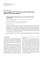
• Equipment
Firstly, the pressure is recognized by a solid-state catheter (probe) which has an outer
diameter of 4.2 mm (figure 1.2a). There are 2 versions of this catheter: The regular probe has 12
circumferential sensors, including ten sensors at 6-mm intervals along the anal canal and two
sensors in the rectal balloon (3.3 cm long, maximum capacity of 400 mL). The small probe lias 8
circumferential sensors in total with 1 balloon sensor. A latex-free rectal balloon that is 3.3 cm
-c -ÍM CỊỈ ugc V
Hl
12
long and lias a maximal capacity of 400 niL is recommended to be used. In both types of probe,
each sensor has 36 circumferentially oriented, pressure-sensing elements that generate an
averaged single value at 35 Hz.
The manometry and topographic images are shown on a computer screen using specialized
software (Figure 1.2b). The system operates at a frequency response of > 20 Hz. and an output
resolution of 0.1 mmHg. The probe is calibrated immediately before the procedure by putting it
in a calibration chamber,
where it becomes zeroed to atmospheric pressure and set to a scope of pressures up to 300
mmHg [17].
-*
a) catheters;
b) all system
Figure 1.4: Higỉì-resolion anorectal manometry
-c -ÍM Qỉ ugc V
Hl
13
At rest, during squeeze, and rectal distention the software recognizes the highest of all
pressures recorded by anal sensors at every point in rime. T1ŨS value is then used to identify the
average and maximum anal resting pressure and the maximum squeeze pressure. In addition,
rectal pressure is also reported to evaluate other anorectal functions.
The patient (who should do an evacuation enema a few hours before the examination) is
placed in left lateral decubitus with overlapping thighs atri bent at 90° on tile trunk: the catheter
is then inserted into the rectum.
• Procedure of high-resolution anorectal manometry.
- Patient preparation: Bowel preparation with Fleet enema or defecation 2 hours before
the test.
-
Patient position: left lateral decubitus with overlapping thighs and bent at 90° on tile
-
Implementing: After digital rectal examination, a lubricated catheter with a balloon
trunk.
attached is then inserted into the rectum until only the 8th sensor is outside the anus.
-
Rest: Give tire patients time to relax for 5 minutes.
-Squeeze: The patient is instructed to squeeze the anal sphincters for 20 seconds or as
long as possible.
-
Push (Bearing-down): Strain as forcefully as possible, hold on 10 to 20 seconds.
-
Cough: The subject is asked to give 5 to 7 coughs
-
Recto-Anal Inhibitory Reflex (RAIR): Inflate the balloon with 10 mL of air through
center canal (balloon was attached to the head of catheter), after 3 to 5 seconds fast withdraw the
air to deflate the balloon totally.
-
Repeat this maneuver several times (in 20-second gaps) and each time, inflate the
balloon with an additional volume of 10 ml air.
- Inflate the balloon to evaluate rectal sensitivity. balloon volume for the first sensation,
for constant desire to defecate, for maximum tolerable sensation.
+ Finish the test: Air withdrawal and catheter extraction, data analysis and then print out
r-u -ÍM CỊỈ ugc V
Hl