Effects of sevoflurane and propofol on the optic nerve sheath diameter in patients undergoing laparoscopic gynecological surgery: A randomized controlled clinical studies
Bạn đang xem bản rút gọn của tài liệu. Xem và tải ngay bản đầy đủ của tài liệu tại đây (951.03 KB, 9 trang )
Geng et al. BMC Anesthesiology
(2021) 21:30
/>
RESEARCH ARTICLE
Open Access
Effects of sevoflurane and propofol on the
optic nerve sheath diameter in patients
undergoing laparoscopic gynecological
surgery: a randomized controlled clinical
studies
Weilian Geng1†, Changxing Chen2†, Xingfeng Sun1 and Shaoqiang Huang1*
Abstract
Background: The results of studies on changes in intracranial pressure in patients undergoing laparoscopic surgery
are inconsistent. Meanwhile, previous neurosurgery studies have suggested that propofol and sevoflurane have
inconsistent effects on cerebral blood flow and cerebrovascular self-regulation. The purpose of this study is to
compare changes in the optic nerve sheath diameter in patients undergoing laparoscopic gynecological surgery
under anesthetic maintenance with propofol versus sevoflurane.
Methods: This study included 110 patients undergoing laparoscopic gynecological surgery with an estimated
operative time of more than 2 h under general anesthesia. The study was a randomized controlled study. The optic
nerve sheath diameter (ONSD) at various time points was measured by ultrasound, including when the patients
entered the operating room (Tawake), after successful anesthesia induction and endotracheal intubation
(Tinduction), when the body position was adjusted to the Trendelenburg position and the CO2 pneumoperitoneum
pressure reached 14 mmHg, which was recorded as T0. Then, measurements were conducted every 15 min for the
first 1 h and then once every hour until the end of the surgery (T15, T30, T45, T1h, T2h …), after the end of surgery
and the tracheal tube was removed (Tend), and before the patients were transferred to the ward (Tpacu).
Results: A significant difference in optic nerve sheath diameter was found between two groups at T15, T30, T45
(4.64 ± 0.48 mm and 4.50 ± 0.29 mm, respectively, p = 0.031;4.77 ± 0.45 mm and 4.62 ± 0.28 mm, respectively, p =
0.036;4.84 ± 0.46 mm and 4.65 ± 0.30 mm, respectively, p = 0.012), while there was no significant difference at Tawake
and other time points.
Conclusion: During laparoscopic gynecological surgery lasting more than 2 h, the optic nerve sheath diameter was
slightly larger in the propofol group than that in the sevoflurane group in the first 45 min. No significant difference
was observed between the two groups 1 h after surgery.
(Continued on next page)
* Correspondence:
†
Weilian Geng and Changxing Chen contributed equally to this work.
1
Department of Anesthesia, Obstetrics and Gynecology Hospital of Fudan
University, No.128, Shenyang RD, Yangpu district, Shanghai 200090, China
Full list of author information is available at the end of the article
© The Author(s). 2021 Open Access This article is licensed under a Creative Commons Attribution 4.0 International License,
which permits use, sharing, adaptation, distribution and reproduction in any medium or format, as long as you give
appropriate credit to the original author(s) and the source, provide a link to the Creative Commons licence, and indicate if
changes were made. The images or other third party material in this article are included in the article's Creative Commons
licence, unless indicated otherwise in a credit line to the material. If material is not included in the article's Creative Commons
licence and your intended use is not permitted by statutory regulation or exceeds the permitted use, you will need to obtain
permission directly from the copyright holder. To view a copy of this licence, visit />The Creative Commons Public Domain Dedication waiver ( applies to the
data made available in this article, unless otherwise stated in a credit line to the data.
Geng et al. BMC Anesthesiology
(2021) 21:30
Page 2 of 9
(Continued from previous page)
Trial registration: clinicaltrials.gov, NCT03498235. Retrospectively registered 1 March 2018.
The manuscript adheres to CONSORT guidelines.
Keywords: Optic nerve sheath diameter, ONSD, sevoflurane, Propofol, CO2 pneumoperitoneum, Trendelenburg
position
Background
During laparoscopic gynecological surgery, due to the
CO2 pneumoperitoneum and the steep Trendelenburg
position (with the head lowered 45 degrees and the feet
raised), cerebral venous recirculation becomes obstructed,
and cerebral venous pressure increases. Meanwhile, intraabdominal pressure increases, cerebrospinal fluid (CSF)
absorption decreases, and intracranial pressure increases
[1, 2]. The CO2 pneumoperitoneum causes hypercapnia,
cerebrovascular dilation, increased intracranial cerebral
blood flow, and increased intracranial pressure [3]. Although these effects rarely result in serious neurological
complications such as cerebral haemorrhage and cerebral
oedema [4], mild neurological complications, such as nausea, vomiting, and headaches, occur sometimes [5].
The results of different studies on changes in intracranial pressure in patients undergoing laparoscopic surgery
are not consistent. Kim et al. compared patients undergoing laparoscopic gynecological surgery and laparoscopic gallbladder surgery under desflurane anesthesia
and found that the pneumoperitoneum can cause a
slight increase in intracranial pressure, but body position
did not affect intracranial pressure, and intracranial
pressure quickly returned to normal [6]. In a study of
patients undergoing robot-assisted laparoscopic prostate
surgery under sevoflurane anesthesia, Verdonck et al.
found that optic nerve sheath diameter (ONSD)
remained unchanged throughout the perioperative
period [7].
Propofol and sevoflurane are commonly used
anesthetic drugs. Previous neurosurgery studies have
suggested that the two drugs have inconsistent effects
on cerebral blood flow and cerebrovascular selfregulation [8]. Propofol dose-dependently contracts
cerebral blood vessels, inhibits the cerebral oxygen metabolic rate, and reduces intracranial pressure [9, 10] but
does not affect self-regulation of cerebral blood flow or
the responsiveness of cerebral blood vessels to CO2 [11,
12]. Unlike propofol, the effect of sevoflurane on cerebral blood vessels depends on the balance between the
direct vasodilating effect and the vasoconstricting effect
caused by the reduction in cerebral metabolism [13].
Meanwhile, sevoflurane at a minimum alveolar concentration (MAC) of 0.5–1.5 does not affect self-regulation
of cerebral blood flow or the reactivity of cerebral blood
vessels to CO2 [14, 15]. It is unclear whether different
anesthetic drugs have different effects on intracranial
pressure because of the postural position and CO2 pneumoperitoneum in laparoscopic gynecological surgery.
The optic nerve sheath is a continuation of the cerebral dura mater with a transverse subarachnoid space,
and its cerebrospinal fluid is also connected to the intracranial subarachnoid space. Therefore, when intracranial
pressure increases, ONSD increases [16]. By artificially
changing intracranial pressure, Hansen et al. [17] found
that there is positive correlation between intracranial
pressure and ONSD. Maissan et al. [18] believed that the
ONSD could reflect changes in intracranial pressure in
real time.
Compared with invasive intracranial pressure measurement, the ONSD measured by ultrasound is simpler,
non-invasive, and convenient for bedside examination,
and changes in intracranial pressure can be observed at
any time [18, 19]. The purpose of this study is to compare the effects of propofol and sevoflurane on ONSD in
patients undergoing laparoscopic gynecological surgery.
Methods
This is a randomized controlled clinical trial. The ethics
committee of Obstetrics and Gynaecology Hospital of
Fudan University approved this study. The study was
registered with clinicaltrials.gov (NCT03498235). A total
of 110 patients who were classified as class I-II according to the standards and guidelines of the American
Society of Anaesthesiologists (ASA) and underwent
elective laparoscopic gynecological surgery under general
anesthesia for an estimated operative time > 2 h from
February 2018 to June 2020 were included in the study.
The patients were randomly divided into the propofol
group (Group P) or the sevoflurane group (Group S). Patients were randomized in a 1:1 ratio occurred by computerized sequence generation. An anesthesiologist, who
was not involved in the study, created sealed opaque envelopes in which groupings were written randomized.
Envelopes were opened in sequential order only after a
patient had signed the consent form. The exclusion criteria were as follow: operative time < 2 h; body mass
index (BMI) < 18.5 kg/m2 or ≥ 24 kg/m2; liver or kidney
disease or abnormal results for related laboratory tests
(C-reactive protein, hemoglobin, electrolytes, liver and
kidney function, international normalized ratio, etc.);
Geng et al. BMC Anesthesiology
(2021) 21:30
neuromuscular disease; allergies to anesthetics; pregnancy; and ophthalmological diseases.
The patients did not receive any preoperative drugs
and were routinely monitored for non-invasive blood
pressure, electrocardiography, and oxygen saturation. All
patients received propofol, sufentanil 0.5 μg/kg and cisatracurium 0.1 mg/kg by intravenous injection for
anesthesia induction and endotracheal intubation. TCI
system was used for propofol, the target concentrations
of propofol during anesthesia induction and maintenance were 4 μg/ml and 3.2 μg/ml, respectively. After successful intubation, mechanical ventilation was initiated
in volumetric control mode, with a tidal volume of 6–8
ml/kg and a respiratory rate of 10–12 breaths/minute
while no PEEP in all patients. The tidal volume and respiratory rate were adjusted to maintain an end-tidal
CO2 of 35–40 mmHg. In the sevoflurane group, sevoflurane was maintained at 1–1.5 minimal alveolar concentration (MAC) in 50% oxygen/ air. Remifentanil at
0.25 μg·kg-1·min-1 and intermittent cisatracurium injections were used for anesthetic maintenance. The infusion rate of propofol or the concentration of sevoflurane
was adjusted according to a Bispectral index (BIS) of
40–60. Thirty minutes before the end of the surgery,
ondansetron was administered to prevent postoperative
nausea and vomiting, and 4 mg of oxycodone was administered intravenously to relieve postoperative pain.
The medications were discontinued immediately upon
completion of the surgery. When the patient was awake,
the tidal volume was greater than 6 L/min, and the respiratory rate was 14 ~ 20 breaths/min with no PEEP;
the endotracheal tube had been removed. Afterwards,
the endotracheal tube was removed, the patient was routinely monitored in the post-anesthetic recovery room
(PACU) for 1 h.
If the intraoperative mean arterial pressure (MAP) was
lower than 90 mmHg or decreased by > 30% from the
baseline value, then a bolus of 100 μg of phenylephrine
was administered. If the heart rate was less than 50
beats/minute, then 0.5 mg of atropine was administered.
The angle of the Trendelenburg position adopted in the
operation was 30°, and the CO2 pneumoperitoneum
pressure was maintained at 14 mmHg. Patients were excluded from analysis due to intraoperative changes in
surgical methods, such as conversion to vaginal surgery
or transabdominal surgery, subcutaneous carbon dioxide
emphysema development intraoperatively, and intraoperative changes in the anesthetic maintenance drugs.
Ultrasound (SonoSite M-Turbo, USA) was used for
ONSD measurement. The patient assumed the supine
position with the head in the middle position and the
eyes gently closed. A disposable transparent patch was
used to protect patient’s eyes. An ultrasound-coupling
agent was evenly applied to both eyes and the ultrasonic
Page 3 of 9
probe. The 6-15 Hz high-frequency ultrasonic probe was
gently placed above the upper eyelids without applying
pressure to the globe. On the ultrasound screen, we can
see a “long strip” hypoechoic area, which is perpendicular to the eyeball. The sheath structure with high echo
can be seen at the edge of hypoechoic area. ONSD refers
to the distance between the high echo sheath structures.
The ONSD was measured at 3 mm behind the lateral
edge of eyeball.
The images of left and right eyes were obtained three
times separately at one time point, and all images were
stored in DICOM and jpeg formats. A trained anaesthesiologist who was blinded for group allocation took
the images of optic nerve sheath in all patients in this
study, and ONSD was measured based on stored images
by an experienced ultrasound doctor, and the average
value was taken, with an accuracy of 0.1 mm. Previous
studies have suggested that no significant difference in
ONSD exists between different surveyors [20], and
trained doctors can also accurately measure the ONSD
by ultrasound at the bedside [21].
The primary outcome is to compare the effects of propofol and sevoflurane on ONSD at different time points.
The time points at which the ONSD was ultrasonically
measured were when the patients entered the operating
room (Tawake), after anesthesia induction and endotracheal intubation (Tinduction), and when the body
position was adjusted to the Trendelenburg position and
the CO2 pneumoperitoneum pressure reached 14
mmHg, which was recorded as T0. Then, the ONSD was
measured every 15 min for the first hour followed by
every hour until the end of the surgery (T15, T30, T45,
T1h, T2h…), after anesthesia and drug discontinuation
and extubation (Tend), and immediately before transfer
from the anesthesia recovery room to the ward (Tpacu).
Each time that the ONSD was measured by ultrasound,
MAP and BIS was recorded.
Statistical analysis
The quantitative data with a normal distribution were
expressed as the mean ± standard deviation, while the
quantitative data with a non-normal distribution were
expressed as the median (interquartile range, IQR).
Analysis of variance was conducted on repeated measurement data within the groups, and the StudentNewman-Keuls (SNK) q test was used for comparisons
between the two groups. P values were adjusted by
Bonferroni correction. P < 0.05 was considered statistically significant.
Calculation of sample size
The early stage of sample size calculation included 15 female patients in the preliminary experiment. The standard deviation of ONSD preoperatively when the patients
Geng et al. BMC Anesthesiology
(2021) 21:30
were conscious was 0.42 mm. According to the research
results of Hansen et al. [17], every 1-mmHg increase in
intracranial pressure corresponds to a 0.025-mm increase
in the ONSD. Consistent with the study of Robba et al.
[22], we believe that variation in the ONSD greater than
0.25 mm is clinically significant. At the levels of α = 0.05
and β = 0.1, the sample size of each group was calculated
to be at least 48 cases. Considering the likelihood that approximately 25% of the patients would withdraw from the
study, 60 cases were needed for each group.
Results
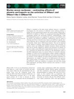
Among the 120 female patients who underwent elective
laparoscopic gynecological surgery, due to not meeting
inclusion criteria or refusing to participate, 116 patients
were included in this study, with 58 patients in each
group. Due to CO2 pneumoderma or changes in surgical
methods, 55 cases in each group were finally analysed
(Fig. 1). The general conditions of the patients are
shown in Table 1.
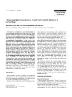
The comparison of the ONSD at each time point
between the two groups is shown in Table 2 and Fig. 2.
No significant difference in the baseline preoperative
ONSD was found between the two groups. After
anesthesia induction, the ONSD values all decreased
compared to the baseline value in both two groups, but
no significant differences between the two groups. At
three time points (Tawake, Tinduction, and T0), the ONSD
was not significantly different between the two groups
(p = 0.984, 0.666, and 0.646, respectively). Over time, at
Fig. 1 Flow diagram
Page 4 of 9
Table 1 Baseline Characteristics
P
Group P (n = 55)
Group S (n = 55)
Age (y)
40.53 ± 11.08
41.15 ± 10.26
0.762
Height (cm)
161.18 ± 4.20
159.79 ± 4.50
0.097
Weight (kg)
59 (54.5, 63)
56 (51.9, 60)
0.057
BMI (kg/m2)
22.74 ± 2.28
22.31 ± 2.15
0.262
Surgery duration (h)
2.33 (2.12, 2.75)
2.5 (2.22, 2.75)
0.165
Total blood loss (ml)
100 (50, 120)
80 (50, 100)
0.424
Urine volume (ml)
400 (300, 500)
400 (400, 500)
0.088
Fluid volume (ml)
1200 (1100, 1500)
1200 (1100, 1500)
0.645
Airway pressure (mmHg)
15 (13, 16)
15 (13, 16)
0.754
The quantitative data with a normal distribution were expressed as
mean ± standard deviation.
The quantitative data with a non-normal distribution were expressed as
median (interquartile range, IQR)
BMI Body mass index
three time points (T15, T30, and T45), significant differences in ONSD were identified between the two groups
(p = 0.031, 0.035, and 0.028, respectively). At T1 h, T2 h,
Tend, and Tpacu, no significant differences in ONSD were
found between the two groups after statistical correction
(p = 0.065, 0.211, 0.368, and 0.646 respectively).
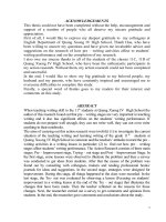
The comparison of the MAP and BIS at each time
point between the two groups is shown in Fig. 3 and
Fig. 4.There are no significant differences in MAP and
BIS at each time point between Group P and Group S.
No hypotension or serious neurological complications
such as cerebral haemorrhage or cerebral oedema occurred in either group.
Geng et al. BMC Anesthesiology
(2021) 21:30
Page 5 of 9
Table 2 Comparison of ONSD at different time points between
two groups
Group P (n = 55)
Group S (n = 55)
P
ONSD Tawake (mm)
4.39 ± 0.37
4.36 ± 0.45
0.694
ONSD TInduction (mm)
4.06 ± 0.45*
4.05 ± 0.45*
0.882
*#
*#
ONSD T0 (mm)
4.53 ± 0.47
4.35 ± 0.40
0.058
ONSD T15 (mm)
4.64 ± 0.48*#
4.50 ± 0.29*#
0.031
ONSD T30 (mm)
*#
4.77 ± 0.45
*#
4.62 ± 0.28
0.036
ONSD T45 (mm)
4.84 ± 0.46*#
4.65 ± 0.30*#
0.012
ONSD T1h (mm)
*#
4.83 ± 0.43
*#
4.66 ± 0.28
0.066
ONSD T2h (mm)
4.82 ± 0.41*#
4.71 ± 0.28*#
0.089
*#
*#
ONSD Tend (mm)
4.84 ± 0.44
4.71 ± 0.34
0.082
ONSD TPACU (mm)
4.40 ± 0.38*#
4.36 ± 0.35*#
0.641
Values were expressed as the mean ± standard variation. * meant comparison
of ONSD intra group at each time points to Tawake. In Group P, values of p
were 0.000 except comparison of Tpacu to Tawake which p value was 0.945; in
Group S, values of p were 0.939、0.035 and 0.884 when compared T0, T15 and
Tpacu to Tawake, while p values were 0.000 at any other time points.# meant
comparison of ONSD intra group at each time points to Tinduction, and all p
values were 0.000 in both two groups
Discussion
This study compared the effects of two general
anesthesia drugs (propofol and sevoflurane) on the
ONSD in patients undergoing laparoscopic gynecological
surgery. The results showed that the ONSD was significantly reduced compared to the baseline value in the patients in the two groups after anesthesia induction and
endotracheal intubation. With establishment of the CO2
pneumoperitoneum and the Trendelenburg position, the
ONSD in both groups increased and exceeded the baseline value. However, at first 45 min, the amplitude of the
increase in the propofol group was greater than that in
the sevoflurane group. Over time, considering the pneumoperitoneum and the Trendelenburg position, no significant difference in the ONSD was found between the
two groups from 1 h after starting the surgery to extubation at the end of the surgery. When leaving the recovery room, the ONSD returned to baseline in the patients
in both groups.
In this study, both groups received propofol and sufentanil for anesthesia induction, and cisatracurium was
used for endotracheal intubation. Both sufentanil and
propofol contract cerebral vessels and reduce the cerebral metabolic rate [9, 10, 23]; therefore, the ONSD after
anesthesia induction was reduced compared to the baseline value.
With establishment of the CO2 pneumoperitoneum
and the Trendelenburg position, the ONSD increased
gradually but recovered to baseline by 1 h after surgery.
The reason may be that the effect of the CO2 pneumoperitoneum and body position on intracranial pressure
exceeds the effect of drugs on intracranial blood flow.
Bilateral internal cervical vessels and vertebral vessels
play an important role in cerebral circulation [24]. After
establishment of the CO2 pneumoperitoneum and the
Trendelenburg position, recirculation in the internal
jugular vein and vertebral vein was obstructed. At the
same time, the mean arterial pressure during establishment of the pneumoperitoneum and positioning was elevated compared to that after anesthesia induction.
Whiteley et al. found that the ONSD was positively correlated with the mean arterial pressure [25].
For the propofol group, our results are basically
consistent with those of Blecha et al. [26]; however, the
amplitude of changes in the optic nerve sheath was
Fig. 2 Comparison of ONSD at each time points between two groups. × meant there were statistical differences between two groups at time
points T15, T30 and T45 (Values of p were 0.031, 0.035 and 0.028). Comparison of ONSD at other time points, values of p were 0.984, 0.666, 0.646,
0.065, 0.211, 0.368 and 0.646 at Tawake, Tinduction, T0, T1h, T2h, Tend and Tpacu
Geng et al. BMC Anesthesiology
(2021) 21:30
Page 6 of 9
Fig. 3 Comparison of MAP at each time points between two groups. There are no significant differences between two groups at each time
points (Values of p were 0.066, 0.312, 0.912, 0.156, 0.125, 0.064, 0.166, 0.095, 0.092 and 0.290). MAP Mean arterial pressure
higher than that in the study of Blecha et al., and maybe
the reason for the difference is that the patients in the
study of Blecha et al. received midazolam before surgery,
which can reduce intracranial pressure. In addition, the
patients in that study were from western countries, and
the ONSD varied among different races. Wang et al.
found that among the Chinese population, the predicted
cut-off value of ONSD that means intracranial pressure
higher than 20 cmH2O was lower than that in
Caucasians [27].
The effects of CO2 pneumoperitoneum and Trendelenburg position establishment on the ONSD in
sevoflurane versus propofol anesthesia are different in
various studies. We found that although the ONSD increased in the sevoflurane group, the amplitude of the
increase was smaller than that in the propofol group at
the early stage of surgery. In the study of Robba et al.
[22], the amplitude of the increase in the ONSD after
sevoflurane anesthesia was consistent with that in our
study. However, the studies of Kim et al. [28] and Chin
et al. [29] showed that the amplitude of the increase in
the ONSD after sevoflurane anesthesia was higher than
that in our study. Verdonck et al. [7] believed that the
ONSD remains unchanged in patients undergoing sevoflurane anesthesia.
The results of this study are generally consistent with
those of Lee et al. [30], while in the first 30 min of operation, the results were inconsistent. The main reason
may be that Lee et al. used midazolam and glinbromide
before operation; on the other hand, study of Lee et al.
only maintained BIS at 40–60 during the whole operation, but did not compare the BIS values between the
two groups. The depth of anesthesia may affect the
changes of cerebral blood flow, thus further affecting the
changes of intracranial pressure and ONSD value.
The inconsistencies across different studies may be
due to the lack of consistency in patients’ anesthetic
depth and sevoflurane blood concentration. Sevoflurane
has a dominant effect on cerebral oxygen metabolism at
a low concentration; while at medium and high
Fig. 4 Comparison of BIS at each time points between two groups. There are no significant differences between two groups at each time points
(Values of p were 0.093、0.065, 0.191, 1.000, 0.970, 0.503 and 0.368). BIS Bispectral index
Geng et al. BMC Anesthesiology
(2021) 21:30
concentrations, it has a direct vasodilatory effect [31].
Propofol reduces cerebral blood flow more because of its
effect on reducing cerebral oxygen metabolism rather
than direct vasoconstriction [32, 33]. Meanwhile, the
ONSD can reflect intracranial pressure in real time;
however, the correlation coefficient between ONSD and
intracranial pressure in previous studies was 0.660–0.820
[19, 34, 35]. Hansen et al. believed that the ONSD and
intracranial pressure have an elastic nonlinear relationship [17]. In other words, the ONSD may better reflect
changing trends in intracranial pressure than specific
values.
We found that although the ONSD increased significantly in both groups, it returned to baseline 1 h after
surgery. Animal studies suggest that with establishment
of the CO2 pneumoperitoneum and the Trendelenburg
position, intracranial pressure increased by 10 mmHg
compared to the baseline value [36]. However, Kalmar
et al. [37] believe that intracranial pressure fluctuations
within the physiological range are regulated by multiple
mechanisms, and that the intracranial pressure increases
exponentially only when these regulatory mechanisms
are exhausted. Notably, the brain has a strong ability to
transfer CSF to the vascular system, and when intracranial pressure increased, CSF moved intrathecally at a
rate of 2 ml/min [38, 39], which is also why the ONSD
returned to baseline 1 h after surgery. On the other way,
a reduction of the intracranial blood volume after termination of the steep Trendelenburg position could be
the reason.
Four limitations exist in this study. First, this study did
not analyse changes in the ONSD in patients with longer
operative times (> 3 h), mainly because few cases required long operative times (> 3 h) in this study. Hansen
et al. suggested that prolonged intracranial hypertension
affected the reversibility of optic nerve sheath changes
[17]. Therefore, we hypothesized that the duration of the
increase in ONSD in patients undergoing prolonged laparoscopic gynecological surgery would be prolonged, but
further studies are needed for confirmation. Second, all
the patients included in this study were female patients
younger than 65 years old, and further studies are
needed to determine whether similar conclusions can be
established for male or elderly patients. Third, in our
study, sevoflurane was maintained at 1–1.5 minimal alveolar concentration (MAC) while we do not record
values of end-tidal concentrations of sevoflurane
(Etsevo). Animal studies suggest that cerebral blood flow
may not be changed when Etsevo is 0.3–1.5MAC [40].
Artru et al. find ICP is not changed when sevoflurane is
0.5MAC, 1.0MAC or 1.5MAC in neurosurgery patients
[41]. We guess that small changes in Etsevo during 0.5–
1.5MAC may not have significant effects on cerebral
blood flow. Fourth, Whiteley et al. suggest that ONSD is
Page 7 of 9
positively correlated with MAP [25]. In our study, there
are no significant differences between Group S and
Group P at different time points and that is why we ignore the effect of blood pressure on ONSD.
In conclusion, we found that when comparing the
two drugs, at the early stage, the ONSD postpneumoperitoneum in the propofol group was slightly
larger than that in the sevoflurane group, and the difference was statistically significant. No significant difference was observed between the two groups 1 h
after surgery.
Abbreviations
ONSD: Optic nerve sheath diameter; ASA: American Society of
Anaesthesiologists; MAP: Mean arterial pressure; BIS: Bispectral index;
MAC: Minimal alveolar concentration; PACU: Post-anesthetic recovery room
Acknowledgements
Not applicable.
Authors’ contributions
WG, first author: Study Design, data collection, interpretation, drafting article,
critical revision of the article and final approval of the version to be
published. CC, co-first author: Study Design, references review, data analysis,
drafting article, critical revision of the article and final approval of the version
to be published. XS, second author: Study Design, critical revision of the
article and final approval of the version to be published. SH, correspondance
author: Study Design, data analysis, critical revision of the article and final
approval of the version to be published.
Funding
Not applicable.
Availability of data and materials
The trial protocol, datasets used and/or analysed during the current study
are available from the corresponding author on reasonable request.
Ethics approval and consent to participate
After approval by the ethics committee of Obstetrics and Gynaecology
Hospital of Fudan University. The committee’s reference number is 2018–
05.We herein confirm written consent obtained from each patient in
accordance with the Declaration of Helsinki in order to report and publish
the individual patient data obtained. A written consent to participate from
each patient in the current study was obtained.
Consent for publication
Not applicable.
Competing interests
The authors declare that they have no competing interests.
Author details
1
Department of Anesthesia, Obstetrics and Gynecology Hospital of Fudan
University, No.128, Shenyang RD, Yangpu district, Shanghai 200090, China.
2
Department of Emergency and Critical Care Medicine, Shanghai General
Hospital, Shanghai Jiao Tong University School of Medicine, Shanghai, China.
Received: 2 August 2020 Accepted: 11 January 2021
References
1. Fahy BG, Barnas GM, Nagle SE, Flowers JL, Njoku MJ, Agarwal M. Effects of
Trendelenburg and reverse Trendelenburg postures on lung and chest wall
mechanics. J Clin Anesth. 1996;8(3):236–44. />2. Halverson A, Buchanan R, Jacobs L, et al. Evaluation of mechanism of
increased intracranial pressure with insufflation. Surg Endosc. 1998;12:266–9.
/>
Geng et al. BMC Anesthesiology
3.
4.
5.
6.
7.
8.
9.
10.
11.
12.
13.
14.
15.
16.
17.
18.
19.
20.
21.
22.
(2021) 21:30
Lassen NA, Christensen MS. Physiology of cerebral blood flow. Br J Anaesth.
1976;48(8):719–34. />Pandey R, Garg R, Darlong V, Punj J, Chandralekha AK. Unpredicted
neurological complications after robotic laparoscopic radical cystectomy
and ileal conduit formation in steep trendelenburg position: two case
reports. Acta Anaesthesiol Belg. 2010;61:163–6 PMID: 21268573.
Cooke SJ, Paterson-Brown S. Association between laparoscopic abdominal
surgery and postoperative symptoms of raised intracranial pressure. Surg
Endosc. 2001;15:723–5. />Kim SH, Kim HJ, Jung KT. Position does not affect the optic nerve sheath
diameter during laparoscopy. Korean J Anesthesiol. 2015;68(4):358–63.
/>Verdonck P, Kalmar AF, Suy K, et al. Optic nerve sheath diameter
remains constant during robot assisted laparoscopic radical
prostatectomy. PLoS One. 2014;9:e111916. />journal.pone.0111916.
Conti A, Iacopino DG, Fodale V, Micalizzi S, Penna O, Santamaria LB. Cerebral
haemodynamic changes during propofol-remifentanil or sevoflurane
anesthesia: transcranial Doppler study under Bispectral index monitoring. Br
J Anaesth. 2006;97(3):333–9. />Oshima T, Karasawa F, Satoh T. Effects of propofol on cerebral blood flow
and the metabolic rate of oxygen in humans. Acta Anaesthesiol Scand.
2002;46:831–5. />Doyle PW, Matta BF. Burst suppression or isoelectric encephalogram for
cerebral protection: evidence from metabolic suppression studies. Br J
Anaesth. 1999;83:580–4. />Eng C, Lam AM, Mayberg TS, Lee C, Mathisen T. The influence of propofol
with and without nitrous oxide on cerebral blood flow velocity and CO,
reactivity in humans. Anesthesiology. 1992;77:872–9. />00000542-199211000-00006.
Matta BF, Lam AM, Strebel S, Mayberg TS. Cerebral pressure autoregulation
and CO-reactivity during propofol-induced EEG suppression. Br J Anaesth.
1995;74:159–63. />Drummond JC, Todd MM, Scheller MS, Shapiro HM. A comparison of the
direct cerebral vasodilating potencies of halothane and isoflurane in the
New Zealand white rabbit. Anesthesiology. 1986;65(5):462–7. />10.1097/00000542-198611000-00002.
Gupta S, Heath K, Matta BF. Effect of incremental doses of sevoflurane on
cerebral pressure autoregulation in humans. Br J Anaesth. 1997;79:469–72.
/>Choi SH, Lee SJ, Rha KH, Shin SK, Oh YJ. The effect of pneumoperitoneum
and Trendelenburg position on acute cerebral blood flow–carbon dioxide
reactivity under sevoflurane anesthesia. Anesthesia. 2008;63(12):1314–8.
/>Killer HE, Laeng HR, Flammer J, Groscurth P. Architecture of arachnoid
trabeculae, pillars, and septa in the subarachnoid space of the human optic
nerve:anatomy and clinical considerations. Br J Ophthalmol. 2003;87(6):777–
81. />Hansen HC, Lagrèze W, Krueger O, Helmke K. Dependence of the optic
nerve sheath diameter on acutely applied subarachnoidal pressure–an
experimental ultrasound study. Acta Ophthalmol. 2011;89(6):528–32. https://
doi.org/10.1111/j.1755-3768.2011.02159.x.
Maissan IM, Dirven PJ, Haitsma IK, Hoeks SE, Gommers D, Stolker RJ.
Ultrasonographic measured optic nerve sheath diameter as an accurate and
quick monitor for changes in intracranial pressure. J Neurosurg. 2015;123(3):
743–7. />Wang LJ, Yao Y, Feng LS, et al. Noninvasive and quantitative intracranial
pressure estimation using ultrasonographic measurement of optic nerve
sheath diameter. Sci Rep. 2017;7(2):42063. />Lochner P, Coppo L, Cantello R, et al. Intra- and interobserver reliability of
transorbital sonographic assessment of the optic nerve sheathdiameter and
optic nerve diameter in healthy adults. J Ultrasound. 2014;19(1):41–5.
/>Hassen GW, Bruck I, Donahue J, et al. Accuracy of optic nerve sheath
diameter measurement by emergency physicians using bedside
ultrasound. J Emerg Med. 2015;48(4):450–7. />jemermed.2014.09.060.
Robba C, Cardim D, Donnelly J, et al. Effects of pneumoperitoneum and
Trendelenburg position on intracranial pressure assessed using different
non-invasive methods. Br J Anaesth. 2016;117(6):783–9. />1093/bja/aew356.
Page 8 of 9
23. Werner C, Hoffman WE, Baughman VL, Albrecht RF, Schulte J. Effects of
sufentanil on cerebral blood flow, cerebral blood flow velocity, and
metabolism in dogs. Anesth Analg. 1991;72:177–81. />00000539-199102000-00006.
24. Smith KJ, Wong LE, Eves ND, et al. Regional cerebral blood flow distribution
during exercise: influence of oxygen. Respir Physiol Neurobiol. 2012;184(1):
97–105. />25. Whiteley JR, Taylor J, Henry M, Epperson TI, Hand WR. Detection of elevated
intracranial pressure in robot-assisted laparoscopic radical prostatectomy
using ultrasonography of optic nerve sheath diameter. J Neurosurg
Anesthesiol. 2015;27(2):155–9. />0000000000000106.
26. Blecha S, Harth M, Schlachetzki F, et al. Changes in intraocular pressure and
optic nerve sheath diameter in patients undergoing robotic-assisted
laparoscopic prostatectomy in steep 45° Trendelenburg position. BMC
Anesthesiol. 2017;17(1):40. />27. Wang L, Feng L, Yao Y, et al. Optimal optic nerve sheath diameter threshold
for the identification of elevated openingpressure on lumbarpuncture in a
Chinese population. PLoS One. 2015;10(2):e0117939. />journal.pone.0117939.
28. Kim MS, Bai SJ, Lee JR, Choi YD, Kim YJ, Choi SH. Increase in intracranial
pressure during carbon dioxide pneumoperitoneum with steep
trendelenburg positioning proven by ultrasonographic measurement of
optic nerve sheath diameter. J Endourol. 2014;28(7):801–6. />10.1089/end.2014.0019.
29. Chin JH, Seo H, Lee EH, et al. Sonographic optic nerve sheath diameter as a
surrogate measure for intracranial pressure in anesthetized patients in the
Trendelenburg position. BMC Anesthesiol. 2015;15:43. />1186/s12871-015-0025-9.
30. Lee YY, et al. Optic nerve sheath diameter changes during gynecologic
surgery in the Trendelenburg position: comparison of propofol-based total
intravenous anesthesia and sevoflurane anesthesia. Anesth Pain Med. 2019;
14:393–400.
31. Doe A, Kumagai M, Tamura Y, Sakai A, Suzuki K. A comparative analysis of
the effects of sevoflurane and propofol on cerebral oxygenation during
steep Trendelenburg position and pneumoperitoneum for robotic-assisted
laparoscopic prostatectomy. J Anesth. 2016;30(6):949–55. />1007/s00540-016-2241-y.
32. Holzer A, Winter W, Greher M, et al. A comparison of propofol and
sevoflurane anesthesia: effects on aortic blood flow velocity and middle
cerebral artery blood flow velocity. Anesthesia. 2003;58(3):217–22. https://
doi.org/10.1046/j.1365-2044.2003.03041.x.
33. Laaksonen L, Kallioinen M, Långsjö J, et al. Comparative effects of
dexmedetomidine, propofol, sevoflurane, and S-ketamine on regional
cerebral glucose metabolism in humans: a positron emission
tomography study. Br J Anaesth. 2018;121(1):281–90. />1016/j.bja.2018.04.008.
34. Raffiz M, Abdullah JM. Optic nerve sheath diameter measurement: a means
of detecting raised ICP in adult traumatic and non-traumatic neurosurgical
patients. Am J Emerg Med. 2017;35(1):150–3. />2016.09.044.
35. Padayachy LC, Padayachy V, Galal U, Gray R, Fieggen AG. The
relationship between transorbital ultrasound measurement of the optic
nerve sheath diameter (ONSD) and invasively measured ICP in
children: part I: repeatability, observer variability and general analysis.
Childs Nerv Syst. 2016;32(10):1769–78. />36. Tatebayashi K, Asai Y, Maeda T, Shiraishi Y, Miyoshi M, Kawai Y. Effects of
head-down tilt on the intracranial pressure in conscious rabbits. Brain Res.
2003;977(1):55–61. />37. Kalmar AF, Dewaele F, Foubert L, et al. Cerebral haemodynamic
physiology during steep Trendelenburg position and CO(2)
pneumoperitoneum. Br J Anaesth. 2012;108(3):478–84. />1093/bja/aer448.
38. Eklund A, Smielewski P, Chambers I, et al. Assessment of cerebrospinal fluid
outflow resistance. Med Biol Eng Comput. 2007;45(8):719–35. https://doi.
org/10.1007/s11517-007-0199-5.
39. Kalmar AF, De Ley G, Van Den Broecke C, Van Aken J, Struys MM, et al.
Influence of an increased intracranial pressure on cerebral and systemic
haemodynamics during endoscopic neurosurgery: an animal model. Br J
Anaesth. 2009;102(3):361–8.
Geng et al. BMC Anesthesiology
(2021) 21:30
40. Kimme P, Ledin T, Sjöberg F. Dose effect of sevoflurane and isoflurane
anesthetics on cortical blood flow during controlled hypotension in the pig.
Acta Anaesthesiol Scand. 2007;51(5):607–13.
41. Artru AA, Lam AM, Johnson JO, Sperry RJ. Intracranial pressure, middle
cerebral artery flow velocity, and plasma inorganic fluoride concentrations
in neurosurgical patients receiving sevoflurane or isoflurane. Anesth Analg.
1997;85(3):587–92.
Publisher’s Note
Springer Nature remains neutral with regard to jurisdictional claims in
published maps and institutional affiliations.
Page 9 of 9