Synthesis, spectroscopic characterization, and biological screening of binuclear transition metal complexes of bicompartmental Schiff bases containing indole and resorcinol
Bạn đang xem bản rút gọn của tài liệu. Xem và tải ngay bản đầy đủ của tài liệu tại đây (12 MB, 21 trang )
Turkish Journal of Chemistry
/>
Research Article
Turk J Chem
(2013) 37: 775 – 795
ă ITAK
c TUB
doi:10.3906/kim-1303-28
Synthesis, spectroscopic characterization, and biological screening of binuclear
transition metal complexes of bicompartmental Schiff bases containing indole and
resorcinol moieties
Mahendra Raj KAREKAL, Mruthyunjayaswamy BENNIKALLU HIRE MATHADA∗
Department of Studies and Research in Chemistry, Gulbarga University, Gulbarga, Karnataka, India
Received: 11.03.2013
•
Accepted: 13.04.2013
•
Published Online: 16.09.2013
•
Printed: 21.10.2013
Abstract: A series of binucleating Cu(II), Ni(II), and Zn(II) complexes of bicompartmental ligands with ONO donors
were prepared. The ligands were synthesized by the condensation of 5-substituted-3-phenyl-1 H -indole-2-carboxyhydrazides
and 4,6-diacetylresorcinol. The newly synthesized ligands and their complexes were characterized by elemental analysis
and various spectral studies like IR,
1
H NMR, ESI-mass, UV-Vis, ESR, thermal studies, magnetic susceptibility, molar
conductance, and powder-XRD data. All the complexes were binuclear and monomeric in nature. Cu(II) complexes have
octahedral geometry, whereas Ni(II) and Zn(II) complexes have square planar and tetrahedral geometry, respectively.
The redox property of the Cu(II) complex was investigated by electrochemical method using cyclic voltammetry. In
order to evaluate the effect of metal ions upon chelation, both the ligands and their metal complexes were screened for
their antibacterial and antifungal activities by minimum inhibitory concentration (MIC) method. The DNA cleaving
capacity of all the complexes was analyzed by agarose gel electrophoresis.
Key words: Indole Schiff bases, binuclear complexes, electrochemical, antimicrobial, DNA cleavage
1. Introduction
The indole structure represents a highly relevant heterocyclic system, since large numbers of indole-containing
synthetic and natural products such as vincristine, indole-micine, reserpine, mitomycin, dolasetron mesylate,
pindolol, indomethacin, and sumatriptan are being used as vital drugs in the treatment of various illnesses.
Large numbers of pharmacological compounds that contain indole nuclei have been reported to possess various
biological properties, viz., anti-inflammatory, 1−3 anticonvulsant, 4 antibacterial, 5 COX-2 inhibitory, 6,7 and
antiviral activities. 8 There are several reports that have described that indole-2-carbohydrazides and related
compounds exhibited MAO inhibitory, 9,10 antihistaminic, 11 and antidepressant activities. 12
The difunctional carbonyl compound 4,6-diacetylresorcinol acts as a precursor for the formation of
various binucleating ligands 13−15 and it is used as primary ligand in the synthesis of various mixed-ligand
complexes. 16,17 Difunctional 4,6-diacetylresorcinol is also employed in the construction of ligands containing
ONS donors by its condensation with various thiosemicarbazides and thiocarbohydrazides. 18 The ligands synthesized by the difunctional carbonyl compound are used to synthesize mono-, bi-, and poly-nuclear complexes
with different binding modes and their structural and functional features were explored in the development
of many biologically active compounds. Studies on binuclear metal complexes have stimulated interest owing
∗ Correspondence:
775
KAREKAL and BENNIKALLU HIRE MATHADA/Turk J Chem
to their unique physicochemical properties. These types of complexes have contributed to a better knowledge
of oxygen transport and activation by metalloenzymes such as hemocyanin (Cu 2 ) and cytochrome-C-oxidase
(CuFe) 19 as well as of some industrial catalytic processes. 20 Deoxyribonucleic acid (DNA) is the primary target
molecule for most antiviral therapies. Small molecule interactions with DNA continue to be intensely and widely
studied for their usefulness as probes of cellular replication and transcriptional regulation and for their potential
pharmaceutical properties. The ability of metallodrugs to bring about DNA-cleavage is an important criterion
in the development of metallodrugs as active chemotherapeutic agents. A number of transition metal complexes showed DNA-cleavage because of their redox behavior. In this study, 4,6-diacetylresorcinol was selected
as precursor for the construction of bicompartmental ligands. In spite of the extensive scientific literature on
Schiff base metal complexes with indole moiety, not much is known about bicompartmental Schiff bases derived
from indole moiety and their metal complexes. In view of these findings and in continuation of our research
work on pharmaceutically active indole molecules, 21−24 we report herein the synthesis, characterization, and
biological evaluation studies of 5-substituted-3-phenyl-1H -indole-2-carboxyhydrazides Schiff bases obtained by
the condensation of 5-substituted-3-phenyl-1H -indole-2-carboxyhydrazides and 4,6-diacetylresorcinol and their
metal complexes in order to obtain new classes of biologically active compounds.
2. Results and discussion
All the synthesized metal complexes are colored solids, amorphous in nature and stable in air. Melting points of
the newly synthesized metal complexes were above 300 ◦ C. The complexes are insoluble in water and common
organic solvents but are soluble in strong coordinating solvents like DMF and DMSO. Elemental analysis and
analytical data of the complexes (Table 1) suggest that the metal to ligand ratio of the complexes is 2:1
stoichiometry of the type [M 2 (L)(Cl) 2 (H 2 O) 4 ] for Cu(II) complexes and [M 2 (L)(Cl) 2 ] for Ni(II) and Zn(II)
complexes of both ligands (1 and 2), where L stands for deprotonated ligand. The molar conductance values
are too low to account for any dissociation of the complexes in DMF (25.23–41.44 Ω−1 cm 2 mol −1 ), indicating
the nonelectrolytic nature of the complexes in DMF. 25
2.1. IR spectral data
The important IR bands of the ligands and their metal complexes are represented in Table 2. In the IR spectra
of ligands 1 and 2, absorption due to phenolic OH exhibited bands at 3406 and 3416 cm −1 , while absorption
due to indole NH and CONH functions displayed bands at 3285 and 3184 cm −1 and 3258 and 3142 cm −1 ,
respectively. The phenolic C-O function of ligands 1 and 2 displayed absorption bands at 1233 and 1236 cm −1 ,
respectively. In both ligands, absorption bands due to carbonyl and azomethine functions appeared at 1657
and 1601 cm −1 and 1651 and 1542 cm −1 , respectively.
The absence of absorption bands due to phenolic OH groups at 3406 and 3416 cm −1 in the IR spectra
of Cu(II), Ni(II), and Zn(II) complexes of ligands 1 and 2 indicates the formation of bonds between metal ion
and phenolic oxygen atom via deprotonation. This is further confirmed by the increase in absorption frequency
of phenolic C-O, which appeared in the region 1258–1271 cm −1 and 1258–264 cm −1 , respectively, in the metal
complexes of both ligands in the present study. The absorption due to indole NH and CONH functions of
the above metal complexes of ligands 1 and 2 displayed bands in the region 3286–3278 cm −1 and 3178–3170
cm −1 and 3261–3242 cm −1 and 3157–3142 cm −1 respectively, which appeared in about the same region as in
the case of the respective ligands, thus confirming the noninvolvement of either indole NH or CONH function
in coordination with the metal ions. The absorption frequency of carbonyl and azomethine functions, which
776
C40 H30 N6 O4 Cl2
Cu2 [C40 H36 N6 O8 Cl4 ]
Ni2 [C40 H28 N6 O4 Cl4 ]
Zn2 [C40 H28 N6 O4 Cl4 ]
C42 H36 N6 O4
Cu2 [C42 H42 N6 O8 Cl2 ]
Ni2 [C42 H34 N6 O4 Cl2 ]
Zn2 [C42 H34 N6 O4 Cl2]
H 2 L1
[Cu2 (L1 )(Cl)2 (H2 O)4 ]
[Ni2 (L1 )(Cl)2 ]
[Zn2 (L1 )(Cl)2 ]
H 2 L2
[Cu2 (L2 )(Cl)2 (H2 O)4 ]
[Ni2 (L2 )(Cl)2 ]
[Zn2 (L2 )(Cl)2 ]
1
1a
1b
1c
2
2a
2b
2c
b
μ total the total magnetic moment of the complex.
μ eff where calculated for one metal ion in the complex.
a
Molecular formula
Ligand/complexes
886.78
873.38
955.08
688
926.78
913.38
995.08
728
Mol. wt.
13.30
(13.24)
13.43
(13.40)
14.74
(14.70)
-
12.77
(12.71)
12.85
(12.82)
14.11
(14.09)
–
M
H
4.12
(4.09)
3.61
(3.58)
3.06
(3.02)
3.02
(3.00)
5.23
(5.19)
4.39
(4.32)
3.89
(3.82)
3.83
(3.81)
C
65.93
(65.91)
48.23
(48.20)
52.55
(52.51)
51.79
(51.72)
73.25
(73.21)
52.77
(52.74)
57.70
(57.65)
56.83
(56.80)
11.53
(11.50)
8.44
(8.41)
9.19
(9.17)
9.06
(9.01)
12.20
(12.17)
8.79
(8.74)
9.61
(9.58)
9.47
(9.43)
N
7.32
(7.28)
8.01
(7.96)
7.89
(7.83)
–
9.61
(9.59)
14.06
(14.01)
15.32
(15.28)
15.10
(15.07)
Cl
Elemental analysis (%) Calcd (Found)
1.52
Dia
mag
Dia
mag
2.68
Dia
mag
Dia
mag
–
Dia
mag
Dia
mag
Dia
mag
Dia
mag
–
1.51
2.66
–
(BM)
(BM)
–
μ bef f
μ atotal
Mag. moment
Table 1. Physical, analytical, and magnetic susceptibility data of ligands 1 and 2 and their complexes.
Orange
Light
brown
Green
Yellowish
orange
Pale
yellow
Brown
Green
Pale
yellow
Color
KAREKAL and BENNIKALLU HIRE MATHADA/Turk J Chem
777
778
1
1a
1b
1c
2
2a
2b
2c
H 2 L1
[Cu2 (L1 )(Cl)2 (H2 O)4 ]
[Ni2 (L1 )(Cl)2 ]
[Zn2 (L1 )(Cl)2 ]
H 2 L2
[Cu2 (L2 )(Cl)2 (H2 O)4 ]
[Ni2 (L2 )(Cl)2 ]
[Zn2 (L2 )(Cl)2 ]
Ligands/complexes
3406
3416
-
νOH
3414
3400
-
νH2O
NH
(indole)
3285
3281
3286
3278
3258
3246
3242
3261
NH
(amide)
3184
3171
3170
3178
3142
3157
3143
3142
C=O
(carbonyl)
1657
1633
1614
1633
1651
1628
1614
1629
C=N
(azomethine)
1601
1584
1585
1585
1542
1518
1536
1538
Table 2. IR spectral data of ligands 1 and 2 and their complexes.
C-O
(phenolic)
1233
1271
1261
1258
1236
1258
1264
1259
540
590
509
590
536
555
M-O
470
439
462
470
439
493
M-N
273
285
266
289
289
293
M-Cl
KAREKAL and BENNIKALLU HIRE MATHADA/Turk J Chem
KAREKAL and BENNIKALLU HIRE MATHADA/Turk J Chem
appeared at 1657 and 1601 cm −1 and 1651 and 1542 cm −1 in the case of ligands 1 and 2, respectively, shifted
to lower frequency by 43–24 and 17–16 cm −1 and 37–22 and 24–4 cm −1 , respectively, in the complexes and
appeared in the region 1633–1614 and 1585–1584 cm −1 and 1629–1614 and 1538–1518 cm −1 , indicating the
involvement of the oxygen atom of carbonyl function as such without undergoing any enolization 26 and the
nitrogen atom of azomethine 27 function in complexation with the metal ions. This is further confirmed by the
appearance of new bands in the region 590–509 and 470–439 cm −1 and 590–536 and 493–439 cm −1 due to M-O
and M-N stretching vibrations 28 in all the complexes of ligands 1 and 2, respectively. The appearance of new
bands in the region 285–266 and 293–289 cm −1 in all the synthesized complexes was due to M-Cl bands. The
broad band due to the coordinated water molecule appeared at 3414 and 3400 cm −1 in the Cu(II) complexes
of ligands 1 and 2, respectively.
2.2.
1
H NMR spectral data
The
1
H NMR data of ligands 1 and 2 and their Zn(II) complexes are presented in Table 3. The
1
H NMR
spectra of ligands 1 (Figure 1) and 2 displayed singlets each at 12.62, 12.29, and 10.48 ppm and 12.62, 12.25,
and 10.25, ppm respectively, due to the 2 protons of amide NH, 2 protons of indole NH, and 2 OH protons of
ligands 1 and 2, respectively. The aromatic protons of ligands 1 and 2 resonated as multiplets in the region
6.35–7.57 ppm (m, 18H, ArH) and 6.35–7.55 ppm (m, 18H, ArH). Six protons of 2 methyl groups attached to
azomethine carbon atoms resonated as distinct singlets at 2.02 ppm and 1.99 ppm, respectively. Six protons of
2 methyl groups attached to the 5-position of 2 indole moieties of ligand 2 appeared as a distinct singlet at 2.63
ppm.
Table 3.
Ligands/Zn(II) complexes
1
(H2 L1 )
1c
[Zn2 (L1 )(Cl)2 ]
2
(H2 L2 )
2c
[Zn2 (L2 )(Cl)2 ]
1
H NMR data of Zn(II) complexes of ligands 1 and 2.
1
H NMR data (ppm)
12.62 (s, 2H, 2 CONH), 12.29 (s, 2H, 2 indole NH), 10.48 (s, 2H, 2 phenolic
OH), 6.35–7.57 (m, 18H, ArH), 2.02 (s, 6H, 2 CH3 )
12.63 (s, 2H, 2 CONH), 12.39 (s, 2H, 2 indole NH), 6.36–7.97 (m, 18H, ArH),
2.03 (s, 6H, 2 CH3 )
12.62 (s, 2H, 2 CONH), 12.25 (s, 2H, 2 indole NH), 10.25 (s, 2H, 2 phenolic
OH), 6.35–7.55 (m, 18H, ArH), 2.63 (s, 6H, 2 CH3 ), 1.99 (s, 6H, 2 CH3 )
12.69 (s, 2H, 2 CONH), 12.30 (s, 2H, 2 indole NH), 7.04–8.07 (m, 18H, ArH),
2.68 (s, 6H, 2 CH3 ), 2.00 (s, 6H, 2 CH3 )
In the case of Zn(II) complexes, the absence of a signal due to the proton of 2 phenolic OH groups confirms
the involvement of bonding of the phenolic oxygen atom to the metal ion via deprotonation. The signals at
12.63 and 12.69 ppm, 12.39 and 12.30 ppm, 6.36–7.97 and 7.04–8.07 ppm, and 2.03 and 2.00 ppm are due to
2 amide NH protons, 2 indole NH protons, aromatic protons, and 6 protons of 2 methyl groups attached to 2
azomethine carbon atoms in each of Zn(II) complexes of ligands 1 and 2, respectively. The singlet that appeared
at 2.68 ppm in the case of the Zn(II) complex of ligand 2 is due to 6 protons of 2 methyl groups attached to the
5-position of the indole moiety. A considerable degree of symmetry is present in these compounds so that the
protons in the 2 halves of the molecules are magnetically equivalent. When compared to the 1 H NMR spectra
of ligands 1 and 2 and their Zn(II) complexes, all the signals due to protons shifted downfield, confirming the
complexation of Zn(II) ions with the ligands. Thus the 1 H NMR data support the assigned structures.
779
KAREKAL and BENNIKALLU HIRE MATHADA/Turk J Chem
Figure 1.
1
H NMR spectrum of ligand 1.
2.3. ESI-mass spectral data
Ligand 1 and its Cu(II), Ni(II), and Zn(II) complexes were studied for their mass spectra. The ESI-mass
spectra of ligand 1 and its Cu(II), Ni(II), and Zn(II) complexes exhibited molecular ion peaks equivalent of
their molecular weight along with other fragmentation peaks. The representative mass spectrum of ligand 1
showed a molecular ion peak due to M +˙ 1 at m/z 729, 731, 733 (20%, 6%, 2.2%). This on loss of hydrogen
radical gave a peak at m/z 728, 730, 732 (10%, 3.2%, 9%), which is equivalent to its molecular weight. Further,
simultaneous loss of C 17 H 13 N 3 OCl radical, OH radical, and H radical gave a fragment ion peak at 400, 402
(100%, 33.3%), which is also a base peak. This fragmentation pattern (Scheme 1) is consistent with its structure.
The ESI-mass spectrum of Cu(II) complex (1a) (Figure 2) of ligand 1 exhibited a molecular ion peak
˙
+
at M 1 995, 997, 999 (21%, 7.2%, 2.3%) which corresponds to its molecular weight, which on loss of 2 water
molecules and a CH 3 radical gave a fragment ion peak recorded at m/z 944, 946, 948 (60%, 32.8%, 6.6%), which
on simultaneous loss of 2 water molecules, chloride radical, chlorine molecule, and 5-chloro-3-pheny-indole-2-yl
radical gave a fragment ion peak recorded at m/z 577 (10.3%). This on further loss of C 4 H 3 radical, CO
molecule, CH 3 radical, 1 hydrogen molecule, and 2 hydrogen radicals gave a fragment ion peak recorded at
m/z 479 (100%), which is also a base peak. This fragmentation pattern (Scheme 2) is in conformity with the
structure of the complex.
Similarly, the mass spectra of Ni(II) and Zn(II) complexes of ligand 1 exhibited a molecular ion peak at
˙
+
M 1 913, 915, 917 (17%, 5.83%, 1.8%) and 926, 928, 930 (31.2%, 10.4%, 3.46%), which corresponds to their
molecular weight. The fragmentation pattern of both complexes is depicted in Schemes 3 and 4, respectively.
780
KAREKAL and BENNIKALLU HIRE MATHADA/Turk J Chem
Ph
Cl
N
H
C
O
Ph
H
N
CH3
CH3
N
N
HO
H
N
Cl
C
O
OH
N
H
(1) M+1 729 ( 20% ), M+2 731 ( 6% ), M+3 733 (2.2% )
-H
Ph
Cl
N
H
C
O
Ph
H
N
CH3
CH3
N
N
HO
H
N
OH
Cl
C
O
N
H
m/z 728 (10%), 730 (3.2%), 733(.9% )
Ph
Cl
N
H
C
O
H
N
N
CH3
C
- OH
-H
Ph
CH3
H
C
N
C
N
O
O
Cl
N
H
m/z 400 (100%), 402 (33.3%)
Scheme 1. Fragmentation pattern of ligand 1.
2.4. Electronic spectral and magnetic susceptibility data
The electronic absorption spectra of Cu(II) and Ni(II) complexes of ligands 1 and 2 were recorded in distilled
DMF (10 −3 M) at room temperature. The band positions of absorption band maxima assignments are listed in
Table 4. The electronic spectra of the Cu(II) complexes of ligands 1 and 2 showed 1 low intensity broad band
and 1 high intensity band each around 627.27 (15,942.10 cm −1 ) and 393.34 (25,423.30 cm −1 ) nm and 630.25
(15,866.72 cm −1 ) and 392.88 (25,453.06 cm −1 ) nm, respectively. The low intensity broad band is assignable
to 2 T 2g ← 2E g transition and the high intensity band observed is due to symmetry forbidden ligand → metal
charge transfer. Based on the electronic spectral data, distorted octahedral geometry around Cu(II) ion is
suggested. 29,30 This was further supported by their magnetic susceptibility measurements. The total magnetic
moment values ( µT otal ) of Cu(II) complexes of ligands 1 and 2 are 2.66 and 2.68, respectively. The calculated
µef f value for each Cu(II) complex is 1.51 and 1.52 (magnetic moment for 1 metal ion) due to the 2 adjacent
781
KAREKAL and BENNIKALLU HIRE MATHADA/Turk J Chem
Cu(II) ions having 1 electron each possessing an antiferromagnetic interaction between them. Thus, based on
the above data, these Cu(II) ions achieve octahedral geometry by the addition of 2 water molecules 31 and this
was further confirmed by the thermal studies.
Figure 2. ESI-mass spectrum of Cu(II) complex (1a).
The electronic spectra of the Ni(II) complexes of ligands 1 and 2 displayed 2 bands each at 710.0
(14,084.51 cm −1 ) and 460.0 (21,739.13 cm −1 ) nm and 710.80 (14,068.66 cm −1 ) and 460.60 (21,710.81 cm −1 )
nm, which are assignable to
1
A 1g →
1
E g and
1
A 1g →1 B 2g transitions, respectively. Since these complexes
are diamagnetic in nature, a square-planar geometry is suggested for the Ni(II) complexes. 32,33
2.5. ESR spectral studies of the Cu(II) complexes of ligands 1 and 2
To obtain information about the hyperfine and superhyperfine structure in order to elucidate the geometry of
the complex and the site of the metal–ligand bonding or environment around the metal ion the X-band ESR
782
KAREKAL and BENNIKALLU HIRE MATHADA/Turk J Chem
Ph
Cl
N
H
C
O
Ph
H
N
CH3
N
H
N
N
O
Cu
H2O
CH3
O
C
O
N
H
Cu
H2O
H2O
Cl
Cl
Cl
H2O
(1a) M 995 (21%), 997 (7.2%), 999 (2.3%)
- 2H2O
- CH3
Ph
Cl
N
H
C
O
Ph
H
N
CH3
C
N
N
O
Cu
O
H
N
Cl
C
O
N
H
Cu
H2O
Cl H2O
Cl
m/z 944 (60%), 946 (32.8%), 948 (6.6%)
Ph
Cl
- 2H2O
- Cl
- Cl2
Ph
N
H
C
O
H
N
N
N
H
CH3
C
N
N
O
O
Cu
Cu
m/z 577 (10.3%)
- HC C C C H
- CO H
- CH 3
- H2
-H
-H
N
H
C
O
N
N
C
C
C
O
H
N
NH
O
O
Cu
Cu
m/z 479 (100%)
Scheme 2. Fragmentation pattern of Cu(II) complex (1a).
783
KAREKAL and BENNIKALLU HIRE MATHADA/Turk J Chem
spectra of Cu(II) complexes [Cu 2 (L 1 )(Cl) 2 (H 2 O) 4 ] (1a) and [Cu 2 (L 2 )(Cl) 2 (H 2 O) 4 ] (2a) were recorded in
the polycrystalline state at room temperature at a frequency of 9.387 GHz with a field set of 3950 G and the
spectral data are given in Table 4. The spin Hamiltonian parameters for the Cu(II) complex were used to derive
the ground state. In octahedral geometry, for the g-tensor parameter with g|| > g⊥ > 2.0023, the unpaired
electron lies in the dx2−y2 orbital in ground state and with g⊥ > g|| > 2.0023, the unpaired electron lies in
Ph
Cl
N
H
C
O
Ph
H
N
CH3
CH3
N
N
O
Ni
O
H
N
Cl
N
H
C
O
Ni
Cl
Cl
(1b) M 913 (17%), 915 (5.83%), 917 (1.8%)
-Cl2
- Cl
- 2H 2
-H
-H
CH3
N
H
C
O
N
N
Ni
Cl
CH3
N
O
O
N
N
H
C
O
Ni
m/z 802 (23.7%), 804 (7.9%)
N
H
H
C
Cl
N
-H
CH3
N
C
O
CH3
C
N
N
Ni
N
O
O
Ni
C
O
m/z 400 (100%)
Scheme 3. Fragmentation pattern of Ni(II) complex (1b).
784
KAREKAL and BENNIKALLU HIRE MATHADA/Turk J Chem
the d2z orbital. 34 The observed measurements for Cu(II) complexes, [Cu 2 (L 1 ) (Cl) 2 (H 2 O) 4 ] (1a), g|| (2.43)
> g⊥ (2.39) > 2.0023 and [Cu 2 (L 2 )(Cl) 2 (H 2 O) 4 ] (1b), g|| (2.44) > g⊥ (2.32) > 2.0023, indicate that the
complexes are axially symmetric and the copper site has a d x2−y2 ground state characteristic of octahedral
geometry for both complexes. 35 The g|| value is an important function for indicating the covalent character of
metal–ligand bond, for ionic g|| < 2.3 and for covalent characters g|| > 2.3, respectively. 36 In the present Cu(II)
complexes the g|| values are more than 2.3, indicating an appreciable covalent character for the metal–ligand
bond. The geometric parameter (G), which is the measure of extent of exchange interaction, is calculated by
Ph
Cl
N
H
Ph
H
N
C
O
CH3
CH3
N
Zn
N
O
O
H
N
Cl
N
H
C
O
Zn
Cl
Cl
(1c) M 926 (31.2%), 928 (10.4%), 930 (3.4%)
Ph
Cl
- Cl2
N
H
C
O
Ph
Cl
N
N
H
NCO
- CH3
Ph
CH3
HN
Zn
C
O
H
N
N
O
C
O
Cl
N
H
Zn
m/z 573 (37.5%), 575 (12.5%)
- C6 H6
- Cl
-H
CH3
N
Zn
N
C
O
N
O
C
O
N
H
Zn
m/z 459 (100%)
Scheme 4. Fragmentation pattern of Zn(II) complex (1c).
785
KAREKAL and BENNIKALLU HIRE MATHADA/Turk J Chem
using g-tensor values by the expression G = g|| − 2/g⊥ − 2. According to Hathaway, 37 if the G value is greater
than 4, the exchange interaction between the copper centers is negligible, whereas if its value is less than 4 the
exchange interaction is noticed. The calculated G-values for the present Cu(II) complexes are 1.116 (1a) and
1.366 (1b), indicating some interaction between Cu(II) centers in solid complex. 38
Table 4. Electronic and EPR data of Cu(II) and Ni(II) complexes of ligands 1 and 2.
Complexes
1a
[Cu2 (L1 )(Cl)2 (H2 O)4 ]
1b
[Ni2 (L1 )(Cl)2 ]
2a
[Cu2 (L2 )(Cl)2 (H2 O)4 ]
2b
[Ni2 (L2 )(Cl)2 ]
Electronic spectral
λmax in nm
(cm−1 )
627.27 (15,942.10)
393.34 (25,423.30)
710.0 (14,084.51)
460.0 (21,739.13)
630.25 (15,866.72)
392.88 (25,453.06)
710.80 (14,068.66)
460.60 (21,710.81)
data
Band
assignments
2
Tg ←2 Eg
L→M
1
A1g →1 Eg
1
A1g →1 B2g
2
Tg ←2 Eg
L→M
1
A1g →1 Eg
1
A1g →1 B2g
Geometry
Distorted
octahedral
Square
planar
Distorted
octahedral
Square
planar
ESR spectral data
g⊥
g||
gavg
G
2.392
2.438
2.408
1.116
-
-
-
-
2.322
2.440
2.361
1.366
-
-
-
-
2.6. Thermal studies
The thermal stabilities were investigated for the Cu(II), Ni(II), and Zn(II) complexes of ligand 1 as a function
of temperature. The proposed stepwise thermal degradation of the complexes with respect to temperature and
the formation of respective metal oxides are given in Table 5. The thermogravimetric curve of Cu(II) complex
shows that the complex is stable up to 192 ◦ C and no weight loss occurs before this temperature. The first stage
of decomposition represents weight loss of 4 coordinated water molecules and a methyl group at 192.8 ◦ C with
practical weight loss of 9.01% (Cald. 8.74%). The resultant complex underwent a second stage of degradation
and gave a break at 350
◦
C with a practical weight loss of 61.39% (Cald. 60.89%), which corresponds to the
decomposition of 2 indole moieties (2C 15 H 10 N 2 OCl) and a methyl group. Further, the complex underwent a
third stage of decomposition and gave a break at 500 ◦ C with a weight loss of 20.49% (Cald. 19.71%), due
to loss of 2 chlorine atoms. Thereafter, the compound showed a gradual decomposition rather than a sharp
decomposition up to 800 ◦ C and onwards due to the loss of the remaining organic moiety. The weight of the
residue corresponds to 2 moles of cupric oxide.
In the thermogram of the Ni(II) complex, the first stage of decomposition represents the weight loss of
2 Cl 2 molecules at 392
◦
C with a practical weight loss of 15.16% (Cald. 15.32%). The complex underwent
further degradation and gave a break at 550
◦
C with a practical weight loss of 64.77% (Cald. 64.39%), which
corresponds to the decomposition of 2 indole moieties (2C 15 H 10 N 2 O). Thereafter, the compound showed a
gradual decomposition up to 850 ◦ C with a weight loss of remaining organic moiety. The weight of the residue
corresponds to 2 moles of nickel oxide. In the case of Zn(II) complex, the first stage of decomposition occurs
at 342.8
◦
C with a practical weight loss of 68.15% (Cald. 68.84%), which represents the loss due to 2 indole
moieties (2C 15 H 10 N 2 OCl), 2 chlorine atoms, and 2 methyl groups. Thereafter, the compound showed a gradual
decomposition up to 800 ◦ C with the weight loss of the remaining organic moiety. The weight of the residue
corresponds to 2 moles of zinc oxide. The percentage metal content in all the complexes as done by elemental
analysis agrees well with the thermal studies.
786
KAREKAL and BENNIKALLU HIRE MATHADA/Turk J Chem
Table 5. Thermal data of the complexes of ligand 1.
Complex
No.
1a
1b
1c
Decomposition
temp (◦ C)
192
% Weight loss
Obsd Cald
9.01
8.74
Metal oxide%
Obsd Cald
–
–
350
61.39
60.89
–
–
500
Up to 900
392
550
Up to 850
342
20.49
–
15.16
64.77
–
68.15
19.71
–
15.32
64.39
–
68.84
–
15.98
–
–
16.38
–
–
15.65
–
–
16.35
–
Up to 800
–
–
17.07
17.56
Inference
Loss of coordinated water molecules and CH3
group
Loss due to 2 indole (2C15 H10 N2 OCl)
molecules and CH3 group
Loss due to 2 chlorine atoms
Loss due to remaining organic moiety
Loss due to 2 chlorine molecules
Loss due to 2 indole (2C15 H10 N2 O) molecules
Loss due to remaining organic moiety
Loss due to 2 indole (2C15 H10 N2 OCl)
molecules, 2 chlorine atoms and 2 CH3 groups.
Loss due to remaining organic moiety
2.7. Powder X-ray diffraction (XRD) studies
Although the synthesized metal complexes were soluble in some polar organic solvents (DMSO and DMF),
crystals that are suitable for single-crystal studies were not obtained. Powder XRD patterns of Cu(II), Ni(II),
and Zn(II) complexes of ligand 1 were studied in order to test the degree of crystallinity of the complexes.
Powder XRD pattern for Cu(II) complex (1a) showed 12 reflections in the range of 3–80 ◦ (2 θ), which arose
from diffraction of X-ray by the planes of complex. The interplanar spacing (d) was calculated by using Bragg’s
equation, n λ = 2d sin θ . The calculated interplanar d-spacing together with relative intensities with respect to
the most intense peak was recorded and is given in Table 6. The unit cell calculations were calculated for cubic
symmetry from all the important peaks and h2 + k 2 + l2 values were determined. The observed interplanar
d-spacing values were compared with the calculated ones and they were found to be in good agreement. The
h2 + k 2 + l2 values were 1, 10, 29, 32, 50, 53, 65, 72, 99, 110, 120, and 161. The presence of forbidden number
120 indicates the Cu(II) complex may belong to hexagonal or tetragonal systems.
Table 6. Powder X-ray data of Cu(II) complex of ligand 1(1a).
Peak
2θ
θ
Sinθ
Sin2 θ
1000
Sin2 θ
1
2
3
4
5
6
7
8
9
10
11
12
4.934
15.716
26.834
28.120
35.506
36.600
40.702
42.821
50.654
53.722
56.131
66.179
2.467
7.858
13.417
14.060
17.753
18.300
20.351
21.410
25.327
26.861
28.065
33.089
0.0430
0.1367
0.2320
0.2429
0.3049
0.3139
0.3477
0.3650
0.4277
0.4518
0.4704
0.5459
0.00184
0.01868
0.05382
0.05900
0.09296
0.09853
0.12089
0.13322
0.18292
0.20412
0.22127
0.29800
1.849
18.68
53.82
59.00
92.96
98.53
120.89
133.22
182.92
204.12
221.27
298.00
1000
Sin2 θ/CF
(h2 + k2 + l2 )
1.00(1)
10.102(10)
29.107(29)
31.909(32)
50.275(50)
53.288(53)
65.381(65)
72.049(72)
98.929(99)
110.394(110)
119.670(120)
161.168(161)
d
hkl
(100)
(310)
(520), (432)
(440)
(550), (710)
(641)
(810)
(660)
(933)
(952)
–
(984)
Obs
Cal
17.897
5.634
3.319
3.170
2.526
2.453
2.214
2.110
1.800
1.704
1.637
1.410
17.906
5.632
3.318
3.170
2.525
2.453
2.214
2.109
1.800
1.704
1.637
1.410
a in
˚
A
17.90
17.90
17.90
17.90
17.90
17.91
17.92
17.90
17.90
17.90
17.90
17.90
787
KAREKAL and BENNIKALLU HIRE MATHADA/Turk J Chem
Similar calculations were performed for Ni(II) and Zn(II) complexes of ligand 1. The Ni(II) complex
showed 15 reflections in the range 3–80 ◦ (2θ) and the Zn(II) complex showed 7 reflections in the range 0–80 ◦
(2θ) . The important peaks of both complexes were indexed and the observed interplanar d-spacing values
were compared with the calculated ones. The unit cell calculations were performed for a cubic system and the
h2 + k 2 + l2 values were determined for both complexes. The presence of forbidden number 7 for the Ni(II)
complex indicates that it may belong to hexagonal or tetragonal systems. Similarly, for the Zn(II) complex, the
absence of forbidden numbers (7, 15, 23 etc.) indicates that the complex has cubic symmetry. The calculated
lattice parameters were a = b = c = 12.93 ˚
A.
2.8. Electrochemistry
The electrochemical behavior of the Cu(II) complex (1a) was investigated in DMF (10 −3 M) solution containing
0.05 M n-Bu 4 N-ClO 4 as a supporting electrolyte by cyclic voltammetry. It is the most versatile electroanalytical
technique for the study of electroactive species. The cyclic voltammogram of the Cu(II) complex (1a) (Figure
3) in DMF at a scan rate of 50 mV/s shows a well-defined redox process corresponding to the formation of
Cu(II)/Cu(I) couple at E pa = –0.5925 V and E pc = –1.0123 V versus Ag/AgCl. The peak separation of this
couple is found to be quasi-reversible with ∆Ep = 0.4198 V and the ratio of anodic to cathodic peak height
was less than 1. The difference between forward and backward peak potential can provide a rough evaluation
of the degree of the reversibility of the one-electron transfer reaction. Thus, the analysis of cyclic voltammetric
response to 50 mV/s, 100 mV/s, and 200 mV/s scan rates gives evidence for a quasi-reversible one-electron
redox process. The ratio of anodic to cathodic peak height was less than 1 and peak current increases with the
increase in the square root of the scan rates, establishing a diffusion-controlled electrode process. 39 From the
peak separation value ∆E p and peak potential increases with higher scan rates, therefore we can suggest that
the electrode process is consistent with the quasi-reversibility of the Cu(II)/Cu(I) couple. 40
Figure 3. Cyclic voltammogram of Cu(II) complex (1a).
2.9. Pharmacological activity results
2.9.1. In vitro antimicrobial activity
The synthesized ligands 1 and 2, and their metal complexes were screened for their antimicrobial activity.
The antibacterial activity was tested against E. coli, S. typhi, B. subtilis, and S. aureus strains and antifungal
788
KAREKAL and BENNIKALLU HIRE MATHADA/Turk J Chem
activity against C. albicans, C. oxysporum, and A. niger strains. The minimum inhibitory concentration (MIC)
values of the compounds against the respective strains are summarized in Table 7. The antimicrobial screening
results of all the synthesized compounds exhibited antimicrobial properties, and it is important to note that the
metal complexes exhibited a more inhibitory effect compared to their respective parent ligands. The enhanced
activity of the complexes over the ligands can be explained on the basis of chelation theory. 41,42 It is known that
chelation makes the ligand a more powerful and potent bactericidal agent, thus killing more of the bacteria than
the ligand. The enhancement in the activity may be rationalized on the basis that ligands mainly possess an
azomethine (C = N) bond. It has been suggested that ligands with hetero donor atoms (nitrogen and oxygen)
inhibit enzyme activity, since the enzymes that require these groups for their activity appear to be especially
more susceptible to deactivation by metal ions on coordination. It is observed that, in a complex, the positive
charge of the metal ion is partially shared with the hetero donor atoms (nitrogen and oxygen) present in the
ligand, and there may be π -electron delocalization over the whole chelating system. 43,44 Thus the increase in
the lipophilic character of the metal chelates favors their permeation through the lipoid layer of the bacterial
membranes and blocking of the metal binding sites in the enzymes of microorganisms. Other factors, namely
solubility, conductivity, and bond length between the metal ion and the ligand, also increase the activity. The
increase in the activity of metal complexes against fungi is due to the formation of a hydrogen bond between the
azomethine nitrogen atom and active centers of the cell constituents, resulting in interference with the normal
cell process.
Table 7. Minimum inhibitory concentration (MIC µ g mL −1 ) of ligands and their metal complexes.
Compound
1
1a
1b
1c
2
2a
2b
2c
Gentamicin
Fluconazole
Zone of
E. coli
50
12.50
25
12.50
50
12.50
25
25
12.50
-
inhibition against bacteria (mm)
S. aureus B. subtilis S. typhi
75
50
100
25
25
50
50
12.50
50
25
25
50
50
75
75
12.50
12.50
25
12.50
25
50
25
25
50
12.50
12.50
12.50
-
Zone of inhibition against fungi (mm)
C. albicans C. oxysporum A. niger
75
50
50
25
12.50
12.50
12.50
25
25
25
12.50
25
25
12.50
25
12.50
25
12.50
25
25
12.50
12.50
25
25
12.50
12.50
12.50
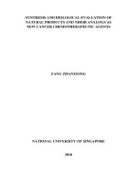
2.9.2. DNA cleavage activity
Ligand 1 and its Cu(II), Ni(II), and Zn(II) complexes, and ligand 2 and its Cu(II) complex were studied for
their DNA cleavage activity by agarose gel electrophoresis against calf-thymus DNA (Cat. No. 105850) and
the gel picture showing cleavage is depicted in Figure 4.
DNA-cleavage studies are used for rational design and to construct new and more efficient drugs that are
targeted to DNA. 45 The cleavage efficiency of all the compounds compared to the control is due to their efficient
DNA-binding ability, which is observed by diminishing of the intensity of the lanes. The DNA-cleavage study by
electrophoresis analysis clearly revealed that the lane ligand 1 and its Zn(II) complex showed partial cleavage,
whereas lane Cu(II) and Ni(II) complexes of ligand 1, ligand 2 and its Cu(II) complex showed complete cleavage
of DNA. The difference was observed in the bands of lanes of compounds compared with the control DNA of
calf-thymus. This shows that the control DNA alone does not show any apparent cleavage, whereas the ligands
789
KAREKAL and BENNIKALLU HIRE MATHADA/Turk J Chem
and metal complexes do. The result indicates the important role of the coordination of nitrogen and oxygen
to the metal ion in these isolated DNA cleavage reactions. On the basis of the cleavage of DNA observed in
the case of ligands 1 and 2 and their Cu(II) and Ni(II) and Zn(II) and Cu(II) complexes, respectively, it can
be concluded that all the compounds in the present study inhibit the growth of pathogenic organism by DNA
cleavage as was observed on the DNA cleavage of calf-thymus.
Figure 4. DNA cleavage of calf-thymus DNA. M, standard molecular weight marker; C, control. Lane 1, 1a, 1b, 1c,
2, and 2a were treated DNA of calf-thymus DNA genome with respective compounds.
Based on these studies, the newly synthesized binuclear ligands and their complexes were characterized by
various spectral studies and analytical data. The coordinating ability of the ligands was proved in complexation
reactions with Cu(II), Ni(II), and Zn(II) ions. In all the complexes both ligands act as a tridentate chelate around
the metallic ion with 2 compartments and provide ONO donating sites to each metal ion in both compartments.
Cu(II) complexes of both ligands have octahedral geometry, whereas Ni(II) and Zn(II) complexes of both the
ligands possess square planar and tetrahedral geometries, respectively. The Cu(II) complex of ligand 1 exhibits
one-electron transfer quasi-reversible redox activity in the applied potential range. The antimicrobial activity
results show that all the complexes exhibited higher activity when compared to their respective ligands. The
DNA cleavage studies revealed that the metal complexes showed good efficiency towards DNA cleavage. Based
on the analytical data and spectral studies, the proposed structures of all the complexes are depicted in Figure
5.
3. Experimental
3.1. Analysis and physical measurements
IR spectra of the newly synthesized compounds were recorded as KBr pellets on a PerkinElmer FT-IR instrument
in the region 4000–350 cm −1 .
1
H NMR spectra of the Zn(II) complexes were recorded in d6 -DMSO using a
Bruker DRX-400 MHz instrument. UV-visible spectra of the Cu(II) and Ni(II) complexes were recorded on an
Elico-SL 164 double beam spectrometer in the range 200–1000 nm in DMF solution (1 × 10 −3 M). Elemental
analysis was obtained from a HERAEUS C, H, N-O rapid analyzer and metal analysis was carried out by
following the standard methods. ESI-MS was recorded on an Agilent 6330 Ion trap-mass spectrophotometer.
790
KAREKAL and BENNIKALLU HIRE MATHADA/Turk J Chem
ESR measurements of Cu(II) complexes in polycrystalline state were obtained on a BRUKER Bio Spin Gmbh
spectrometer at a microwave frequency of 9.903 GHz. The experiment was carried out by using DPPH as
reference with the field set at 3950 G. Electrochemistry of the Cu(II) complex was recorded on a 600 D series
model electrochemical analyzer in DMF using n -Bu 4 N-ClO 4 as a supporting electrolyte. Powder-XRD of the
complexes was recorded using a Bruker AXS D8 Advance (Cu, wavelength 1.5406 ˚
A source). Molar conductivity
measurements were recorded on an ELICO CM-180 conductivity bridge in dry DMF (10 −3 M) solution using a
dip-type conductivity cell fitted with a platinum electrode, and the magnetic susceptibility measurements were
made at room temperature on a Gouy balance using Hg[Co(NCS) 4 ] as the calibrant.
Ph
R
N
H
C
O
Ph
CH3
H
N
N
N
O
Cu
H2O
CH3
H2O
Cl
O
H
N
C
O
R
N
H
Cu
H2O
Cl
H2O
R= Cl, CH3
Ph
R
N
H
C
O
Ph
CH3
H
N
CH3
N
N
O
O
H
N
M
M
Cl
Cl
C
O
R
N
H
R= Cl, CH3
Where, M= Ni or Zn
Figure 5. Suggested structure for Cu(II), Ni(II), and Zn (II) of ligand 1 and 2.
3.2. Methods
All the chemicals used were of reagent grade and procured from Hi-media and Sigma Aldrich. The solvents were
dried and distilled before use. Melting points of the newly synthesized compounds were determined by electrothermal apparatus using open capillary tubes. The metal and chloride contents of the metal complexes were
determined as per standard procedures. 46 5-Substituted-3-phenyl-1H -indole-2-carboxyhydrazide was prepared
by the literature method. 47 The 4,6-diacetyl resorcinol was procured from Sigma Aldrich.
3.2.1. Synthesis of ligands 1 and 2
A mixture of 5-substituted-3-phenyl-1H -indole-2-carboxyhydrazide (0.002 mol) and 4,6-diacetylresorcinol (0.001
mol) with a catalytic amount of glacial acetic acid (1–2 drops) in ethanol (20 mL) was refluxed on a water bath
for about 7–8 h. The reaction was monitored by TLC. The pale yellow colored solid separated was filtered,
washed with a little ethanol, dried, and recrystallized from dioxane (Scheme 5).
791
KAREKAL and BENNIKALLU HIRE MATHADA/Turk J Chem
CH3
CH3
Ph
R
2
N
H
C
O
H
N
O
NH2
O
HO
OH
AcOH/ EtOH
Ph
R
N
H
1
C
O
Ph
H
N
CH3
N
HO
CH3
N
OH
H
N
C
O
R
N
H
2
R= Cl, CH3
Scheme 5. Synthesis of ligands 1 and 2.
Mol. For. = C 40 H 30 N 6 O 4 Cl 2 , mp = 305 ◦ C, yield = 71% (Schiff base 1)
Mol. For. = C 42 H 36 N 6 O 4 , mp = 310 ◦ C, yield = 68% (Schiff base 2)
3.2.2. Synthesis of Cu(II), Ni(II), and Zn(II) complexes of Schiff bases 1 and 2
To a hot solution of 5-substituted-N ’-(1-(5-1-(2-(5-substituted-3a,7a-dihydro- 1H -indole-2-carbonyl)hydrazono)ethyl)2,4-dihydroxyphenyl)ethylidene)-1H -indole-2-carbohydrazide (1 and 2) (0.001 mol) in ethanol (30 mL) was
added a hot ethanolic solution (15 mL) of respective metal chlorides (0.002 mol). The reaction mixture was
then refluxed on a water bath for about 4–5 h. An aqueous alcoholic solution of sodium acetate (0.5 g) was
added to the reaction mixture to maintain a neutral pH and refluxing was continued for about 1 h more. The
reaction mixture was poured into distilled water and the separated solid complexes were collected by filtration,
washed with a sufficient quantity of distilled water, then with hot ethanol to apparent dryness, and dried in a
vacuum over anhydrous calcium chloride in a desiccator.
3.3. Pharmacological activity
3.3.1. Antimicrobial assays
The biological activities of the synthesized Schiff bases 1 and 2 and their Cu(II), Ni(II), and Zn(II) complexes
were studied for their antibacterial and antifungal activities by the disk and well diffusion methods, respectively.
The in vitro antibacterial activities of the compounds were tested against 2 gram-negative (E. coli and S. typhi )
and 2 gram-positive (B. subtilis and S. aureus) bacteria. The in vitro antifungal activities were tested against
C. albicans, C. oxysporum, and A. niger. 48,49 Stock solutions of the test chemicals (1 mg mL −1 ) were prepared
by dissolving 10 mg of each test compound in 10 mL of distilled DMSO solvent. Different concentrations of
the test compounds (100, 75, 50, 25, and 12.5 µ g mL −1 ) were prepared by diluting the stock solution with
the required amount of distilled DMSO. Further, the controlled experiments were carried out by using DMSO
solvent alone.
792
KAREKAL and BENNIKALLU HIRE MATHADA/Turk J Chem
3.3.2. Antibacterial screening
Mueller–Hinton agar medium was used for the antibacterial studies. The pure dehydrated Mueller–Hilton agar
(38 g) was dissolved in 1000 mL of distilled water. Pure cultures of the bacterial strains E. coli, S. aureus, B.
subtilis, and S. typhi were subcultured by inoculating in the nutrient broth and they were incubated at 37 ◦ C
for about 18 h. The agar plates were prepared by using the above Mueller–Hinton agar medium and wells were
dug with the help of a 6-mm sterile metallic cork borer. Each plate was inoculated with an 18-h-old bacterial
culture (100 µ L) using a micropipette and spread uniformly using a bent glass rod on each plate. The drug
gentamicin was used as standard. Different concentrations of the test compounds were incorporated into the
wells using a micropipette and the plates were incubated at 37 ◦ C for 24 h. Soon after the completion of
the incubation period, the diameter of the inhibition zone generated by each test compound against bacterial
growth was measured using an antibiogram zone measuring scale.
3.3.3. Antifungal screening
Potato dextrose agar (PDA) medium was used for the antifungal studies. The following ingredients were used
to prepare the medium: potatoes (sliced, washed, unpeeled) 200 g, dextrose 20 g, agar 20 g in 1000 mL of
distilled water. Pure cultures of C. albicans, C. oxysporum, and A. niger were inoculated on PDA slants.
These slants were incubated at 32 ◦ C for 7 days. To these 7-day-old slants of fungal strains, 10 mL of 0.1%
Tween-80 solution was added and the cultures were scraped with a sterile inoculating loop to get uniform spore
suspension. The agar plates were prepared using the above PDA medium and wells were dug with the help of
a 6-mm sterile metallic cork borer. Each plate was inoculated with a 7-day-old spore suspension of each fungal
culture (100 µ L) using a micropipette and spread uniformly using a bent glass rod on each plate. Each well
was incorporated with the test compound solution of different concentrations. The drug fluconazole was used
as standard. All the inoculated plates were incubated at 32 ◦ C for about 48 h. Soon after the completion of the
incubation period the diameter of the inhibition zone generated by each test compound against fungal growth
was measured using an antibiogram zone measuring scale.
3.3.4. DNA cleavage experiment
The extent to which the newly synthesized ligands and their metal complexes could function as DNA cleavage
agents was examined using calf-thymus DNA (Cat. No. 105850) as a target. Electrophoresis was employed to
study the efficiency of cleavage by the synthesized compounds. Nutrient broth medium was used (Peptone 10
g, NaCl 10 g, and yeast extract 5 g L −1 ) for culturing calf-thymus. The electrophoresis of the test compounds
was done according to the literature method. 50
The freshly prepared calf-thymus culture (1.5 mL) was centrifuged, and the pellets obtained were then
dissolved in 0.5 mL of lysis buffer (50 mM EDTA, 100 mM Tris pH 8.0, 50 mM lysozyme). To this, 0.5 mL
of saturated phenol was added and the resulting mixture was incubated at 55 ◦ C for 10 min. Soon after the
incubation the solution was centrifuged at 10,000 rpm for 10 min and to the supernatant liquid an equal volume
of chloroform:isoamyl alcohol (24:1) and 1/20 volume of 3 M sodium acetate (pH 4.8) were added. Again the
solution was centrifuged at 10,000 rpm for 10 min and the supernatant layer was collected and then mixed
with 3 volumes of chilled absolute alcohol, and the DNA precipitates. The precipitated DNA was separated
by centrifugation and the pellet was dried and dissolved in Tris buffer (10 mM Tris pH 8.0) and stored in cold
conditions.
793
KAREKAL and BENNIKALLU HIRE MATHADA/Turk J Chem
Agarose (250 mg) was dissolved in hot Tris–acetate–EDTA (TAE) buffer (25 mL) (4.84 g Tris base, pH
8.0, 0.5 M EDTA L −1 ) and heated to boil for a few minutes. When the gel was approximately 55 ◦ C, it
was poured into a gas cassette fitted with a comb. Slowly the gel was allowed to solidify by cooling to room
temperature and then carefully the comb was removed. The solidified gel was placed in the electrophoresis
chamber containing TAE buffer. Test compounds (1 mg mL −1 ) were prepared in DMSO. The test compounds
(25 µ g) were added to the isolated DNA of calf-thymus and they were incubated for 2 h at 37
◦
C. Soon after
the incubation period the DNA sample (20 µ L) was mixed with bromophenol blue dye in equimolar ratio and
along with standard DNA marker containing TAE buffer was loaded carefully into the wells and a constant 50
V of electricity was supplied for about 30 min. Later, the gel was removed and stained with ethidium bromide
solution (0.01 M) for 15–20 min and then the bands were observed and photographed under a UV-illuminator.
Acknowledgements
The authors are grateful to the Professor and Chairman, Department of Chemistry, Gulbarga University,
Gulbarga, for providing the laboratory facilities. We also thank SAIF, STIC Cochin University, Chairman,
Department of Material Science Gulbarga University, Gulbarga, for providing spectral data, and BioGenics
Research and Training Centre in Biotechnology, Hubli, for biological activities.
References
1. Chavan, R. S.; More, H. N.; Bhosale, A. V. Torpical J. Pharm. Res. 2011, 10, 463–473.
2. Misra, U.; Hitkari, A.; Saxena, A. K.; Gurtu, S.; Shanker, K. Eur. J. Med. Chem. 1996, 31, 629–634.
3. Preeti, R.; Srivastava, V. K.; Ashok, K. Eur. J. Med. Chem. 2004, 39, 449–452.
4. El-Gendy Adel, A.; Abdou Naida, A.; Sarhan El-Taher, Z.; El-Banna Hosney, A. Alexandria J. Pharma. Sci. 1993,
7, 99–103.
5. Dandia, A.; Sehgal, V.; Singh, P. Indian J. Chem. 1993, 32B, 1288–1291.
6. Kalgutkar, A. S.; Crews, B. C.; Saleh, S.; Prudhomnae, D.; Marnett, L. J. Bioorg. Med. Chem. 2005, 13, 6810–6822.
7. Sureyya, O.; Dogu, N. I. L. Farmaco. 2002, 57, 677–683.
8. Leneva, I. A.; Fadeeva N. I.; Fedykina, I. T. Abstract 187, In 7th International Conference on Antiviral Research,
1994.
9. Ergenc, N.; Gunay, N. S.; Demirdamar, R. Eur. J. Med. Chem. 1998, 33, 143–148.
10. Louis. H. A. P.; Jacobas, P. P.; Sarel, F. M. Eur. J. Med. Chem. 2010, 45, 4458–4466.
11. Merwade, A. Y.; Rajur, S. B.; Basngoudar, L. D. Indian J. Chem. 1990, 29B, 1113–1117.
12. Fernandez, A. E.; Monge, V. A. Span. Pat. 400, 436. Chem Abstract 1975, 83, 1142059.
13. Gangadharmath, U. B.; Revankar, V. K.; Mahale, V. B. Spectrochim. Acta. Part A. 2002, 58, 2651–2657.
14. Seleem, H. S.; El-Shetary, B. A.; Khalil, S. M. E.; Mostafa, M.; Shebl, M. J. Coord. Chem. 2005, 58, 479–493.
15. Shebl, M. Spectrochim. Acta. Part A. 2009, 73, 313–323.
16. Liu, S. L.; Wen, C. L.; Qi, S. S.; Liang, E. X. Spectrochim. Acta. Part A. 2008, 69, 664–669.
17. Taha, A. Spectrochim. Acta. Part A. 2003, 59, 1611–1620.
18. Seleem, H. S.; El-Shetary, B. A.; Shebl, M. Heteroatom. Chem. 2007, 18, 100–107.
19. Solomon, E. I. Pure Appl. Chem. 1983, 55, 1069–1088.
20. Niederhoffer, C. E.; Tommons, J. H.; Martell, A. G. Chem. Rev. 1984, 84, 137–203.
794
KAREKAL and BENNIKALLU HIRE MATHADA/Turk J Chem
21. Jadegoud, Y.; Ijare, O. B.; Mallikarjuna, N. N.; Angandi, S. D.; Mruthyunjayaswamy, B. H. M. J. Indian Chem.
Soc. 2002, 79, 921–924.
22. Mruthyunjayaswamy, B. H. M.; Ijare, O. B.; Jadegoud, Y. J. Brazilian Chem. Soc. 2005, 16, 783–789.
23. Mruthyunjayaswamy, B. H. M.; Jadegoud, Y.; Ijare, O. B.; Patil, S. G.; Kudari, S. M. Trans. Metal Chem. 2005,
30, 234–242.
24. Rahaman, F.; Ijare, O. B.; Jadegoud, Y.; Mruthyunjayaswamy, B. H. M. J. Coord. Chem. 2009, 1, 1–11.
25. Geary, W. J. Coord. Chem. Rev. 1971, 7, 81–122.
26. Roy, S.; Mandal, T. N.; Das, K.; Butcher, R. J.; Rheingold, A. L.; Kar, S. K. J. Coord. Chem. 2010, 63, 2146–2157.
27. Sulekha; Lokesh, K. G. Spectrochim. Acta. Part A. 2005, 61A, 269–272.
28. Dholakiya, P. P.; Patel, M. N. Synth. React. Inorg. Metal-Org. Chem. 2002, 32, 753–762.
29. Liu, H.; Wang, H.; Gao, F.; Niu, D.; Lu, Z. J. Coord. Chem. 2007, 60, 2671–2678.
30. Koji, A.; Kanako, M.; Ohba, M.; Okawa, H. Inorg. Chem. 2002, 41, 4461– 4467.
31. Azza, A. A. A. J. Coord. Chem. 2006, 59, 157–176.
32. Mishra, A. P.; Mishra, R. K.; Shrivastava, S. P. J. Serb. Chem. Soc. 2009, 74, 523–535.
33. Shriver, D. F.; Atkins, P. W.; Langford, C. H. Inorganic Chemistry, Oxford University Press: Oxford, 1990, pp.
434–468.
34. Balasubramanian, S.; Krishnan, C. N. Polyhedron 1986, 5, 669–679.
35. Speier, G.; Csihony, J.; Whalen, A. M.; Pierpont, C.G. Inor. Chem. 1996, 35, 3519–3524.
36. Kilveson, D. J. Phys. Chem. B. 1997, 101, 8631–8634.
37. Hathaway, B. J.; Billing, D. E. Coord. Chem. Rev. 1970, 5, 143–207.
38. Bencini, A.; Gattechi, D. EPR of Exchange Coupled System; Springer-Verlag: Berlin, 1990.
39. Bard, A. J.; Faulkner, L. R. Electrochemical Methods; 2nd ed. Wiley. New York, 2001.
40. Patil, S. A.; Naik, V. H.; Kulkarni, A. D.; Badami, P. S. J. Sulphur Chem. 2010, 31, 109–121.
41. Chohan, Z. H.; Arif, M.; Akhtar, M. A.; Supuran, C. T. Bioinorg. Chem. Appl. 2006, 1–13.
42. Thimmaiah, K. N.; Lioyd, W. D.; Chandrappa, G. T. Inorg. Chim. Acta. 1985, 160, 81–85.
43. Wahab, Z. H. A.; Mashaly, M. M.; Salman, A. A.; El-Shetary, B. A.; Faheim, A. A. Spectrochim. Acta. Part A.
2004, 60, 2861–2864.
44. Meyer, B. N.; Ferrigni, N. R.; Putnam, J. E.; Jacobsen, L. B.; Nichols, D. E.; McLaughlin, J. L. Planta Med. 1982,
45, 31–34.
45. Waring, M. J. Drug Action at the Molecular Level; Roberts, G. C. K. Ed, Macmillan: London, 1977.
46. Vogel, A. I. A Text Book of Quantitative Inorganic Analysis; 3rd edn. Longman ELBS, London, 1968.
47. Hiremath, S. P.; Mruthyunjayaswamy, B. H. M.; Purohit, M. G. Indian J. Chem. 1978, 16B, 789–792.
48. Walker, R. D. Antimicrobial susceptibility testing and interpretation of results. In J. F. Prescott, J. D. Baggot
& R. D. Walker, (Eds.), Antimicrobial Therapy in Veterinary Medicine. Ames, IA, Iowa State University Press.
2000. pp. 12–26.
49. Sadana, A. K.; Miraza, Y.; Aneja, K. R.; Prakash, O. Eur. J. Med. Chem. 2003, 38, 533–536.
50. Sambrook, J.; Fritsch, E. F.; Maniatis, T. Molecular Cloning, A Laboratory Manual ; 2nd edn. Cold Spring Harbor
Laboratory, Cold Spring Harbor, New York, 1989.
795