Synthesis and X-ray powder diffraction, electrochemical, and genotoxic properties of a new azo-Schiff base and its metal complexes
Bạn đang xem bản rút gọn của tài liệu. Xem và tải ngay bản đầy đủ của tài liệu tại đây (2.03 MB, 20 trang )
Turkish Journal of Chemistry
/>
Research Article
Turk J Chem
(2014) 38: 222 – 241
ă ITAK
c TUB
doi:10.3906/kim-1306-28
Synthesis and X-ray powder diffraction, electrochemical, and genotoxic properties
of a new azo-Schiff base and its metal complexes
1
ă
Mustafa BAL1 , Gă
okhan CEYHAN1 , Barás AVAR2 , Muhammet KOSE
,
3
1,
Ahmet KAYRALDIZ , Mă
ukerrem KURTOGLU
1
Department of Chemistry, Faculty of Science and Arts, Kahramanmaraás Să
utácu
ă Imam
University,
Kahramanmaraás, Turkey
2
Department of Metallurgy and Materials Engineering, Bă
ulent Ecevit University, Incivez,
Zonguldak
3
˙
Department of Biology, Faculty of Science and Arts, Kahramanmaraás Să
utácu
ă Imam
University,
Kahramanmaraás, Turkey
Received: 12.06.2013
ã
Accepted: 18.08.2013
ã
Published Online: 14.03.2014
ã
Printed: 11.04.2014
Abstract: A new, substituted 2-[( E) -{[4-(benzyloxy)phenyl]imino} methyl]-4-[( E) -(4-nitrophenyl)diazenyl]phenol azoazomethine ligand (mbH) was synthesized from 2-hydroxy-5-[(4-nitrophenyl)diazenyl]benzaldehyde and 4-benzyloxyanilinehydrochloride in ethyl alcohol solution. These mononuclear Mn(II), Co(II), Ni(II), Cu(II), and Zn(II) complexes of
the ligand were prepared and their structures were proposed by elemental analysis, and infrared and ultraviolet-visible
spectroscopy; the proton NMR spectrum of the mbH ligand was also recorded. The azo-azomethine ligand, mbH, behaves
as a bidentate ligand coordinating through the nitrogen atom of the azomethine (–CH=N–) and the oxygen atom of
the phenolic group. Elemental analyses indicated that the metal:ligand ratio was 1:2 in the metal chelates. Powder
X-ray diffraction parameters suggested a monoclinic system for the mbH ligand and its Ni(II), Cu(II), Co(II), and Zn(II)
complexes, and an orthorhombic system for the Mn(II) complex. Electrochemical properties of the ligand and its metal
complexes were investigated in 1 × 10 −3 –1 × 10 −4 M DMF and CH 3 CN solvent in the range 200, 250, and 500 mV
s −1 scan rates. The ligand showed both reversible and irreversible processes at these scan rates. In addition, genotoxic
properties of the ligand and its complexes were examined.
Key words: Azo dye, Schiff base, transition metal complexes, electrochemistry, X-ray powder diffraction, genotoxicity
1. Introduction
Schiff bases, first reported by Hugo Schiff in 1864, are condensation products of primary amines with carbonyl
compounds. 1 The common structural feature of these compounds is the azomethine group with a general
formula R–HC=N–R. These compounds are an important class of ligands in coordination chemistry and have
found extensive application in various fields of science. d-Block metal complexes of Schiff bases have expanded
enormously and embraced wide and diversified subjects comprising vast areas of organometallic compounds and
various aspects of biocoordination chemistry. 2−5 A number of Schiff base derivatives have shown interesting
biological activities such as antibacterial, antifungal, anticonvulsant, antimalarial, and anticancer. 6−9 Schiff
base ligands and their metal complexes have also been investigated due to their interesting and important
features, such as their ability to reversibly bind oxygen, and their use in catalyses for oxygenation and oxidation
reactions of organic compounds and electrochemical reduction reactions. 10−13
∗ Correspondence:
222
BAL et al./Turk J Chem
Azo dyes form an important class of organic colorants, consisting of at least a conjugated azo (–N=N–)
chromophore, and are the largest and most versatile class of dyes. Azo compounds have received considerable
attention due to their impressive and useful chemical physical properties. These compounds belong to one of the
most intensively studied groups for nonlinear optics, optical information storage, and optical switching. 14−17
Azo-azomethines have been extensively used as dyestuffs for wool, leather, and synthetic fabrics because of their
extraordinary coloring properties and in photonic devices, electro-optic modulators, and components of optical
communication systems due to their second-order nonlinear optical properties. 18,19
Previously, we obtained and characterized various bidentate and/or polydentate ligands containing N
and O donors. 6,20−27 In continuation of these studies, we discuss the synthesis of a new azo-azomethine ligand
(mbH) and its mononuclear complexes with Mn(II), Co(II), Ni(II), Cu(II), and Zn(II). All the synthesized
compounds were characterized by using various spectral (IR, 1 H NMR, and UV-Vis) and physico-chemical
techniques. The elemental analysis, type of chelation of ligand, and the geometry of the metal complexes are
discussed in detail.
2. Experimental
2.1. Chemicals
All reagents and solvents were purchased from commercial sources and used without further purification
unless otherwise noted. 2-Hydroxy-5-[(E )-(4-nitrophenyl)diazenyl]benzaldehyde was prepared according to a
previously published procedure. 28
2.2. Physical measurements
Infrared spectra were obtained using KBr discs (4000–400 cm −1 ) on a PerkinElmer FT-IR spectrophotometer.
The electronic absorption spectra of the compound in the 200–800 nm range were measured in DMSO on a
T80+ UV-Vis spectrophotometer (PG Instruments Ltd). Carbon, hydrogen, and nitrogen elemental analyses
were performed with a model LECO CHNS 932 elemental analyzer. 1 H NMR spectrum of the ligand was
obtained in CDCl 3 as solvent on a Bruker FT-NMR AC-400 (400 MHz) spectrometer. All chemical shifts are
reported in δ (ppm) relative to the tetramethylsilane as internal standard. Powder X-ray diffraction analysis was
performed by PANanalytical X’Pert PRO instrument with Cu–K α radiation (wavelength 0.154 nm) operating
at 40 kV and 30 mA. Measurements were scanned for diffraction angles (2θ) ranging from 20 o to 90 ◦ with a
step size of 0.02 ◦ and a time per step of 1 s. Melting points were obtained with a Electrothermal LDT 9200
apparatus in open capillaries. Cyclic voltammograms studies were recorded according to the literature method
on an Iviumstat Electrochemical workstation equipped with a low current module (BAS PA-1) recorder. 29
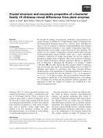
2.3. Synthesis of 2-[(E )-{[4-(benzyloxy)phenyl]imino} methyl]-4-[(E )-(4-nitrophenyl)diazenyl]phenol, (mbH).1/2H 2 O
A solution of 4-benzyloxyanilinehydrochloride (433.50 mg, 2.176 mmol) in ethyl alcohol (10 mL) was mixed
with a solution of 2-hydroxy-5-[(4-nitrophenyl)diazenyl]benzaldehyde (498.64 mg, 1.84 mmol) in ethyl alcohol
(50 mL) and the reaction mixture was refluxed for 24 h. The dark yellow product formed was dissolved in ethyl
alcohol (25 mL) and left for crystallization at room temperature for a day. Then orange crystals were collected,
washed with cold ethyl alcohol, and dried in air. Yield, 650.00 mg (77%). Mp: 209–210 ◦ C. Elemental analyses
223
BAL et al./Turk J Chem
for C 26 H 21 N 4 O 4,5 (461.46 g/mol): Found: C, 67.42; H, 4.34; N, 12.09%. Calcd.: C, 67.67; H, 4.59; N, 12.14%.
IR (cm −1 ): 3448 υ (O–H hydrated water), 1637 υ (C=N), 1520 υ (–N=N–), 1342 υ (C=C), 1104 υ (C–O–C).
2.4. Synthesis of di(aqua)bis{2-[(E )-{[4-(benzyloxy)phenyl]imino} methyl]-4-[(E )-(4-nitrophenyl)
diazenyl]phenolato} mangan(II), [Mn(mb) 2 (H 2 O) 2 ].4H 2 O
A solution of MnCl 2 .4H 2 O (1.40 mg, 0.011 mmol) in methyl alcohol (10 mL) was added to a solution of mbH
(10.00 mg, 0.022 mmol) in dichloromethane (20 mL). The mixture was then heated in a water bath for another
30 min to complete the precipitation. The red complex was filtered, washed with cold ethyl alcohol, and dried.
Yield, 7.34 mg (64%). Mp: > 250 ◦ C. Elemental analyses for C 52 H 50 MnN 8 O 14 (1065.93 g/mol): Found:
C, 58.72; H, 3.90; N, 10.48%. Calcd.: C, 58.59; H, 4.73; N, 10.51%. IR (cm −1 ): 3419 υ (O–H hydrated water),
1630 υ (C=N), 1523 υ (–N=N–), 1342 υ (C=C), 1106 υ (C–O–C), 850 (coordinated water), ∼650 υ (Mn–O),
545 υ (Mn–N).
2.5. Synthesis of di(aqua)bis{2-[(E )-{[4-(benzyloxy)phenyl]imino} methyl]-4-[(E )-(4-nitrophenyl)
diazenyl]phenolato} nickel(II), [Ni(mb) 2 (H 2 O) 2 ].3H 2 O
2-[(E)-{[4-(benzyloxy)phenyl]imino} methyl]-4-[(E) -(4-nitrophenyl)diazenyl]phenol ligand (10.00 mg, 0.022
mmol) was dissolved in dichloromethane (20 mL) at room temperature (Figure 1). A solution of NiCl 2 .6H 2 O
(2.70 mg, 0.011 mmol)) in methyl alcohol (10 mL) was added dropwise into the solution of the ligand with
continuous stirring. The mixture was refluxed for 3 h; the volume of the solution was then reduced to 10 mL
and left to cool down to room temperature. On addition of ethyl alcohol (10 mL) a precipitate formed and
-
O
+
N
O
O
N
N
O
H
.HCl
+
OH
H2N
EtOH
reflux
O
-
+
N
O
O
0.5 H2O
N
N
N
OH
Figure 1. Synthesis of 2-[(E )-{[4-(benzyloxy)phenyl]imino} methyl]-4-[(E )-(4-nitrophenyl)diazenyl]phenol (mbH).
224
BAL et al./Turk J Chem
was collected and washed with a small amount of ethyl alcohol. The orange product was recrystallized from
hot ethyl alcohol and it was dried at room temperature. Yield, 8.00 mg (70%). Mp: 266–267 ◦ C. Elemental
analyses for C 52 H 48 N 8 NiO 13 (1051.67 g/mol): Found: C, 59.29; H, 4.00; N, 10.29%. Calcd.: C, 59.39; H,
4.60; N, 10.65%. IR (cm −1 ) : 3409 υ (O–H hydrated water), 1627 υ (C=N), 1510 υ (–N=N–), 1376 υ (C=C),
1104 υ (C–O–C), 845 (coordinated water), 610 υ (Ni–O), ∼540 υ (Ni–N).
2.6. Synthesis of di(aqua)bis{2-[(E )-{[4-(benzyloxy)phenyl]imino} methyl]-4-[(E )-(4-nitrophenyl)
diazenyl]phenolato} copper(II), [Cu(mb) 2 (H 2 O) 2 ].3H 2 O
Cu(CH 3 COO) .2 H 2 O (2.20 mg, 0.011 mmol) was dissolved in methyl alcohol (10 mL) and stirred under reflux
for 45 min, followed by the addition of the mbH Schiff base (10.00 mg, 0.022 mmol) in dichloromethane (20 mL),
and the reaction mixture was refluxed for 3 h. The brown precipitate obtained was filtered, washed with methyl
alcohol, and dried in air. Yield, 5.90 mg (51%). Mp: 250–251 ◦ C. Elemental analyses for C 52 H 48 N 8 CuO 13
(1056.53 g/mol): Found: C, 58.43; H, 3.86; N, 10.52%. Calcd.: C, 59.11; H, 4.58; N, 10.61%. IR (cm −1 ) : 3375
υ (O–H hydrated water), 1630 υ (C=N), ∼ 1520 υ (–N=N–), 1340 υ (C=C), 1105 υ (C–O–C), 855 (coordinated
water), 691 υ (Cu–O), 546 υ (Cu–N).
2.7. Synthesis of di(aqua)bis{2-[(E )-{[4-(benzyloxy)phenyl]imino} methyl]-4-[(E )-(4-nitrophenyl)
diazenyl]phenolato} cobalt(II), [Co(mb) 2 (H 2 O) 2 ].8H 2 O
A methanolic solution (10 mL) of Co(CH 3 COO) .2 4H 2 O (2.60 g, 0.011 mmol) was added gradually to a
dichloromethane solution (20 mL) of the ligand (10.00 mg, 0.022 mmol). The solution was stirred for 2 h and
a reddish brown precipitate formed. The product was filtered and washed with ethyl alcohol and then diethyl
ether, and finally dried in air. Yield, 7.40 mg (59%). Mp: 254 ◦ C. Elemental analyses for C 52 H 58 CoN 8 O 18
(1141.99 g/mol): Found: C, 54.74; H, 4.70; N, 9.78%. Calcd.: C, 54.69; H, 5.12; N, 9.81%. IR (cm −1 ) : 3390
υ (O-H/hydrated water), 1632 υ (C=N), ∼1520 υ (–N=N–), 1342 υ (C=C), 1106 υ (C–O–C), 857 (coordinated
water), ∼ 650 υ (Co–O), 547 υ (Co–N).
2.8. Synthesis of di(aqua)bis{2-[(E )-{[4-(benzyloxy)phenyl]imino} methyl]-4-[(E )-(4-nitrophenyl)
diazenyl]phenolato} zinc(II), [Zn(mb) 2 (H 2 O) 2 ].H 2 O
The red colored compound was prepared by the addition of Zn(CH 3 COO) 2 .2H 2 O (2.40 mg, 0.011 mmol) in
methyl alcohol (10 mL) to a refluxing mixture of the ligand (10.00 mg, 0.022 mmol) mbH in dichloromethane (20
mL). The red compound was separated out via
filtration, washed with cold ethyl alcohol, and dried in vacuo.
◦
Yield, 6.10 mg (55%). Mp: 286–287 C. Elemental analyses for C 52 H 44 N 8 O 11 Zn (1022.36 g/mol): Found: C,
61.00; H, 3.83; N, 10.83%. Calcd.: C, 61.09; H, 4.34; N, 10.96%. IR (cm −1 ) : 3395 υ (O–H hydrated water),
1625 υ (C=N), ∼ 1515 υ (N=N), 1340 υ (C=C), 1103 υ (C–O–C),
845 ( coordinated water),698 υ (Zn–O), 545
υ (Zn–N).
2.9. Salmonella/microsome test (Ames)
2.9.1. Bacterial strains
Histidine deficient (his–) tester strains TA98 and TA100 of Salmonella typhimurium were provided by LK
Nakamura (Microbiologist Emeritus, Microbial Properties Research, Department of Agriculture, Peoria, Illinois,
225
BAL et al./Turk J Chem
USA). The TA98 strain was used to detect the frameshift mutagens and the TA100 strain for the detection of
base pair substitution mutagens. Each strain used for testing was checked for the presence of strain-specific
marker as described by Maron and Ames. 30
2.9.2. Mutagenicity assay and preparation of S9
The standard plate-incorporation assay was examined with Salmonella typhimurium TA98 and TA100 strains
in the presence and absence of S9 mix according to Maron and Ames. 30 Mutagenicity tests and preparation of
S9 for the compounds were performed according to the literature. 30,31 For the test, the mbH bidentate ligand
and its metal complexes were dissolved in DMSO and used as 0.06, 0.12, 0.24, 0.49, and 0.98 mg per plate.
Each sample was evaluated with 3 replicate plates and all tests were performed twice. Fresh S9 mix was used
for each mutagenicity assay.
2.9.3. Statistical significance
The significance between control revertants and revertants of treated groups were also compared by t-test.
Dose-response relationships were evaluated by using regression and correlation (r) test systems.
3. Results and discussion
3.1. Synthesis
2-[(E)-{[4-(benzyloxy)phenyl]imino} methyl]-4-[(E)-(4-nitrophenyl)diazenyl]phenol (mbH) was prepared by the
reaction of 2-hydroxy-5-[(4-nitrophenyl)diazenyl]benzaldehyde with 4-benzyloxyanilinehydrochloride in ethyl alcohol. The product of the condensation reaction of 2-hydroxy-5-[(4-nitrophenyl)diazenyl]benzaldehyde salt
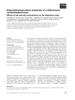
with 4-benzyloxyanilinehydrochloride is depicted in Figure 1. The new azo-azomethine ligand, 2-[(E) -{[4(benzyloxy)phenyl]imino} methyl]-4-[(E)-(4-nitrophenyl)diazenyl]phenol (mbH), resulted in mononuclear complexes (Figure 2) with Mn(II), Co(II), Ni(II), Cu(II), and Zn(II) as follows:
dichloromethane
MX 2 + 2 mbH −−−−−−−−−−−→ [M(mb) 2 (H 2 O) 2 ].nH 2 O + 2 HX
ref lux
mbH: 2-[( E)-{[4-(benzyloxy)phenyl]imino} methyl]-4-[(E) -(4-nitrophenyl)diazenyl]phenol
M = Mn(II) (n = 4); Co(II) (n = 8); Ni(II) (n = 3); Cu(II) (n = 3); Zn(II) (n = 1)
Experimental results of the elemental analyses of the synthesized ligand and its metal chelates are in
good agreement with theoretical values. The elemental analyses of the complexes indicate that the metal–
ligand ratios are 1:2 in the [M(mb) 2 (H 2 O) 2 ].nH 2 O [M = Mn(II), n = 4; Co(II), n = 8; Ni(II), n = 3; Cu(II),
n = 3; or Zn(II), n = 1] metal complexes. The purity of the compounds was checked by TLC using silica gel G
as adsorbent. The ligand and its mononuclear complexes are not soluble in water. Single crystals of the new
azo-azomethine ligand and its transition metal chelates could not be isolated from any organic solvent; thus, no
definite structures could be described.
However, structures of the compounds were proposed based on the analytical and spectroscopic data as
shown in Figures 1 and 2. The analytical and spectroscopic data showed that M(II) ions are 6-coordinate,
bonded to 2 nitrogen (C=N) and 2 phenolic oxygen atoms of 2 azo-azomethine ligands and 2 water molecules.
Coordination geometry around the metal centers is octahedral. M–N and M–O bonds are expected to be trans in
226
BAL et al./Turk J Chem
-
O
O
N
+
N
N
O
N
O
M
O
.nH2O
N
O
N
N
N
-
O
+
O
Figure 2. The proposed structure of metal complexes of the azo-azomethine ligand (mbH).
configuration due to steric reasons and this trans configuration was also observed for similar complexes reported
in the literature. 20−28
3.2.
1
H NMR spectrum of the ligand
For further information about the azo-azomethine ligand the 1 H NMR was recorded in CDCl 3 . NMR shifts of
the ligand are shown in Table 1. The
1
H NMR spectrum confirms that the ligand is intact in solution. The
hydrogen atom of the azomethine group (–CH=N–) was observed at δ 8.67 ppm as a singlet. 6 The aromatic
protons were observed in the range of δ 6.98–8.67 ppm as a multiplet. Benzyl (C19) protons were assigned
to a singlet peak at δ 5.05 ppm. A shift at δ 14.29 ppm could be assigned to phenolic proton (O(16)H). 32
Additionally, water protons were observed at 1.52 ppm. The presence of water in the structure was also
confirmed by infrared spectroscopy and elemental analysis.
227
BAL et al./Turk J Chem
Table 1. The
1
H NMR data (ppm) of the azo-azomethine (mbH) ligand in CDCl 3 .
33
34
25
OH
26
27
28
31
30
H
16
15
14
29
O
24
32
19
17
21
N
23
22
20
13
18
12
N
11
N
10
7
6
8
9
5
4
N
-
O
3
2
+
O
1
Chemical shifts, δT M S (ppm)
14.29
8.67
8.32
8.03
7.98
7.92
7.39
7.35
7.30
7.27
7.09
6.98
5.05
1.52
a
Assignmentsa
[s, 1H] (16)
[s, 1H] (19)
[d, 2H] (9, 5)
[d, 2H] (8, 6 )
[s, 1H] (18)
[d, 1H] (13)
[d, 2H] (30, 34)
[t, 2H] (31, 33)
[t, 1H] (32)
[d, 1H] (14)
[d, 2H] (22, 26)
[d, 2H] (23, 25)
[s, 2H] (28)
[s, 2H] (H2 O)
J(Hz)
8.84
8.87
8.87
7.30
7.95
8.81
7.44
8.92
8.88
-
s: singlet; d: doublet and t: triplet.
3.3. FT-IR spectra
In order to study the bonding of the mbH azo-Schiff base to the metal, the infrared spectrum of the mbH was
compared with spectra of the corresponding metal chelates. The infrared spectra provided valuable information
regarding the nature of the functional groups attached to the metal ion. The main infrared bands and their
assignments are given in the experimental section. In the spectrum of azo-azomethine ligand (mbH), a strong
band at 1637 cm −1 is attributed to the C=N (azomethine) group. 33 Upon coordination, this band C=N
(azomethine) shifted to a lower frequency due to a shift of lone pair density toward the metal ion, indicating
coordination of azomethine nitrogen to the metal center. 34−36 The spectrum of the mbH ligand exhibits a broad
228
BAL et al./Turk J Chem
band at 3448 cm −1 due to phenolic and water –OH. 37 The phenolic –OH stretch disappears in the spectra of
metal complexes, indicating that upon coordination of the ligand to metal centers the phenolic oxygen atoms are
deprotonated. The spectra of the metal chelates exhibited broad bands at 3448–3375 cm −1 that are attributed
to OH of the crystal water molecules, while the bands observed at approximately 857–845 cm −1 are assigned to
coordinated water molecules. 32,37 A comparison between infrared spectra of mbH and the [M(mb) 2 ] complexes
also shows that a band, characteristic of ν (C–O) at 1315 cm −1 , is shifted to 1345–1325 cm −1 , due to C–
O–M bond formation. Bands at 2920–2885 cm −1 are assigned to CH 2 asymmetric and symmetric stretching
vibrations. The azo-Schiff base mbH showed a band at 1342 cm −1 for ν (C=C) of aromatic rings, while its
metal complexes shift to 1376–1340 cm −1 . In addition, all the metal complexes show 2 new bands at 698–610
and 547–540 cm −1 due to formation of M–O and M–N bonds, further confirming formation of coordination
complexes. 38 All the vibrational data suggest that the metal ion bonded to the azo-azomethine ligand through
the phenolic oxygen and the imino nitrogen atoms.
3.4. Electronic spectra
The electronic spectra of the mbH ligand and its metal chelates were recorded in DMSO between 200 and 800
nm. The compared dates of the UV-Vis spectra for the azo-azomethine dye and its metal chelates are shown in
Table 2. The UV-Vis spectra of the ligand and its Ni(II) chelate in DMSO solution are shown in Figures 3 and
4.
Table 2. UV-Vis data of the ligand and its metal complexes in DMSO.
Compounds
mbH
[Mn(mb)2 (H2 O)2 ].4H2 O
[Co(mb)2 (H2 O)2 ].8H2 O
[Ni(mb)2 (H2 O)2 ].3H2 O
[Cu(mb)2 (H2 O)2 ].3H2 O
[Zn(mb)2 (H2 O)2 ].H2 O
λmax (nm)
238, 292, 361
234, 372, 535
240, 391, 493
238, 314, 414, 516
237, 327, 531
240, 410, 540
Figure 3. The UV-Vis spectrum of mbH.1/2H 2 O ligand
in DMSO.
Transitions
π − π*, n–π*
π − π*, n–π*,
π − π*, n–π*,
π − π*, n–π*,
π − π*, n–π*,
π − π*, n–π*,
d–d
d–d
d–d
d–d
CT
Figure 4. The UV-Vis spectrum of [Ni(mb) 2 (H 2 O) 2 ].
3H 2 O complex in DMSO.
229
BAL et al./Turk J Chem
The absorption of the synthesized ligand (mbH) displays mainly 3 bands in DMSO solution at room
temperature within the studied range. The band at 238 nm was assigned to the π → π * transition of
aromatic rings, while the band at 292 nm as a shoulder is due to the low energy π → π * transition of the
–CH=N– and –N=N– groups. 39,40 The peaks belonging to the π → π * transitions in the spectra of the
[Mn(mb) 2 (H 2 O) 2 ].4H 2 O, [Ni(mb) 2 (H 2 O) 2 ].3H 2 O, [Co(mb) 2 (H 2 O) 2 ].8H 2 O, [Cu(mb) 2 (H 2 O) 2 ].3H 2 O, and
[Zn(mb) 2 (H 2 O) 2 ].H 2 O coordination compounds were observed at 234, 238, 240, 237, and 240 nm, respectively.
The band at 361 nm was assigned to the n→ π * transitions of the –CH=N– and –N=N– azo chromophore groups.
The peaks belonging to these groups in the spectra of the [Mn(mb) 2 (H 2 O) 2 ].4H 2 O, [Ni(mb) 2 (H 2 O) 2 ].3H 2 O,
[Co(mb) 2 (H 2 O) 2 ].8H 2 O, [Cu(mb) 2 (H 2 O) 2 ].3H 2 O, and [Zn(mb) 2 (H 2 O) 2 ].H 2 O complexes appeared at 372,
314, 391, 327, and 410 nm, respectively. Furthermore, d–d transition bands in the spectra of the Mn(II), Co(II),
Ni(II), and Cu(II) chelates were observed at 493–535 nm. The bands at 414 nm of Ni(II) and 540 nm of the
Zn(II) chelates can be assigned to charge-transfer transitions. The spectroscopic data obtained in this work
agreed well with those in previous work. 41
3.5. X-ray powder diffraction analysis
Growth of single crystals of azo-azomethine compounds from various solvents including DMF, ethyl alcohol,
chloroform etc. failed and so they were characterized by XRD. 42,43 X-ray powder diffraction analysis of the
mbH ligand and its metal complexes was carried out to determine the type of crystal system, lattice parameters,
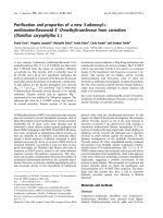
and the cell volume. As shown in Figure 5 the XRD patterns indicate a crystalline nature for the mbH ligand
and its metal complexes. Indexing of the diffraction patterns was performed using HighScore Plus software. For
the Mn(II) and Co(II) complexes, for example, their Miller indices (hkl) along with observed and calculated
2θ angles, d values, and relative intensities are given in Tables 3 and 4. From the indexed data the unit
cell parameters were also calculated and are listed in Table 5. The powder XRD patterns of the compounds
are completely different from those of the starting materials, demonstrating the formation of coordination
compounds. It is found that mbH ligand and Ni(II), Cu(II), Co(II), and Zn(II) complexes have monoclinic
structures, while Mn(II) complex has an orthorhombic structure. The crystal structures of similar type of
samples were reported as monoclinic and orthorhombic. 32,44−46 Moreover, using the diffraction data, the mean
crystallite sizes of the complexes, D , were determined according to the Scherrer equation (D = 0.9 λ /(β cos θ),
´ ), θ is Bragg diffraction angle, and β is the full width at half maximum
where λ is X-ray wavelength (1.5406 ˚
A
of the diffraction peak). 47,48 The average crystallite sizes of all the samples were found to be ∼ 38–75 nm and
the values are given in Table 5.
3.6. Cyclic voltammograms
Cyclic voltammograms of the ligand and its complexes were run in DMF and CH 3 CN solutions at room
temperature using Bu 4 NBF 4 as supporting electrolyte at 293 K. All potentials quoted refer to measurements
run at a scan rate (v) of 200, 250, and 500 mV s −1 and against an internal ferrocene–ferrocenium standard,
unless otherwise stated. In order to investigate the effect of the ligand concentration, the electrochemical studies
were performed in 1 × 10 −3 and 1 × 10 −4 M solutions of the ligand and its complexes. The voltammograms
were recorded in the range from –2.0 to 2.0 V vs. Ag + /AgCl. The electrochemical data of the ligand and its
complexes are summarized in Tables 6 and 7.
230
BAL et al./Turk J Chem
Table 3. XRD data of the [Mn(mb) 2 (H 2 O) 2 ].4H 2 O metal complex.
P.No.
1
2
3
4
5
6
7
8
9
10
11
12
13
14
15
16
17
18
19
20
21
22
h
3
3
2
0
4
3
0
3
5
3
0
6
3
3
3
7
8
1
2
7
4
6
k
1
0
0
3
1
1
0
3
0
1
4
2
4
1
2
1
0
0
0
4
6
0
l
0
1
2
1
1
2
3
1
1
3
2
0
2
4
4
2
0
5
5
1
2
5
2Th.(o) [◦ ]
20.1641
21.2311
23.9018
25.8232
28.0398
28.8295
30.5684
31.9817
32.8441
37.0964
38.1481
41.0199
42.6428
46.5069
48.6772
49.8599
50.837
52.9586
54.1668
56.4883
59.4269
66.4442
2Th.(c) [◦ ]
20.164
21.1882
23.9011
25.8256
28.0364
28.8319
30.7969
31.9846
32.8522
37.0892
38.0359
40.9886
42.6343
46.5263
48.7038
49.8176
50.8986
52.9488
54.1816
56.5039
59.458
66.3865
´]
d-sp.(o) [˚
A
4.400241
4.181457
3.719926
3.447334
3.179653
3.094321
2.922147
2.796165
2.724693
2.421542
2.357172
2.198532
2.118539
1.951116
1.869077
1.827475
1.794621
1.727614
1.691897
1.627748
1.55408
1.405949
´]
d-sp.(c) [˚
A
4.400274
4.189827
3.720043
3.447019
3.180032
3.094069
2.900988
2.79592
2.724033
2.421999
2.363864
2.200137
2.11894
1.950348
1.868118
1.828927
1.792594
1.727912
1.69147
1.627335
1.55334
1.40703
Rel. Int. [%]
100
12.16
19.32
72.06
33.54
33.93
47.85
77.6
31.54
58.66
35.51
44.13
46.76
8.07
12.73
18.46
16
20.64
12.33
19.68
7.77
12.96
Table 4. XRD data of the [Co(mb) 2 (H 2 O) 2 ].8H 2 O metal complex.
P.No.
1
2
3
4
5
6
7
h
2
1
3
4
1
3
2
k
1
1
1
0
1
2
1
l
1
2
1
–3
–4
–1
4
2Th.(o) [◦ ]
20.1819
20.8981
24.6466
25.7842
28.7745
31.676
35.2553
2Th.(c) [◦ ]
20.1919
20.8929
24.6676
25.7678
28.7878
31.6676
35.2476
´]
d-sp.(o) [˚
A
4.396392
4.247317
3.609173
3.452471
3.100117
2.82245
2.543671
´]
d-sp.(c) [˚
A
4.394257
4.248372
3.606158
3.454619
3.098718
2.823184
2.544208
Rel. Int. [%]
16.43
13.87
31.09
100
27.42
8.72
8.16
Table 5. XRD parameters of the mbH ligand and its metal complexes.
Sample
(1 ) (mbH)
(2 )[Ni(mb)2 (H2 O)2 ].3H2 O
(3 )[Mn(mb)2 (H2 O)2 ].4H2 O
(4 )[Cu(mb)2 (H2 O)2 ].3H2 O
(5 )[Co(mb)2 (H2 O)2 ].8H2 O
(6 )[Zn(mb)2 (H2 O)2 ].H2 O
Lattice parameters
a (˚
A)
b (˚
A)
9.5494
15.4706
11.9487 3.9729
14.3386 11.2711
13.4747 11.9007
15.8585 6.6875
17.3896 8.5036
c (˚
A)
7.3935
10.5231
8.6961
10.1995
14.0527
14.2796
β (◦ )
98.0241
100.6310
90
113.4330
108.1620
120.0730
Volume
(˚
A3 )
1081.5890
490.97
1405.40
1500.54
1416.09
1827.34
Crystallite
size D (nm)
60
75
38
37
64
52
Crystal system
Monoclinic
Monoclinic
Orthorhombic
Monoclinic
Monoclinic
Monoclinic
All complexes show strong cathodic peaks in the range from –0.5 to 1.0 V. The complexes have 2 anodic
peaks in the 1.4–2.0 V range. The anodic and cathodic peaks are irreversible. The complexes show irreversible
cathodic peak potentials in the 1.0–1.4 V range.
231
BAL et al./Turk J Chem
(f)
(e)
Intensity [a.u.]
(d)
(c)
(b)
(a)
20
30
40
50
60
70
80
90
2 Theta [Degree]
Figure 5. The XRD diffraction patterns of (a) (mbH), (b) [Ni(mb) 2 (H 2 O) 2 ].3H 2 O, (c) [Mn(mb) 2 (H 2 O) 2 ].4H 2 O, (d)
[Cu(mb) 2 (H 2 O) 2 ].3H 2 O, (e) [Co(mb) 2 (H 2 O) 2 ].8H 2 O, and (f) [Zn(mb) 2 (H 2 O) 2 ].H 2 O.
The [Co(mb)(H 2 O) 2 ].8H 2 O complex shows the reversible process (Ipa:Ipc = 1.0) in the 1 × 10 −3 M
DMF and CH 3 CN solutions at the 250 and 500 mV s −1 scan rates. Their potential ranges change from 0.35 V
to 1.28 V (Epc) and from 0.35 V to 1.34 V (Epa). The [Cu(mb)(H 2 O) 2 ].3H 2 O complex shows the irreversible
process (Ipa:Ipc ̸= 1.0) in the 1 × 10 −3 M CH 3 CN solution at the 250 and 500 mV s −1 scan rates. Their
potential ranges change from –0.25 V to 1.60 V (Epc) and from 0.21 V to 1.54 V (Epa). At the 250 mV s −1 scan
rate, the [Cu(mb)(H 2 O) 2 ].3H 2 O complex shows the reversible process (Ipa:Ipc ≈ 1.0) at 1.60 V (Epc) and
1.54 V (Epa). All processes at other potentials are irreversible in the 1 × 10 −3 M CH 3 CN and DMF solutions.
The electrochemical curves of the [Co(mb)(H 2 O) 2 ].8H 2 O, [Cu(mb)(H 2 O) 2 ].3H 2 O, [Mn(mb)(H 2 O) 2 ].4H 2 O,
[Ni(mb)(H 2 O) 2 ].3H 2 O, and [Zn(mb)(H 2 O) 2 ].H 2 O complexes at 200, 250, and 500 mV s −1 scan rates in the
1 × 10 −3 M DMF solutions are shown in Figures 6a–e.
The [Mn(mb)(H 2 O) 2 ].4H 2 O complex shows the reversible process (Ipa:Ipc = 1.0) in the 1 × 10 −4 M
CH 3 CN solution at the 500 mV s −1 scan rate. Their potential ranges change from –1.35 V to –0.76 V (Epc)
and from –1.35 V to 0.61 V (Epa). The [Mn(mb)(H 2 O) 2 ].4H 2 O complex show the irreversible process (Ipa :
Ipc ̸= 1.0) in the 1 × 10 −4 M DMF solution at the 250 and 500 mV s −1 scan rates. All processes at other
potentials are irreversible in the 1 × 10 −4 M CH 3 CN and DMF solutions.
232
0.21, 0.82, 1.54
–0.40, 1.68
–0.59, 1.60
–0.70, 0.35
–0.18, 1.70
–0.90, 0.34, 1.78
–0.71, –0.38, 0.90
DMF
AN
DMF
AN
DMF
AN
DMF
–0.72, 0.29, 1.64
–1.12, –0.69, 1.01
0.35, 1.28
–0.60, 1.55
–0.23, 0.36, 1.29
AN
DMF
AN
DMF
AN
E pa (V)
i
Solvent
E pc (V)
1.76, –0.62, –0.81
–0.51, –0.90, –1.42
1.28, 0.90, 0.35
0.59, –0.65
0.89, –0.10
1.66, 1.12,
–0.25
1.31, –0.61
1.12, –0.69, –0.89
1.40, –0.40
1.23, –0.80
1.21, –0.60, –1.09
1.24, –0.63
i
–
–
–
–
–
–
0.23*
–
–
0.35*
0.62*
–
E 1/2
(mV)
0.37
0.68
0.95
0.47
0.56
0.25
0.46
0.09
0.21
0.38
0.05
0.40
Ep
E pa (V)
–0.69, 0.32, 0.84
–1.11, –0.66, 105
0.30, 1.32
–0.65, 1.71
–0.20, 0.31, 1.31
0.20, 0.80,
1.50
–0.31, 1.67
–0.50, 1.70
–0.89, 0.30
–0.20, 1.68
–0.91, 0.35, 1.79
–0.73, –0.20, 0.98
ii
E pc (V)
1.25, –0.62
1.20, –0.70, –0.90
1.31, 0.63
1.18, –0.84
1.30, –0.62, –1.10
1.20, –0.70
1.64, 1.10, –0.20
1.47, –0.65, –1.09
–0.51, –0.90, –1.45
1.33, 0.89, 0.28
0.80, –0.71
0.90, –0.12
ii
0,71*
-
0.29*
-
E 1/2
(mV)
0.42
0.50
0.33
0.70
0.49
0.50
0.40
0.33
0.24
0.23
0.06
0.40
Ep
rate 250 mV s − 1 . Other data (ii) have been obtained by scan rate 500 mV s − 1 . *: Reversible
and Epc are anodic and cathodic potentials, respectively. E1/2 = 0.5 × ( Epa + Epc ) , ∆ Ep = Epa – Epc . (i): These data have been obtained from scan
Supporting electrolyte: [NBu 4 ](BF 4 ) (0.1 M); concentrations of the compounds: 1 × 10 − 3 M. All the potentials are referenced to Ag + /AgCl, where Epa
[Zn(mb)(H2O)2].H2O
[Ni(mb)(H2O)2].3H2O
[Mn(mb)(H2O)2].4H2O
[Cu(mb)(H2O)2].3H2O
[Co(mb)(H2O)2].8H2O
mbH.1/2H2O
Compound
Table 6. Electrochemical data of the azo-Schiff base ligand and its metal complexes (1 × 10 − 3 M).
BAL et al./Turk J Chem
233
234
0.26, 0.76, 1.61
–0.97, –0.32, 1.5
–0.29, 0.23
–1.16, –0.89, 1.57
–0.33, 0.27, 1.28
–0.42, 1.44
–1.47, –0.85, –0.33
–1.09, –0.68, –0.33
0.32, 1.66
–0.3
–0.33, 0.32
–0.36, 1.54
AN
DMF
AN
DMF
AN
DMF
AN
DMF
AN
DMF
AN
DMF
E pc(V)
1.87
–0.6, –1.31
–0.68, –1.03
–1.34
–0.65, –1.14
0.73, –0.62
1.82, –0.68, –1.44
1.29, –0.79, –1.24
1.87, 1.45
–0.59
1.43, –0.62
1.78, –0.6
i
E 1/2
(mV)
–
–
–
–
–
0.52
0.50
–
–
–
–
–
0.26
0.28
0.39
0.18
0.33
0.20
0.35
0.11
0.21
0.29
0.29
0.24
Ep
E pa (V)
–1.43, –0.9, –0.3
–0.3
–1.16, –0.89
–1.80, –0.86, 1.61
–0.26, 1.28
–0.87, 0.96
–1.35, 0.31, 0.61
–1.07, –0.6, 1.66
0.27, 1.63
–0.34
–0.32, 0.3, 1.62
–0.37, 1.58
ii
E pc(V)
1.82, –0.69, –0.88
1.08
1.57, –1.34
1.29, –0.61–1.53
–0.65, –1.17
1.27, –0.69–1.56
–0.76, –1.35
1.29
1.87
–0.61
1.79, –0.63, –1.1
–0.63, 1.24
ii
E 1/2
(mV)
–
–
–
–
–
–
1.00
–
–
–
–
0.50
0.39
–
0.18
0.32
0.39
0.69
–
0.37
–
0.27
0.31
0.26
Ep
rate 250 mV s − 1 . Other data (ii) have been obtained by scan rate 500 mV s − 1
and Epc are anodic and cathodic potentials, respectively. E1/2 = 0.5 × ( Epa + Epc ) , ∆ Ep = Epa – Epc . (i): These data have been obtained from scan
Supporting electrolyte: [NBu 4 ](BF 4 ) (0.1 M); concentrations of the compounds: 1 × 10 − 4 M. All the potentials are referenced to Ag + /AgCl, where Epa
[Zn(mb)2(H2O)2].H2O
[Ni(mb)2(H2O)2].3H2O
[Mn(mb)2(H2O)2].4H2O
[Cu(mb)2(H2O)].3H2O
[Co(mb)2(H2O)2].8H2O
mbH.1/2H2O
E pa (V)
i
Solvent
Table 7. Electrochemical data of the azo-Schiff base ligand and its metal complexes (1 × 10 − 4 M).
BAL et al./Turk J Chem
BAL et al./Turk J Chem
Figure 6. a–e The electrochemical curves of the metal complexes at 200, 250, and 500 mV s −1 scan rates in DMF
solution (1 × 10 −3 M).
The Zn(II) complex of the azo-Schiff base ligand mbH shows the reversible process at the –0.70 V
(Epc ) and –0.73 V (Epa ) potentials at the 500 mV s −1 scan rate in the 1 × 10 −3 M solution.
The
[Zn(mb) 2 (H 2 O) 2 ].H 2 O chelate shows the reversible process at the –0.70 and 1.20 V ( Epc ) and –0.73, –0.20, and
0.98 V (Epa ) potentials at the 500 mV s −1 scan rate in the 1 × 10 −3 M solution. The [Zn(mb)(H 2 O) 2 ].H 2 O
235
BAL et al./Turk J Chem
complex shows the irreversible process (Ipa:Ipc ̸= 1.0) in the 1 × 10 −3 M and 1 × 10 −4 M CH 3 CN solutions at the 250 and 500 mV s −1 scan rates. The electrochemical curves of the [Co(mb)(H 2 O) 2 ].8H 2 O,
[Cu(mb)(H 2 O) 2 ].3H 2 O, [Mn(mb)(H 2 O) 2 ].4H 2 O, [Ni(mb)(H 2 O) 2 ].3H 2 O, and [Zn(mb)(H 2 O) 2 ].H 2 O complexes at 200, 250, and 500 mV s −1 scan rates in the 1 × 10 −3 M CH 3 CN solution are shown in Figures
7a–e.
Figure 7. a–e The electrochemical curves of the metal complexes at 200, 250, and 500 mV s −1 scan rates in CH 3 CN
solution (1 × 10 −3 M).
236
BAL et al./Turk J Chem
As the ligand has an electron donating benzyloxy group, the cathodic and anodic peak potentials were
shifted to the negative regions. However, the ligand has a nitro group. As the nitro group has the electron
accepting property, the redox potentials in the metal complexes shifted to the positive regions due to the –
NO 2 groups in the complexes, and the reduction and oxidation potentials were shifted to the higher positive
regions. 49 The quinoid process would involve self-protonation reactions where the benzyloxy group acts as a
proton donor. Oxidation–reduction peaks of the ligands at the different scan rates shifted to lower or higher
potentials. 45 This process is shown below:
M II + e − ↔ M I
The mbH ligand showed the quinoid forms (Figure 8).
O
+
O
-
O
N
N
O
M
O
O
-
N
O
N
-
N
O
N
N
N
+
N
+
O
+
M
-2e - 2H
O
-
O
+
O
N
+
+2e + 2H
N
N
N
N
+
O
-
N
+
O
O
-
N
O
Figure 8. Reversible reduction–oxidation processes of the azo-Schiff base metal complexes in DMF solution.
3.7. Genotoxicity
The azo-azomethine (mbH) ligand was mutagenic on S. typhimurium TA98 but not mutagenic on S. typhimurium TA100 in the presence and absence of S9 mix (Table 8). In addition, mutagenic activity of the
mbH ligand on TA98 increased with increasing dose in the presence and absence of S9 mix (Figure 9, r =
0.95924; Figure 10, r = 0.96762).
237
BAL et al./Turk J Chem
Table 8. The mutagenicity of mbH ligand and its metal complexes on S. typhimurium TA98 and TA100 in the presence
or absence of S9 mix.
Test substances
Spontaneous control
4-NPD
2-AF
SA
(1) mbH.1/2H2 O)
(2) [Ni(mb)2 (H2 O)2 ].3H2 O
(3) [Mn(mb)2 (H2 O)2 ].4H2 O
(4) [Cu(mb)2 (H2 O)2 ].3H2 O
(5) [Co(mb)2 (H2 O)2 ].8H2 O
(6) [Zn(mb)2 (H2 O)2 ].H2 O
Concentration
mg/plate
200 µ g/mL
20 µ g/mL
1 µ g/mL
0.98
0.49
0.24
0.12
0.06
0.98
0.49
0.24
0.12
0.06
0.98
0.49
0.24
0.12
0.06
0.98
0.49
0.24
0.12
0.06
0.98
0.49
0.24
0.12
0.06
0.98
0.49
0.24
0.12
0.06
TA98
− S9
10.50 ± 2.33
+ S9
11.67 ± 2.51
3111 ± 225
3025 ± 172
73.33 ± 4.03***
46.17 ± 3.50***
40.83 ± 4.32***
33.17 ± 4.74**
31.33 ± 3.51**
42.00 ± 3.54***
32.50 ± 402**
25.33 ± 2.56**
20.00 ± 3.43*
15.83 ± 1.97*
27.17 ± 2.57***
25.67 ± 1.82***
20.50 ± 1.65***
14.33 ± 2.06
13.00 ± 1.98
29.00 ± 3.39**
31.33 ± 3.36**
26.83 ± 2.98**
21.67 ± 2.30**
17.67 ± 2.63*
114.33 ± 7.49***
100.33 ± 9.78***
67.3 ± 11.6**
61.00 ± 9.65**
33.17 ± 7.13*
36.33 ± 1.78***
28.33 ± 5.17*
15.33 ± 1.23*
19.33 ± 2.54*
14.33 ± 3.28
57.17 ± 5.22***
54.00 ± 6.58***
34.33 ± 4.69**
30.83 ± 4.78**
28.83 ± 3.65**
39.00 ± 5.14**
34.83 ± 4.75**
22.17 ± 3.24*
18.50 ± 2.59*
17.17 ± 2.99
27.33 ± 3.19**
32.17 ± 1.76***
20.55 ± 1.84**
14.83 ± 3.35
12.50 ± 1.61
28.83 ± 4.30**
32.50 ± 4.15**
27.17 ± 3.41**
18.83 ± 2.50*
18.67 ± 2.26*
110.17 ± 7.40***
110.67 ± 9.81***
84.7 ± 10.5***
68.2 ± 13.3**
45.0 ± 10.2*
36.83 ± 2.33***
31.67 ± 4.60**
21.33 ± 2.70*
19.00 ± 1.81**
15.00 ± 2.37
TA100
− S9
106.7 ± 12.9
+ S9
99.3 ± 10.1
691.0 ± 25.7
651.8 ± 48.5
124.00 ± 5.77*
122.0 ± 19.7
101.50 ± 4.23
84.33 ± 3.57**
87.50 ± 13.2
180.7 ± 21.5*
141.0 ± 29.2
137.7 ± 18.6
112.7 ± 15.2
79.83 ± 6.67**
127.00 ± 4.12**
132.00 ± 7.82*
117.8 ± 9.9
107.33 ± 9.06
78.81 ± 6.18**
120.67 ± 6.96
111.3 ± 13.8
97.33 ± 5.41
76.17 ± 5.16*
54.67 ± 4.55***
213.7 ± 36.3**
162.5 ± 27.4*
123.2 ± 15.0
117.3 ± 19.8
78.67 ± 6.90**
121.17 ± 6.32
90.33 ± 3.99**
93.50 ± 7.98
74.0 ± 10.3*
59.00 ± 5.99***
126.8 ± 16.4
116.3 ± 17.1
101.67 ± 6.11
105.33 ± 4.52
71.17 ± 9.44*
158.7 ± 18.2*
124.8 ± 12.2
111.3 ± 10.2
124.0 ± 20.4
91.8 ± 12.3
122.00 ± 5.28**
133.00 ± 8.04**
106.83 ± 7.35
112.0 ± 10.5
85.83 ± 9.46
114.0 ± 5.74
103.0 ± 6.66
94.33 ± 6.26
74.00 ± 5.74**
58.50 ± 8.13**
164.2 ± 36.8*
222.8 ± 36.3**
158.8 ± 30.9*
151.0 ± 32.0
97.0 ± 13.2
106.00 ± 7.35
95.17 ± 7.11
93.17 ± 5.51
76.50 ± 5.79*
67.2 ± 10.9*
*: P < 0.05; **: P < 0.01; ***: P < 0.001
NPD: 4-nitro-o-phenylenediamine, 2AF: 2-Aminoflourene, SA: Sodium azide
Similarly, Cu(II), Ni(II), and Zn(II) metal complexes of (mbH) ligand [(4)[Cu(mb) 2 (H 2 O) 2 ].3H 2 O,
(2)[Ni(mb) 2 (H 2 O) 2 ].3H 2 O, and (6)[Zn(mb) 2 (H 2 O) 2 ].H 2 O] were also mutagenic on S. typhimurium TA98 but
not mutagenic on S. typhimurium TA100 in the absence or presence of S9 mix. In addition, mutagenic activity of
(2)[Ni(mb) 2 (H 2 O) 2 ].3H 2 O on S. typhimurium TA98 increased with increasing dose in the presence or absence
of S9 mix (Figure 9, r = 0.98882; Figure 10, r = 0.95068) and mutagenic activity of (6)[Zn(mb) 2 (H 2 O) 2 ].H 2 O
on S. typhimurium TA98 increased with increasing dose in the presence of S9 mix (Figure 10, r = 0.97699).
Co(II) and Mn(II) metal complexes of (mbH) ligand [(5)[Co(mb) 2 (H 2 O) 2 ].8H 2 O, (3)[Mn(mb) 2 (H 2 O) 2 ].
4H 2 O] exerted strong mutagenic activity on S. typhimurium TA98 but weak mutagenic activity on S. typhimurium TA100 in the absence or presence of S9 mix. Moreover, mutagenic activity of (5)[Co(mb) 2 (H 2 O) 2 ].
8H 2 O and (3)[Mn(mb) 2 (H 2 O) 2 ].4H 2 O on S. typhimurium TA98 increased with increasing dose in the absence
os S9 mix (Figure 9, r = 0.99735; Figure 10, r = 0.9768).
238
BAL et al./Turk J Chem
70
140
y = 10.5373x + 1.8046, r = 0.95924
y = 5.84901x + 3.8696, r = 0.98882
y = 3.76135x + 5.4351, r = 0.9768
y = 21.2112x - 10.403, r = 0.99735
100
y = 8.8632x + 4.6592, r = 0.96762
y = 4.9427x + 5.7956, r = 0.95068
y = 5.0633x + 4.6655, r = 0.97699
60
Number of revertant colonies
Number of revertant colonies
120
80
60
40
20
50
40
30
20
10
0
0
1
2
3
4
5
6
Doses
Figure 9.
Dose-dependent increase in the mutagenic activity of (mbH) ligand and its metal complexes on S.
of S9 mix.
typhimurium TA98 in the absence
Square, circle, triangle and upside
down triangle represent, respectively, (mbH) ligand,
1
2
3
4
5
6
Doses
Figure 10. Dose dependent increase in the mutagenic
activity of mbH ligand and its metal complexes on S.
typhimurium TA98 in the presence of S9 mix. Square,
circle, and triangle represent, respectively, mbH ligand,
[Ni(mb) 2 (H2O) 2 ].3H 2 O, and [Zn(mb)2(H 2 O) 2 ].H 2 O.
[Ni(mb) 2 (H 2 O) 2 ].3H 2 O, [Mn(mb) 2 (H 2 O) 2 ].4H 2 O, and
[Cu(mb) 2 (H 2 O) 2 ].3H 2 O.
4. Conclusion
In this work an azo chromophore group containing a Schiff base ligand, 2-[(E) -{[4-(benzyloxy)phenyl]imino}
methyl]-4-[(E)-(4-nitrophenyl)diazenyl]phenol derived from 2-hydroxy-5-[(4-nitrophenyl)diazenyl]benzaldehyde
with 4-benzyloxyaniline hydrochloride in ethyl alcohol and some of its transition metal complexes were prepared.
The analytical data and the spectroscopic studies suggested that the complexes had the general formula
[M(mb) 2 (H 2 O) 2 ].nH 2 O, where M is manganese(II), cobalt(II), nickel(II), copper(II), or zinc(II). According
to the UV-Vis and IR data of the nitrophenylazo linked Schiff base ligand, mbH was coordinated to the metal
ion through the azomethine nitrogen (–CH=N–) and phenolic oxygen atom. From the XRD results, it was
found that the mbH ligand and Ni(II), Cu(II), Co(II), and Zn(II) complexes have monoclinic structures, while
the Mn(II) complex has a orthorhombic structure. In the electrochemical studies of the ligand and its metal
chelates, reversible and irreversible redox processes were shown. Based on the above results, the structure of
the coordination compounds under investigation can be formulated as in Figure 2.
According to data obtained from the salmonella/microsome test, the mbH ligand and its 5 transition
metal complexes tested and their metabolites induced frameshift mutation (TA98). Generally, the effect of the
mbH azo-azomethine ligand and its complexes on TA98 was greater than that on TA100.
Acknowledgments
This work was supported by the KSU Research Fund (No: 2010/2–22YLS). The authors wish to express
their thanks to Prof Musa Găo
gebakan for the use of the X-ray diffractometer, and Prof Mehmet Tă
umer for
electrochemistry measurements and his valuable discussion.
239
BAL et al./Turk J Chem
References
1. Schiff, H. Ann. Chem. 1864, 131, 118–119.
2. Trujillo, A.; Fuentealba, M.; Carrillo, D.; Ledoux-Rak, I.; Hamon, J. R.; Saillard, J. Y. Inorg. Chem. 2010, 49,
2750–2764.
3. Fuentealba, M.; Garland, M. T.; Carrillo, D.; Manzur, C.; Hamon, J. R.; Saillard, J. Y. Dalton Trans. 2008, 49,
77–86.
4. Osinsky, S. P.; Levitin, I. Y.; Sigan, A. L.; Bubnovskaya, L. N.; Ganusevich, I. I.; Campanella, L.; Wardman, P.
Russ. Chem. Bull. 2003, 52, 2636–2645.
5. Beinert, H.; Kennedy, M. C.; Stout, C. D. Chem. Rev. 1996, 96, 2335–2374.
6. Kurtoglu, M.; Ispir, E.; Kurtoglu N.; Serin, S. Dyes Pigments 2008, 77, 75–80.
7. Dimiza, F.; Papadopoulos, A. N.; Tangoulis, V.; Psycharis, V.; Raptopoulou, C. P.; Kessissoglou, D. P.; Psomas,
G. Dalton Trans. 2010, 39, 4517–4528.
8. Harpstrite, S. E.; Collins, S. D.; Oksman, A.; Goldberg, D. E.; Sharma, V. Med. Chem. 2008, 4, 392–395.
9. Abd-Elzaher, M. M.; Moustafa, S. A.; Labib, A. A.; Ali, M. M. Monatsh. Chem. 2010, 141, 387–393.
10. Park, S.; Mathur, V. K.; Planap, R. P. Polyhedron 1998, 17, 325–330.
11. Nashinaga, A.; Ohara, H.; Tomita, H.; Matsuura, T. Tetrahedron Lett. 1983, 24, 213–216.
12. Pletcher, D.; Thompson, H. J. Electroanal. Chem. 1999, 464, 168–175.
13. Kianfara, A. H.; Paliz, M.; Roushani, M.; Shamsipur, M. Spectrochim. Acta A 2011, 82, 44–48.
14. Ho, M. S.; Barrett, C.; Paterson, J.; Esteghamatian, M.; Natansohn, A.; Rochon, P. Macromolecules 1996, 29,
4613–4618.
15. Yin, S.; Xu, H.; Shi, W.; Gao, Y.; Song, Y.; Wing, J. Polymer 2005, 46, 7670–7677.
16. Ho, M. S.; Natansohn, A. Macromolecules 1995, 28, 6124–6127.
17. Nabeshima, Y.; Shishido, A.; Kanazawa, A.; Shiono, T.; Ikeda, T.; Hiyama, T. Chem. Mater. 1997, 9, 1480–1487.
18. Kamel, M.; Galil, F.; Abdelwahab, L.; Osman, A. J. Prakt. Chem. 1971, 313, 1011–1021.
19. Gopal, J.; Srinivasan, M. J. Polym. Sci. Polym. Chem. Ed. 1986, 24, 2789–2796.
20. Serin, S.; Kurtoglu, M. Analyst 1994, 119, 2213–2215.
21. Kurtoglu, M.; Birbicer, N.; Kimyonsen, U.; Serin, S. Dyes Pigments 1999, 41, 141–143.
22. Birbicer, N.; Kurtoglu, M.; Serin, S. Synth. React. Inorg. Met. Org. Chem. 1999, 29, 1353–1364.
23. Kurtoglu, N.; Kurtoglu, M.; Serin, S. Synth. React. Inorg. Met. Org. Chem. 1999, 29, 1779–1791.
24. Kurtoglu, M.; Serin, S. Synth. React. Inorg. Met. Org. Chem. 2001, 31, 1129–1139.
25. Kurtoglu, M. Synth. React. Inorg. Met. Org. Chem. 2004, 34, 967–977.
26. Kurtoglu, M.; Baydemir, S. A. J. Coord. Chem. 2007, 60, 655–665.
27. Kurtoglu, M.; Serin, S. Synth. React. Inorg. Met. Org. Chem. 2002, 32, 629–637.
28. Khanmohammadi, H.; Darvishpour, M. Dyes Pigments 2009, 81, 167–173.
29. Ceyhan, G.; Kose, M.; McKee, V. J. Lumin. 2012, 132, 850–857.
30. Maron, D. M.; Ames, B. N. Mutation Research 1983, 113, 173–215.
31. Bal, M. Master’s Thesis, Institute of Science, KSU, 2010, Kahramanmara¸s, Turkey.
32. Kara, Y.; Avar, B.; Kayraldiz, A.; Gă
uzel, B.; Kurtoglu, M. Heteroatom Chem. 2011, 22, 119–130.
33. Karipcin, F.; Dede, B.; Ozkorucuklu, S. P.; Kabalcilar, E. Dyes Pigments 2010, 84, 14–18.
34. Gulcan, M.; Sonmez, M.; Berber, I. Turk. J. Chem. 2012, 36, 189–200.
35. Halli, M. B.; Patil, V. B.; Bevinamarada, S. R. Turk. J. Chem. 2011, 35, 393–404.
240
BAL et al./Turk J Chem
36. Kulaksizoglu, S.; Gokce, C.; Gup, R. Turk. J. Chem. 2012, 36, 717–733.
37. Alghool, S.; Hanan, S. A.; El-Halim, F. A.; Dahshan, A. J. Mol. Struct. 2010, 983, 32–38.
38. Ispir, E. Dyes Pigments 2009, 82, 13–19.
39. Khedr, A. M.; Gaber, M.; Issa, R. M.; Erten, H. Dyes Pigments 2005, 67, 117–126.
40. Kurtoglu, N. J. Serb. Chem. Soc. 2009, 74, 917–926.
41. Kilincarslan, R.; Erdem, E.; Kocaokutgen, H. Trans. Met. Chem. 2007, 32, 102–106.
42. Chavan, S. S.; Sawant, V. A. J. Mol. Struct. 2010, 965, 1–6.
43. Ide, S.; Ancın, N.; Oztas, S. G.; Tuzun, M. J. Mol. Struct. 2001, 562, 1–9.
44. Joseph, J.; Mehta, B. H. J. Coord. Chem. 2007, 33, 124–129.
45. Roy, G. B. Inorg. Chim. Acta 2009, 362, 1709–1714.
46. Munde, A. S.; Jagdale, A. N.; Jadhav, S. M.; Chondhekar, T. K. J. Serb. Chem. Soc. 2010, 75, 349–359.
47. Baranwal, B. P.; Fatma, T.; Varma, A. J. Mol. Struct. 2009, 920, 472–477.
48. Kolmas, J.; Jaklewicz, A.; Zima, A.; Bucko, M.; Paszkiewicz, Z.; Lis, J.; Sloarczyk, A.; Kolodziejski, W. J. Mol.
Struct. 2011, 987, 40–50.
49. Ceyhan, G.; Celik, C.; Urus, S.; Demirta¸s, I.; Elmastas, M.; Tumer, M. Spectrochim. Acta A 2011, 81, 184–198.
241