Effects of propofol and sevoflurane on hepatic blood flow: A randomized controlled trial
Bạn đang xem bản rút gọn của tài liệu. Xem và tải ngay bản đầy đủ của tài liệu tại đây (851.25 KB, 11 trang )
van Limmen et al. BMC Anesthesiology
(2020) 20:241
/>
RESEARCH ARTICLE
Open Access
Effects of propofol and sevoflurane on
hepatic blood flow: a randomized
controlled trial
Jurgen van Limmen1* , Piet Wyffels1, Frederik Berrevoet2, Aude Vanlander2, Laurent Coeman1, Patrick Wouters1,
Stefan De Hert1 and Luc De Baerdemaeker1
Abstract
Background: Maintaining adequate perioperative hepatic blood flow (HBF) supply is essential for preservation of
postoperative normal liver function. Propofol and sevoflurane affect arterial and portal HBF. Previous studies have
suggested that propofol increases total HBF, primarily by increasing portal HBF, while sevoflurane has only minimal
effect on total HBF. Primary objective was to compare the effect of propofol (group P) and sevoflurane (group S) on
arterial, portal and total HBF and on the caval and portal vein pressure during major abdominal surgery. The study
was performed in patients undergoing pancreaticoduodenectomy because - in contrast to hepatic surgical
procedures - this is a standardized surgical procedure without potential anticipated severe hemodynamic
disturbances, and it allows direct access to the hepatic blood vessels.
Methods: Patients were randomized according to the type of anesthetic drug used. For both groups, Bispectral
Index (BIS) monitoring was used to monitor depth of anesthesia. All patients received goal-directed hemodynamic
therapy (GDHT) guided by the transpulmonary thermodilution technique. Hemodynamic data were measured,
recorded and guided by Pulsioflex™. Arterial, portal and total HBF were measured directly, using ultrasound transit
time flow measurements (TTFM) and were related to hemodynamic variables.
Results: Eighteen patients were included. There was no significant difference between groups in arterial, portal and
total HBF. As a result of the GDHT, pre-set hemodynamic targets were obtained in both groups, but MAP was
significantly lower in group S (p = 0.01). In order to obtain these pre-set hemodynamic targets, group S necessitated
a significantly higher need for vasopressor support (p < 0.01).
Conclusion: Hepatic blood flow was similar under a propofol-based and a sevoflurane-based anesthetic regimen.
Related to the application of GDHT, pre-set hemodynamic goals were maintained in both groups, but sevofluraneanaesthetized patients had a significantly higher need for vasopressor support.
Trial registration: Study protocol number is AGO/2017/002 – EC/2017/0164. EudraCT number is 2017–000071-90.
Clin.trail.gov, NCT03772106, Registered 4/12/2018, retrospective registered.
Keywords: Propofol, Sevoflurane, Liver circulation
* Correspondence:
1
Department of Anaesthesiology and Perioperative Medicine, Ghent
University Hospital, Corneel Heymanslaan 10, 9000 Ghent, Belgium
Full list of author information is available at the end of the article
© The Author(s). 2020 Open Access This article is licensed under a Creative Commons Attribution 4.0 International License,
which permits use, sharing, adaptation, distribution and reproduction in any medium or format, as long as you give
appropriate credit to the original author(s) and the source, provide a link to the Creative Commons licence, and indicate if
changes were made. The images or other third party material in this article are included in the article's Creative Commons
licence, unless indicated otherwise in a credit line to the material. If material is not included in the article's Creative Commons
licence and your intended use is not permitted by statutory regulation or exceeds the permitted use, you will need to obtain
permission directly from the copyright holder. To view a copy of this licence, visit />The Creative Commons Public Domain Dedication waiver ( applies to the
data made available in this article, unless otherwise stated in a credit line to the data.
van Limmen et al. BMC Anesthesiology
(2020) 20:241
Background
Maintaining adequate perioperative hepatic blood flow
supply is essential for preservation of postoperative normal liver function, especially during major hepatic surgery [1] and liver transplantation for both graft [2–4]
and patient [5, 6] survival. HBF is unique because it receives a dual blood flow from both the hepatic artery
and the portal vein [7–9]. Regulation of the HBF is complex and depends on many factors [9–11]. As a consequence, any pharmacological intervention may critically
interfere with this complex control [12]. Surprisingly,
the clinical impact of any pharmacological modulation
of the hepatic circulation remains ill-defined. This includes the potential effects of routinely used anesthetic
agents, such as propofol and sevoflurane.
Anesthetic agents have been shown to influence HBF
[7]. Results from animal studies suggested that both
volatile and intravenous anesthetic agents modulate
HBF. Animal studies have indicated that propofol increases total HBF. This increase seemed primarily related to the increased portal HBF [13–15]. Only one
human study has observed similar effects of propofol on
hepatic circulation [16].
The effects of sevoflurane on HBF remain unclear. All
volatile anesthetics reduce mean arterial blood pressure
(MAP) and cardiac output (CO) in a dose-dependent
manner. This has an effect on hepatic circulation. Studies
in dogs showed no effect of sevoflurane on total HBF but
it was assumed that sevoflurane reduced portal HBF,
resulting in a reactive increase of arterial HBF [13, 17, 18].
Based on these data, we hypothesized that during goaldirected hemodynamic therapy (GDHT), propofol
anesthesia would be associated with a higher total HBF as
compared with sevoflurane anesthesia. To address this
question, we compared the effects of a propofol-based
anesthesia versus a sevoflurane-based anesthesia on HBF
and pressure in the portal and caval vein in patients undergoing pancreaticoduodenectomy. We chose this type of
surgery because – in contrast to hepatic surgical procedures
– pancreaticoduodenectomy is a standardized procedure
without potential anticipated severe hemodynamic disturbances. In addition, during the surgical procedure there is
an easy access to the hepatic blood vessels.
Methods
Design and patients
The study was approved by the ethical committee of the
University Hospital Ghent (AGO/2017/002 – EC/2017/
0164) and registered under EudraCT number: 2017–
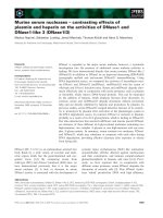
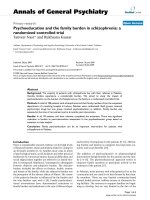
000071-90. This study adheres to the CONSORT guidelines, an additional file with the CONSORT diagram is
available (Fig. 1). Adult patients (age > 18 years) of both
gender scheduled for a pancreaticoduodenectomy
(Whipple’s procedure) in Ghent University Hospital and
Page 2 of 11
with an American Society of Anesthesiologists (ASA)
physical status of I to III were included. Exclusion criteria were allergy to the medication, renal insufficiency
(serum creatinine > 2 mg dL− 1), severe heart failure
(ejection fraction < 25%), pre-operative hemodynamic instability, atrial fibrillation, sepsis, body mass index > 40
kg m− 2,
severe
coagulopathy
(INR > 2),
thrombocytopenia (< 80 × 103 μL− 1) or history of severe
postoperative nausea and vomiting (PONV).
After written informed consent, patients were randomly allocated to two groups. Group P received total
intravenous anesthesia using a propofol target controlled
infusion (TCI), group S received inhalation anesthesia
using sevoflurane. An anesthesia co-worker, not involved
in the study, performed a simple randomization using
sealed pre-numbered envelopes. After randomization of
all patients, drop-outs which occurred during the trial
were replaced in order of their appearance.
Primary objective was to compare the effect of propofol (group P) and sevoflurane (group S) on arterial, portal and total HBF and on the caval and portal vein
pressure during pancreaticoduodenectomy. The secondary objectives were to compare the need for inotropic
and vasopressor support, the amount of fluids administered, plasma lactate levels and blood loss during surgery
between both groups.
Anesthetic procedure
All patients received standard anesthesia care according
to the departmental protocol. Patients received ASA
standard anesthesia monitoring. The departmental
protocol for this type of procedures includes placement
of an epidural catheter for postoperative analgesia. This
catheter was placed before induction of anesthesia but
only used after all experimental measurements had been
performed, which was at the end of surgery. Depth of
anesthesia was measured using Bispectral Index (BIS™,
Covidien, MA, USA) monitoring and titrated to remain
between 40 and 60. After induction of anesthesia, central
venous and arterial catheters were placed. A 5-Fr PiCCO
catheter (Maquet, Getinge Group, Germany) placed in
the left femoral artery was used for the additional
hemodynamic assessment in the current study.
Before induction of anesthesia 4 mg intravenous dexamethasone was administered for prevention of PONV.
Induction and maintenance of general anesthesia differed in both groups. In group S, induction of anesthesia
was obtained with propofol 1–2 mg kg− 1 until loss of
consciousness. Anesthesia was maintained with sevoflurane. In group P, induction and maintenance were performed using propofol TCI (Schnider Model), starting at
an effect site concentration of 5.0 mcg ml− 1. In both
groups, anesthesia was titrated to obtain a BIS between
40 and 60. For intraoperative analgesia, TCI remifentanil
van Limmen et al. BMC Anesthesiology
(2020) 20:241
Page 3 of 11
Fig. 1 CONSORT. CONSORT flow diagram
(Minto Model) was used in both groups. TCI remifentanil was started at an effect site concentration of 5 ng
ml− 1 and titrated according heart rate and blood pressure. Neuromuscular blockade was achieved using rocuronium, 1 mg kg− 1 at induction and intermittent boluses
during surgery. Before each experimental measurement,
an additional bolus of rocuronium 10 mg was given.
After tracheal intubation and lung recruitment, mechanical ventilation was started with a tidal volume 6–8 ml
kg− 1 ideal body weight, respiratory rate 12–14 min− 1
and a positive end-expiratory pressure of 5 cmH2O.
Ventilation was adjusted according to the data of the arterial blood gas analysis. All patients received an individualized goal-directed hemodynamic therapy (GDHT)
according to the departmental written procedure. A
baseline crystalloid infusion (Plasmalyte A, Baxter S.A.,
Lessines, Belgium) of 3 ml kg− 1 h− 1 was administered.
The hemodynamic goal was a cardiac index (CI) > 2.2 L
min− 1 m− 2 with a mean arterial pressure (MAP) > 60
mmHg and a pulse pressure variation (PPV) < 12%.
When PPV was > 12% a bolus of 200 ml colloid
(Volulyte A, Fresinius Kabi NV, Schelle Belgium) was
administered. When CI was > 2.2 L min− 1 m− 2 in the
presence of a MAP < 60 mmHg, a noradrenaline infusion was started at 0.1 mcg kg− 1 min− 1 and titrated
according to the MAP. To temporarily bridge the latency of effect the noradrenaline infusion, boluses of
ephedrine 3 mg were administered when heart rate
was less than 60 beats per minute or phenylephrine
0.1 mg, if heart rate was > 60 beats/min. At the end
of surgery, all patients received 1 g paracetamol and
10–15 ml
ropivacaine
0.15%
epidurally
for
postoperative analgesia. A nerve stimulator was used
to assess the evoked muscle response with doubleburst-stimulation (DBS) or train-of-four (TOF). Reversal of neuromuscular block was done with sugammadex, guided by the twitch response to DBS or TOF.
Measurements
Hemodynamic variables were measured using Pulsioflex™ (Maquet, Getinge Group, Germany). After placement of the 5-Fr arterial catheter in the femoral artery,
the pulse contour analysis was calibrated using 3 boluses
of 20 ml of cold saline. The hemodynamic variables measured were heart rate (HR), central venous pressure
(CVP), MAP, CI and PPV. To assess the performance of
the GDHT protocol, we calculated the percentage of
time, during which the hemodynamic goals were within
the limits of the targets set (CI > 2.2 L min− 1 m− 2 with
PPV < 12% and MAP > 60 mmHg).
During surgery, 3 flow measurements were performed
by the surgeon, at predefined times, while systemic
hemodynamic variables were recorded. Pancreaticoduodenectomy is a standardized surgical procedure, which
we divided in three different stages. The first flow measurements were made after transection of the gastroduodenal artery (T1). The second flow measurement
(T2) was performed after pancreatectomy. The last flow
measurement (T3) was performed before surgical reconstruction and minimal 10 min after T2. Blood flow measurements at the hepatic artery and portal vein were
obtained using perivascular ultrasound transit time flow
probes (TTFM, Medi-Stim AS, Oslo, Norway) [19]. Different probe sizes were used according to the type and
van Limmen et al. BMC Anesthesiology
(2020) 20:241
size of the vessel (range 2–12 mm). Blood flow was
expressed in ml min− 1. At the same time, the pulsatility
index (PI) was calculated by the TTFM. PI quantifies
pulsatility of a blood flow wave which represents vascular resistance of the blood vessel downstream. PI is calculated by maximum volumetric peak flow minus
minimum volumetric peak flow divided by mean volumetric volume [20]. Simultaneously with flow measurements, additional pressure measurements were
performed in the portal and caval vein. A 25-gauge needle was directly placed in the vein and connected to a
pressure transducer. Systemic hemodynamic, regional
hepatic flow and portocaval pressure measurements
were performed simultaneously during apnea to
minimize the effect of ventilation. The relative blood
flow over the hepatic artery or portal vein was calculated
by dividing arterial or portal HBF by CO.
Statistical analysis
To the best of our knowledge, no previous studies are
available comparing the effect of propofol and sevoflurane on HBF. Therefore, we could not rely on previous
publications to determine the exact sample size needed
to compare the effects of both anesthetics on HBF. As
such, the current study is also a feasibility study and the
information provided can be used for sample size calculation of future studies assessing HBF using TTFM. The
publication of Sand Bown et al. [21] was used to define a
clinically relevant reduction of HBF. Based on this publication, a 30% reduction in arterial and portal HBF was
considered clinically significant. G*Power 3.1.9.2 was used
to calculate the sample size [22]. For an alpha error of 5%,
a beta error of 20%, SD of 0.25 and an effect size F of 0.6,
each group necessitated 9 patients to detect a flow reduction of 30%. After testing for normal distribution with the
Shapiro-Wilk normality test, data between both groups
were compared using a two-way ANOVA for repeated
measurements, or its non-parametric equivalent where appropriate. Pairwise comparisons were done using paired ttest with Bonferroni correction for significance.
Numbers of patients necessitating vasopressor support
were compared using Fisher exact test. All statistical
tests were performed using R (version 3.3.3) [23].
Page 4 of 11
because of in-operability (n = 4), technical failure of the
registration device (n = 2), unexpected portal hypertension (n = 1) and investigator unavailability (n = 1). In
group P, 1 patient dropped out due to technical failure
of the registration device. Finally, data of 9 patients in
each group were analyzed (Fig. 1). Patient characteristics
are listed in Table 1. Both groups were comparable with
respect to age, gender, length, weight, BMI, ASA physical status, pre-operative blood pressure and heart rate,
and smoking status.
Hemodynamic variables
Hemodynamic variables are listed in Table 2. All patients
received individualized GDHT as described above. The
pre-set hemodynamic targets were obtained in both
groups, but MAP was lower in group S (p = 0.01). Successful achievement of the hemodynamic targets, as defined
by the cumulative time within pre-set hemodynamic goals
were met, and expressed as a percentage of total study
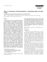
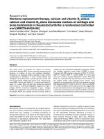
duration, it was higher in group P (p = 0.046) (Fig. 2a). In
group P mean percentage of time in range was 89% (SD
5.5%) while in group S, a mean of 76% (SD 18.2%) was
achieved. The total dose of vasopressors needed to obtain
these pre-set targets however was higher in group S than
for group P ephedrine respectively 10.4 mg (SD 5.6 mg)
versus 5.3 mg (SD 3.3 mg) (p = 0.04) and noradrenaline infusion 2809 mcg (SD 2197 mcg) versus 227 mcg (SD 237
mcg) (p 0.0004) (Fig. 2b). All patients required noradrenaline in group S, as compared to only 3 patients in group
P (p = 0.009). A rise in blood lactate levels over time was
observed in both groups (p = 0.0003) but the increase was
significantly more pronounced in group S (p = 0.04).
Fluid management
Intraoperative characteristics are listed in Table 3. The
total amount of administered crystalloids was similar between group P and group S respectively 1974 ml (SD 440
ml) versus 2308 ml (SD 471 ml) (p = 0.14). The total
amount of administered colloids was similar between both
groups, 1067 ml (SD 500 ml) for group P versus 1078 ml
(441 ml) for group S (p = 0.96). Surgical time was
Table 1 Patient characteristics
Propofol Group (n = 9) Sevoflurane Group (n = 9)
Results
Patient characteristics
Between June 2017 and January 2018, a total of 35 patients were assessed for eligibility to participate in the
study. Six patients were excluded based on the exclusion
criteria. Twenty- nine patients were included. Two patients were additionally excluded due to unexpected coagulopathy and investigator unavailability. A total of 27
patients were randomized of whom 10 in group P and
17 in group S. In group S, 8 patients dropped out
Age (year)
63.6 (5.4)
63.9 (12.0)
Gender (F/M)
4/5
3/6
Height (cm)
169.1 (8.8)
169.8 (7.9)
Weight (kg)
72.0 (7.5)
67.4 (9.9)
−2
BMI (kg m )
25.2 (2.5)
23.3 (2.5)
Smoker
1
5
ASA class (I/II/III) 1/3/5
0/6/3
Data are presented as mean (SD). F/M Female/male ratio, Body mass index
(BMI), American Society of Anesthesiologist physical status (ASA)
van Limmen et al. BMC Anesthesiology
(2020) 20:241
Page 5 of 11
Table 2 Hemodynamic data
MAP
(mmHg)
HR
(bpm)
CVP
(mmHg)
Propofol Group
(n = 9)
Sevoflurane Group
(n = 9)
Between group difference
T1
82 (9)
69 (10)
13 [4–22]a
T2
76 (5)
74 (9)
1 [−7–9]
T3
82 (5)
76 (9)
6 [−2–13]
T1
75 (12)
78 (14)
-2 [− 19–14]
T2
79 (10)
80 (11)
-1 [− 15–13]
T3
77 (8)
79 (12)
-2 [−16–11]
T1
5 (2)
5 (2)
0 [−2–2]
T2
5 (1)
5 (4)
-1 [−4–2]
T3
4 (2)
6 (2)
-2 [−4–1]
T1
2.7 (0.4)
3.1 (0.8)
−0.3 [−1.1–0.4]
T2
3.0 (0.5)
3.3 (0.6)
−0.3 [−1–0.4]
T3
3.0 (0.4)
3.2 (0.6)
−0.2 [− 0.9–0.4]
T1
1267 (231)
991 (230)
276 [−52–604]
T2
1084 (209)
1000 (316)
84 [−301–469]
T3
1168 (178)
1031 (261)
136 [−166–439]
PPV
(%)
T1
9 (4.0)
8 (4.6)
1.2 [−3.6–6.1]
T2
9 (1.7)
10 (5.6)
−0.9 [−4.9–3.1]
T3
8 (3.0)
8 (5.8)
0.1 [−3.9–4.1]
Lactacte
(mg.dL−1)
T1
8.9 (1.5)
12.2 (3.8)
−3.3 [− 6.0 – − 0.6]a
T2
10.1 (2.7)b
16.3 (6.8)b
−6.2 [− 10.9 – − 1.6]a
T3
10.5 (2.9)
b
b
18.2 (8.4)
−7.7 [− 13.9 – − 1.5]a
PaCO2
(mmHg)
T1
42 (4.5)
42 (5.9)
0.1 [−5.2–5.4]
T2
41 (6.1)
43 (4.8)
−2.1 [−8.2–4.1]
T3
42 (3.3)
42 (5.1)
−0.3 [−3.8–4.5]
pH
T1
7.36 (0.05)
7.34 (0.05)
0.02 [−0.04–0.08]
T2
7.35 (0.05)
7.33 (0.05)
0.01 [−0.04–0.07]
T3
7.34 (0.05)
7.35 (0.06)
−0.01 [− 0.06–0.03]
CI
(L.min−1.m−2)
SVR
(dyn.sec.cm− 5)
Data are presented as mean (SD). MAP Mean arterial pressure, HR Heart rate, CVP Central venous pressure, CI Cardiac index, SVR Systemic vascular resistance, PPV
Pulse pressure variation, PaCO2 Arterial carbon dioxide tension. Bonferroni corrected significance are marked as a for between group comparisons and b for
significant within group difference compared to T1
significantly longer in group S, 534 min (SD 98 min) compared with group P, 465 min (SD 68 min) (p = 0.0002).
This was related to variability in the time to obtain a diagnosis from the intraoperative frozen section. However,
there was no difference in the delivered amount of crystalloid per minute between both groups, 4.7 ml.min− 1 (SD
2.3 ml.min− 1) for group S compared with 4.3 ml.min− 1
(SD 0.8 ml.min− 1) for group P (p = 0.63). Urinary output
was similar in both groups, respectively 779 ml (SD 602
ml) for group S compared with 463 ml (SD 198 ml) (p =
0.17). Also, blood loss was comparable in both groups
(p = 0.47).
Flow measurements
Flow measurements are summarized in Table 4. Total
HBF was similar in both groups at all points of
measurement (p = 0.76). There was no difference in portal
HBF (p = 0.85) and arterial HBF (p = 0.70) between both
groups at all time points. There was no difference between
the relative blood flow in the hepatic artery (p = 0.67) and
in the portal vein (p = 0.85) between both groups. The ratio portal over arterial HBF also showed no difference between groups (p = 0.22). Portal and caval vein pressures
were similar in both groups at all measurement times.
The PI of both portal vein (p = 0.38) and hepatic artery
(p = 0.61) showed no difference between groups.
Discussion
In this study we compared the effect of a propofol- and
sevoflurane-based anesthesia on HBF during GDHT.
Our results showed that portal, arterial and total HBF
were similar in propofol- and sevoflurane-anesthetized
van Limmen et al. BMC Anesthesiology
(2020) 20:241
Page 6 of 11
Fig. 2 a: Maintenance of hemodynamic targets. Efficacy of goal-directed hemodynamic therapy during procedure: percentage of time within
hemodynamic goals as defined in the departmental protocol between propofol titrated-patients (group P) and sevoflurane-titrated patients
(group S). * P < 0.05. b: Noradrenaline infusion. Noradrenaline infusion related to observation periods
van Limmen et al. BMC Anesthesiology
(2020) 20:241
Page 7 of 11
Table 3 Intraoperative characteristics
Propofol Group (n = 9)
Sevoflurane Group (n = 9)
Crystalloids (ml)
1974 (440)
2308 (471)
Crystalloids (ml.min− 1)
4.3 (0.8)
4.7 (2.3)
Colloids (ml)
1067 (500)
1078 (441)
Blood loss (ml)
567 (212)
689 (448)
Urinary Output (ml)
463 (198)
779 (602)
Ephedrine (mg) *
5.3 (3.3)
10.3 (5.6)
Phenylephrine (mcg)
0.16 (0.11)
0.37 (0.38)
Noradrenaline (mcg) *
227 (237)
2809 (2197)
Patients requiring vasopressor support
3
9
Surgical time (min) *
465 (68)
534 (98)
Remifentanil (mcg)
3915 (1520)
3826 (1068)
Patients receiving Somatostatin infusion
4
5
Intraoperative characteristics are expressed in mean (SD)
* = statistically significant difference (p < 0.05) between group comparison
patients. Due to the application of a GDHT protocol
similar hemodynamic variables were observed in both
groups. However, patients in group S required a significantly higher administration of vasopressor to maintain
adequate MAP.
To our knowledge there are no previous human trials,
assessing and comparing HBF with direct flow measurements under propofol- and sevoflurane-based anesthesia.
Clinical practice guidelines on liver transplantation are
lacking advice for the choice of anesthetic technique for
maintenance [24]. Previous studies have suggested that
sevoflurane compared to propofol may attenuate the effects of ischemia-reperfusion injury after liver resection
[25]. However, a similar study comparing effects of propofol on sevoflurane on hepatic graft survival yielded no different effects between both anesthetic agents [26].
Maintaining adequate HBF is important for allograft [2–4]
and patient survival [5, 6]. Yet, potential effects of
anesthetic agents on HBF in the clinical setting remain
largely unexplored. Both sevoflurane and propofol have an
effect on HBF [7]. Conflicting results about the effect of
propofol on HBF have been described. Previous studies
have suggested that propofol increases total HBF. However, the putative mechanism for this increase in HBF differs between the studies. A study in rats showed an
increase in total HBF by an increase of both arterial and
portal HBF. Propofol reduced hepatic arterial resistance
and portal venous resistance in an identical manner [14].
A study in dogs showed similar results. However, in this
study, there was only a transient increase in total HBF by
propofol which was mediated primarily by an increased
arterial HBF [13]. A study in rabbits showed an increased
total HBF with propofol, primarily by an increased portal
HBF [15]. Conversely, one study in sheep showed a reduction in total HBF [27]. Only one human study was
performed. In this study, desflurane and propofol were
compared in 20 patients using a cross-over design. Total
HBF was significantly higher in propofol-treated patients
compared to desflurane-treated patients [16]. The mechanism behind the observed effects of propofol on HBF remains unclear. It was assumed that the metabolization of
propofol increases hepatic oxygen consumption. To maintain hepatic oxygen balance, there would be then a compensatory increased oxygen delivery primarily by
increasing portal HBF [14, 15].
The effect of sevoflurane on HBF remains unclear.
Animal studies suggested that sevoflurane has only minimal effects on total HBF. A study in dogs showed that
sevoflurane resulted in a hepatic vasodilation with a reduction in portal HBF at 1.2 and 2.0 MAC but a significant increased arterial HBF was only seen at 2.0 MAC
[17]. Other animal studies confirmed this finding. Sevoflurane maintained total HBF, and although portal HBF
was reduced, arterial HBF increased, resulted in sufficient HBF to maintain hepatic oxygen delivery [18, 28].
Results from human studies are conflicting. Hongo et al.
showed a reduction in total HBF in sevoflurane but
Kanaya et al. on the contrary found no effect on HBF
with sevoflurane [29, 30].
The previous studies, both animal and human, used different techniques to measure arterial, portal and total HBF.
HBF can be measured both directly and indirectly [31]. Indirect measurements are less invasive but also less accurate.
Examples of indirect measurements are radio-labelled microspheres [14] or indicator substance such as sodium
bromsulphthalein [27] and the indocyanine green (ICG)
clearance test [16, 29, 30]. Propofol interacts with ICG and
inhibits the hepatic clearance of ICG which may consequently lead to an underestimation of true HBF [32, 33].
Recently, total HBF was measured indirectly by calculating
van Limmen et al. BMC Anesthesiology
(2020) 20:241
Page 8 of 11
Table 4 Hepatic blood flow and pressures
Total HBF
(ml.min−1)
T1
Propofol Group
(n = 9)
Sevoflurane Group
(n = 9)
Between group difference
997 (344)
1003 (411)
−5 [− 395–384]
T2
937 (231)
860 (318)
77 [− 272–426]
T3
998 (264)
943 (225)
55 [− 252–362]
Portal HBF
(ml.min− 1)
T1
760 (275)
790 (317)
−30 [− 352–291]
T2
687 (203)
612 (218)
75 [− 163–313]
T3
714 (210)
704 (137)
10 [− 234–254]
Arterial HBF
(ml.min− 1)
T1
237 (150)
212 (138)
25 [− 130–180]
T2
249 (110)
247 (199)
2 [−212–216]
T3
284 (101)
239 (149)
45 [−110–200]
T1
20.0 (6.0)
19.9 (12.1)
0.1 [−7.4–7.6]
T2
17.4 (3.4)
15.5 (7.6)
1.8 [−5.2–8.9]
T3
18.4 (4.1)
17.4 (6.7)
1.0 [−4.8–6.7]
T1
15.2 (5.2)
15.9 (9.4)
−0.6 [−8.1–6.9]
T2
12.8 (3.7)
11.1 (5.3)
1.6 [−3.7–7.0]
T3
13.1 (3.8)
12.8 (3.8)
0.3 [−3.7–4.3]
T1
4.8 (2.8)
4.1 (3.4)
0.7 [−1.4–2.8]
T2
4.6 (1.9)
4.4 (3.7)
0.2 [−3.6–4.0]
T3
5.3 (2.0)
4.6 (3.5)
0.7 [−2.6–4.0]
T1
10 (6)
9 (3)
1 [−4–7]
T2
6 (3)
10 (4)
−4 [−7–0]
T3
8 (4)
9 (5)
0 [−4–3]
T1
6 (3)
5 (3)
1 [−2–4]
T2
5 (3)
7 (4)
−1 [−5–2]
T3
7 (4)
7 (3)
0 [−5–4]
T1
0.4 (0.2)
0.5 (0.3)
−0.1 [−0.4–0.2]
T2
0.3 (0.2)
0.5 (0.3)
−0.2 [− 0.4–0]
T3
0.5 (0.2)
0.5 (0.2)
0 [−0.1–0.2]
Relative Total HBF
(% of CO)
Relative Portal HBF
(% of CO)
Relative Arterial HBF
(% of CO)
Portal Vein Pressure
(mmHg)
Caval Vein Pressure
(mmHg)
PI Portal Vein
PI Hepatic Artery
T1
1.4 (0.9)
1.7 (0.9)
−0.3 [− 0.9–0.4]
T2
1.4 (0.6)
1.5 (0.7)
−0.1 [− 0.8–0.5]
T3
1.4 (0.7)
1.5 (0.5)
− 0.1[− 0.7–0.6]
Data are presented as mean (SD) for both groups. Mean estimated between group differences with their confidence intervals (95%) are provided in the right
column. No Bonferroni corrected significant difference (p < 0.05) were found for between or within group comparisons. Hepatic blood flow (HBF). PI = pulsatility
index. CO = cardiac output. No significant differences (p < 0.05) were found for between or within group comparisons
blood flow at the hepatic vein using transesophageal echocardiography [34]. Direct measurement of HBF is a fast and
accurate technique but is also more invasive. Previous studies used Doppler or electromagnetic flow probes which
were directly placed around the hepatic artery and portal
vein [13, 15, 17].
During liver transplantation, assessment of the graft
blood flow by TTFM plays an important role in the
assessment of the survival chances of the allograft
[35, 36]. If flow measurements are needed, TTFM is
very reliable and is considered to be the ăgold standardă for measuring blood flow [19]. As our study
demonstrated, measuring HBF using TTFM is feasible
in a clinical steady state.
The results of the present study should be interpreted
within the constraints of the methodological protocol. First,
as a predefined GDHT was used to maintain patient’s
hemodynamic stability, the current data should not be
interpreted as a direct independent effect of both propofol
and sevoflurane on the hepatic circulation. Indeed,
hemodynamic targets were achieved in both groups, but to
achieve this, a significantly higher vasopressor support was
needed in sevoflurane-titrated patients, while propofoltitrated patients had higher MAP, well above target MAP
without vasopressor support. As both groups were
van Limmen et al. BMC Anesthesiology
(2020) 20:241
comparable of depth of anesthesia, a possible explanation
could be a more profound vasodilation with sevoflurane
than with propofol. As this vasodilating effect was compensated by the vasopressor therapy, it cannot be excluded that
at the same time a vasodilatory effect at the level of the
hepatic circulation was also blunted. In the present study
noradrenaline was used to maintain adequate MAP. The effect of noradrenaline on HBF during surgery remains complex and unclear. The splanchnic circulation has a wide
variety and distribution of adrenergic receptors [37] and
therefore noradrenaline may affect HBF. Previous animal
studies suggested that noradrenaline reduced HBF [38], primarily by reducing arterial HBF [39]. However, a recent
study in pigs showed that noradrenaline infusion - used to
correct hypotension - did not affect HBF during abdominal
surgery [40]. The current observations do not allow to
make inferences of potential independent effects of
noradrenaline on HBF. Interestingly, lactate levels in
the present study were higher in group S. Although
we do not have a straightforward explanation for this
phenomenon, it might be seen as indication that despite the GDHT-related stability in hemodynamic variables, global tissue oxygenation was jeopardized more
than in group P.
Secondly, the data obtained may have been influenced by other factors related to intra-operative patient care. A total of 9 patients received – on surgical
indication - somatostatin at 250 mcg h− 1 (4 in group
P and 5 in group S) to reduce pancreatic secretion.
Previous animal studies suggested that somatostatin
may affect portal HBF and portal pressure primarily
in the presence of portal hypertension [41, 42]. We
cannot exclude that the use of somatostatin had an
influence on the results, but the number of patients
treated were equally divided between both groups. In
addition, a post-hoc sub-analysis comparing patients
with and without somatostatin treatment revealed no
differences in hemodynamic or hepatic flow profiles.
Thirdly, selecting the correct size of the probe is of
crucial importance to obtain reliable flow data, as the
use of an oversized probe may lead to overestimation
of the blood flow [43]. In our institution TTFM is a
routinely used procedure during major liver surgery
and liver transplantation. The size of the probe was
meticulously assessed by the participating surgeons
who are highly experienced in the use of this technique. Fourthly, a total of 9 patients dropped out during the trial. These patients were replaced after
randomization in order of their dropout appearance.
This may impose a risk for allocation bias. As most
dropouts occurred due to inoperability, this could not
be influenced by the researcher. Replacement of dropouts was done in order of their dropout appearance,
which could not be influenced by the researcher.
Page 9 of 11
Therefore, the risk for allocation bias as such seems
limited. Fifthly, no previous studies were available to
assess differences in HBF between sevoflurane- and
propofol-anesthetized patients. Therefore, we could
not rely on previous publications to determine the
exact sample size needed to compare the effects of
both anesthetics on HBF and we relied on the publication of Sand Bown et al. [21] to determine the clinically
relevant reduction of HBF. However, a reduction of 30%
in portal and arterial HBF is probably an overestimation
of the real effect size. This may impose a risk for insufficient power of the study. To address this issue, we conducted a post-hoc power analysis with our current results.
We observed a mean total HBF for propofol of 977
ml.min− 1 (SD 260 ml.min− 1) and for sevoflurane of 935
ml.min− 1 (SD 300 ml.min− 1). When using the results of
Meierhenrich [16], who had an effect size f of 0.54, we calculated a post hoc power of 75% which is slightly lower
than the a priori set power of 80%. As such, the current
study should be considered as a pilot study, performed to
check the feasibility of assessing HBF during goal-directed
hemodynamic therapy and to provide clinically relevant
data on HBF under anesthesia, which may be used, to explore effect size assessments in future trials.
Conclusion
The results of the present study indicate that when
applying a GDHT, aiming at stable hemodynamic variables, HBF during propofol- and sevoflurane-based
anesthesia was similar. However, to maintain these
identical
hemodynamic
goals,
sevofluraneanaesthetized patients necessitated a significantly
higher need for vasopressor support and blood lactate
levels were higher in comparison to patients receiving
propofol-based anesthesia.
Abbreviations
BMI: Body Mass Index; CI: Cardiac Index; CO: Cardiac Output; CVP: Central
Venous Pressure; DBS: Double Burst Stimulation; GDHT: Goal-directed
Hemodynamic Therapy; HBF: Hepatic Blood Flow; HR: Heart Rate; MAP: Mean
Arterial Pressure; PI: Pulsatility index; PONV: Postoperative Nausea and
Vomiting; PPV: Pulse Pressure Variation; PVP: Portal Venous Pressure;
PVR: Portal Venous Resistance; SVR: Systemic Vascular Resistance; TCI: Target
Control Infusion; TOF: Train-of-four; TTFM: Transit Time Flow Measurement
Acknowledgements
The authors wish to thank miss Ann De Bruyne (study nurse) and Luis Abreu
De Carvalho, M.D. for their support in this trial.
Authors’ contributions
JVL: study design – patient recruitment – data collection – data analysis –
writing manuscript. PW: study design – patient recruitment – data analysis –
statistical analysis – revising manuscript. FB: study design – patient
recruitment – data collection – revising manuscript. AVL: patient recruitment
– data collection – revising manuscript. LC: study design - patient
recruitment – data collection. PW: data analysis – writing manuscript –
revising manuscript. SDH: study design – patient recruitment – data
collection – data analysis – statistical analysis – writing manuscript. LDB:
study design – patient recruitment – data collection – data analysis – writing
manuscript. The authors have read and approved the manuscript.
van Limmen et al. BMC Anesthesiology
(2020) 20:241
Funding
JVL received an educational non-restricted grant from the Society of
Anesthesia and Resuscitation of Belgium (SARB) in 2017 for this study. JVL is
the principal investigator of this trial and contributed in all aspects of the
study, as mentioned below.
Availability of data and materials
The datasets used and/or analyzed during the current study are available
from the corresponding author on reasonable request.
Ethics approval and consent to participate
This study was approved by the ethical committee of the University Hospital
Ghent, Belgium. Study protocol number is AGO/2017/002 – EC/2017/0164.
EudraCT number is 2017–000071-90. Clin.trail.gov is NCT03772106. Patients
provided written informed consent.
Consent for publication
Not applicable.
Competing interests
JVL received 50 vials of propolipid (Fresenius-Kabi, Schelle, Belgium) without
any restrictions nor obligations.
Author details
Department of Anaesthesiology and Perioperative Medicine, Ghent
University Hospital, Corneel Heymanslaan 10, 9000 Ghent, Belgium.
2
Department of General and Hepatic-pancreatico-biliary Surgery and Liver
transplantation, Ghent University Hospital, Corneel Heymanslaan 10, Ghent
9000, Belgium.
1
Received: 8 June 2020 Accepted: 7 September 2020
References
1. Kin Y, Nimura Y, Hayakawa N, Kamiya J, Kondo S, Nagino M, et al. Doppler
analysis of hepatic blood flow predicts liver dysfunction after major
hepatectomy. World J Surg. 1994;18:143–9. />BF00348207.
2. Pratschke S, Meimarakis G, Mayr S, Graeb C, Rentsch M, Zachoval R, et al.
Arterial blood flow predicts graft survival in liver transplant patients. Liver
Transpl. 2011;17:436–45. />3. Lominchar PL, Orue-Echebarria MI, Martín L, Lisbona CJ, Salcedo MM,
Olmedilla L, et al. Hepatic flow is an intraoperative predictor of early
allograft dysfunction in whole-graft deceased donor liver transplantation: an
observational cohort study. World J Hepatol. 2019;11:689–700. https://doi.
org/10.4254/wjh.v11.i9.689.
4. Kelly DM, Shiba H, Nakagawa S, Irefin S, Eghtesad B, Quintini C, et al.
Hepatic blood flow plays an important role in ischemia-reperfusion injury.
Liver Transpl. 2011;17:1448–56. />5. Spitzer AL, Dick AAS, Bakthavatsalam R, Halldorson JB, Salvalaggio PR, Reyes
JD, et al. Intraoperative portal vein blood flow predicts allograft and patient
survival following liver transplantation. Hpb. 2010;12:166–73. />10.1111/j.1477-2574.2009.00137.x.
6. Marambio A, Ton JMC, Gómez LMM, Martínez JMA, Bellido CB, Artacho
GS, et al. Intraoperative portal vein flow > 123 mL/min per 100 g predicts a
better survival of patients after liver transplantation. Transplant Proc. 2018;
50:3582–6. />7. Gelman S. General anesthesia and hepatic circulation. Can J Physiol
Pharmacol. 1987;65:1762–79. />8. Lautt WW. Mechanism and role of intrinsic regulation of hepatic arterial
blood flow: hepatic arterial buffer response. Am J Physiol Gastrointest Liver
Physiol. 1985;12. />9. Parks DA, Jacobson ED. Physiology of the splanchnic circulation. Arch Intern
Med. 1985;145:1278–81. />00360070158027.
10. Gelman S, Mushlin PS. Catecholamine-induced changes in the splanchnic
circulation affecting systemic hemodynamics. Anesthesiology. 2004;100:434–
9. />11. Richardson PDI, Withrington P. Physiological regulation of the hepatic
circulation. Annu Rev Physiol. 1982;44:57–69. />annurev.ph.44.030182.000421.
Page 10 of 11
12. Nakamoto S, Tatara T, Okamoto T, Hirose M. Complex effects of continuous
vasopressor infusion on fluid responsiveness during liver resection: a
randomised controlled trial. Eur J Anaesthesiol. 2019;36:667–75. https://doi.
org/10.1097/EJA.0000000000001046.
13. Wouters PF, Van de Velde MA, Marcus MAE, Deruyter HA, Van Aken H.
Hemodynamic changes during induction of anesthesia with Eltanolone and
Propofol in dogs. Anesth Analg. 1995;81:125–31. />00000539-199507000-00025.
14. Carmichael FJ, Crawford MW, Khayyam N, Saldivia V. Effect of propofol
infusion on splanchnic hemodynamics and liver oxygen consumption in
the rat: a dose-response study. Anesthesiology. 1993;79:1051–60. https://doi.
org/10.1097/00000542-199311000-00024.
15. Zhu T, Pang Q, McCluskey SA, Luo C. Effect of propofol on hepatic blood
flow and oxygen balance in rabbits. Can J Anesth. 2008;55:364–70. https://
doi.org/10.1007/BF03021492.
16. Meierhenrich R, Gauss A, Mühling B, Bracht H, Radermacher P, Georgieff M,
et al. The effect of propofol and desflurane anaesthesia on human hepatic
blood flow: a pilot study. Anaesthesia. 2010;65:1085–93. />1111/j.1365-2044.2010.06504.x.
17. Bernard JM, Doursout MF, Wouters P, Hartley CJ, Merin RG, Chelly JE. Effects
of sevoflurane and isoflurane on hepatic circulation in the chronically
instrumented dog. Anesthesiology. 1992;77:541–5. />00000542-199209000-00021.
18. Frink EJ, Morgan SE, Coetzee A, Conzen PF, Brown BR. The effects of
sevoflurane, halothane, enflurane, and isoflurane on hepatic blood flow and
oxygenation in chronically instrumented greyhound dogs. Anesthesiology.
1992;76:85–90. />19. Beldi G, Bosshard A, Hess OM, Althaus U, Walpoth BH. Transit time flow
measurement: experimental validation and comparison of three different
systems. Ann Thorac Surg. 2000;70:212–7. />20. Aleksic M, Heckenkamp J, Gawenda M, Brunkwall J. Pulsatility index
determination by flowmeter measurement: a new indicator for vascular
resistance? Eur Surg Res. 2004;36:345–9. />21. Sand Bown L, Ricksten SE, Houltz E, Einarsson H, Söndergaard S, Rizell M,
et al. Vasopressin-induced changes in splanchnic blood flow and hepatic
and portal venous pressures in liver resection. Acta Anaesthesiol Scand.
2016;60:607–15. />22. Erdfelder E, FAul F, Buchner A, Lang AG. Statistical power analyses using
G*power 3.1: tests for correlation and regression analyses. Behav Res
Methods. 2009;41:1149–60. />23. Team R. R-studio User’s manual; 2018.
24. Burra P, Burroughs A, Graziadei I, Pirenne J, Valdecasas JC, Muiesan P, et al.
EASL clinical practice guidelines: liver transplantation. J Hepatol. 2016;64:
433–85. />25. Beck-Schimmer B, Breitenstein S, Bonvini JM, Lesurtel M, Ganter M, Weber A,
et al. Protection of pharmacological postconditioning in liver surgery: results
of a prospective randomized controlled trial. Ann Surg. 2012;256:837–45.
/>26. Beck-Schimmer B, Bonvini JM, Schadde E, Dutkowski P, Oberkofler CE,
Lesurtel M, et al. Conditioning with sevoflurane in liver transplantation:
results of a multicenter randomized controlled trial. Transplantation. 2015;
99:1606–12. />27. Runciman WB, Mather LE, Selby DG. Cardiovascular effects of propofol and
of thiopentone anaesthesia in the sheep. Br J Anaesth. 1990;65:353–9.
/>28. Takeda S, Sato N, Tomaru T. Haemodynamic and splanchnic organ blood flow
responses during sevoflurane-induced hypotension in dogs. Eur J Anaesthesiol.
2002;19:442–6. />29. Kanaya N, Nakayama M, Fujita S, Namiki A. Comparison of the effects of
sevoflurane, isoflurane and halothane on indocyanine green clearance. Br J
Anaesth. 1995;74(2):164–7. />30. Hongo T. Sevoflurane reduced but isoflurane maintained hepatic blood
flow during anesthesia in man. J Anesth. 1994;8:55–9. />1007/BF02482756.
31. Chow PKH, Yu WK, Soo KC, Chan STF. The measurement of liver blood flow:
a review of experimental and clinical methods. J Surg Res. 2003;112:1–11.
/>32. Sear JW, Diedericks J, Foex P. Continuous infusions of propofol administered
to dogs: effects on icg and propofol disposition. Br J Anaesth. 1994;72:451–
5. />
van Limmen et al. BMC Anesthesiology
(2020) 20:241
33. Lange H, Stephan H, Rieke H, Kellermann M, Sonntag H, Bircher J. Hepatic and
extrahepatic disposition of propofol in patients undergoing coronary bypass
surgery. Br J Anaesth. 1990;64:563–70. />34. Schütz W, Meierhenrich R, Träger K, Gauss A, Radermacher P, Georgieff M. Is
it feasible to monitor total hepatic blood flow by use of transesophageal
echography? An experimental study in pigs. Intensive Care Med. 2001;27:
580–5. />35. Rasmussen A, Hjortrup A, Kirkegaard P. Intraoperative measurement of graft
blood flow - a necessity in liver transplantation. Transpl Int. 1997;10:74–7.
36. Lisik W, Gontarczyk G, Kosieradzki M, Lagiewska B, Pacholczyk M, Adadyński
L, et al. Intraoperative blood flow measurements in organ allografts can
predict postoperative function. Transplant Proc. 2007;39:371–2. https://doi.
org/10.1016/j.transproceed.2007.01.046.
37. Guimarães S, Moura D. Vascular adrenoceptors: an update. Pharmacol Rev.
2001;53:319–56.
38. Turk LN, Shoemaker WC. Hepatic vascular response to norepinephrine. Am J
Physiol Content. 1962;202:1175–8. />202.6.1175.
39. Hirsch LJ, Ayabe T, Glick G. Direct effects of various catecholamines on liver
circulation in dogs. Am J Physiol. 1976;230:1394–9. />ajplegacy.1976.230.5.1394.
40. Hiltebrand LB, Koepfli E, Kimberger O, Sigurdsson GH, Brandt S. Hypotension
during fluid-restricted abdominal surgery: effects of norepinephrine
treatment on regional and microcirculatory blood flow in the intestinal
tract. Anesthesiology. 2011;114:557–64. />0b013e31820bfc81.
41. Hessheimer AJ, Escobar B, Muñoz J, Flores E, Gracia-Sancho J, Taurá P, et al.
Somatostatin therapy protects porcine livers in small-for-size liver
transplantation. Am J Transplant. 2014;14:1806–16. />ajt.12758.
42. Mohkam K, Darnis B, Schmitt Z, Duperret S, Ducerf C, Mabrut JY. Successful
modulation of portal inflow by somatostatin in a porcine model of smallfor-size syndrome. Am J Surg. 2016;212:321–6. />amjsurg.2016.01.043.
43. Amin S, Werner RS, Madsen PL, Krasopoulos G, Taggart DP. Intraoperative
bypass graft flow measurement with transit time Flowmetry: a clinical
assessment. Ann Thorac Surg. 2018;106:532–8. />athoracsur.2018.02.067.
Publisher’s Note
Springer Nature remains neutral with regard to jurisdictional claims in
published maps and institutional affiliations.
Page 11 of 11