Preprocedural ultrasound versus landmark techniques for spinal anesthesia performed by novice residents in elderly: A randomized controlled trial
Bạn đang xem bản rút gọn của tài liệu. Xem và tải ngay bản đầy đủ của tài liệu tại đây (584.97 KB, 8 trang )
Rizk et al. BMC Anesthesiology
(2019) 19:208
/>
RESEARCH ARTICLE
Open Access
Preprocedural ultrasound versus landmark
techniques for spinal anesthesia performed
by novice residents in elderly: a
randomized controlled trial
Marwan S. Rizk1, Carine A. Zeeni1, Joanna N. Bouez1, Nathalie J. Bteich1, Samia K. Sayyid2, Waseem S. Alfahel1 and
Sahar M. Siddik-Sayyid1*
Abstract
Background: Spinal anesthesia using the midline approach might be technically difficult in geriatric population. We
hypothesized that pre-procedural ultrasound (US)-guided paramedian technique and pre-procedural US-guided
midline technique would result in a different spinal anesthesia success rate at first attempt when compared with
the conventional landmark-guided midline technique in elderly patients.
Methods: In this prospective, randomized, controlled study, one hundred-eighty consenting patients scheduled for
elective surgery were randomized into the conventional surface landmark-guided midline technique (group LM),
the pre-procedural US-guided paramedian technique (group UP), or the pre-procedural US-guided midline technique
(group UM) with 60 patients in each group. All spinal anesthesia were performed by a novice resident.
Results: The successful dural puncture rate on first attempt (primary outcome) was higher in groups LM and UM (77
and 73% respectively) than in group UP (42%; P < 0.001). The median number of attempts was lower in groups LM and
UM (1 [1] and 1 [1–1.75] respectively) than in group UP (2 [1, 2]; P < 0.001). The median number of passes was lower in
groups LM and UM (2 [0.25–3] and 2 [0–4]; respectively) than in group UP (4 [2–7.75]; P < 0.001). The time taken to
perform the spinal anesthesia was not different between groups LM and UM (87.24 ± 79.51 s and 116.32 ± 98.12 s,
respectively) but shorter than in group UP (154.58 ± 91.51 s; P < 0.001).
Conclusions: A pre-procedural US scan did not improve the ease of midline and paramedian spinal anesthesia as
compared to the conventional landmark midline technique when performed by junior residents in elderly population.
Trial registration: Retrospectively registered at Clinicaltrials.gov, registration number NCT02658058, date of
registration: January 18, 2016.
Keywords: Spinal anesthesia, Ultrasound imaging, Geriatric
Background
Spinal anesthesia is traditionally performed using the
palpation of bony landmarks to identify the level and
point of the needle insertion, together with haptic feedback during needle insertion. Ultrasound (US) imaging
has become an increasingly popular tool among anesthesiologists to guide neuraxial blockade. Studies have
* Correspondence:
1
Department of Anesthesiology, American University of Beirut Medical
Center, P.O. Box 11-0236, Beirut, Lebanon
Full list of author information is available at the end of the article
shown that pre-procedural US facilitates the performance of spinal anesthesia in patients in whom technical
difficulties are expected [1, 2] and is not of significant
benefit over the traditional landmark technique when it
is performed for patients without lumbar spine abnormalities [3–5].
According to a practice survey amongst UK anesthesiologists, the conventional midline approach is the most
commonly used technique for spinal anesthesia [6].
However, the paramedian palpation approach has shown
to be useful in geriatric population, since it is less
© The Author(s). 2019 Open Access This article is distributed under the terms of the Creative Commons Attribution 4.0
International License ( which permits unrestricted use, distribution, and
reproduction in any medium, provided you give appropriate credit to the original author(s) and the source, provide a link to
the Creative Commons license, and indicate if changes were made. The Creative Commons Public Domain Dedication waiver
( applies to the data made available in this article, unless otherwise stated.
Rizk et al. BMC Anesthesiology
(2019) 19:208
influenced by spinal osteoarthritic changes, and is associated with a higher success rate than palpation midline
approach in the elderly (85% vs. 45%) [7].
Most of the studies on pre-procedural US-guided
neuraxial techniques are limited to a midline approach
using a transverse median (TM) view. The parasagittal
oblique (PSO) view allows for a wider ultrasound window of the epidural space, providing an enhanced visibility of the neuraxis and surrounding structures compared
to the TM view [8]. However, it is still not evident
whether these superior PSO views lead to an easier paramedian needle insertion. In the literature, there are no
studies directly comparing the US-guided paramedian
approach using the PSO view, the US-guided midline
approach using the TM view, and the conventional landmark midline approach to perform spinal anesthesia by
novice residents in elderly patients.
In this prospective, randomized, controlled study, we
hypothesized that both midline and paramedian preprocedural US-guided spinal anesthesia would result in
different success rates at first attempt when compared
with the conventional landmark-guided midline technique in elderly patients. All procedures were executed
by first year clinical anesthesia residents (CA-1) under
direct staff anesthesiologist supervision.
Methods
This study was approved by the American University of
Beirut Institutional Review Board and written informed
consent was obtained from all patients and from the 14
residents participating in the trial. The study adheres to
the CONSORT guidelines and was retrospectively registered at clinicaltrials.gov (NCT02658058, principal investigator: Sahar Siddik-Sayyid, date of registration: January
18, 2016).
Patients scheduled for surgery under spinal anesthesia,
were more than 60 years old, with American Society of
Anesthesiologists physical status 1 to 4, were considered
eligible for enrollment. Patients who were unable to give
informed consent, refused spinal anesthesia or had contraindications to spinal anesthesia, including allergy to
local anesthetics or a bleeding diathesis were excluded.
After obtaining informed consent, a computer-generated
block randomization schedule was used to randomize
patients to receive spinal anesthesia into one of three treatment groups: the conventional surface landmark-guided
midline technique (group LM), the pre-procedural USguided paramedian technique (group UP), or the preprocedural US-guided midline technique (group UM).
Group allocation was concealed from study investigators
until the procedure time. Due to the nature of the study,
blinding of the residents performing the procedure and observer collecting data was not possible. Only patients were
blinded to the study group.
Page 2 of 8
Baseline patients characteristics recorded were: age,
gender, body mass index, and presence of any spinal abnormalities (including significant scoliosis on physical
exam and previous spine operations with instrumentation). Upon arrival to the operating room, standard
monitoring (three-lead electrocardiogram, noninvasive
blood pressure, and pulse oximeter) and intravenous access were established. The operator performing the procedure was a CA-1 under direct supervision of one
attending anesthesiologist (MR) with fifteen years of
clinical experience. All US imaging of the lumbar spine
were performed by the same attending anesthesiologist
trained and experienced in US-assisted neuraxial block.
The Sonosite (TM, Bothell, WA 98021 USA) with a low
frequency (2 to 5 MHz) curvilinear probe was used for
this study. The pre-procedural spinal US was performed
in a nonsterile manner. Thereafter, all spinal procedures
were carried out with the patient in the sitting position
and under sterile technique. All patients were requested
to maintain a lumbar flexion posture. The lumbar interspaces selected were presumably between L2 and L5.
Each resident was randomly allocated procedures in
subject allocation blocks of six. Each subject allocation
block contained randomly two landmark-guided midline
techniques, two pre-procedural US-guided paramedian
techniques, and two pre-procedural US-guided midline
techniques. Each resident did six spinal blocks in a row
and had to complete two to three subject allocation
blocks. Residents chosen were novices who had performed less than five spinal attempts since the beginning
of their residency. They were instructed about the three
spinal techniques by watching 3 cases of each before the
beginning of the study, in addition to the standardized
teaching about spinal anesthesia that included teaching
videos and reading material.
Study interventions
In group LM, spinal anesthesia was performed using the
conventional surface anatomic landmark-guided technique and a midline approach. The resident palpated
first the surface anatomic landmarks (iliac crests, lumbar
spinous processes and interspinous spaces) with landmark identification confirmed by the attending
anesthesiologist. The quality of surface landmarks was
graded by the attending anesthesiologist according to
the overall ease of palpation on a 4-point scale: easy,
moderate, difficult or impossible. The lumbar interspace
that appeared widest was chosen for the first attempt,
and the site of needle insertion was marked on the patient’s skin.
In both US groups, the resident palpated the surface
anatomical landmarks, and the quality was graded as described above. Then the investigator (MR) performed
the pre-procedural US examination, demonstrating
Rizk et al. BMC Anesthesiology
(2019) 19:208
explicitly the lumbar spine view for the resident (who
was present at all times during US visualization). The
quality of the scan at each level was recorded and the
level at which it was optimal was chosen as the interspace for the first attempt. Also, the PSO and TM views
were graded as good (both the ligamentum flavum-dura
mater complex (LFD) and posterior longitudinal ligament (PLL) visible), intermediate (either LFD or PLL visible), or poor (both LFD and PLL not visible).
In group UP, the probe was oriented longitudinally to
obtain a parasagittal oblique view of the lumbosacral
spine, in which the L2–L3 to L4–L5 interlaminar spaces
were identified by counting upward from the sacrum.
The locations of the interlaminar spaces were identified
by visualizing the LFD and the PLL. The angulation at
which LFD and PLL were best visualized was considered
the optimal angle for needle insertion, and was clearly
communicated to the resident, in addition to the distance from skin to dura. The interlaminar space was
then centered on the US screen and a skin mark was
made on the patient’s back at the intersection point of 2
lines joining the midpoints of long and short borders of
the probe.
In group UM, the transducer was applied in the parasagittal plane, and after identification of the intervertebral levels as described above, the probe was rotated 90
degrees to obtain the TM view. Similarly, the angle at
which the LFD and PLL were best visualized was noted.
The resident was also informed about the direction of
the probe and depth to the dura. A skin mark was
placed on the patient’s back at the intersection point of
2 lines joining the midpoints of long and short borders
of the probe.
For all three groups, if the first attempt was unsuccessful, further attempts could be made at the same interspace. No more than 3 attempts were permitted to the
residents, after which the attending anesthesiologist was
given the option to use an alternative technique and/or
another interspace. All residents used a 25 G Whitacre
90-mm, pencil-point spinal needle through a 20 gauge
introducer, and patients received heavy bupivacaine 0.5%
(12–15 mg).
Data collection
The primary outcome measure was the rate of successful
dural puncture on the first needle insertion attempt.
Any additional needle attempt is defined as a complete
withdrawal of the introducer needle from the skin and
subsequent reinsertion. This differs from a needle redirection which is defined as an incomplete withdrawal of
the needle from the patient’s skin and change in its insertion path.
The secondary outcomes included the following: number of needle insertion attempts required for successful
Page 3 of 8
dural puncture, number of needle passes (insertion + redirection attempts required for successful dural puncture), time taken to perform the spinal anesthesia
(defined as the time from the first insertion of the introducer needle till withdrawal of the spinal needle after
intrathecal injection of the anesthetic solution), patient
satisfaction (rated immediately after spinal block completion as very good, good, or satisfactory), periprocedural pain score (rated by patients immediately
after spinal block completion on a scale from 0 to 10),
success of spinal anesthesia (defined as a sensory block
level above T10 within 30 min of administration of the
local anesthetic), requirement for verbal assistance by
the attending anesthesiologist while the resident is doing
the spinal block, and complications such as bloody tap
or paresthesia.
All data were measured and recorded by one of the research team members who was not involved in the case’s
anesthetic management.
Statistics
Sample size calculation was based on the aim to improve
successful dural puncture on the first needle insertion attempt (the primary outcome) from 60% with the
landmark-guided technique to 84% with the preprocedural US-guided techniques, as per a recent study in
the elderly population [9]. The used method was JavaStat
-- Binomial Proportion Differences (o/
proppowr.html). We concluded that 54 patients would be
required in each group to achieve a power of 0.8 and a
type 1 error rate of less than 0.05. The sample size was increased to 60 per group to compensate for potential subject loss that may occur during the course of the study
(180 patients in total).
The primary outcome (successful dural puncture on
first attempt) was expressed as numbers and percentages and was analyzed using Chi square or Fisher’s
exact test as appropriate. For secondary outcomes,
categorical data (ease of landmark palpation, grading
by US, successful dural puncture, successful dural
puncture on first pass, patient satisfaction, verbal
attending assistance, and complications) were reported
as numbers and percentages and were analyzed using
Chi square or Fisher’s exact test as appropriate. Non
parametric data (number of attempts, number of
passes, and pain scores) were reported as medians
and interquartile ranges and were analyzed using
Mann-Whitney U-test. Continuous data (time taken
to perform spinal anesthesia) were reported as means
± standard deviations and were analyzed using
ANOVA test using Tukey. P < 0.05 was considered
significant. We used SPSS version 23 (SPSS Inc.,
Chicago, IL) for our statistical analysis.
Rizk et al. BMC Anesthesiology
(2019) 19:208
Page 4 of 8
Results
Twelve of the 14 residents did two subject allocation
blocks (4 procedures of each group) and the remaining
two did 3 subject allocation blocks (6 procedures of each
group). We randomized 209 patients of whom twentynine did not receive the allocated intervention for the
following reasons: the surgical procedure was canceled
by the surgical team (4 patients), the attending
anesthesiologist with the expertise in the US-guided
technique was unavailable (11 patients), it was deemed
that there was insufficient time to perform study assessments (3 patients), or there was a change in the
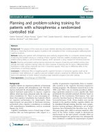
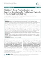
anesthesia type (11 patients). The final number of patients included was 60 patients for each group (Fig. 1).
No patients were lost to follow-up. The three groups
were similar regarding baseline demographics and type
of surgery (Table 1). None of our patients had scoliosis
or previous spine operations.
The successful dural puncture rate on first attempt
(primary outcome) was higher in groups LM and UM
(77 and 73% respectively) than in group UP (42%;
P < 0.001). The median number of attempts was lower
Enrollment
in groups LM and UM (1 [1] and 1 [1–1.75] respectively)
than in group UP (2 [1–2]; P < 0.001). Also, the median
number of passes was lower in groups LM and UM (2
[0.25–3] and 2 [0–4]; respectively) than in group UP (4
[2–7.75]; P < 0.001) (Table 2).
The ease of landmark palpation was similar between
the three groups, and grading by US was similar between
the US groups. The time taken to perform the spinal
anesthetic was not different between groups LM and
UM (87.24 ± 79.51 sand 116.32 ± 98.12 s respectively)
but shorter than in group UP (154.58 ± 91.51 s;
P < 0.001). Less verbal attending assistance was required
in groups LM and UM (53 and 48% respectively) compared to group UP (85%; P < 0.001) (Table 2).
More complications (paresthesia, blood tap, and radicular pain) occurred in patients in group UP, and patients
with complications were followed-up to 24 h as per hospital protocol with no consequences. Also, percentage of
patients satisfied during the procedure was less in group
UP (Table 2). All spinal anesthetics were successful and
patients achieved complete sensory block to the T7
dermatome or higher. All failed dural punctures by the
Assessed for eligibility (n=260)
Excluded (n=80)
Not meeting inclusion criteria (n=51)
Declined to participate (n=10)
Other reasons (n=19
Randomized (n=180)
Allocation
Allocated to Group LM (n=60)
Received allocated intervention
(n=60)
Allocated to Group UP (n=60)
Received allocated intervention
(n=60)
Allocated to Group UM (n=60)
Received allocated intervention
(n=60)
Follow-Up
Lost to follow-up (n= 0)
Lost to follow-up (n=0)
Lost to follow-up (n=0)
Discontinued intervention (n=0)
Discontinued intervention (n=0)
Discontinued intervention (n=0)
Analysis
Analysed (n=60)
Analysed (n=60)
Analysed (n=60)
Excluded from analysis (n=0)
Excluded from analysis (n=0)
Excluded from analysis (n=0)
Fig. 1 Consort flow diagram
Rizk et al. BMC Anesthesiology
(2019) 19:208
Page 5 of 8
Table 1 Baseline characteristics
Group LM (n = 60)
Group UM (n = 60)
Group UP (n = 60)
Age (y)
73.51 ± 7.99
72.37 ± 7.83
71.42 ± 7.54
Weight (kg)
77.72 ± 11.48
81.73 ± 17.09
78.44 ± 15.13
Height (cm)
166.13 ± 8.86
167.83 ± 7.12
167.48 ± 8.19
BMI (kg/m2)
28.21 ± 4.30
28.98 ± 6.01
28.21 ± 4.67
M
47 (78)
50 (83)
49 (82)
F
13 (22)
10 (17)
11 (18)
Gender
Type of surgery
Urology
47 (78)
47 (78)
51 (85)
Orthopedics
6 (10)
7 (12)
8 (13)
General surgery
7 (12)
6 (10)
1 (2)
Values are mean ± SD or numbers (%)
Group LM landmark-guided midline technique, group, UP Ultrasound-guided paramedian technique, group, UM Ultrasound -guided midline technique
residents were achieved successfully by the attending
anesthesiologist using the same technique as the resident
except for one patient in group UP and three patients in
group UM who had the spinal procedure using the LM
technique.
Discussion
Our study showed that both preprocedural US-assisted
modalities (midline or paramedian) did not prove more
efficacious than the landmark-based midline approach in
facilitating spinal anesthesia performed by CA-1 residents in elderly patients. In fact, while the first attempt
and overall success rates of dural puncture in the midline US and the conventional landmark groups were not
significantly different, they were higher than the rates
achieved with preprocedural US-guided paramedian
spinal technique. Furthermore, time to perform spinal
anesthesia, need for verbal attending assistance, as well
as the number of attempts and passes were all significantly less in the midline US and conventional groups
compared to the paramedian US group.
Metanalyses suggest that preprocedural US leads to reduction of the risk of failure and a lower number of needle passes compared to conventional palpation approach
[10–12]. This is particularly true in patients with whom
technical difficulties are expected such as those with
high body mass indices, nonpalpable landmarks, or difficult spinal anatomy. Of note, most of these studies are
limited to a midline approach using a TM view.
Geriatric population, similar to our study population,
also may present with higher likelihood of technical difficulties during spinal anesthesia due to narrowed interspinous spaces and interlaminar spaces as a result of
ossification of the interspinous ligaments and hypertrophy of the facet joints respectively [1]. However, our
findings did not demonstrate an improved outcome with
the US midline vs landmark. This may be explained by
the fact that the spinal procedures in our elderly population using the landmark midline technique were not difficult enough and easier than expected, thus limiting the
benefit of a preprocedural US. In fact, the first success
rate in the LM and UM groups (77 and 73% respectively) were higher than that reported in a previous study
conducted by Chin et al. in a nonobstetric patient population with difficult anatomic landmarks (32% in landmark group and 65% in US group), even though
operator in all cases in the aforementioned study was a
clinical fellow in regional anesthesia or consultant with
more than 5 year clinical experience [1].
As for the paramedian approach, it should theoretically be valuable in the elderly since the interlaminar
spaces are less affected by aging offering a better view of
LFD and PLL compared to midline view [13], and it does
not require flexing of the spine (an advantage in elderly
patients with fractures). Even with these advantages, a
lower first attempt success rate with the paramedian approach was obtained compared to both landmark- and
US-guided midline approaches. As a matter of fact, the
paramedian approach has been shown to be superior to
the midline approach in some previous studies [7, 14,
15] but not in others [16–18]. It must be noted that we
found only two studies in the literature that describe the
use of paramedian US techniques to facilitate spinal
anesthesia with contradictory results [4, 9]. Lim et al.
compared an US-assisted paramedian vs a conventional
paravertebral approach for orthopedic and other types of
surgery and found no intergroup difference in first skin
puncture-success rate, number of needle redirections,
and complications [4]. In another study, Srinivasan et al.
compared an US-assisted paramedian vs a palpation at
midline approach for an elderly orthopedic population
and showed that US technique required fewer passes
Rizk et al. BMC Anesthesiology
(2019) 19:208
Page 6 of 8
Table 2 Clinical outcomes
Group LM (n = 60)
Group UP (n = 60)
Group UM (n = 60)
P
46 (77)
*P < 0.001
25 (42)
44 (73)
†P < 0.001
< 0.001
Primary outcome
Successful dural puncture on first attempt
Secondary Outcomes
Successful dural puncture on first pass
15 (33)
5 (20)
19 (43)
Successfull dural puncture
53 (88)
*P = 0.001
38 (63)
48 (80)
†P = 0.043
Number of attempts
1 (1–1)
* P = 0.001
2 (1–2)
1 (1–1.75)
† P = 0.003
0.001
Number of passes
2 (0.25–3)
*P < 0.001
4 (2–7.75)
2 (0–4)
† P < 0.001
< 0.001
Time taken to perform spinal anesthesia (s)
87.24 ± 79.51
*P < 0.001
154.58 ± 91.51
116.32 ± 98.12
† P = 0.006
< 0.001
Ease of landmark palpation
0.14
< 0.001
0.89
Easy
38 (63)
37 (62)
36 (60)
Moderate
19 (32)
18 (30)
18 (30)
Difficult
3 (5)
5 (8)
6 (10)
Impossible
0 (0)
0 (0)
0 (0)
40 (70)
34 (65)
Grading by ultrasound
0.50
Good
Intermediate
16 (28)
18 (35)
Poor
1 (2)
0 (0)
Verbal attending assistance
32 (53)
*P < 0.001
51 (85)
29 (48)
† P < 0.001
Number of patients who developed complications
10 (17)
20 (33)
9 (15)
Number of complications
10
26
9
Paresthesia
0
3
0
Blood tap
9
15
8
Radicular pain
1
* P = 0.035
8
1
† P = 0.019
Periprocedural pain score
0 (0–0)
* P = 0.009
0 (0–1)
0 (0–1)
† P = 0.042
Very good
51 (85.00)
31 (51.67)
44 (73.33)
Good
8 (13.33)
27 (45.00)
15 (25.00)
Satisfactory
1 (1.67)
* P = 0.0004
2 (3.33)
1 (1.67)
† P = 0.049
Satisfaction
< 0.001
0.026
0.017
0.002
Values are mean ± SD, numbers (%), or medians and interquartile ranges
Group LM landmark-guided midline technique, group, UP Ultrasound-guided paramedian technique, group, UM Ultrasound -guided midline technique
* Group LM vs Group UP; † Group UM vs Group UP
and attempts to reach the subarachnoid space [9]. We
found a much lower success rate at first attempt in the
US paramedian group (42%) compared to those in the
Lim et al. and Srinivasan et al. studies (64% vs 84% respectively). This may be explained by that our operators
were CA-1 residents as opposed to the other two studies
where operators were either trainees at variable level of
experience (up to three years of experience) [4] or
attending anesthesiologists [9].
In our study, the higher number of attempts in the UP
group resulted in more blood taps, paresthesia, and radicular
pain than in the other two groups. Also, patients in the UP
group had more periprocedural pain and lower satisfaction
than the other two groups, probably because the needle has
to cross the erector spinae muscle before reaching the dura.
These findings can be due to the fact that the paramedian
approach to the neuraxis is intrinsically more difficult to
perform and is utilized by few anesthesiologists.
Rizk et al. BMC Anesthesiology
(2019) 19:208
Studies with junior residents acting as operators during preprocedural US scanning of the spine are present
in the obstetric literature with mixed results. All investigators used the midline approach prior to the spinal
block. While Sahin et al. found a high level of success in
the prepuncture US-determined insertion point by
anesthesia residents [2], Turkstra et al. reported similar
results to our findings with no observed benefit to preprocedural US examination for junior residents performing spinal anesthesia [5].
Limitations in this study include lack of blinding of the
residents and observer collecting data causing a potential
bias. Second, the design of the study did not completely
eliminate both patient and operator bias. However, we assumed that computer-generated randomization would
equally distribute patients of different levels of spinal
anesthesia difficulty and thus would decrease patient bias.
Anyhow, results showed that none of our patients had
scoliosis or previous spine operations, both of which are
common features that would further increase the difficulty
of spinal anesthesia. In addition, the ease of landmark palpation was not different among the three groups. Although
operator bias cannot be ruled out completely, we believe
that our operator group is a homogenous one consisting of
junior residents with less than 5 spinal anesthesia experience and at the lower end of the learning spectrum.
Third, the potential learning effect over the course of the
study could be present; however, it was mitigated by performing all spinal blocks in a row and minimizing the number of procedures done outside study. Yet, it must be noted
that as per Kopacz et al., notable improvement in the spinal
anesthesia technique among novice residents require at
least 20 procedures to be performed [17]. In our study, 12
residents performed each 12 spinal blocks and only 2
residents performed each 18 spinal blocks, all of which are
below minimal required number.
Finally, we found some challenges with the use of the
US-assisted approach in general: the difficulty to mark
the point for needle insertion that can change if the patient moves between the time of skin marking and the
actual procedure, the tissue distortion particularly in the
elderly with mobile and loose skin, and to guarantee that
the trainee will follow the exact direction of the probe
despite the clear instruction about the angle at which
the needle should be inserted. It is important to highlight on the different definitions of attempts, passes, and
redirections in the literature as a redirection without
withdrawal is sometimes considered in some reports as
an additional attempt, thus making comparison of
measured outcomes difficult among studies.
Conclusions
In conclusion, our study showed that preprocedural US
scanning did not improve the ease of midline and
Page 7 of 8
paramedian spinal anesthesia as compared to the conventional landmark midline technique when performed
by junior residents in an elderly population. Thus, there
was no observed additional value to preprocedural US,
whether using the TM or the PSO views for anesthesiologists in training. Our results should be confirmed with
the use of the newer modality of real-time US guidance
in the future.
Abbreviations
CA-1: First year clinical anesthesia residents; LFD: Ligamentum flavum-dura
meter complex; LM: Landmark-guided midline; PLL: Posterior longitudinal
ligament; PSO: Parasagittal oblique; TM: Transverse median; UM: Ultrasound
guided midline technique; UP: Ultrasound guided paramedian;
US: Ultrasound
Acknowledgements
I would like to extend special thanks to Ms. Fadia Shebbo for her assistance
with analyzing the data and finalizing this manuscript.
Authors’ contributions
MSR, CAZ and SMS helped design the study, collect and analyze the data,
and write the manuscript. JNB helped collect and analyze the data and write
the manuscript. NJB helped design the study and write the manuscript. SKS
helped analyze the data and write the manuscript. WSF helped design the
study and write the manuscript. All authors have read and approved the
manuscript.
Funding
This research study was supported by the Department of Anesthesiology,
American University of Beirut Medical Center. The funding body contributed
with research equipment, but had no role in the design of this study,
analyses, interpretation of the data or writing the manuscript.
Availability of data and materials
The datasets used and/or analyzed during the current study are available
from the corresponding author on reasonable request.
Ethics approval and consent to participate
This study was approved by the Institutional Review Board of the American’s
University of Beirut Medical Center (No. ANES.SS.13). All patients provided
written informed consent.
Consent for publication
Not applicable.
Competing interests
The authors declare that they have no competing interests.
Author details
1
Department of Anesthesiology, American University of Beirut Medical
Center, P.O. Box 11-0236, Beirut, Lebanon. 2Division of Musculoskeletal
Imaging, Emory University Hospital, Atlanta, USA.
Received: 24 May 2019 Accepted: 30 October 2019
References
1. Chin KJ, Perlas A, Chan V, Brown-Shreves D, Koshkin A, Vaishnav V.
Ultrasound imaging facilitates spinal anesthesia in adults with difficult
surface anatomic landmarks. Anesthesiology. 2011;115:94–101.
2. Sahin T, Balaban O, Sahin L, Solak M, Toker K. A randomized controlled trial
of preinsertion ultrasound guidance for spinal anaesthesia in pregnancy:
outcomes among obese and lean parturients. J Anesth. 2014;28:413–9.
3. Ansari T, Yousef A, El Gamassy A, Fayez M. Ultrasound-guided spinal
anaesthesia in obstetrics: is there an advantage over the landmark
technique in patients with easily palpable spines? Int J Obstet Anesth. 2014;
23:213–6.
Rizk et al. BMC Anesthesiology
4.
5.
6.
7.
8.
9.
10.
11.
12.
13.
14.
15.
16.
17.
18.
(2019) 19:208
Lim YC, Choo CY, Tan KTJ. A randomised controlled trial of ultrasoundassisted spinal anaesthesia. Anaesth Intensive Care. 2014;42:191–8.
Turkstra TP, Marmai KL, Armstrong KP, Kumar K, Singh SI. Preprocedural
ultrasound assessment does not improve trainee performance of spinal
anesthesia for obstetrical patients: a randomized controlled trial. J Clin
Anesth. 2017;37:21–4.
Wantman A, Hancox N, Howell PR. Techniques for identifying the epidural
space: a survey of practice amongst anaesthetists in the UK. Anaesthesia.
2006;61:370–5.
Rabinowitz A, Bourdet B, Minville V, Chassery C, Pianezza A, Colombani A,
et al. The paramedian technique: a superior initial approach to continuous
spinal anesthesia in the elderly. Anesth Analg. 2007;105:1855–7.
Grau T, Leipold RW, Horter J, Conradi R, Martin EO, Motsch J. Paramedian
access to the epidural space: the optimum window for ultrasound imaging.
J Clin Anesth. 2001;13:213–7.
Srinivasan KK, Iohom G, Loughnane F, Lee PJ. Conventional landmarkguided midline versus preprocedure ultrasound-guided paramedian
techniques in spinal anesthesia. Anesth Analg. 2015;121:1089–96.
Elgueta MF, Duong S, Finlayson RJ, Tran DQ. Ultrasonography for neuraxial
blocks: a review of the evidence. Minerva Anestesiol. 2017;83(5):512–23.
Perlas A, Chaparro LE, Chin KJ. Lumbar neuraxial ultrasound for spinal and
epidural anesthesia. A systematic review and meta-analysis. Reg Anesth Pain
Med. 2016;41(2):251–60.
Shaikh F, Brzezinski J, Alexander S, Arzola C, Carvalho JC, Beyene J, et al.
Ultrasound imaging for lumbar punctures and epidural catheterisations:
systematic review and meta-analysis. BMJ. 2013;346:f1720.
Chin KJ, Ramlogan R, Arzola C, Singh M, Chan V. The utility of ultrasound
imaging in predicting ease of performance of spinal anesthesia in an
orthopedic patient population. Reg Anesth Pain Med. 2013;38:34–8.
Blomberg RG, Jaanivald A, Walther S. Advantages of the paramedian
approach for lumbar epidural analgesia with catheter technique. A clinical
comparison between midline and paramedian approaches. Anaesthesia.
1989;44:742–6.
Leeda M, Stienstra R, Arbous MS, Dahan A, Th Veering B, Burm AG, et al.
Lumbar epidural catheter insertion: the midline vs. the paramedian
approach. Eur J Anaesthesiol. 2005;22:839–42.
Sprung J, Bourke DL, Grass J, Hammel J, Mascha E, Thoman P, et al.
Predicting the difficult neuraxial block: a prospective study. Anesth Analg.
1999;89:384–9.
Kopacz DJ, Neal JM, Pollock JE. The regional anesthesia ‘learning curve’:
what is the minimum number of epidural and spinal blocks to reach
consistency? Reg Anesth Pain Med. 1996;21:182–90.
Cook TM. Combined spinal-epidural techniques. Anaesthesia. 2000;55:42–64.
Publisher’s Note
Springer Nature remains neutral with regard to jurisdictional claims in
published maps and institutional affiliations.
Page 8 of 8