Efficacy and safety of pulsed radiofrequency as a method of dorsal root ganglia stimulation for treatment of nonneuropathic pain: A systematic review
Bạn đang xem bản rút gọn của tài liệu. Xem và tải ngay bản đầy đủ của tài liệu tại đây (1.16 MB, 21 trang )
Vuka et al. BMC Anesthesiology
(2020) 20:105
/>
RESEARCH ARTICLE
Open Access
Efficacy and safety of pulsed
radiofrequency as a method of dorsal root
ganglia stimulation for treatment of nonneuropathic pain: a systematic review
Ivana Vuka1, Svjetlana Došenović2, Tihana Marciuš1, Lejla Ferhatović Hamzić3, Katarina Vučić4,
Damir Sapunar1,5† and Livia Puljak5*†
Abstract
Background: We systematically reviewed the evidence on the efficacy and safety of dorsal root ganglion (DRG)
targeted pulsed radiofrequency (PRF) versus any comparator for treatment of non-neuropathic pain.
Methods: We searched MEDLINE, CINAHL, Embase, PsycINFO, clinicaltrials.gov and WHO clinical trial register until
January 8, 2019. All study designs were eligible. Two authors independently conducted literature screening. Primary
outcomes were pain intensity and serious adverse events (SAEs). Secondary outcomes were any other pain-related
outcome and any other safety outcome that was reported. We assessed the risk of bias using the Cochrane tool
and Risk of Bias In Non-randomized Studies of Interventions (ROBINS-I). We conducted narrative evidence synthesis
and assessed the conclusiveness of included studies regarding efficacy and safety.
Results: We included 17 studies with 599 participants, which analyzed various pain syndromes. Two studies were
randomized controlled trials; both included participants with low back pain (LBP). Non-randomized studies included
patients with the following indications: LBP, postsurgical pain, pain associated with herpes zoster, cervicogenic
headache, complex regional pain syndrome type 1, intractable vertebral metastatic pain, chronic scrotal and
inguinal pain, occipital radiating pain in rheumatoid arthritis and chronic migraine. In these studies, the PRF was
usually initiated after other treatments have failed. Eleven studies had positive conclusive statements (11/17) about
efficacy; the remaining had positive inconclusive statements. Only three studies provided conclusiveness of
evidence statements regarding safety – two indicated that the evidence was positive conclusive, and one positive
inconclusive. The risk of bias was predominantly unclear in randomized and serious in non-randomized studies.
(Continued on next page)
* Correspondence: ;
†
Damir Sapunar and Livia Puljak contributed equally to this work.
5
Center for Evidence-Based Medicine and Health Care, Catholic University of
Croatia, Ilica 242, 10000 Zagreb, Croatia
Full list of author information is available at the end of the article
© The Author(s). 2020 Open Access This article is licensed under a Creative Commons Attribution 4.0 International License,
which permits use, sharing, adaptation, distribution and reproduction in any medium or format, as long as you give
appropriate credit to the original author(s) and the source, provide a link to the Creative Commons licence, and indicate if
changes were made. The images or other third party material in this article are included in the article's Creative Commons
licence, unless indicated otherwise in a credit line to the material. If material is not included in the article's Creative Commons
licence and your intended use is not permitted by statutory regulation or exceeds the permitted use, you will need to obtain
permission directly from the copyright holder. To view a copy of this licence, visit />The Creative Commons Public Domain Dedication waiver ( applies to the
data made available in this article, unless otherwise stated in a credit line to the data.
Vuka et al. BMC Anesthesiology
(2020) 20:105
Page 2 of 21
(Continued from previous page)
Conclusion: Poor quality and few participants characterize evidence about benefits and harms of DRG PRF in
patients with non-neuropathic pain. Results from available studies should only be considered preliminary. Not all
studies have reported data regarding the safety of the intervention, but those that did, indicate that the
intervention is relatively safe. As the procedure is non-destructive and early results are promising, further
comparative studies about PRF in non-neuropathic pain syndromes would be welcomed.
Keywords: Chronic pain, Non-neuropathic pain, Pulsed radiofrequency, Dorsal root ganglion
Background
Chronic pain is one of the major public health issues
worldwide and is one of the leading causes of years lived
with disability [1]. Estimates on the prevalence of
chronic pain in the general population vary, ranging
from 11% [2] up to 64% [3]. These different estimates
are mostly due to differences in the definition of chronic
pain regarding the duration of symptoms (3 vs. 6
months) and the wording of questions used for assessing
chronic pain [4]. Besides its major clinical impact and
costs for the healthcare system, chronic pain impairs patients’ quality of life, as well as their ability to work and
function, causing massive indirect socioeconomic costs
worldwide [5]. Chronic pain asserts this major impact
on individuals, health systems and society because of inadequate treatment modalities.
Pulsed radiofrequency (PRF) emerged as a therapeutic
treatment for various painful conditions, including both
neuropathic and non-neuropathic pain [6–8]. PRF has
been described as “a non-neurodestructive therapy in
pain management ”[9]. PRF is a minimally invasive intervention, which involves the application of pulses of electric current, created at the tip of an electrode, without a
harmful increase in the temperature [9].
It has been suggested that a dorsal root ganglion
(DRG) is a desirable target for the treatment of pain
[10]. PRF application close to dorsal root could alleviate
neuropathic pain [11]. However, we have observed an increasing number of studies on chronic pain, reporting
use of DRG targeted PRF treatment of non-neuropathic
pain in humans. Therefore, we aimed to conduct a systematic review about the evidence on the efficacy and
safety of DRG targeted PRF treatment of nonneuropathic pain.
Methods
Study design
We published a systematic review protocol a priori in
the PROSPERO database (registration number:
CRD42017076502). Since the original protocol covered
extremely wide scope and heterogeneous interventions,
subsequently we divided the original protocol into a separate assessment of DRG targeted electrical field stimulation
(EFS) [12] and PRF. The systematic review was performed
following the PRISMA statement and Center for Reviews
and Dissemination (CRD) manuals.
Eligibility criteria
Participants, intervention and study designs
We included primary studies with participants suffering
from various painful conditions which are not currently
classified as purely of neuropathic origin (i.e. nonneuropathic pain). In case that condition was defined of
both origins, neuropathic and non-neuropathic, such as
post-surgical pain or low back pain we included such
condition. We excluded studies where PRF treatment
was used for neuropathic pain as it is defined in the
guidelines of the International Association for the Study
of Pain (IASP). We used the IASP classification of
chronic pain for ICD-11. We chose to include both randomized controlled trials (RCTs) as well as nonrandomized study designs (NRSDs) because we expected
a few RCTs in this research area, and we wanted to provide a comprehensive picture of evidence in this field of
research. We used Cochrane Handbook for Reviews of
Interventions to define the design of included studies.
Manuscripts that included more than 10 participants
were classified as case series, while those that included
less than 10 participants were defined as case reports
[13]. We only included studies where PRF treatment was
directed to the DRG, including a combination of PRF
with other therapies. If the study only reported results
about efficacy, and safety was not reported, we still included such a study to get comprehensive evidence synthesis regarding efficacy.
Outcome measures
Primary outcomes were: pain intensity and serious adverse events (SAEs). For primary outcome, we reported
any outcome measures, as reported in included manuscripts. Secondary outcomes for efficacy were any other
pain-related outcomes, and for safety any other safety
data, including non-serious adverse events and other
complications regarding tested intervention.
Search strategy and information source
We searched four databases: MEDLINE via Ovid,
Embase via Ovid, CINAHL and PsycINFO via
Vuka et al. BMC Anesthesiology
(2020) 20:105
EBSCOhost (Supplementary Table 1). Databases were
searched from the date of inception until January 8,
2019 with no restriction regarding the language. Records were then exported to the EndNote X5 citation
software (Clarivate Analytics, Boston, MA, USA) and
duplicates removed. Furthermore, reference lists of all
included studies and their citations were downloaded
from Web of Science and screened to find additional
eligible studies. ClinicalTrials.gov and World Health
Organization’s International Clinical Trial Registry Platform (WHO ICTRP) were searched to identify ongoing
studies.
Study selection
Reviewers independently screened each title/abstract of
retrieved records as well as full-texts of retrieved studies
for possible inclusion (authors LFH, IV, TM and SD participated in screening). Discrepancies were resolved by
another author (DS).
Data extraction
Independent data extraction was performed by two authors for each data point (authors: IV, and TM or KV).
We extracted the following data: the surname of the first
author, year of publication, study design, details about
intervention (treatment protocol and device used), comparator, inclusion and exclusion criteria, number of participants, baseline characteristics of participants, followup period, DRG level treated and outcomes about efficacy and safety.
Risk of bias assessment
We used the Cochrane Risk of Bias (RoB) tool (version
from 2011) to assess RoB in RCTs and the Risk of Bias
In Non-randomized Studies of Interventions (ROBINS-I)
tool for cohort type studies. RoB was analyzed independently by two authors (IV, and SD or KV). Discrepancies
were resolved by another author (LP).
Synthesis of results
Due to the heterogeneity of included studies, it was not
possible to conduct a meta-analysis, even though we
have planned to do it in our study protocol. For this reason, we conducted a narrative and tabular synthesis of
results. We also conducted an analysis of conclusiveness
about efficacy and safety of the treatment in the abstracts of included studies. We divided conclusiveness
statements into five categories: positive conclusive (favorable conclusion in favor of PRF), positive inconclusive (favorable conclusion, but with a note about
insufficient or low quality evidence), negative conclusive
(PRF not beneficial), negative inconclusive (PRF not
beneficial, but with a note about insufficient or low quality evidence) and not reported.
Page 3 of 21
Results
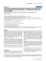
The flow chart in Fig. 1 shows the number of records
analyzed in each screening phase. We screened 63 manuscripts in full text, and we finally included 17 manuscripts in this systematic review. Excluded studies, and
reasons for their exclusion, are listed in Supplementary
Table 2. The characteristics of the included studies are
detailed in Table 1.
Among 17 included studies there were two randomized
controlled trials [14, 15] and 15 non-randomized studies
(Table 1). The total number of participants in these studies was 599; the median number of participants was 28
(range: 1 to 127) (Table 1). Both RCTs included participants with low back pain (LBP) [14, 15]. Non-randomized
studies included patients with the following indications:
LBP [16–18], postsurgical pain [19–21], pain associated
with herpes zoster [6], cervicogenic headache [22, 23],
complex regional pain syndrome type 1 [24, 25], intractable vertebral metastatic pain [26], chronic scrotal and inguinal pain [27], occipital radiating pain in rheumatoid
arthritis [28] and chronic migraine [29] (Table 1). These
studies had highly heterogeneous parameters of stimulation (Table 2). Detailed information about inclusion and
exclusion criteria, as well as baseline characteristics of included participants, are listed in Table 3.
Low back pain
In this group, there were 5 studies with a total of 328
participants, including two RCTs with 28 participants in
one [14] and 60 participants in another one [15], one
retrospective cohort study including 29 participants [16],
and two before and after comparisons with 84 participants in one [17] and 127 participants in another [18].
Trial by Holanda et al. [14] included 28 participants
which were randomized in three groups: PRF treatment
group with the probe directed through the needle in the
second lumbar intervertebral foramen (N = 11), lidocaine
injection group (N = 7) and laser irradiation treatment
group (N = 10). All participants from the lidocaine injection group and laser irradiation group reported a 100%
reduction in visual analogue scale (VAS) scores immediately after the treatment, while participants from the
PRF group reported a 62.5% reduction in pain. At 1month follow-up laser irradiation group had a 55.5%
reduction in pain; lidocaine injection group 62.5% reduction and PRF group only 20% [14].
An RCT by Lee et al. [15] analyzed predictive value
and cost-effectiveness of the use of diagnostic blocks before PRF treatment. They included 60 participants suffering from LBP with or without lower-limb pain,
randomized into two groups. In one group (N = 30) participants received DRG blocks with 1 ml of 2% bupivacaine and 1 ml of 2% triamcinolone, and those who had
at least 50% improvement were scheduled for PRF
Vuka et al. BMC Anesthesiology
(2020) 20:105
Page 4 of 21
Fig. 1 Flow chart of study inclusion
treatment. The other group (N = 30) received only PRF
treatment without DRG blocks. Limited low back pain
was treated with DRG block or PRF applied to the L2
DRG; lower -limb pain was treated with PRF applied to
the L3–S1 DRG. The authors concluded that DRG
blocks had no statistically significant impact on the results of PRF treatment, while their application resulted
in overall higher medical costs [15].
Yang et al. [16] reported results of a retrospective cohort study that aimed to develop a patient-mounted navigated intervention (PaMNI) system for spinal diseases
to evaluate the success of the PRF treatment. The study
also included a pilot clinical trial were the new PaMNI
system (N = 16) was compared to conventional fluoroscopy (N = 13). In all patients, PRF treatment was delivered on the L4 DRG. Both groups showed a reduction in
VAS scores 1 month after the treatment with no statistically significant difference between groups (P = 0.238).
However, the study showed the feasibility and efficacy of
the PaMNI system [16].
Before and after comparison by Hsu et al. [17]
followed 84 participants up to 3 years to investigate the
correlation between different types of lumbar lordosis
with the outcomes of PRF treatment applied to L2 DRG
in chronic low back pain. The analysis showed that after
3-year follow-up participants had a statistically significant reduction in low back pain, regardless of the type of
lumbar lordosis [17]. The study by Tsou et al. [18], also
followed participants for up to 3 years. They included
participants who had low back pain with lower -limb
pain (N = 78) or without it (N = 49). LBP was treated
with PRF applied to the L2 DRG and lower-limb pain
was treated with PRF applied to the L3–S1 DRG. Percentage of participants achieving at least 50% improvement in VAS scores was similar in both groups at 1-year
follow-up, with 20 out of 45 participants (44.44%) in the
group without lower -limb pain and 34 out of 74 participants (45.95%) in the group with lower -limb pain [18].
None of the studies reported serious adverse events. Two
studies reported minor complications: mild discomfort
No other
intervention
Diagnostic block
+ PRF group
received
diagnostic block
with 1 ml of 2%
bupivacaine and
1 ml of 2%
triamcinolone.
Lee 2018 [15]
RCT / randomized,
prospective, and
comparative study
Interventions
prior to PRF
treatment
Holanda 2016 [14]
RCT / pilot study
Low back pain (LBP)
Author and year/Study
design (Cochrane
handbook and study
authors)
Diagnostic block +
PRF group (n = 30);
PRF group (n = 30)
PRF treatment
group n = 11;
lidocaine injection
group n = 7;
laser irradiation
treatment group
n = 10
Number of
participants (for
each pain
condition treated)
2 weeks, 1, 3
and 6 months
5 min and 1
month
Follow-up
Table 1 Characteristics about efficacy and safety of included studies
1. Pain intensity by
NRS
2. Functional
disabilities by ODI
1. Lumbar pain
intensity by VAS
presented as
percentage of
relative difference
2. Chronic LBP
relief by PRS
Outcome measures
Diagnostic
block + PRF group:
Baseline NRS: 8 (range
5–9);
NRS at 2 weeks: 2 (range
1–7);
NRS at 1 month: 2
(range 1–8);
NRS at 3 months: 3
(range 1–8);
NRS at 6 months: 4
(range 1–8).
PRF alone group:
Baseline NRS: 7.5 range
(3–10);
NRS at 2 weeks: 2 (range
1–9);
NRS at 1 month: 2
(range 1–9);
NRS at 3 months: 3
(range 1–9);
NRS at 6 months: 4
(range 1–9).
P values comparison
between groups:
NRS at 2 weeks: P =
0.302
NRS at 1 month: P =
0.690
NRS at 3 moths: P =
0.957
PRF group:
VAS relative difference
at 5 min: 62.5%
VAS relative difference
at 1 month: 20%
Lidocaine injection
group: VAS relative
difference at 5 min:
100%
VAS relative difference
at 1 month: 62.5%
Laser treatment
group:
VAS relative difference
at 5 min: 100%
VAS relative difference
at 1 month: 55%
Results: efficacy for pain
intensity
No SAEs
occurred
Not reported
Positive
inconclusive
Results: serious
adverse events
Positive
conclusive
Conclusion
statement
about efficacy
Not reported
Some patients
experienced
only mild
discomfort
during
procedure
Results: any
other safety
data
Not reported
Not reported
Conclusion
statement
about safety
Vuka et al. BMC Anesthesiology
(2020) 20:105
Page 5 of 21
No other
intervention
No other
intervention
Hsu 2017 [17]
BA / retrospective
study
Tsou 2010 [18]
BA / not stated
Albayrak 2017 [19]
In the PRF
No other
intervention
Yang 2010 [16]
RCS / in vivo clinical
trial
PRF group (TENS +
Group A (CLBP
without lower-limb
pain) n = 49;
Group B (CLBP with
lower-limb pain)
n = 78
84
PaMNI system n =
16;
conventional
fluoroscopy n = 13
Number of
participants (for
each pain
condition treated)
15 days and 1-
From 1 week up
to 3 years postoperatively
1 week after the
treatment and
at 3, 6, 9, 12
months and
yearly
postoperatively
(for 3 years in
total)
1 month
Follow-up
1. Pain intensity by
1. Pain intensity by
VAS
2. Adverse events
1. Pain intensity by
VAS
2. Functional
disabilities by ODI
1. Pain intensity by
VAS
Outcome measures
PRF group activity:
Group A, L2
treatment:
≥50% VAS
improvement:
at 1 week: 25/49
(51.02%);
at 3 months: 27/49
(55.1%);
at 1 year 20/45 (44.44%).
Group B, L2 treatment:
≥50% VAS
improvement:
at 1 week: 34/78
(43.59%);
at 3 months: 37/78
(47.44%);
at 1 year: 34/74 (45.95%).
Analysis of VAS scores
for pain indicated
significant reductions of
low back pain during
the 3-year follow-up for
patients with all 4 types
of lumbar lordosis.
PaMNI group:
Baseline VAS: 5.8 (±2.3);
VAS at 1 month: 4.1 (±
2.1).
P = 0.005.
Fluoroscopy group:
Baseline VAS: 6.5 (±2.2);
VAS at 1 month: 5.3 (±
2.8).
P = 0.067.
No statistical difference
between groups at 1
month (P = 0.238).
NRS at 6 months: P =
0.673
Results: efficacy for pain
intensity
Positive
Positive
conclusive
Positive
conclusive
Positive
conclusive
Conclusion
statement
about efficacy
No SAEs
No SAEs
occurred
No SAEs
occurred
Not reported
Results: serious
adverse events
No
No obvious
complications
were observed
Cerebral spinal
fluid leaking
from the
cannulas of two
patients while
the needle was
being directed
toward the
DRG. This
leakage ceased
immediately
after adjusting
the location of
the needle tip.
Not reported
Results: any
other safety
data
Not reported
Positive
conclusive
Specific
adverse
events
mention, no
overall
conclusion
about safety
Not reported
Conclusion
statement
about safety
(2020) 20:105
Postsurgical pain
Interventions
prior to PRF
treatment
Author and year/Study
design (Cochrane
handbook and study
authors)
Table 1 Characteristics about efficacy and safety of included studies (Continued)
Vuka et al. BMC Anesthesiology
Page 6 of 21
Interventions
prior to PRF
treatment
group
participants
received
prognostic
diagnostic block
prior to
involvement.
Author and year/Study
design (Cochrane
handbook and study
authors)
PCS / retrospective
study of prospectively
collected
data
exercise + PRF) n =
22;
TENS group (TENS
+ exercise) n = 17
Number of
participants (for
each pain
condition treated)
month post
treatment and
following last
control examination. The mean
follow-up time
was 253.8 ± 109
days; for TENS
group: 217 ±
114 days and for
PRF group:
282.2 ± 97 days
Follow-up
VAS
2. Degree of
neuropathic pain
reduction by DN4
3. Change in knee
flexion by ROM
4. Functional status
by WOMAC
5. Patient
satisfaction
Success was
defined as at least
50% reduction to
the VAS (activity,
rest, night)
Outcome measures
Table 1 Characteristics about efficacy and safety of included studies (Continued)
baseline VAS: 6.6 (±1.5);
VAS at 15 days: 3 (±1.4);
VAS at 1 month: 3.9 (±2);
VAS at last control: 3.5
(±2.4).
PRF group rest:
baseline VAS: 4.3 (±1.7);
VAS at 15 days: 1.8 (±
0.9);
VAS at 1 month: 2.6 (±
1.5);
VAS at last control: 2 (±
1.6).
PRF group night:
baseline VAS: 3.8 (±2.2);
VAS at 15 days: 1.5 (±
1.1);
VAS at 1 month: 2.1 (±
1.4);
VAS at last control: 1.7
(±1.4).
TENS group activity:
baseline VAS: 5.9 (±1.9);
VAS at 15 days: 3.8 (±
2.2);
VAS at 1 month: 4.3 (±
2.2);
VAS at last control: 4.4
(±2.1).
TENS group rest:
baseline VAS: 5 (±2.4);
VAS at 15 days: 2.6 (±
2.4);
VAS at 1 month: 3.4 (±
2.5);
VAS at last control: 2.8
(±2.1).
TENS group night:
baseline VAS: 4.3 (±2.6);
VAS at 15 days: 2.1 (±
2.7);
VAS at 1 month: 2.7 (±
2.6);
VAS at last control: 2.6
(±1).
Significant difference
achieved in an
improvement of at least
50% on the VAS scores
at activity following the
Results: efficacy for pain
intensity
conclusive
Conclusion
statement
about efficacy
occurred
Results: serious
adverse events
complications
were observed
Results: any
other safety
data
Conclusion
statement
about safety
Vuka et al. BMC Anesthesiology
(2020) 20:105
Page 7 of 21
No other
intervention
Steroid
injections with
1 ml of
bupivacaine
0.25% and 1 ml
of
dexamethasone
4 mg in a total
volume of 2 ml
immediately
after PRF
procedure.
Cohen 2006 [20]
RCS / retrospective
data analysis
Fam 2018 [21]
BA / single arm
intervention study
Kim 2017 [6]
RCS / retrospective
No other
intervention
PRF group n = 20;
continuous
n = 100
PRF DRG group
n = 13;
PRF ICN group n =
15;
MM group n = 21
Number of
participants (for
each pain
condition treated)
1, 3 and 6
months
1 week, 1, 3 and
6 months
6 weeks, 3
months
Follow-up
1. Pain intensity by
NRS
1. Pain intensity by
VAS
2. Quality of life by
QOLS
3. Change in use of
pain medication
4. Adverse effects
5. Patient
satisfaction
1. Pain intensity by
VAS
2. Answers to 2
questions
evaluating patient
satisfaction and
functional
improvement
Successful was
defined as ≥50%
pain reduction on
VAS and affirmative
answer to 2
questions
Outcome measures
PRF group:
baseline NRS: 6.30 ± 0.98
Baseline VAS: 7.48 ± 1.46
(median: 8);
VAS at 1 week: 5.01 ±
2.61 (median: 5) (P =
0.032344);
VAS at 1 month: 3.26 ±
2.37 (median: 3) (P <
0.0001);
VAS at 3 months: 4.44 ±
2.8 (median: 4) (P =
0.00139);
VAS at 6 months: 4.7 ±
2.88 (median: 4) (P =
0.0057).
No separate VAS scores
shown in manuscript,
success was achieved as
follows.:
PRF DRG group:
6 weeks: 61.5%
3 months: 53.8%
PRF ICN group:
6 weeks: 21.4%
3 months: 6.7%
MM group:
6 weeks: 27.3%
3 months: 19.9%
Effect did not reach
statistical significance at
6 weeks (P = 0.12).
At 3 months, success
rate for PRF DRG group
was significantly greater
than for those patients
treated with PRF ICN
(P = 0.01), and
approached significance
when compared with
MM (P = 0.06).
last control examination
between the two
groups (P = 0.006), but
not on the VAS scores
at rest and night (P >
0.05).
Results: efficacy for pain
intensity
Positive
conclusive
Positive
inconclusive
Positive
conclusive
Conclusion
statement
about efficacy
No SAEs
occurred
No SAEs
occurred
Small incidental
pneumothorax
was found
during a routine
scan of the lung
fields after PRF
DRG. This
patient was not
symptomatic
and was treated
conservatively
with
observation.
Results: serious
adverse events
1 patient
complained of
Pain at the
needling site,
fever of
unknown
etiology at the
night of
intervention,
mild to
moderate
elevation of
glucose level in
portion of
diabetic
participants
No other
complications
occurred
Results: any
other safety
data
Not reported
Positive
inconclusive
Not reported
Conclusion
statement
about safety
(2020) 20:105
Pain associated with herpes zoster
Interventions
prior to PRF
treatment
Author and year/Study
design (Cochrane
handbook and study
authors)
Table 1 Characteristics about efficacy and safety of included studies (Continued)
Vuka et al. BMC Anesthesiology
Page 8 of 21
Diagnostic
blocks with
1.5% lidocaine.
Positive
response was
considered as
90% pain relief
lasting for 30
min.
Zhang 2011 [23]
CR/CR
n=2
n=1
No other
intervention
No other
intervention
Apiliogullari 2015 [25]
CR / CR
n=2
n = 14
epidural group
(ropivacaine) n = 22
Number of
participants (for
each pain
condition treated)
Albayrak 2016 [24]
CR / CS
Complex regional pain syndrome
Diagnostic block
prior to
involvement.
Participants with
> 50% pain
relief received
PRF.
Interventions
prior to PRF
treatment
van Zundert 2003 [22]
BA / clinical audit
Cervicogenic headache
cohort study
Author and year/Study
design (Cochrane
handbook and study
authors)
1. Pain intensity by
VAS
1. Pain intensity by
VAS
2. ROM degree
1. Pain intensity by
NRS
1.Satisfactory pain
relief (GPE: defined
as a score of 6 or 7
points on the Likert
scale; at least 50%
pain relief
2. Duration of the
effect
3. Other treatments
4. Change in use of
pain medication
2. Dose of
anticonvulsants
and analgesics
Outcome measures
Baseline VAS: 100;
VAS at 1 day (PRF on
Patient 1:
Baseline VAS during
movement: 80;
VAS at 3 days: 30;
VAS at 2 and 10 months:
20.
Patient 2:
Baseline VAS during
movement: 100;
VAS at 3 days: 30;
VAS at 2 months: 20;
VAS at 5 months: 10.
Patient 1:
Baseline NRS: 5;
NRS at 6 months: 0.
Patient 2:
Baseline NRS: 4;
NRS at 6 months: 0.
Data about pain relief
(GPE):
9/14 patients (64%)
reported successful pain
reduction (6 or 7 points
on the GPE Likert scale).
Continuous epidural
group:
baseline NRS: 6.73 ± 0.88
NRS values were
significantly lower in
PRF group from 1 to 3
months and 6 months
after the procedure (P =
0.029) than those in
continuous epidural
group.
Results: efficacy for pain
intensity
Positive
inconclusive
Positive
inconclusive
Positive
inconclusive
Positive
conclusive
Conclusion
statement
about efficacy
No SAEs
occurred
No SAEs
occurred
No significant
complications
occurred
No SAEs
occurred
Results: serious
adverse events
No significant
complications
No
complications
were observed
No significant
complications
occurred
No other
complications
observed
pain at the
procedure site,
and it improved
within few days
Results: any
other safety
data
Not reported
Not reported
Not reported
Positive
conclusive
Conclusion
statement
about safety
(2020) 20:105
1 day after
treatment (2
1 and 3 days
after PRF, 2 and
5 or 10 months
(different last
follow-up time
point for 2
patients)
6 months
2 months and 6
months after
the last patient
were included.
Mean follow-up
was 19.4
months (±8.9
months), maximum 2.5 years.
Follow-up
Table 1 Characteristics about efficacy and safety of included studies (Continued)
Vuka et al. BMC Anesthesiology
Page 9 of 21
Interventions
prior to PRF
treatment
No other
intervention
Diagnostic block
with 1 ml of
levobupivacaine
0.25%
n=1
n = 15
Number of
participants (for
each pain
condition treated)
Diagnostic block
with 1 ml of 2%
lidocaine
Diagnostic block
with 0.3 ml of
0.75%
levobupivacaine
and 1 mg
triamcinolone
n=1
n=1
1 year
6 months
12 months
0, 1, 7, 21, 28, 35
and 42 days
weeks after first
PRF the
treatment was
repeated), 6
months
Follow-up
1. Pain intensity by
VAS
1. Pain intensity by
VAS
1. Pain intensity by
VAS
1. Pain intensity by
NRS at rest and
while moving
Outcome measures
Baseline VAS: 8;
VAS at 1 year: complete
pain relief.
Baseline VAS: 10;
VAS at 6 months: 0.
Baseline VAS scores: 7–8;
VAS initially after
intervention: 4;
VAS at 12 months: 0–1.
NRS at rest:
baseline NRS: from 1 to
4 (median 3);
NRS at day 1: median 2;
NRS at day 7: median 1;
NRS at day 21: median
1.
Significant decrease in 3
weeks (P < 0.0001).
NRS while moving:
baseline NRS from 5 to
10 (median 8);
NRS at day 1: median 4;
NRS at day 7: median 4;
NRS at day 21: median
3.
Significant decrease in 3
weeks (P < 0.0001).
L5): 50;
VAS at 2 weeks (after
repeated PRF on L4): 10.
The patient had
symptoms relief for over
6 months.
Results: efficacy for pain
intensity
Positive
inconclusive
Positive
conclusive
Positive
conclusive
Positive
conclusive
Conclusion
statement
about efficacy
Not reported
Not reported
Not reported
No SAEs
occurred
Results: serious
adverse events
Not reported
Not reported
Not reported
No other
complications
occurred
occurred
Results: any
other safety
data
Not reported
Not reported
Not reported
Not reported
Conclusion
statement
about safety
(2020) 20:105
Abbreviations: BA before and after, CR case report, CS case series, CLBP chronic low back pain, DRG dorsal root ganglion, GPE global perceived effect, ICN intercostal nerves, MM medical management, NRS
numerical rating scale, ODI Oswestry disability index, PaMNI system patient-mount navigated intervention, PRF pulsed radiofrequency, PRS pain relief scale QOLS quality of life scale, RCS retrospective
cohort study, ROM range of motion, TENS transcutaneous electrical nerve stimulation, SAEs serious adverse events, VAS visual analogue scale, WOMAC functional status by Western Ontario and McMaster
universities osteoarthritis index
Li 2018 [29]
CR / CR
Chronic migraine
Lee 2015 [28]
CR / CR
Occipital radiating pain in rheumatoid arthritis
Hofmeester 2013 [27]
CR / CR
Chronic scrotal and inguinal pain
Arai 2015 [26]
CS / CR
Intractable vertebral metastatic pain
Author and year/Study
design (Cochrane
handbook and study
authors)
Table 1 Characteristics about efficacy and safety of included studies (Continued)
Vuka et al. BMC Anesthesiology
Page 10 of 21
Diagnostic block + PRF
2 implantation techniques
No comparator
No comparator
Lee 2018 [15]
Yang 2010 [16]
Hsu 2017 [17]
Tsou 2010 [18]
No comparator
Fam 2018 [21]
Pulse width: 20 ms;
Frequency: 2 Hz;
Amplitude: 45 V;
Duration: 360 s;
Temperature: 42 °C
Duration: 120 s;
Temperature: 42 °C
2 cycles performed
Pulse width: 20 ms in 1 s cycle;
Frequency: 2 Hz;
Amplitude: 45 V;
Duration: 120 s;
Temperature: 42 °C.
The procedure was repeated 4
times, for a total duration of 8
min.
Pulse width: 20 ms active and
480 ms silent periods;
Frequency: 2 Hz;
Amplitude: 45 V;
Duration: 120 s;
Temperature: 42 °C
Frequency: 2 Hz;
Amplitude: 45 V;
Duration: 120 s;
Temperature: 42 °C
Frequency: 2 Hz;
Amplitude: 45 V;
Duration: 120 s;
Temperature: 42 °C
No stimulation parameters given
Amplitude: 100 V;
Duration: 240 s;
Temperature: 40–42 °C
Pulse width: 20 ms with washout period of 480 ms;
Frequency: 50 Hz;
Amplitude: 45 V;
Duration: 5 min (with wash out
periods of 300 ms);
Temperature: 42 °C
Protocol used for treatment
22-gauge 10 cm electrode with 10 mm active tip
(Radionics Inc., Burlington, MA, USA); RF generator
not specified.
22-gauge, 10 cm, curved cannula with 10 mm
active tip (Baylis Medical Company, Montreal,
Canada). RF generator used not reported.
10 cm electrode with a 5 mm active tip (PMC22–
100-5, Baylis Medical, Montreal, Quebec, Canada);
PMG-115-TD, V2.0A RF generator (Baylis Medical
Company, Montreal, Canada).
22-gauge cannula (OWL RF cannula 100 mm) with
5 mm active tip electrode (Diros Technology Inc.,
Canada); NeuroTherm 1100 RF generator
(NeuroTherm, Wilmington, MA, USA).
10-cm, 22-gauge, curved-tip cannula with a 1 cm
active tip electrode (company not specified); RF
generator (Baylis Medical Co., Montreal, Canada).
10-cm 22-gauge sliced-tip cannula with 1 cm active tip (company is not specified); RF generator
(Baylis Medical Company, Montreal, Canada).
22-gauge, SMK-C10 (Radionics Inc., Burlington, MA,
USA). RF generator not specified.
20-gauge cannula and Cosman
Four-Electrode Radiofrequency Generator (G4)
(Cosman Medical, Burlington, MA, USA).
150 mm RF probe with 5 mm active tip (company
is not specified); COSMAN G4 pulse generator
(Cosman Medical, Burlington, MA, USA).
Device used
Cervical, thoracic,
lumbosacral (exact DRGs
not specified)
T2 and T3
Exact DRGs are not
specified
L4
L2-L5 and S1
L2
L4
L2 - L5 and S1
L2
Position of the electrode
(2020) 20:105
Cervicogenic headache
Kim 2017 [6]
Continuous epidural block
(ropivacaine)
Intercostal nerve
stimulation and medical
management
Cohen 2006 [20]
Pain associated with herpes zoster
TENS + exercise vs. TENS
+ exercise + PRF
Albayrak 2017 [19]
Postsurgical pain
PRF treatment and
lidocaine injection vs.
laser irradiation
Comparator
Holanda 2016 [14]
Low back pain
Author and year
Table 2 Parameters of pulsed radiofrequency treatment of dorsal root ganglion
Vuka et al. BMC Anesthesiology
Page 11 of 21
No comparator
No comparator
van Zundert 2003 [22]
Zhang 2011 [23]
No comparator
No comparator
No comparator
Pulse width: 20 ms;
Frequency: 2 Hz;
Amplitude: 45 V;
Duration: 900 s;
Temperature: 42 °C
Duration: 120 s, three cycles
performed;
Temperature: 42 °C
Pulse width: 8 ms;
Frequency: 2 Hz;
Amplitude: 45 V;
Duration: 480 s;
Temperature: 42 °C
Pulse width: 20 ms active and
480 ms silent periods;
Frequency: 2 Hz;
Amplitude: 40 V;
Duration: 120 s;
Temperature: 42 °C
Pulse width: 20 ms active and
480 ms silent periods;
Frequency: 2 Hz;
Amplitude: 45 V;
Duration: 120 s;
Temperature: 42 °C
Pulse width: 20 ms active and
480 ms silent periods;
Frequency: 2 Hz;
Amplitude: 40 V;
Duration: 120 s;
Temperature: 42 °C
Duration: 360 s;
Temperature: 42 °C
Pulse width: 20 ms;
Amplitude: 45 V;
Duration: 120 s (20 ms current
and 480 ms without current);
Temperature: 42 °C
Protocol used for treatment
22-gauge needle, RF generator G4 (Cosman
Medical, Burlington, MA, USA).
21-gauge 10 cm insulated needle (company is not
specified); RF generator not specified.
Information not given.
5 mm active tip KT, guiding needle (Hakko Co. Ltd.,
Tokyo, Japan); RF generator JK-3 NeuroTherm
(Morgan Automation Ltd., Liss, UK).
22-gauge cannula (OWL RF cannula 54 mm) with
4 mm active tip electrode (Diros Technology Inc.,
Canada); NeuroTherm 1100 RF generator
(NeuroTherm, Wilmington, MA, USA).
22-gauge cannula (OWL RF cannula 54 mm) with
4 mm active tip electrode (Diros Technology Inc.,
Canada); NeuroTherm 1100 RF generator
(NeuroTherm, Wilmington, MA, USA).
Information not given.
54 mm, 22-gauge SMK Pole needle with 4 mm active tip (Cotop International BV, Amsterdam,
Netherlands); RFG 3C Plus RF generator (Radionics
Inc. Burlington, MA, USA).
Device used
C2
C2
T12, L1 and L2
On each metastatic
vertebral body, L1–5 and
Th 7, 9–12
L4 and L5
C5 and C6
C2
Cervical DRG
Position of the electrode
(2020) 20:105
Abbreviations: DRG dorsal root ganglion; PRF pulsed radiofrequency; RF radiofrequency
Li 2018 [29]
Chronic migraine
Lee 2015 [28]
Occipital radiating pain in rheumatoid arthritis
Hofmeester 2013 [27]
Scrotal and inguinal pain
Arai 2015 [26]
No comparator
No comparator
Apiliogullari 2015 [25]
Intractable vertebral metastatic pain
No comparator
Albayrak 2016 [24]
Complex regional pain syndrome
Comparator
Author and year
Table 2 Parameters of pulsed radiofrequency treatment of dorsal root ganglion (Continued)
Vuka et al. BMC Anesthesiology
Page 12 of 21
- low back pain for > 3 months
- age 20 years or older
- predominantly axial low back pain for > 3
months
- medication therapy for > 3 months without
benefit
- physical rehabilitation for > 3 months without
benefit
- chronic LBP with focal neurologic symptoms
for > 3 months
- age 20 years or older
- LBP for > 6 months that worsened upon
prolonged sitting or standing
- failed to improve after at least 3 months of
conservative treatment
- chronic LBP with or without lower-limb pain for
> 6 months
- conservative treatment for > 3 months without
benefit
- participants with symptoms of nerve root
compromise due to mild or moderate bulging
disc also included
Lee 2018 [15]
Yang 2010 [16]
Hsu 2017 [17]
Tsou 2010 [18]
Inclusion criteria / Previous treatment
Holanda 2016 [14]
Low back pain
ID
Table 3 Inclusion and exclusion criteria and baseline characteristics of participants
Not given
- sagittal imbalance
- spinal listhesis
- infection
- tumor
- stenosis
- disc herniation causing nerve root compression
- spinal disorders
- coagulopathy
- concomitant medical or psychiatric illness
- an identified etiology of low back pain (i.e.,
grade II or III spondylolisthesis)
- positive response to previous spine
interventions such as epidural steroids or
sacroiliac joint blocks
- previous facet interventions, lumbar spine
fusion
- untreated coagulopathy
- concomitant medical (e.g., unstable angina or
degenerative osteoarthritis of knee), or
psychiatric
conditions
- concurrent lumbar pain generator (i.e.,
muscular/fascial pain, or organs within the
abdominal cavity) that could confound the
diagnosis of low back pain
- cancer in lumbar region
- coagulation disturbance
- infection
- neurologic deficits
Exclusion criteria
LBP without lower limb pain group:
- 26 males, 23 females
- men age: 62.94 ± 12.39 years
- level treated: L2: 49
LBP with lower limb pain group:
- 33 males, 45 females
- men age: 63.88 ± 14.00 years
- levels treated: L2: 78, L3: 14, L4: 33, L5:
72, S1: 21
- 29 males, 55 females
- mean age: 56.03 ± 9.04 years
PaMNI group:
- 5 males, 11 females
- mean age: 55.5 ± 13.9 years
Fluoroscopy group:
- 2 males, 11 females
- mean age: 57.2 ± 14.7 years
TFESI DRG block + PRF treatment
group:
- median age: 74 years, range 53–90
years
- median duration of symptoms: 26
months, range: 3–58 months
PRF treatment alone group:
- median age: 75 years, range 33–93
years
- median duration of symptoms: 25
months, range: 3–125 months
PRF treatment group:
- 2 males, 9 females
- age range: 42–86 years
- pain duration range: 3–144 months
Lidocaine injection group:
- 3 males, 4 females
- age range: 33–82 years
- pain duration range: 3–48 months
Laser treatment group:
- 3 males, 7 females
- age range: 35–84 years
- pain duration range: 14–120 months
Baseline characteristics
Vuka et al. BMC Anesthesiology
(2020) 20:105
Page 13 of 21
- between 18 and 65 years
- refractory to morphine sulfate (MST) and
pregabalin
Fam 2018 [21]
van Zundert 2003 [22]
Cervicogenic headache
Kim 2017 [6]
- 18 years or older
- chronic pain in the cervical region
for > 6 months
- pharmacotherapy, physical or manual therapy,
TENS, and/or rehabilitation program without
benefit
- temporary pain relief of at least 50% on 7-point
Likert scale after a diagnostic segmental nerve
block
- ability to understand the information provided
- informed consent
- participants who underwent the procedure
between 30 and 180 days after zoster onset
- age 18 years or older
- duration of pain ≥3 months
- VAS score ≥ 5
- pain deemed to be of neuropathic origin based
on history and physical examination
Cohen 2006 [20]
Pain associated with herpes zoster
- VAS score of ≥3 during activity
- pain lasting for ≥2 months
- no improvement with physical medicine and
rehabilitation
- refractory to pharmacological therapies
including paracetamol 2 g/day and the
maximum tolerable dose of nonsteroidal antiinflammatory drugs for 1 week and pregabalin
300 mg/day for 2 weeks
Inclusion criteria / Previous treatment
Albayrak 2017 [19]
Postsurgical pain
ID
- systemic disease
- tumor
- clinically demonstrable neurologic deficit
- signs of radicular compression
- trigeminal-nerve-involved zoster
- follow-up loss within 6 months after the
procedure
- participants who did not receive appropriate
antiviral treatment during the acute phase of
herpes zoster
- cases where both procedures were performed
between 30 and 180 days of zoster onset
- bleeding tendency
- local infection at the site of the intervention
- psychological disorders
- disturbed anatomy (congenital, traumatic, and
postsurgical)
- allergy to used medication (local anesthetics
and contrast)
- inability to lie comfortably during the
intervention as the cardiopulmonary distress
- presence of pathology that could account for a
majority of persistent symptoms (e.g. recurrent
cancer)
- untreated coagulopathy
- unstable medical or psychiatric condition
- any pathological features, such as acute strain
or sprain
- stroke/central nervous system disease
- serious psychiatric disorders
- sciatic pain
- fibromyalgia
- mental impairment affecting ability to
understand tests/measures
Exclusion criteria
Table 3 Inclusion and exclusion criteria and baseline characteristics of participants (Continued)
- 5 males, 13 females
- age range: 27–77 years
- duration of pain prior to treatment: <
1–40 years
- DRG level treated: C2: 4, C3: 2, C4: 2,
C5: 4, C6: 3, C7: 3
PRF treatment group:
- 11 males, 9 females
- mean age: 68.10 ± 7.99 years
- days from zoster onset: 68.20 ± 40.53
Continuous epidural block group:
- 6 males, 16 females
- mean age: 70.41 ± 10.25 years
- days from zoster onset: 74.09 ± 44.50
Not given
PRF treatment group:
- 6 males, 7 females
- mean age: 45.8 ± 4.7 years
Intercostal nerve stimulation:
- 7 males, 8 females
- mean age: 50.8 ± 4.0 years
Medical management group:
- 9 males, 12 females
- mean age: 48.6 ± 2.4 years
PRF + TENS + exercise group:
- 2 (9.1%) males, 20 (90.9%) females
- mean age: 62.1 ± 4.9 years
TENS + exercise group:
- 2 males (11.8%), 15 (88.2%) females
- mean age: 65.8 ± 6.5 years
Baseline characteristics
Vuka et al. BMC Anesthesiology
(2020) 20:105
Page 14 of 21
- an orchidopexy performed
- test block of the relevant DRG with 1 ml of
levobupivacaine 0.25%
- failure of pharmacological therapy and stellate
ganglion block
- diagnostic C2 block with 1 mL of 2% lidocaine
with 75–100% pain relief for only 4 days
- right 3rd occipital and right 4th,
- 5th, and 6th cervical medial branch blocks with
levobupivacaine (0.3 mL; 0.75%) and
triamcinolone (1 mg) were injected at each level
NA
NA
- 34-years-old female
- 10 years of chronic migraine
- 74-years-old female
- pain lasting for 2–3 years
- 13-years-old boy
- 9 males, 6 females
- age range: 34–82 years
16-years-old girl
Patient 1:
- 69-years-old women
- 9 months of previous pain
Patient 2:
- 48-years-old women
Patient 1:
- 40-years-old woman
- pain lasting for 5 years
Patient 2:
- 66-years-old women
- pain lasting for 1 year
Baseline characteristics
Abbreviations: DRG dorsal root ganglion, LBP low back pain, NA not applicable, PaMNI system patient-mount navigated intervention, PRF pulsed radiofrequency, RF radiofrequency, TENS transcutaneous
electrical nerve stimulation, TFESI transforaminal epidural steroid injection, VAS visual analogue scale
Li 2018 [29]
Chronic migraine
Lee 2015 [28]
NA
- neurological deficit
- coagulopathy
- significant cardiovascular disease
NA
NA
- confirmed to have vertebral metastases by
bone scintigraphy, computed tomography, and
magnetic resonance imaging
- systemic analgesics did not provide a sound
pain relief
NA
Occipital radiating pain in rheumatoid arthritis
Hofmeester 2013 [27]
Scrotal and inguinal pain
Arai 2015 [26]
Intractable vertebral metastatic pain
Apiliogullari 2015 [25]
Albayrak 2016 [24]
NA
Exclusion criteria
- no improvement with the combined use of
medical therapy, physical therapy, and the
rehabilitation program
- initial diagnostic selective the greater occipital
nerve blocks with 1.5% lidocaine
- pain relief of 90% or more lasting for at least
30 min.
Zhang 2011 [23]
Complex regional pain syndrome
Inclusion criteria / Previous treatment
ID
Table 3 Inclusion and exclusion criteria and baseline characteristics of participants (Continued)
Vuka et al. BMC Anesthesiology
(2020) 20:105
Page 15 of 21
Vuka et al. BMC Anesthesiology
(2020) 20:105
during the procedure [14] and leakage of the cerebrospinal
fluid [17]. One study reported that there were no complications [18]. Two studies from this group did not report any
outcomes regarding safety [15, 16], but one of them provided a general warning about the radiation dose exposure
[16].
In this group all studies reported positive statements
regarding the efficacy of the treatment, four studies had
positive conclusive statements [14, 16–18] while one
study had positive inconclusive statement [15]. Only one
study reported a positive conclusive statement about
safety [18], one reported only specific adverse events that
occurred [17], while others did not report any conclusion statements (Table 1 and Supplementary Table 3).
Page 16 of 21
ropivacaine at the rate of 1 ml per hour, while concentration and rate of administration depended on pain relief and adverse effects (mean concentration of
ropivacaine and infusion rates used were 0.22 ± 0.07%
and 1.82 ± 0.65 ml/hr). When satisfactory pain relief was
achieved catheter was removed. Reduction in pain was
significantly higher in the PRF group compared to a continuous epidural block group (P = 0.029) up to 6 months
after the treatment [6]. From the safety aspect, only procedural pain was reported [6]. The study abstract had a
positive conclusive statement about efficacy, while safety
conclusion was not reported [6] (Table 1 and Supplementary Table 3).
Cervicogenic headache
Post-surgical pain
Three studies explored PRF in postsurgical pain, with a
total of 188 participants. In a cohort study of Albayrak
et al. [19] there were 39 participants with postsurgical
pain after total knee arthroplasty. In another cohort
study, Cohen et al. [20] included 49 participants suffering from thoracic postsurgical pain. Fam et al. [21] included 100 women suffering from intercostobrachial
neuralgia (ICBN) postmastectomy in a study designed as
before and after comparison. Despite different etiology
of postsurgical pain the majority of participants experienced a reduction in pain after the treatment (details are
given in Table 1).
One participant from the study of Cohen et al. [20]
had a serious adverse event that could not be related to
procedure or treatment. Small pneumothorax was found
during a routine scan after the PRF procedure. This participant was treated conventionally and monitored [20].
Pain at the site of the procedure was reported as a mild
complication [21]. The third study reported that complications were not observed [19].
Two studies from this group reported positive conclusive statement for efficacy, while the conclusion for
safety was not reported [19, 20]. The study by Fam et al.
[21] reported positive inconclusive statements for both
efficacy and safety [21] (Table 1 and Supplementary
Table 3).
The before and after comparison by van Zundert et al.
[22] included 18 participants, of which 14 had pain related to non-neuropathic origin (their characteristics
were reported separately in Table 1). Participants were
followed for a mean time of 19.4 months (maximum
follow-up time 2.5 years) [22]. Before study inclusion,
participants received diagnostic nerve blocks with 0.5
mL of 2% lidocaine. Treatment outcomes were scored
using a 7-point Likert scale.
Participants who had at least 50% pain relief were included in the study and received PRF treatment. Successful PRF treatment was defined as 6 (≥ 50%
improvement) or 7 (≥ 75% improvement) points on 7point Likert scale (Global Perceived Effect good or very
good). Participants from the group of non-neuropathic
pain origin had successful treatment in 9 cases while
treatment was not successful in 5 cases. The case report
by Zhang et al. [23] described 2 participants who reported 100% pain relief lasting for 6 months after the
treatment. Both studies reported that no complications
occurred (Table 1).
The study by van Zundert et al. [22] reported positive
conclusive statements about both, safety and efficacy,
while Zhang et al. [23] reported positive inconclusive
statement about efficacy, while safety was not reported
(Table 1 and Supplementary Table 3).
Complex regional pain syndrome
Pain associated with herpes zoster
A retrospective cohort study by Kim et al. [6] with 42
participants addressed PRF of DRG for pain associated
with herpes zoster but before post-herpetic neuralgia
(PHN) was established. The study analyzed two groups
of participants; one received continuous epidural block
(N = 22), and the other received PRF treatment (N = 20)
after the acute phase of herpes zoster, but before it
was well established, meaning between 30 and 180 days
of the herpes zoster diagnosis. Participants from the
continuous epidural block group received 0.187%
This group included only two case reports [24, 25] with
three participants included. Albayrak et al. [24] reported
cases of two women with post-stroke complex regional
pain syndrome (CRPS). Both patients used multiple
treatment modalities before the PRF treatment, including medical therapy, physical therapy, rehabilitation program and transcutaneous electrical nerve stimulation
(TENS). After PRF treatment, both participants had an
immediate resolution of their symptoms that lasted up
to 5 and 10 months which were final follow-up time
points [24].
Vuka et al. BMC Anesthesiology
(2020) 20:105
Apiliogullari et al. [25] reported a case of a 16-year-old
girl suffering from CRPS due to sequelae of poliomyelitis, who did not respond to non-steroidal antiinflammatory drugs. However, after two PRF treatments
(first applied at L5 and repeated after 2 weeks at L4
DRG) she reported immediate pain relief, with VAS
scores going from 100 points down to 10, this effect
remained for over 6 months of follow-up [25]. Both
studies reported that no complications occurred (Table
1).
Both studies from this group reported positive inconclusive statements about efficacy, while the conclusion
about safety was not reported [24, 25] (Table 1 and Supplementary Table 3).
Intractable vertebral metastatic pain
The case series of Arai et al. [26] included 15 cases with
vertebral metastatic pain, which demonstrated pain relief, defined as a 50% pain reduction from baseline
values. Values on the numerical rating scale (NRS), measured during rest and upon movement, were significantly lower 3 weeks after the PRF treatment (P <
0.0001) [26]. From the safety aspect, there were no SAEs
or other complications (Table 1). The study reported
positive conclusive statements about efficacy, while conclusion about safety was not reported [26] (Table 1 and
Supplementary Table 3).
Chronic scrotal and inguinal pain
Hofmeester et al. [27] reported the first case of using
PRF to treat scrotal and inguinal pain after orchidopexy
in a 13-year boy. PRF was performed at three levels (T12
-L2) after other treatment modalities have failed. The
PRF of DRG led to an immediate and lasting pain alleviation of more than 70% as reported by the patient [27].
Information about safety was not reported. The study reported positive conclusive statements about efficacy,
while the conclusion about safety was not reported [27]
(Table 1 and Supplementary Table 3).
Occipital radiating pain in rheumatoid arthritis
Lee et al. [28] reported PRF of the C2 DRG to treat occipital radiating headache in a 74-year old woman with
rheumatoid arthritis. The patient has not complained of
any occipital radiculopathy for 6 months, and the posterior neck pain has since been reduced to a visual
analogue scale (VAS) score of three, from initial 6/10.
Information about safety were not reported [28]. This
study also reported positive conclusive statements about
efficacy, while the conclusion about safety was not reported [28] (Table 1 and Supplementary Table 3).
Page 17 of 21
Chronic migraine
Li et al. [29] reported a case of a 34-year old woman
who suffered from chronic migraine with occipital pain.
She underwent PRF treatment after the failure of other
treatment modalities. The patient had complete pain relief with no symptoms 1 year after the treatment [29].
Details are given in Table 1. The study did not report
conclusion about safety, while the conclusion about efficacy was positive inconclusive [29] (Table 1 and Supplementary Table 3).
Parameters of PRF treatment
Low back pain was a painful condition which had the
most different treatment parameters among included
studies, with a range of different values for amplitude
(45 and 100 V), frequency (2 and 50 Hz) and duration of
treatment (120, 240 and 300 s). Pulse width was only reported in one study [14] (Table 2). In other studies parameters were similar, the majority had a pulse width of
20 ms, the amplitude of 45 V, frequency of 2 Hz and duration of 120 s (Table 2). The temperature at the electrode tip was constant parameter, same in all studies,
and set to 42 °C in order to avoid tissue damage.
Participants’ inclusion criteria
More than a half of included studies were before and
after comparisons, case series or case reports where participants were included and scheduled for PRF treatment
after failure of other treatment modalities and as a last
treatment option (Table 3). On the other side, higherquality studies, such as RCTs and cohort type studies
had clearly defined inclusion and exclusion criteria as
well as described participants’ baseline characteristics
(Table 3).
Summary on the conclusiveness of the evidence
Among 17 included studies, 11 studies had positive conclusive statements about efficacy; the remaining had
positive inconclusive statements. The majority of the
studies did not provide conclusive statements regarding
safety in the manuscript abstracts. Only three studies
provided safety conclusiveness statements – two indicated that the evidence was positive conclusive, and one
positive inconclusive (Table 1 and Supplementary Table
3).
Risk of bias
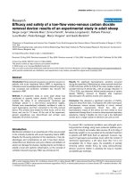
Analysis of two included RCTs, with Cochrane Rob tool,
indicated that the majority of the domains were judged
with unclear RoB due to insufficient information about
the used methodology (Supplementary Table 4, Fig. 2).
Four non-randomized studies were eligible for assessment with ROBINS-I. The most common judgment for
analyzed domains (12 domains out of 28 domains judged
Vuka et al. BMC Anesthesiology
(2020) 20:105
Page 18 of 21
Fig. 2 Risk of bias assessment for randomized controlled studies and cohort type studies
for these four studies) was serious RoB. Ten domains
were judged with moderate RoB, and only 6 domains
with low RoB (Supplementary Table 5, Fig. 2).
Studies awaiting classification
One RCT, which aims to study DRG thermal RF versus
PRF for metastatic pain in the thoracic vertebral body
on 69 participants, is classified as completed on July 30,
2018. Results were reported to Clinical Trials.gov but
were returned to the authors after the quality control review so results are still not publicly available
(NCT03204942). A trial that aimed to study superior
hypogastric plexus block versus PRF for chronic pelvic
cancer pain on 40 participants is classified as ‘Not yet
recruiting’ since June 26, 2018 (NCT03228316). Studies
awaiting classification are described in Supplementary
Table 6.
Discussion
In this systematic review, we included 17 studies about
the treatment of several non-neuropathic chronic pain
conditions with PRF directed to DRG. All studies presented positive conclusions (both conclusive and inconclusive) about the efficacy of the treatment. However, the
studies were mostly non-randomized, with small sample
sizes, and issues related to the risk of bias. Therefore, their
results should only be considered as preliminary.
PRF was developed as a less destructive pain relief modality alternative to conventional radiofrequency (CRF)
which can selectively block delta and C fibers [30]. The
first report about the clinical effects of PRF on DRG was
published relatively recently, in 1998. Due to its theoretical benefits, it was postulated that PRF could be particularly helpful in neuropathic pain [31]. However, we have
observed in the literature that clinicians and researchers
apply PRF to non-neuropathic chronic pain as well.
Vuka et al. BMC Anesthesiology
(2020) 20:105
Despite the number of studies found in the literature
about the treatment of non-neuropathic chronic pain in
humans with PRF, their findings cannot be generalized.
In the studies that we have found, the PRF was usually
initiated after other treatments have failed. We reported
a similar issue in our recent systematic review in which
we studied the efficacy and safety of EFS of DRG [12]. In
that systematic review, we found only one RCT among
29 included studies; most of the studies were low-level
of evidence – non-randomized study designs, including
case series and case reports. The review about EFS of
DRG also included few participants, with a median of 6
participants per study [12]. In this systematic review
there were 17 included studies, with a median of 28
participants.
The paucity of large and high-quality studies in the
field of DRG neuromodulation is likely due to the relative novelty of this approach for the treatment of pain.
In this systematic review, about PRF of DRG in nonneuropathic pain, only two of 17 included studies were
RCTs, and RoB judgment for the majority of their methodological aspects was unclear. Likewise, the most common assessment in non-randomized studies assessed
with the ROBINS-I tool was that there was a serious risk
of bias. Besides their suboptimal methodological reporting, the analyzed studies were relatively small. Even the
two included randomized controlled trials were small;
one included a total of 28 patients in 3 groups, and the
other one included 60 patients in two groups.
The highest number of studies was found for the low
back pain indication. However, we were not able to perform a meta-analysis due to clinical heterogeneity of the
studies, as can be seen from characteristics of included
studies, different comparators used in included trials,
and different stimulation parameters. Differences in
treatment approaches can result in different clinical
outcomes.
Despite the low level of evidence, all of the analyzed
studies sent positive conclusions to the research community in their abstracts. The majority of these conclusions were conclusive, i.e. they did not mention the need
to conduct further studies on this subject. Despite the
authors’ positive conclusions regarding the tested interventions, caution is needed when advising DRG targeted
PRF to chronic pain patients, because of the paucity of
high-quality and high-level evidence. This intervention
should be tested in large-scale, high-quality RCTs to
truly test whether the intervention has expected benefits
and harms. Until then, these studies should be treated as
preliminary evidence only.
A broad focus of this systematic review could be considered as a limitation of this review, as we included any
pain condition that fits the IASP criteria of nonneuropathic pain. Furthermore, we acknowledge that the
Page 19 of 21
examined studies included patients with various clinical
conditions, and thus there is a possibility that the effectiveness of the treatment depends on underlying pathogenic mechanisms. However, as can be observed from
our results, there were very few studies in each group of
indications; the highest number of studies (five) was
found for low back pain. Therefore, focusing on every
single one of these indications in a separate systematic
review would result in a high number of systematic reviews, with minimal results included. Furthermore, with
this approach, we are giving readers a very wide and informative picture of all the non-neuropathic pain conditions that were reported in the literature as treated with
DRG targeted PRF.
We have used IASP classification for definitions of
non-neuropathic pain; these classifications are evolving and changing, so the included conditions may be
categorized differently, depending on the time of
categorization and reference classification used. Previous versions of chronic pain classification were to
some extent insufficient for chronic neuropathic pain
conditions since some conditions were not defined
properly or were missing so we decided to use the
updated version of classification since it is crucial to
get the comprehensive evidence synthesis. According
to the newest IASP classification that we used (ICD11) when deciding about study inclusion we might
have included some studies that in previous versions
of classification were classified either as neuropathic
pain or as the pain of mixed origin. We have included CRPS 1 [25], which is not considered neuropathic pain. In the study of Kim et al. [6] the authors
studied the effects of DRG PRF beyond the acute
phase of zoster, bur before PHN was well established
(from 30 days to 180 days after zoster onset). The
study of van Zundert [22] has excluded “signs of radicular compression”.
It has been questioned before what is the value of systematic review including poor evidence and small studies
[32]. However, such systematic reviews are valuable because they are highlighting the paucity of evidence and
the low quality of available information [33]. Our systematic review is such a case. We even included two
case reports with only one participant which may be
considered anecdotal rather than firm evidence. It could
be argued that such studies should not even be included
in systematic reviews; however, we did not set any restrictions regarding number of participants in our study
eligibility criteria. By showing that many clinicians and
researchers have published small studies, with low-level
evidence, about potential benefits of PRF in chronic
non-neuropathic pain, we hope that trialists will be inspired to explore this intervention in studies that are
considered high-level evidence.
Vuka et al. BMC Anesthesiology
(2020) 20:105
Page 20 of 21
Conclusion
Even though PRF of DRG was primarily studied for
neuropathic pain, we have found as many as 17 published studies that have reported the use of DRG targeted PRF in non-neuropathic pain conditions. Although
all of these studies reported positive information regarding the analyzed interventions, considerable caution is
needed when interpreting these results as anything more
than preliminary. The quality of evidence is low, as there
were only two randomized controlled trials among included studies, and the risk of bias was predominantly
unclear in RCTs and severe among non-randomized
studies. The majority of studies included patients that
have failed other therapies so these results cannot be
generalized. PRF treatment needs to be tested in new,
high-quality and large-scale trials, to confirm the efficacy
of this intervention.
Funding
The study was funded by the Croatian Science Foundation (HRZZ) grant for
Young Scientist Career Development (HRZZ-DOK-2015-10-2774) and HRZZ
grant for Treating Neuropathic Pain with Dorsal Root Ganglion Stimulation
awarded to Prof Damir Sapunar (HRZZ-IP-2013-11-4126). The funders had no
role in study design, data collection and analysis, decision to publish, or
preparation of the manuscript.
Supplementary information
Additional file 3: Supplementary Table 3. Conclusion statements
presented in the abstracts of included studies.
Author details
Laboratory for Pain Research, University of Split School of Medicine,
Šoltanska 2, 21000 Split, Croatia. 2Department of Anesthesiology,
Reanimatology and Intensive Care, University Hospital Split, Spinčićeva 1,
21000 Split, Croatia. 3Center for Translational and Clinical Research,
Department of Proteomics, University of Zagreb School of Medicine, Šalata 3,
10000 Zagreb, Croatia. 4Department for Safety and Efficacy Assessment of
Medicinal Products, Agency for Medicinal Products and Medical Devices,
Ksaverska cesta 4, 10000 Zagreb, Croatia. 5Center for Evidence-Based
Medicine and Health Care, Catholic University of Croatia, Ilica 242, 10000
Zagreb, Croatia.
Additional file 4: Supplementary Table 4. Individual Cochrane risk of
bias judgments for randomized controlled trials.
Received: 20 March 2020 Accepted: 26 April 2020
Supplementary information accompanies this paper at />1186/s12871-020-01023-9.
Additional file 1: Supplementary Table 1. Search strategies for four
bibliographic databases searched.
Additional file 2: Supplementary Table 2. Characteristics of excluded
studies.
Additional file 5: Supplementary Table 5. Individual ROBINS
judgments for non-randomized studies.
Additional file 6: Supplementary Table 6. Details about studies
awaiting classification.
Abbreviations
AEs: adverse events; BA: before and after comparison; CLBP: chronic low back
pain; CR: case report; CRPS: complex regional pain syndrome; CRD: center for
reviews and dissemination; CS: case series; DRG: dorsal root ganglion;
EFS: electrical field stimulation; GPE: global perceived effect IASP –
International Association for the Study of Pain; ICN: intercostal nerves;
LBP: low back pain; MM: medical management; NA: not applicable;
NRS: numeric rating scale; NRSD: non-randomized study designs;
ODI: Oswestry disability index; PaMNI: patient-mounted navigated
intervention; PHN: post-herpetic neuralgia; PRF: pulsed radiofrequency;
QOLS: quality of life scale; RCS: retrospective cohort study; RCT: randomized
controlled trial; RoB: risk of bias; ROBINS-I: Risk of Bias In Non-randomized
Studies of Interventions; ROM: range of motion; SAEs: serious adverse events;
TENS: transcutaneous electrical nerve stimulation; TFESI: transforaminal
epidural steroid injection; VAS: visual analogue scale; WHO ICTRP: World
Health Organization’s International Clinical Trial Registry Platform;
WOMAC: functional status by Western Ontario and McMaster universities
osteoarthritis index
Acknowledgements
We are very grateful to Ms. Ana Utrobicic, expert librarian, for reviewing our
search strategies and providing valuable advice.
Authors’ contributions
IV, SD, DS, LP: study design. IV, TM, SD, LFH, KV: data collection and analysis.
IV, TM, SD, LFH, KV, DS, LP: manuscript writing, approval of final version of
the manuscript
Availability of data and materials
The datasets used and/or analyzed during the current study are available
from the corresponding author on reasonable request.
Ethics approval and consent to participate
In this study we analyzed only data from publicly available published articles;
for this reason, ethic approval and consent of participants to participate are
not applicable.
Consent for publication
Not applicable.
Competing interests
None.
1
References
1. Vos T, Flaxman AD, Naghavi M, et al. Years lived with disability (YLDs) for
1160 sequelae of 289 diseases and injuries 1990-2010: a systematic analysis
for the global burden of disease study 2010. Lancet. 2012;380(9859):2163–
96.
2. Goldberg DS, Mcgee SJ. Pain as a global public health priority. BMC Public
Health. 2011;11:770.
3. Harstall C, Ospina M. How prevalent is chronic pain? Pain Clinical Updates.
2003;11:1–4.
4. Landmark T, Romundstad P, Dale O, Borchgrevink PC, Kaasa S. Estimating
the prevalence of chronic pain: validation of recall against longitudinal
reporting (the HUNT pain study). Pain. 2012;153(7):1368–73.
5. Breivik H, Eisenberg E, O'brien T. The individual and societal burden of
chronic pain in Europe: the case for strategic prioritisation and action to
improve knowledge and availability of appropriate care. BMC Public Health.
2013;13:1229.
6. Kim ED, Lee YI, Park HJ. Comparison of efficacy of continuous epidural block
and pulsed radiofrequency to the dorsal root ganglion for management of
pain persisting beyond the acute phase of herpes zoster. PLoS ONE. 2017;
12(8):e0183559. />7. Chang MC, Cho YW, Ahn SH. Comparison between bipolar pulsed
radiofrequency and monopolar pulsed radiofrequency in chronic
lumbosacral radicular pain: a randomized controlled trial. Medicine. 2017;
96(9):e6236.
8. Lin WL, Lin BF, Cherng CH, et al. Pulsed radiofrequency therapy for relieving
neuropathic bone pain in cancer patients. J Med Sci (Taiwan). 2014;34(2):
84–7.
9. Snidvongs S, Mehta V. Pulsed radio frequency: a non-neurodestructive
therapy in pain management. Current Opinion Supportive Palliative Care.
2010;4(2):107–10.
10. Sapunar D, Kostic S, Banozic A, Puljak L. Dorsal root ganglion - a potential
new therapeutic target for neuropathic pain. J Pain Res. 2012;5:31–8.
Vuka et al. BMC Anesthesiology
(2020) 20:105
11. Vuka I, Marcius T, Dosenovic S, Ferhatovic Hamzic L, Vucic K, Sapunar D,
Puljak L. Efficacy and safety of pulsed radiofrequency of the dorsal root
ganglia in patients with neuropathic pain: a systematic review. Pain Med.
/>12. Vuka I, Marcius T, Dosenovic S, et al. Neuromodulation with electrical field
stimulation of dorsal root ganglion in various pain syndromes: a systematic
review with focus on participant selection. J Pain Res. 2019;12:803–30.
13. Higgins JPT, Green S (editors). Cochrane Handbook for Systematic Reviews
of Interventions Version 5.1.0 [updated March 2011]. London: The Cochrane
Collaboration; 2011. Available from />archive/v5.1/.
14. Holanda VM, Chavantes MC, Silva DF, et al. Photobiomodulation of the
dorsal root ganglion for the treatment of low back pain: a pilot study.
Lasers Surg Med. 2016;48(7):653–9.
15. Lee CC, Chen CJ, Chou CC, et al. Lumbar dorsal root ganglion block as a
prognostic tool before pulsed radiofrequency: a randomized, prospective,
and comparative study on cost-effectiveness. World Neurosurgery. 2018;112:
e157–64.
16. Yang CL, Yang BD, Lin ML, Wang YH, Wang JL. A patient-mount navigated
intervention system for spinal diseases and its clinical trial on percutaneous
pulsed radiofrequency stimulation of dorsal root ganglion. Spine. 2010;
35(21):E1126–32.
17. Hsu HT, Chang SJ, Huang KF, Tai PA, Li TC, Huang CJ. Correlation between
lumbar lordosis and the treatment of chronic low back pain with pulsed
radiofrequency applied to the L2 dorsal root ganglion. Formosan J Surg.
2017;50(4):125–30.
18. Tsou H-K, Chao S-C, Wang C-J, et al. Percutaneous pulsed radiofrequency
applied to the L-2 dorsal root ganglion for treatment of chronic low-back
pain: 3-year experience clinical article. J Neurosurg-Spine. 2010;12(2):190–6.
19. Albayrak I, Apiliogullari S, Dal CN, Levendoglu F, Ozerbil OM. Efficacy of
pulsed radiofrequency therapy to dorsal root ganglion adding to TENS and
exercise for persistent pain after Total knee Arthroplasty. J Knee Surg. 2017;
30(2):134–42.
20. Cohen SP, Sireci A, Wu CL, Larkin TM, Williams KA, Hurley RW. Pulsed
radiofrequency of the dorsal root ganglia is superior to pharmacotherapy or
pulsed radiofrequency of the intercostal nerves in the treatment of chronic
postsurgical thoracic pain. Pain Physician. 2006;9(3):227–35.
21. Fam BN, El-Sayed GGE, Reyad RM, Mansour I. Efficacy and safety of pulsed
radiofrequency and steroid injection for intercostobrachial neuralgia in
postmastectomy pain syndrome - a clinical trial. Saudi J Anaesth. 2018;12(2):
227–34.
22. Van Zundert J, Lame IE, De Louw A, et al. Percutaneous pulsed
radiofrequency treatment of the cervical dorsal root ganglion in the
treatment of chronic cervical pain syndromes: a clinical audit.
Neuromodulation. 2003;6(1):6–14.
23. Zhang J, Shi D-S, Wang R. Pulsed radiofrequency of the second cervical
ganglion (C2) for the treatment of cervicogenic headache. J Headache Pain.
2011;12(5):569–71.
24. Albayrak I, Apiliogullari S, Onal O, Gungor C, Saltali A, Levendoglu F. Pulsed
radiofrequency applied to the dorsal root ganglia for treatment of poststroke complex regional pain syndrome: a case series. J Clin Anesth. 2016;
33:192–7.
25. Apiliogullari S, Aydin BK, Onal O, Kirac Y, Celik JB. Pulsed radiofrequency of
dorsal root ganglia for the treatment of complex regional pain syndrome in
an adolescent with poliomyelitis sequel: a case report. Pain Med. 2015;16(7):
1369–72.
26. Arai Y-CP, Nishihara M, Yamamoto Y, et al. Dorsal root ganglion pulsed
radiofrequency for the management of intractable vertebral metastatic pain:
a case series. Pain Med. 2015;16(5):1007–12.
27. Hofmeester I, Steffens MG, Brinkert W. Chronic scrotal and inguinal pain
after orchidopexy in a 13-year-old boy, treated by pulsed radiofrequency of
the dorsal ganglia. J Pediatric Urol. 2013;9(6 Pt B):e155–6.
28. Lee SY, Jang DI, Noh C, Ko YK. Successful treatment of occipital radiating
headache using pulsed radiofrequency therapy. J Korean Neurosurgical
Society. 2015;58(1):89–92.
29. Li J, Yin Y, Ye L, Zuo Y. Pulsed radiofrequency of C2 dorsal root ganglion
under ultrasound guidance for chronic migraine: a case report. J Pain Res.
2018;11:1915–9.
30. Letcher FS, Goldring S. The effect of radiofrequency current and heat on
peripheral nerve action potential in the cat. J Neurosurg. 1968;29(1):42–7.
Page 21 of 21
31. Byrd D, Mackey S. Pulsed radiofrequency for chronic pain. Curr Pain
Headache Rep. 2008;12(1):37–41.
32. Roberts I, Ker K. How systematic reviews cause research waste. Lancet. 2015;
386(10003):1536.
33. Handoll HH, Langhorne P. In defence of reviews of small trials:
underpinning the generation of evidence to inform practice. Cochrane
Database Systematic Reviews doi: />ED000106(11), ED000106 (2015).
Publisher’s Note
Springer Nature remains neutral with regard to jurisdictional claims in
published maps and institutional affiliations.