Mô mềm quanh răng và Implants - Jan Lindhe, Jan L. Wennström, and Tord Berglundh
Bạn đang xem bản rút gọn của tài liệu. Xem và tải ngay bản đầy đủ của tài liệu tại đây (1.03 MB, 18 trang )
Chương 3
Mơ mềm quanh răng
và Implants
Jan Lindhe, Jan L. Wennstrưm, and Tord Berglundh
Nướu, 69
Khoảng sinh học, 69
Kích thước mơ mặt ngoài, 69
Dimensions of the interdental papilla, 71
The peri-implant mucosa, 71
Biologic width, 72
Quality, 76
Vascular supply, 77
Probing gingiva and peri-implant mucosa, 78
Dimensions of the buccal soft tissue at implants, 80
Dimensions of the papilla between teeth and implants, 81
Dimensions of the “papilla” between adjacent implants, 82
Nướu
Khoảng sinh học
Thuật từ thường được sử dụng để mơ tả kích thước mơ mềm
đối diện răng là khoảng sinh học của bám dính mơ mềm. Quan
Khe/túi nướu
niệm khoảng sinh học phát triển dựa trên những nghiên cứu và
phân tích của Gottlieb (1921), Orban và Kohler (1924), và Sicher
(1959). Những nghiên cứu này đã chứng minh, mô mềm bám
CEJ
dính vào răng bao gồm 2 phần, mơ sợi và biểu mơ bám dính.
Biểu mơ bám dính
Mơ liên kết
bám dính
Nghiên cứu của Gargiulo và cs (1961), có tên gọi “Kích thước và
các vấn đề liên quan đến kết nối răng nướu ở người”, khảo sát
những lát cắt từ mẫu sinh thiết nguyên khối ở những giai đoạn
“mọc răng thụ động” (nghĩa là sự phá hủy mô nha chu) khác
nhau. Đánh giá đo đạc sinh học được tiến hành để mô tả chiều
dài của rãnh nướu (không nằm trong phần kết nối), bám dính
biểu mơ (ngày nay được gọi là biểu mơ nối) và kết nối mơ liên
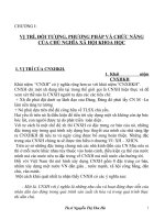
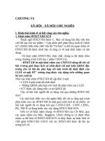
Hình. 3-1 Hình vẽ minh họa “khoảng sinh học” của bám dính mơ
mềm tại mặt ngồi của răng có mơ nha chu lành mạnh. Tổng chiều
dài của biểu mơ nối (bám dính biểu mơ) và kết nối mô liên kết được
gọi là “khoảng sinh học” của bám dính mơ mềm. Lưu ý rãnh nướu
khơng nằm trong phần bám dính.
kết (Hình 3-1). Kết quả khảo sát cho thấy, chiều dài của kết nối
mô liên kết thay đổi trong 1 giới hạn nhỏ (1.06 - 1.08 mm) trong
khi chiều dài của bám dính biểu mơ vào khoảng 1.4 mm ở
những vị trí có mơ nha chu bình thường, 0.8 mm tại vị trí có phá
Kích thước mơ mặt ngồi
hủy mơ nha chu trung bình và 0.7 mm ở những vị trí có sự phá
Đặc điểm hình thái của nướu liên hệ với kích thước mào
hủy nặng của mơ nha chu. Nói cách khác, (1) khoảng sinh học
xương ổ, hình dạng (giải phẫu) của răng, các biến cố xảy ra
của bám dính thay đổi trong khoảng 2.5 mm đối với những
trong quá trình mọc răng, và vị trí cũng như chiều hướng
trường hợp bình thường và 1.8 đối với những trường hợp bệnh
của răng đã mọc đầy đủ (Wheeler 1961; O’Connor & Biggs
nặng, và (2) khác biệt nhiều nhất trong phần bám dính mơ mềm
1964; Weisgold 1977). Ochenbein và Ross (1969), …..
là chiều dài của bám dính biểu mô (biểu mô nối).
70
Giải phẩu học
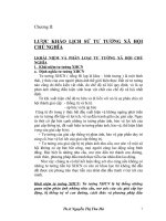
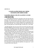
Hình. 3-2 Hình ảnh lâm sàng của cá thể có dạng sinh học
“uốn lượn”. Thân răng tương đối dài và thon. Gai nướu dài,
viền nướu mỏng và dải nướu sừng hóa hẹp.
Becker và cs. (1997) đã đề nghị (1) giải phẫu của nướu liên
quan với đường viền của mào xương ổ, và (2) tồn tại 2 dạng
cấu trúc nướu cơ bản được gọi là dạng sinh học “uốn lượn” và
“bằng”.
Những cá thể thuộc dạng sinh học “uốn lượn” có răng dài,
thon với thân răng dạng thn, cổ răng lồi nhẹ, vùng tiếp cận
hẹp và tiếp điểm nằm gần cạnh cắn (Hình 32). Nướu rời bao
quanh các răng trước hàm trên ở những cá thể này mỏng và
bờ nướu nằm ngang hoặc về phía chóp so với đường nối
men-xê măng. Vùng nướu hẹp với đường viền rất uốn lượn
(Olsson và cs. 1993). Ngược lại, những cá thể thuộc dạng
sinh học nướu “bằng” có các răng cửa với thân răng vng
và vùng cổ răng rất lồi (Hình 3-3). Nướu ở những cá thể này
rộng và dày hơn, vùng kẽ răng rộng và tiếp điểm nằm về
phía chóp hơn, gai nướu ngắn. Các báo cáo cho thấy, những
cá thể có nướu rất uốn lượn thường có sự tụt mơ mềm ở
vùng răng trước hàm trên trầm trọng hơn so với những cá
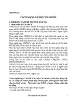
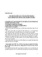
Hình. 3-3 Hỉnh ảnh lâm sàng của cá thể có dạng sinh học nướu
“bằng”. Thân răng tương đối ngắn và rộng. Gai nướu tương đối ngắn
nhưng dày, dải nướu sừng hóa rộng.
cá thể thuộc loại dạng sinh học bằng trung bình khoảng
4.5 mm. Trong khi đó, những cá thể thuộc dạng sinh học
rất uốn lượn có kích thước tương ứng nhỏ hơn đáng kể
(3.8 mm). Điều này khẳng định rằng, những cá thể thuộc
loại dạng sinh học bằng có thể tính mơ mềm vùng tiếp
giáp giữa mặt ngồi và mặt bên lớn hơn so với loại dạng
sinh học uốn lượn.
Pontoriero và Carnevale (2001) tiến hành đánh giá sự
sửa chữa của đơn vị nướu ở mặt ngoài các răng được
bộc lộ trong phẫu thuật làm dài thân răng có mài chỉnh
xương. Tại thời điểm 1 năm sau phẫu thuật, mô mềm đo
từ vị trí mào xương được mài chỉnh ở những bệnh nhân
dạng sinh học dày (bằng) có kích thước lớn hơn so với
dạng sinh học mỏng (uốn lượn), (3.1 mm so với 2.5 mm).
Nghiên cứu này không đánh giá sự thay đổi vị trí xương
giữa thời điểm phẫu thuật và thời điểm tái khám. Tuy
nhiên, phải xác định rằng có thể có sự tiêu xương trong
q trình lành thương và có sự tái thiết lập khoảng sinh
học của kết nối mơ liên kết mới phía trên (về phía thân
răng) vị trí xương đã được mài chỉnh.
thể có nướu bằng (Olsson & Lindhe 1991).
Kan và cs. (2003) đo kích thước của nướu - xác định
bằng cách thăm dò xuyên nướu (bone sounding) - tại
mặt ngoài-gần và ngoài-xa răng trước hàm trên. Thăm
dị xun nướu xác định khoảng cách từ viền mơ mềm
tới đỉnh xương và đưa đến kết quả ước tính lớn hơn 1
mm so với phương pháp đo túi thông thường. Các tác
giả đã báo cáo rằng độ dày của nướu thay đổi tùy theo
cá thể và dạng sinh học nướu. Vì vậy, chiều cao nướu
ở vị trí tiếp giáp giữa mặt ngồi và mặt bên ở những
Kích thước của nướu mặt ngồi cũng bị ảnh hưởng bởi
vị trí ngồi - trong của răng trong xương ổ. Di chuyển vị
trí răng về phía mặt ngồi làm giảm kích thước nướu mặt
ngoài và ngược lại (Coatoam và cs. 1981; Andlin-Sobocki
& Brodi 1993). Trong một nghiên cứu đánh giá sự khác
biệt độ dày của nướu mặt ngoài ở những người trưởng
thành trẻ, Muller và Knonen (2005) đã chứng minh rằng,
sự khác biệt độ dày nướu chủ yếu là do vị trí răng quyết
định, còn ảnh hưởng của sự khác biệt giữa các cá thể
(nghĩa là dạng sinh học dày hay uốn lượn) có vai trị rất
hạn chế.
The Mucosa at Teeth and Implants
71
B
B
P
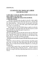
Fig. 3-4 Tarnow et al. (1992) measured the distance between
the contact point (P) between the crowns of the teeth and the
bone crest (B) using sounding (transgingival probing).
P
Dimensions of the interdental papilla
The interdental papilla in a normal, healthy dentition
has one buccal and one lingual/palatal component
that are joined in the col region (Chapter 1; Figs.
1-1–1-9). Experiments performed in the 1960s (Kohl
& Zander 1961; Matherson & Zander 1963) revealed
that the shape of the papilla in the col region was not
determined by the outline of the bone crest but by
the shape of the contact relationship that existed
between adjacent teeth.
Tarnow et al. (1992) studied whether the distance
between the contact point (area) between teeth and
the crest of the corresponding inter-proximal bone
could influence the degree of papilla fill that occurred
at the site. Presence or absence of a papilla was determined visually in periodontally healthy subjects. If
there was no space visible apical of the contact point,
the papilla was considered complete. If a “black
space” was visible at the site, the papilla was considered incomplete. The distance between the facial
level of the contact point and the bone crest (Fig. 3-4)
was measured by sounding. The measurement thus
included not only the epithelium and connective
tissue of the papilla but in addition the entire supraalveolar connective tissue in the inter-proximal area
(Fig. 3-5). The authors reported that the papilla was
always complete when the distance from the contact
point to the crest of the bone was≤5 mm. When this
distance was 6 mm, papilla fill occurred in about 50%
of cases and at sites where the distance was≥7 mm,
the papilla fill was incomplete in about 75% of cases.
Considering that the supracrestal connective tissue
attachment is about 1 mm high, the above data indicate that the papilla height may be limited to about
4 mm in most cases. Interestingly, papillae of similar
height (3.2–4.3 mm) were found to reform following
surgical denudation procedures (van der Velden
1982; Pontoriero & Carnevale 2001), but to a greater
Fig. 3-5 Mesio-distal section of the interproximal area
between the two central incisors. Arrows indicate the location
of the cemento-enamel junction. Dotted line indicates the
outline of the marginal bone crest. The distance between the
contact point (P) between the crowns of the teeth and the
bone crest (B) indicates the height of the papilla.
height in patients with a thick (flat) than in those with
a thin (pronounced scalloped) biotype.
Summary
· Flat gingival (periodontal) biotype: the buccal marginal gingiva is comparatively thick, the papillae
are often short, the bone of the buccal cortical wall
is thick, and the vertical distance between the
interdental bone crest and the buccal bone is short
(about 2 mm).
· Pronounced scalloped gingival (periodontal) biotype:
the buccal marginal gingiva is delicate and may
often be located apical of the cemento-enamel
junction (receded), the papillae are high and
slender, the buccal bone wall is often thin and the
vertical distance between the interdental bone
crest and the buccal bone is long (4 mm).
The peri-implant mucosa
The soft tissue that surrounds dental implants is
termed peri-implant mucosa. Features of the periimplant mucosa are established during the process of
wound healing that occurs subsequent to the closure
of mucoperiosteal flaps following implant installation (one-stage procedure) or following abutment
connection (two-stage procedure) surgery. Healing
of the mucosa results in the establishment of a soft
tissue attachment (transmucosal attachment) to the
72
Anatomy
implant. This attachment serves as a seal that prevents products from the oral cavity reaching the bone
tissue, and thus ensures osseointegration and the
rigid fixation of the implant.
The peri-implant mucosa and the gingiva have
several clinical and histological characteristics in
common. Some important differences, however, also
exist between the gingiva and the peri-implant
mucosa.
Biologic width
The structure of the mucosa that surrounds implants
made of titanium has been examined in man and
several animal models (for review see Berglundh
1999). In an early study in the dog, Berglundh et al.
(1991) compared some anatomic features of the
gingiva (at teeth) and the mucosa at implants. Since
the research protocol from this study was used in
subsequent experiments that will be described in this
chapter, details regarding the protocol are briefly
outlined here.
The mandibular premolars in one side of the mandible were extracted, leaving the corresponding teeth
in the contralateral jaw quadrant. After 3 months
of healing following tooth extraction (Fig. 3-6) the
fixture part of implants (Brånemark system®, Nobel
Biocare, Gothenburg, Sweden) were installed (Fig.
3-7) and submerged according to the guidelines given
in the manual for the system. Another 3 months later,
abutment connection was performed (Fig. 3-8) in a
second-stage procedure, and the animals were placed
in a carefully monitored plaque-control program.
Four months subsequent to abutment connection, the
dogs were exposed to a clinical examination following which biopsy specimens of several tooth and all
implant sites were harvested.
The clinically healthy gingiva and peri-implant
mucosa had a pink color and a firm consistency (Fig.
3-9). In radiographs obtained from the tooth sites it
Fig. 3-7 Three titanium implants (i.e. the fixture part and
cover screw; Brånemark System®) are installed.
Fig. 3-6 The edentulous mandibular right premolar region 3
months following tooth extraction (from Berglundh et al.
1991).
a
Fig. 3-8 Abutment connection is performed and the mucosa
sutured with interrupted sutures.
b
Fig. 3-9 After 4 months of careful plaque control the gingiva (a) and the peri-implant mucosa (b) are clinically healthy.
The Mucosa at Teeth and Implants
73
Fig. 3-10 Radiograph obtained from the premolars in the left
side of the mandible.
Fig. 3-12 Microphotograph of a cross section of the buccal
and coronal part of the periodontium of a mandibular
premolar. Note the position of the soft tissue margin (top
arrow), the apical cells of the junctional epithelium (center
arrow) and the crest of the alveolar bone (bottom arrow).
The junctional epithelium is about 2 mm long and the
supracrestal connective tissue portion about 1 mm high.
Fig. 3-11 Radiograph obtained from the implants in the right
side of the mandible.
was observed that the alveolar bone crest was located
about 1 mm apical of a line connecting the cementoenamel junction of neighboring premolars (Fig. 3-10).
The radiographs from the implant sites disclosed that
the bone crest was close to the junction between the
abutment and the fixture part of the implant (Fig.
3-11).
Histological examination of the sections revealed
that the two soft tissue units, the gingiva and the
peri-implant mucosa, had several features in common.
The oral epithelium of the gingiva was well keratinized and continuous with the thin junctional epithelium that faced the enamel and that ended at the
cemento-enamel junction (Fig. 3-12). The supraalveolar connective tissue was about 1 mm high and
the periodontal ligament about 0.2–0.3 mm wide. The
principal fibers were observed to extend from the
root cementum in a fan-shaped pattern into the soft
and hard tissues of the marginal periodontium (Fig.
3-13).
The outer surface of the peri-implant mucosa was
also covered by a keratinized oral epithelium, which
in the marginal border connected with a thin barrier
epithelium (similar to the junctional epithelium at the
teeth) that faced the abutment part of the implant
(Fig. 3-14). It was observed that the barrier epithelium was only a few cell layers thick (Fig. 3-15) and
Fig. 3-13 Higher magnification of the supracrestal connective
tissue portion seen in Fig. 3-12. Note the direction of the
principal fibers (arrows).
that the epithelial structure terminated about 2 mm
apical of the soft tissue margin (Fig. 3-14) and 1–
1.5 mm from the bone crest. The connective tissue in
the compartment above the bone appeared to be in
direct contact with the surface (TiO2) of the implant
(Figs. 3-14, 3-15, 3-16). The collagen fibers in this connective tissue apparently originated from the periosteum of the bone crest and extend towards the margin
of the soft tissue in directions parallel to the surface
of the abutment.
74
Anatomy
Fig. 3-16 Microphotograph of a section (buccal–lingual) of
the implant–connective tissue interface of the peri-implant
mucosa. The collagen fibers invest in the periosteum of the
bone and project in directions parallel to the implant surface
towards the margin of the soft tissue.
Fig. 3-14 Microphotograph of a buccal–lingual section of the
peri-implant mucosa. Note the position of the soft tissue
margin (top arrow), the apical cells of the junctional
epithelium (center arrow), and the crest of the marginal bone
(bottom arrow). The junctional epithelium is about 2 mm
long and the implant–connective tissue interface about
1.5 mm high.
Fig. 3-17 Implants of three systems installed in the mandible
of a beagle dog. Astra Tech Implants® Dental System (left),
Brånemark System® (center) and ITI® Dental Implant System
(right).
Fig. 3-15 Higher magnification of the apical portion of the
barrier epithelium (arrow) in Fig. 3-14.
The observation that the barrier epithelium of the
healthy mucosa consistently ended at a certain distance (1–1.5 mm) from the bone is important. During
healing following implant installation surgery, fibroblasts of the connective tissue of the mucosa apparently formed a biological attachment to the TiO2 layer
of the “apical” portion of the abutment portion of the
implant. This attachment zone was evidently not recognized as a wound and was therefore not covered
with an epithelial lining.
In further dog experiments (Abrahamsson et al.
1996, 2002) it was observed that a similar mucosal
attachment formed when different types of implant
systems were used (e.g. Astra Tech Implant System,
Astra Tech Dental, Mưlndal, Sweden; Brånemark
System®, Nobel Biocare, Göteborg, Sweden; Strau-
75
The Mucosa at Teeth and Implants
a
b
c
Fig. 3-18 Microphotographs illustrating the mucosa (buccal–lingual view) facing the three implant systems. (a) Astra. (b)
Brånemark. (c) ITI.
mann® Dental Implant System, Straumann AG, Basel,
Switzerland; 3i® Implant System, Implant Innovation
Inc., West Palm Beach, FL, USA). In addition, the
formation of the attachment appeared to be independent of whether the implants were initially submerged or not (Figs. 3-17, 3-18).
In another study (Abrahamsson et al. 1998), it was
demonstrated that the material used in the abutment
part of the implant was of decisive importance for the
location of the connective tissue portion of the transmucosal attachment. Abutments made of aluminumbased sintered ceramic (Al2O3) allowed for the
establishment of a mucosal attachment similar to that
which occurred at titanium abutments. Abutments
made of a gold alloy or dental porcelain, however,
provided conditions for inferior mucosal healing.
When such materials were used, the connective tissue
attachment failed to develop at the abutment level.
Instead, the connective tissue attachment occurred in
a more apical location. Thus, during healing following the abutment connection surgery, some resorption of the marginal peri-implant bone took place to
expose the titanium portion of the fixture (Brånemark
System®) to which the connective tissue attachment
was eventually formed.
The location and dimensions of the transmucosal
attachment were examined in a dog experiment by
Berglundh and Lindhe (1996). Implants (fixtures) of
the Brånemark System® were installed in edentulous
premolar sites and submerged. After 3 months of
healing, abutment connection was performed. In the
left side of the mandible the volume of the ridge
mucosa was maintained while in the right side the
vertical dimension of the mucosa was reduced to
≤2 mm (Fig. 3.19) before the flaps were replaced and
sutured. In biopsy specimens obtained after another
6 months, it was observed that the transmucosal
Flap adaptation and suturing
OE
OE
4 mm
2 mm
B
Test
B
Control
Fig. 3-19 Schematic drawing illustrating that the mucosa at
the test site was reduced to about 2 mm. From Berglundh &
Lindhe (1996).
attachment at all implants included one barrier epithelium that was about 2 mm long and one zone of
connective tissue attachment that was about 1.3–
1.8 mm high.
A further examination disclosed that at sites
with a thin mucosa, wound healing consistently
had included marginal bone resorption to establish
space for a mucosa that eventually could harbor
both the epithelial and the connective tissue components of the transmucosal attachment (Figs. 3-20,
3-21).
The dimensions of the epithelial and connective
tissue components of the transmucosal attachment at
implants are established during wound healing following implant surgery. As is the case for bone
healing after implant placement (see Chapter 5), the
wound healing in the mucosa around implants is a
delicate process that requires several weeks of tissue
remodeling.
76
Anatomy
In a recent animal experiment, Berglundh et al.
(2007) described the morphogenesis of the mucosa
attachment to implants made of c.p. titanium. A nonsubmerged implant installation technique was used
and the mucosal tissues were secured to the conical
marginal portion of the implants (Straumann® Dental
Implant System) with interrupted sutures. The
sutures were removed after 2 weeks and a plaquecontrol program was initiated. Biopsies were performed at various intervals to provide healing periods
extending from day 0 (2 hours) to 12 weeks. It was
reported that large numbers of neutrophils infiltrated
and degraded the coagulum that occupied the compartment between the mucosa and the implant during
6 months
PM
PM
2.0
aJE
2.1
aJE
The junctional and barrier epithelia are about 2 mm
long and the zones of supra-alveolar connective
tissue are between 1 and 1.5 mm high. Both epithelia
are attached via hemi-desmosomes to the tooth/
implant surface (Gould et al. 1984). The main attachment fibers (the principal fibers) invest in the root
cementum of the tooth, but at the implant site the
equivalent fibers run in a direction parallel with the
implant and fail to attach to the metal body. The soft
tissue attachment to implants is properly established
several weeks following surgery.
Quality
B
Control
Fig. 3-20 Schematic drawing illustrating that the peri-implant
mucosa at both control and test sites contained a 2 mm long
barrier epithelium and a zone of connective tissue that was
about 1.3–1.8 mm high. Bone resorption occurred in order to
accommodate the soft tissue attachment at sites with a thin
mucosa. From Berglundh & Lindhe (1996).
Test
a
Conclusion
1.8
B
1.3
Test
the initial phase of healing. The first signs of epithelial proliferation were observed in specimens representing 1–2 weeks of healing and a mature barrier
epithelium was seen after 6–8 weeks. It was also
demonstrated that the collagen fibers of the mucosa
were organized after 4–6 weeks of healing. Thus,
prior to this time interval, the connective tissue is not
properly arranged.
The quality of the connective tissue in the supraalveolar compartments at teeth and implants was
examined by Berglundh et al. (1991). The authors
observed that the main difference between the mesenchymal tissue present at a tooth and at an implant
site was the occurrence of a cementum on the root
surface. From this cementum (Fig. 3-22), coarse
dento-gingival and dento-alveolar collagen fiber
bundles projected in lateral, coronal, and apical
Control
b
Fig. 3-21 Microphotograph
illustrating the peri-implant mucosa
of a normal dimension (left) and
reduced dimension (right). Note the
angular bone loss that had occurred
at the site with the thin mucosa.
The Mucosa at Teeth and Implants
Fig. 3-22 Microphotograph of a tooth with marginal
periodontal tissues (buccal–lingual section). Note on the tooth
side the presence of an acellular root cementum with
inserting collagen fibers. The fibers are orientated more or
less perpendicular to the root surface.
directions (Fig. 3-13). At the implant site, the collagen
fiber bundles were orientated in an entirely different
manner. Thus, the fibers invested in the periosteum
at the bone crest and projected in directions parallel
with the implant surface (Fig. 3-23). Some of the
fibers became aligned as coarse bundles in areas
distant from the implant (Buser et al. 1992).
The connective tissue in the supra-crestal area at
implants was found to contain more collagen fibers,
but fewer fibroblasts and vascular structures, than
the tissue in the corresponding location at teeth.
Moon et al. (1999), in a dog experiment, reported that
the attachment tissue close to the implant (Fig. 3-24)
contained only few blood vessels but a large number
of fibroblasts that were orientated with their long
axes parallel with the implant surface (Fig. 3-25). In
more lateral compartments, there were fewer fibroblasts but more collagen fibers and more vascular
structures. From these and other similar findings it
may be concluded that the connective tissue attachment between the titanium surface and the connective tissue is established and maintained by
fibroblasts.
77
Fig. 3-23 Microphotograph of the peri-implant mucosa and
the bone at the tissue/titanium interface. Note that the
orientation of the collagen fibers is more or less parallel (not
perpendicular) to the titanium surface.
Fig. 3-24 Microphotograph of the implant/connective tissue
interface of the peri-implant mucosa. A large number of
fibroblasts reside in the tissue next to the implant.
Vascular supply
The vascular supply to the gingiva comes from two
different sources (Fig. 3-26). The first source is represented by the large supraperiosteal blood vessels, that
put forth branches to form (1) the capillaries of the
connective tissue papillae under the oral epithelium
and (2) the vascular plexus lateral to the junctional
epithelium. The second source is the vascular plexus
of the periodontal ligament, from which branches run
in a coronal direction and terminate in the supra-
Fig. 3-25 Electron micrograph of the implant–connective
tissue interface. Elongated fibroblasts are interposed between
thin collagen fibrils (magnification24 000).
78
Anatomy
alveolar portion of the free gingiva. Thus, the blood
supply to the zone of supra-alveolar connective tissue
attachment in the periodontium is derived from two
apparently independent sources (see also Chapter
1).
Berglundh et al. (1994) observed that the vascular
system of the peri-implant mucosa of dogs (Fig. 3-27)
originated solely from the large supra-periosteal blood
vessel on the outside of the alveolar ridge. This vessel
that gave off branches to the supra-alveolar mucosa
and formed (1) the capillaries beneath the oral epithelium and (2) the vascular plexus located immedi-
ately lateral to the barrier epithelium. The connective
tissue part of the transmucosal attachment to titanium implants contained only few vessels, all of
which could be identified as terminal branches of the
supra-periosteal blood vessels.
Summary
The gingiva at teeth and the mucosa at dental
implants have some characteristics in common, but
differ in the composition of the connective tissue, the
alignment of the collagen fiber bundles, and the distribution of vascular structures in the compartment
apical of the barrier epithelium.
Probing gingiva and
peri-implant mucosa
Fig. 3-26 A buccal–lingual section of a beagle dog gingiva.
Cleared section. The vessels have been filled with carbon.
Note the presence of a supraperiosteal vessel on the outside
of the alveolar bone, the presence of a plexus of vessels
within the periodontal ligament, as well as vascular
structures in the very marginal portion of the gingiva.
a
b
It was assumed for many years that the tip of the
probe in a pocket depth measurement identified the
most apical cells of the junctional (pocket) epithelium
or the marginal level of the connective tissue attachment. This assumption was based on findings by, for
example, Waerhaug (1952), who reported that the
“epithelial attachment” (e.g. Gottlieb 1921; Orban
& Köhler 1924) offered no resistance to probing.
Waerhaug (1952) inserted, “with the greatest caution”,
thin blades of steel or acrylic in the gingival pocket
of various teeth of100 young subjects without signs
of periodontal pathology. In several sites the blades
were placed in approximal pockets, “in which position radiograms were taken of them”. It was
concluded that the insertion of the blades could be
performed without a resulting bleeding and that the
device consistently reached to the cemento-enamel
junction (Fig. 3.28). Thus, the epithelium or the
epithelial attachment offered no resistance to the
insertion of the device.
Fig. 3-27 (a) A buccal–lingual cleared
section of a beagle dog mucosa facing
an implant (the implant was positioned
to the right). Note the presence of a
supraperiosteal vessel on the outside
of the alveolar bone, but also that there
is no vasculature that corresponds to
the periodontal ligament plexus. (b)
Higher magnification (of a) of the
peri-implant soft tissue and the bone
implant interface. Note the presence
of a vascular plexus lateral to the
junctional epithelium, but the absence
of vessels in the more apical portions
of the soft tissue facing the implant
and the bone.
The Mucosa at Teeth and Implants
79
2 mm
a
b
c
Fig. 3-28 An acrylic strip with a blue zone located 2 mm from the strip margin (a) prior to and (b) after its insertion into a
buccal “pocket”. The strip could with a light force be inserted 2 mm into the “pocket”. (c) Thin blades of steel were inserted in
pockets at approximal sites of teeth with healthy periodontal conditions. In radiographs, Waerhaug (1952) could observe that the
blades consistently reached the cemento-enamel junction.
In subsequent studies it was observed, however,
that the tip of a periodontal probe in a pocket depth
measurement only identified the base of the dentogingival epithelium by chance. In the absence of an
inflammatory lesion the probe frequently failed to
reach the apical part of the junctional epithelium (e.g.
Armitage et al. 1977; Magnusson & Listgarten 1980).
If an inflammatory lesion, rich in leukocytes and poor
in collagen, was present in the gingival connective
tissue, however, the probe penetrated beyond the
epithelium to reach the apical–lateral border of
the infiltrate.
The outcome of probing depth measurements at
implant sites was examined in various animal models.
Ericsson and Lindhe (1993) used the model by Berglundh et al. (1991) referred to above and, hence, had
both teeth and implants available for examination.
The gingiva at mandibular premolars and the mucosa
at correspondingly positioned implants (Brånemark
System®) were, after extended periods of plaque
control, considered clinically healthy. A probe with
a tip diameter of 0.5 mm was inserted into the buccal
“pocket” using a standardized force of 0.5 N. The
probe was anchored to the tooth or to the implant
and biopsies from the various sites were performed.
The histologic examination of the biopsy material
revealed that probing the dento-gingival interface
had resulted in a slight compression of the gingival
tissue. The tip of the probe was located coronal to
the apical cells of the junctional epithelium. At the
implant sites, probing caused both compression and
a lateral dislocation of the peri-implant mucosa, and
the average “histologic” probing depth was markedly deeper than at the tooth site: 2.0 mm versus
0.7 mm. The tip of the probe was consistently
positioned deep in the connective tissue/abutment
interface and apical of the barrier epithelium. The
distance between the probe tip and the bone crest
at the tooth sites was about 1.2 mm. The corresponding distance at the implant site was 0.2 mm. The
findings presented by Ericsson and Lindhe (1993)
regarding the difference in probe penetration in
healthy gingiva and peri-implant mucosa are not in
agreement with data reported in subsequent animal
experiments.
Lang et al. (1994) used beagle dogs and prepared
the implant (Straumann® Dental Implant System)
sites in such a way that at probing some regions were
healthy, a few sites exhibited signs of mucositis, and
some sites exhibited peri-implantitis. Probes with different geometry were inserted into the pockets using
a standardized probing procedure and a force of only
0.2 N. The probes were anchored and block biopsy
specimens were harvested. The probe locations were
studied in histologic ground sections. The authors
reported that the mean “histologic” probing depth at
80
Anatomy
healthy sites was about 1.8 mm, i.e. similar to the
depth (about 2 mm) recorded by Ericsson and Lindhe
(1993). The corresponding depth at sites with mucositis and peri-implantitis was about 1.6 mm and
3.8 mm respectively. Lang et al. (1994) further stated
that at healthy and mucositis sites, the probe tip
identified “the connective tissue adhesion level” (i.e.
the base of the barrier epithelium) while at periimplantitis sites, the probe exceeded the base of the
ulcerated pocket epithelium by a mean distance of
0.5 mm. At such peri-implantitis sites the probe
reached the base of the inflammatory cell infiltrate.
Schou et al. (2002) compared probing measurements at implants and teeth in eight cynomolgus
monkeys. Ground sections were produced from tooth
and implant sites that were (1) clinically healthy,
(2) slightly inflamed (mucositis/gingivitis), and (3)
severely inflamed (peri-implantitis/periodontitis)
and in which probes had been inserted. An electronic
probe (Peri-Probe®) with a tip diameter 0.5 mm and
a standardized probing force of 0.3–0.4 N was used.
It was demonstrated that the probe tip was located
at a similar distance from the bone in healthy tooth
sites and implant sites. On the other hand, at implants
exhibiting mucositis and peri-implantitis, the probe
tip was consistently identified at a more apical position than at corresponding sites at teeth (gingivitis
and periodontitis). The authors concluded that (1)
probing depth measurements at implant and teeth
yielded different information, and (2) small alterations in probing depth at implants may reflect changes
in soft tissue inflammation rather than loss of
supporting tissues.
Recently, Abrahamsson and Soldini (2006) evaluated the location of the probe tip in healthy periodontal and peri-implant tissues in dogs. It was reported
that probing with a force of 0.2 N resulted in a probe
penetration that was similar at implants and teeth.
Furthermore, the tip of the probe was often at or close
to the apical cells of the junctional/barrier epithelium. The distance between the tip of the probe and
the bone crest was about 1 mm at both teeth and
implants (Figs. 3-29, 3-30). Similar observations were
reported from clinical studies in which different
implant systems were used (Buser et al. 1990;
Quirynen et al. 1991; Mombelli et al. 1997). In these
studies the distance between the probe tip and the
bone was assessed in radiographs and was found to
vary between 0.75 and 1.4 mm when a probing force
of 0.25–0.45 N was used.
By comparing the findings from the studies
reported above, it becomes apparent that probing
depth and probing attachment level measurements
are also meaningful at implant sites. When a “normal”
probing force is applied in healthy tissues the probe
seems to reach similar levels at implant and tooth
sites. Probing inflamed tissues both at tooth and
implant sites will, however, result in a more advanced
probe penetration and the tip of the probe may come
closer to the bone crest.
Fig. 3-29 Buccal–lingual ground section from a tooth site
illustrating the probe tip position in relation to the bone crest
(from Abrahamsson & Soldini 2006).
Fig. 3-30 Buccal–lingual ground section from an implant site
illustrating the probe tip position in relation to the bone crest
(from Abrahamsson & Soldini 2006).
Dimensions of the buccal
soft tissue at implants
Chang et al. (1999) compared the dimensions of the
periodontal and peri-implant soft tissues of 20 subjects who had been treated with an implantsupported single-tooth restoration in the esthetic
zone of the maxilla and had a non-restored natural
tooth in the contralateral position (Fig. 3-31). In
The Mucosa at Teeth and Implants
a
81
b
Fig. 3-31 Clinical photographs of (a) an implant-supported single tooth replacement in position 12 and (b) the natural tooth in
the contralateral position (from Chang et al. 1999).
Dimensions of the papilla between
teeth and implants
4
Tooth
Implant
mm
*
2
*
0
Mucosa
thickness
Probing depth
Fig. 3-32 Comparison of mucosa thickness and probing
depth at the facial aspect of single-implant restorations and
the natural tooth in the contralateral position (from Chang
et al. 1999).
comparison to the natural tooth, the implant-supported crown was bordered by a thicker buccal
mucosa (2.0 mm versus 1.1 mm), as assessed at a
level corresponding to the bottom of the probeable
pocket, and had a greater probing pocket depth
(2.9 mm versus 2.5 mm) (Fig. 3-32). It was further
observed that the soft tissue margin at the implant
was more apically located (about 1 mm) than the gingival margin at the contralateral tooth.
Kan et al. (2003) studied the dimensions of the
peri-implant mucosa at 45 single implants placed
in the anterior maxilla that had been in function for
an average of 33 months. Bone sounding measurements performed at the buccal aspect of the implants
showed that the height of the mucosa was 3–4 mm
in the majority of the cases. Less than 3 mm of mucosa
height was found at only 9% of the implants. It
was suggested that implants in this category were
(1) found in subjects that belonged to a thin periodontal biotype, (2) had been placed too labially, and/
or (3) had an overcontoured facial prosthetic emergence. A peri-implant soft tissue dimension of4 mm
was usually associated with a thick periodontal
biotype.
In a study by Schropp et al. (2003) it was demonstrated that following single tooth extraction the
height of the papilla at the adjacent teeth was reduced
about 1 mm. Concomitant with this reduction (recession) of the papilla height the pocket depth was
reduced and some loss of clinical attachment
occurred.
Following single tooth extraction and subsequent
implant installation, the height of the papilla in the
tooth–implant site will be dependent on the attachment level of the tooth. Choquet et al. (2001) studied
the papilla level adjacent to single-tooth dental
implants in 26 patients and in total 27 implant sites.
The distance between the apical extension of the
contact point between the crowns and the bone crest,
as well as the distance between the soft tissue level
and the bone crest, was measured in radiographs.
The examinations were made 6–75 months after
the insertion of the crown restoration. The authors
observed that the papilla height consistently was
about 4 mm, and, depending on the location of the
contact point between adjacent crowns papilla, fill
was either complete or incomplete (Fig. 3-33). The
closer the contact point was located to the incisal
edge of the crowns (restorations) the less complete
was the papilla fill.
Chang et al. (1999) studied the dimensions of
the papillae at implant-supported single-tooth
restorations in the anterior region of the maxilla and
at non-restored contralateral natural teeth. They
found that the papilla height at the implantsupported crown was significantly shorter and
showed less fill of the embrasure space than the
papillae at the natural tooth (Fig. 3-34). This was particularly evident for the distal papilla of implant-supported restorations in the central incisor position,
both in comparison to the distal papilla at the contralateral tooth and to the papilla at the mesial aspect of
the implant crown. This indicates that the anatomy
of the adjacent natural teeth (e.g. the diameter of the
root, the proximal outline/curvature of the cementoenamel junction/connective tissue attachment
82
Anatomy
8
mm
6
4
2
0
0
1
2
3
Papilla index
Fig. 3-33 Soft tissue height adjacent to
single-tooth dental implants in
relation to the degree of papilla fill
(from Choquet et al. 2001).
6
Tooth
Implant
4
*
*
2
0
Papilla height
Papilla fill
Fig. 3-34 Comparison of papilla height and papilla fill
adjacent to single-implant restorations and the natural tooth
in the contralateral position (from Chang et al. 1999).
level) may have a profound influence on the dimension of the papilla lateral to an implant. Hence, the
wider facial–lingual root diameter and the higher
proximal curvature of the cemento-enamel junction
of the maxillary central incisor – in comparison to
corresponding dimensions of the lateral incisor
(Wheeler 1966) – may favor the maintenance of the
height of the mesial papilla at the single-implant
supported restoration.
Kan et al. (2003) assessed the dimensions of the
peri-implant mucosa lateral to 45 single implants
placed in the anterior maxilla and the 90 adjacent
teeth using bone sounding measurements. The bone
sounding measurements were performed at the
mesial and distal aspects of the implants and at the
mesial and distal aspects of the teeth. The authors
reported that the thickness of the mucosa at the
mesial/distal surfaces of the implant sites was on the
average 6 mm while the corresponding dimension at
the adjacent tooth sites was about 4 mm. It was
further observed that the dimensions of the peri-
implant mucosa of subjects who belonged to the thick
periodontal biotype were significantly greater than that
of subjects of a thin biotype.
The level of the connective tissue attachment on
the adjacent tooth surface and the position of the
contact point between the crowns are obviously key
factors that determine whether or not a complete
papilla fill will be obtained at the single-tooth
implant-supported restoration (Fig. 3.35). Although
there are indications that the dimensions of the
approximal soft tissue may vary between individuals
having thin and thick periodontal biotypes, the height
of the papilla at the single-implant restoration seems
to have a biological limit of about 4 mm (compare the
dimension of the interdental papilla). Hence, to
achieve a complete papilla fill of the embrasure space,
a proper location of the contact area between the
implant crown and the tooth crown is mandatory. In
this respect it must also be recognized that the papilla
fill at single-tooth implant restorations is unrelated
to whether the implant is inserted according to a oneor two-stage protocol and whether a crown restoration is inserted immediately following surgery or
delayed until the soft tissues have healed (Jemt 1999;
Ryser et al. 2005).
Dimensions of the “papilla”
between adjacent implants
When two neighboring teeth are extracted, the papilla
at the site will be lost (Fig. 3-36). Hence, at replacement of the extracted teeth with implant-supported
restorations the topography of the bone crest and the
thickness of the supracrestal soft tissue portion are
the factors that determine the position of the soft
tissue margin in the inter-implant area (“implant
papilla”). Tarnow et al. (2003) assessed the height
above the bone crest of the inter-implant soft tissue
(“implant papilla”) by transmucosal probing at 136
anterior and posterior sites in 33 patients who had
maintained implant-supported prostheses for at least
The Mucosa at Teeth and Implants
a
83
b
Fig. 3-35 See text for details.
a
b
c
Fig. 3-36 See text for details.
2 months. It was found that the mean height of the
“papillae” was 3.4 mm, with 90% of the measurements in the range of 2–4 mm.
The dimension of the soft tissues between adjacent
implants seems to be independent of the implant
design. Lee et al. (2006) examined the soft tissue
height between implants of two different systems
(Brånemark Implant® and Astra Tech Implant®
systems) as well as the potential influence of the horizontal distance between implants. The height of the
inter-implant “papilla”, i.e. the height of soft tissue
coronal to the bone crest measured in radiographs,
was about 3.1 mm for both implant systems. No difference was found regarding the “papilla” height for
any of the implant systems with regard to sites with
3 mm and≥3 mm in horizontal distance between
the implants. Gastaldo et al. (2004) evaluated the
presence or absence of “papilla” at 96 inter-implant
sites in 58 patients. It was reported that the “papilla”
filled the entire space between the implants only
when the distance from the bone crest to the base
of the contact point between the crown restorations,
assessed by sounding, was4 mm. Thus, taken
together these observations indicate that the soft
tissue between two implants will have a maximum
height of 3–4 mm, and that the location of the contact
point between the crown restorations in relation to
the bone crest level determines whether a complete
soft tissue fill will be obtained in the embrasure space
between two implants (Fig. 3-37).
84
Anatomy
2 months
a
6 months
6 months
b
c
12 months
d
e
Fig. 3-37 See text for details.
References
Abrahamsson, I., Berglundh, T., Glantz, P.O. & Lindhe, J.
(1998). The mucosal attachment at different abutments. An
experimental study in dogs. Journal of Clinical Periodontology
25, 721–727.
Abrahamsson, I., Berglundh, T., Wennström, J. & Lindhe, J.
(1996). The peri-implant hard and soft tissues at different
implant systems. A comparative study in the dog. Clinical
Oral Implants Research 7, 212–219.
Abrahamsson, I., Zitzmann, N.U., Berglundh, T., Linder, E.,
Wennerberg, A. & Lindhe, J. (2002). The mucosal attachment to titanium implants with different surface characteristics: an experimental study in dogs. Journal of Clinical
Periodontology 29, 448–455.
Abrahamsson, I. & Soldini, C. (2006). Probe penetration in periodontal and peri-implant tissues: an experimental study in
the beagle dog. Clinical Oral Implants Research 17, 601–605.
Andlin-Sobocki, A. & Bodin, L. (1993). Dimensional alterations
of the gingiva related to changes of facial/lingual tooth
position in permanent anterior teeth of children. A 2-year
longitudinal study. Journal of Clinical Periodontology 20,
219–224.
Armitage, G.C., Svanberg, G.K. & Löe, H. (1977). Microscopic
evaluation of clinical measurements of connective tissue
attachment levels. Journal of Clinical Periodontology 4,
173–190.
Becker, W., Ochenbein, C., Tibbets, L. & Becker, B.E. (1997).
Alveolar bone anatomic profiles as measured from dry
skulls. Journal of Clinical Periodontology 24, 727–731.
Berglundh, T. (1999). Soft tissue interface and response to
microbial challenge. In: Lang, N.P., Lindhe, J. & Karring,
T., eds. Implant Dentistry. Proceedings from 3rd European
Workshop on Periodontology. Berlin: Quintessence, pp.
153–174.
Berglundh, T. & Lindhe, J. (1996). Dimensions of the periimplant mucosa. Biological width revisited. Journal of Clinical Periodontology 23, 971–973.
Berglundh, T., Lindhe, J., Ericsson, I., Marinello, C.P.,
Liljenberg, B. & Thomsen, P. (1991). The soft tissue barrier
at implants and teeth. Clinical Oral Implants Research 2,
81–90.
Berglundh, T., Lindhe, J., Jonsson, K. & Ericsson, I. (1994). The
topography of the vascular systems in the periodontal and
peri-implant tissues dog. Journal of Clinical Periodontology 21,
189–193.
Berglundh, T., Abrahamsson, I., Welander, M., Lang, N.P. &
Lindhe, J. (2007). Morphogenesis of the periimplant mucosa.
An experimental study in dogs. Clinical Oral Implants
Research (in press).
Buser, D., Weber, H.P. & Lang, N.P. (1990). Tissue integration
of non-submerged implants. 1-year results of a prospective
The Mucosa at Teeth and Implants
study on 100 ITI-hollow-cylinder and hollow-screw
implants. Clinical Oral Implants Research 1, 225–235.
Buser, D., Weber, H.P., Donath, K., Fiorellini, J.P., Paquette,
D.W. & Williams, R.C. (1992). Soft tissue reactions to nonsubmerged unloaded titanium implants in beagle dogs.
Journal of Periodontology 63, 226–236.
Chang, M., Wennstrưm, J., Ưdman, P. & Andersson, B. (1999).
Implant supported single-tooth replacements compared to
contralateral natural teeth. Clinical Oral Implants Research 10,
185–194.
Choquet, V., Hermans, M., Adriaenssens, P., Daelemans, P.,
Tarnow, D. & Malevez, C. (2001). Clincal and radiographic
evaluation of the papilla level adjacent to single-tooth
dental implants. A retrospective study in the maxillary
anterior region. Journal of Periodontology 72, 1364–1371.
Coatoam, G.W., Behrents, R.G. & Bissada, N.F. (1981). The
width of keratinized gingiva during orthodontic treatment:
its significance and impact on periodontal status. Journal of
Periodontology 52, 307–313.
Ericsson, I. & Lindhe, J. (1993). Probing depth at implants and
teeth. Journal of Clinical Periodontology 20, 623–627.
Gargiulo, A.W., Wentz, F.M. & Orban, B. (1961). Dimensions
and relations of the dentogingival junction in humans.
Journal of Periodontology 32, 261–267.
Gastaldo, J.F., Cury, P.R. & Sendyk, W.R. (2004). Effect of the
vertical and horizontal distances between adjacent implants
and between a tooth and an implant on the incidence
of interproximal papilla. Journal of Periodontology 75, 1242–
1246.
Gottlieb, B. (1921). Der Epithelansatz am Zahne. Deutsche
monatschrift führ Zahnheilkunde 39, 142–147.
Gould, T.R.L., Westbury, L. & Brunette, D.M. (1984).
Ultrastructural study of the attachment of human gingiva
to titanium in vivo. Journal of Prosthetic Dentistry 52,
418–420.
Jemt, T. (1999). Restoring the gingival contour by means of
provisional resin crowns after single-implant treatment.
International Journal of Periodontics and Restorative Dentistry
19, 21–29.
Kan, J., Rungcharassaeng, K., Umezu, K. & Kois, J. (2003).
Dimensions of the periimplant mucosa: An evaluation of
maxillary anterior single implants in humans. Journal of
Periodontology 74, 557–562.
Kohl, J. & Zander, H. (1961). Morphology of interdental gingival tissue. Oral Surgery, Oral Medicine and Oral Pathology 60,
287–295.
Lang, N.P., Wetzel, A.C., Stich, H. & Caffesse, R.G. (1994).
Histologic probe penetration in healthy and inflamed
peri-implant tissues. Clinical Oral Implants Research 5,
191–201.
Lee, D-W., Park, K-H. & Moon, I-S. (2006). Dimension of interproximal soft tissue between adjacent implants in two
distinctive implant systems. Journal of Periodontology 77,
1080–1084.
Magnusson, I. & Listgarten, M.A. (1980). Histological evaluation of probing depth following periodontal treatment.
Journal of Clinical Periodontology 7, 26–31.
Matherson, D. & Zander, H. (1963). Evaluation of osseous
surgery in monkeys. Journal of Dental Research 42, 116.
Mombelli, A., Mühle, T., Brägger, U., Lang, N.P. & Bürgin, W.B.
(1997). Comparison of periodontal and peri-implant probing
by depth-force pattern analysis. Clinical Oral Implants
Research 8, 448–454.
Moon, I-S., Berglundh, T., Abrahamsson, I., Linder, E. & Lindhe,
J. (1999). The barrier between the keratinized mucosa and
85
the dental implant. An experimental study in the dog.
Journal of Clinical Periodontology 26, 658–663.
Müller, H.P. & Könönen, E. (2005). Variance components
of gingival thickness. Journal of Periodontal Research 40,
239–244.
O’Connor, T.W. & Biggs, N. (1964). Interproximal craters.
Journal of Periodontology 35, 326–330.
Olsson, M. & Lindhe, J. (1991). Periodontal characteristics in
individuals with varying forms of upper central incisors.
Journal of Clinical Periodontology 18, 78–82.
Olsson, M., Lindhe, J. & Marinello, C. (1993). On the relationship between crown form and clinical features of the gingiva
in adolescents. Journal of Clinical Periodontology 20,
570–577.
Orban, B & Köhler, J. (1924). Die physiologische Zanhfleischetasche, Epithelansatz und Epitheltiefenwucherung. Zeitschrift
für Stomatologie 22, 353.
Oschenbein, C. & Ross, S. (1969). A reevaluation of osseous
surgery. In: Dental Clinics of North America. Philadelphia,
PA: W.B. Saunders, pp. 87–102.
Pontoriero, R. & Carnevale, G. (2001). Surgical crown lengthening: A 12-month clinical wound healing study. Journal of
Periodontology 72, 841–848.
Quirynen, M., van Steenberge, D., Jacobs, R., Schotte, A. &
Darius, P. (1991). The reliability of pocket probing around
screw-type implants. Clinical Oral Implants Research 2,
186–192.
Ryser, M.R., Block, M.S. & Mercante, D.E. (2005). Correlation
of papilla to crestal bone levels around single tooth implants
in immediate or delayed crown protocols. Journal of Maxillofacial Surgery 63, 1184–1195.
Schou, S., Holmstrup, P., Stolze, K., Hjørting-Hansen, E., Fien,
N.E. & Skovgaard, L.T. (2002). Probing around implants
and teeth with healthy or inflamed marginal tissues. A histologic comparison in cynomolgus monkeys (Macaca fascicularis). Clinical Oral Implants Research 13, 113–126.
Schropp, L., Wenzel, A., Kostopoulos, L. & Karring, T. (2003).
Bone healing and soft tissue contour changes following
singe-tooth extraction: A clinical and radiographic 12month prospective study. International Journal of Periodontics
and Restorative Dentistry 23, 313–323.
Sicher, H. (1959). Changing concepts of the Supporting Dental
Structure. Oral Surgery, Oral Medicine and Oral Pathology 12,
31–35.
Tarnow, D., Elian, N., Fletcher, P., Froum, S., Magner, A., Cho,
S-C., Salama, M., Salama, H. & Garber, D.A. (2003). Vertical
distance from the crest of bone to the height of the interproximal papilla between adjacent implants. Journal of
Periodontology 74, 1785–1788.
Tarnow, D., Magner, A. & Fletcher, P. (1992). The effect of the
distance from the contact point to the crest of bone on the
presence or absence of the interproximal dental papilla.
Journal of Periodontology 63, 995–996.
van der Velden, U. (1982). Regeneration of the interdental soft
tissues following denudation procedures. Journal of Clinical
Periodontology 9, 455–459.
Weisgold, A. (1977). Contours of the full crown restoration.
Alpha Omegan 7, 77–89.
Waerhaug, J. (1952). Gingival pocket: anatomy, pathology,
deepening and elimination. Odontologisk Tidskrift 60 (Suppl
1).
Wheeler, R.C. (1961). Complete crown form and the periodontium. Journal of Prosthetic Dentistry 11, 722–734.
Wheeler, R. C. (1966). An Atlas of Tooth Form. Philadelphia: W.B.
Saunders Co, pp. 24–25.
Thank you for evaluating Wondershare PDF Converter.
You can only convert 3 pages with the trial version.
To get all the pages converted, you need to purchase the software from:
/>