THÀNH PHẦN hợp CHẤT AXÍT béo và CAROTENOID của gấc fatty acid and carotenoid composition of gac (momordica cochinchinensisspreng) fruit
Bạn đang xem bản rút gọn của tài liệu. Xem và tải ngay bản đầy đủ của tài liệu tại đây (72.66 KB, 6 trang )
Fatty Acid and Carotenoid Composition of Gac (Momordica
cochinchinensis Spreng) Fruit
BETTY K. ISHIDA,* CHARLOTTA TURNER,MARY H. CHAPMAN, AND
THOMAS A. MCKEON
Western Regional Research Center, Agricultural Research Service, United States Department of
Agriculture, 800 Buchanan Street, Albany, California 94710
In this study, we analyzed fatty acid and carotenoid composition of fruit tissues, including seed (which
are surrounded by a bright red, oily aril) of
Momordica cochinchinensis
Spreng, known as gac in
Vietnam. Carotenoid content was analyzed by reversed-phase HPLC, using a C
30
column and a
method separating cis- and trans-isomers of the major carotenoids in this fruit. Mean values obtained
in aril tissues were 1342 µg trans-, 204 µg cis-, and 2227 µg total lycopene; 597 µg trans-, 39 µg
cis-, and 718 µg total β-carotene; and 107 µg R-carotene/g FW. Mesocarp contained 11 µg trans-, 5
µg cis
-
β-carotene/g FW, trace amounts of R-carotene, and no lycopene. Gac aril contained 22%
fatty acids by weight, composed of 32% oleic, 29% palmitic, and 28% linoleic acids. Seeds contained
primarily stearic acid (60.5%), smaller amounts of linoleic (20%), oleic (9%), and palmitic (5-6%)
acids, and trace amounts of arachidic, cis-vaccenic, linolenic, and palmitoleic, eicosa-11-enoic acids,
and eicosa-13-enoic (in one fruit only) acids.
KEYWORDS:
Momordica cochinchinensis
Spreng; fatty acids; carotenoids; HPLC; lycopene; β-carotene;
aril; mesocarp; seed; oil.
INTRODUCTION
Momordica cochinchinensis Spreng, a Cucurbitaceae,is
indigenous throughout Asia and used as food and for medicinal
purposes. The fruit, called gac in Vietnam, are only picked there
at maturity from August through February when they are red
and the seeds are hardened. Aril, the oily, red, fleshy pulp
surrounding the seeds, has a palatable, bland to nutty taste and
is cooked along with seeds to impart its red color and flavor to
a rice dish, xoi gac, served at festive occasions (e.g., weddings)
in Vietnam (1). Seeds are used in Chinese traditional medicine.
Early recognition of the value of gac fruit focused on β-carotene
concentration (2). West and Poortvliet (3) measured 188.10 µg
of β-carotene and 891.50 µg total carotenoids/g fresh weight
(FW) in gac aril. Chemical analyses by Vuong et al. (1) showed
that gac aril contained 175 µgofβ-carotene and 802 µgof
lycopene/g FW (1). Lycopene concentration in gac aril is in
marked contrast to the 40-60 µg lycopene/g FW found in field-
grown tomatoes (4, 5), which is the major source of lycopene
in the Western diet. Lycopene, of course, is of interest, because
of the correlation of reduced risk of certain cancers, such as
prostate (6-8) and lung (7, 9), with the consumption of tomato
products, which is attributed to protection by free radical-
quenching lycopene (10, 11). In addition, studies on African-
American men having prostate cancer show that daily con-
sumption of lycopene from tomato sauce significantly increased
lycopene content of plasma and the prostate gland, decreased
their prostate-specific antigen levels (a marker for prostate
cancer), and showed significant clinical and metabolic improve-
ments (12). Antioxidants seem to have protective effects against
cardiovascular diseases (13-16) and a number of common eye
diseases, such as cataracts and age-related macular degeneration
(17-20). In addition, because β-carotene is a precursor to
Vitamin A, gac fruit is a potentially valuable source of this
vitamin and could be extremely useful in fighting Vitamin A
deficiency, which is common in third world countries (1).
According to a report by Vuong et al. (1), gac aril also
contains 102 mg oil/g of FW. These authors also found that, of
the total fatty acids in gac aril, 69% are unsaturated, and 35%
of these are polyunsaturated (21). Vuong and King (21) reported
that the oil in gac aril contains significant amounts of Vitamin
E (334 µg/mL), as well as 3020 µg of lycopene and 2710 µgof
β-carotene (and isomers)/mL, making gac aril with its oil a
valuable potential source of antioxidants.
Gac seed composition is of interest because of its use in
traditional Chinese medicine. Recently, a pentacyclic triterpenoid
ester was isolated from the seed (22).
Since the completion of this study, a report on carotenoid
pigments in gac fruit was published (23). Our study, in addition
to identification of major carotenoids, includes carotenoid
profiles, measuring both trans- and cis-isomers of lycopene and
β-carotene. We also provide a detailed fatty acid analysis of
gac seed and aril, as well as the weight distribution of anatomical
components of the fruit.
* To whom correspondence should be addressed. Tel.: 510-559-57267.
Fax: 51-0-559-6166. E-mail:
274 J. Agric. Food Chem. 2004, 52, 274−279
10.1021/jf030616i This article not subject to U.S. Copyright. Published 2004 by the American Chemical Society
Published on Web 12/30/2003
MATERIALS AND METHODS
Fatty Acid Analysis. Materials. Gac fruit were purchased from two
Asian markets (Vinh Phat and Shun Fat) in Sacramento, California.
Fruit had been shipped frozen by commercial exporters from Vietnam
to California (storage temperature during transport unknown) and were
left frozen in a -20 °C freezer until ready for analyses. Gac fruit were
divided carefully into its anatomical components: skin, mesocarp,
connective tissue, aril, and seed. Most of the seeds used for analyses
were taken from purchased, frozen fruit that had been shipped from
Vietnam; a few were a gift from the Guangzhi Province in Western
China.
Trifluoroacetic anhydride, 3-pyridyl carbinol, 4-(dimethyl amino)
pyridine, and cyclohexane were obtained from Sigma-Aldrich (St. Louis,
MO). Nonadecanoic acid methyl ester and GLC-68 fatty acid methyl
ester (FAME) standard mixture were obtained from Nu-Chek Prep, Inc.
(Elysian, MN). Heptadecanoic acid methyl ester and anhydrous acetyl
chloride were purchased from Alltech (Deerfield, IL), and butylated
hydroxytoluene (BHT) was obtained from Spectrum Chemical MFG
Corp. (Gardena, CA). Anhydrous sodium sulfate was purchased from
J. T. Baker Inc. (Philipsburg, NJ), and 2-propanol, methanol, hexane,
toluene, diethyl ether, and dichloromethane were obtained from Fisher
Scientific (Fair Lawn, NJ). Potassium hydroxide, sodium thiosulfate,
sodium chloride, and potassium bicarbonate were obtained from
Mallinckrodt Laboratory Chemicals (Philipsburg, NJ). Ethanol was
purchased from AAPER Alcohol and Chemical Co. (Shelbyville, KY).
The water used was double distilled, and all chemicals and solvents
used were of reagent grade.
Method. Gac aril and mesocarp were thoroughly homogenized using
a household-type coffee grinder (Mr. Coffee, Cleveland, OH; Model
IDS59) and then dried using a vacuum centrifuge (7-8% dry weight).
Gac seed was homogenized using a mortar and pestle. Gac sample (0.05
g) was accurately weighed into 10-mL glass tubes. The lipids were
extracted using 2 mL of hexane/2-propanol (8:2, v/v) containing 50
µg/mL of BHT. Internal standard (nonadecanoic acid methyl ester) was
added, and the extraction took place at 55 °C for 30 min with shaking
every 10 min. Extracts were filtered and dried over sodium sulfate,
and the solvent was evaporated under nitrogen. Oil weight was
determined gravimetrically. Toluene (0.5 mL) was then added, and the
lipids were methylated for1hat80°C using methanolic hydrogen
chloride (3%), as described by Christie (24). Resulting FAMEs were
dissolved in 10 mL of cyclohexane (0.01% BHT) for GC analysis.
Quantitative analysis was carried out by GC-FID using a Hewlett-
Packard 6890 GC system with split injection connected to a 7673
automatic liquid sampler (Agilent Technologies, Palo Alto, CA).
Separation was achieved on a DB-WAX column (20-m × 0.12-mm
i.d., 0.18-µm film thickness) purchased fromJ&WScientific, Agilent
Technologies. The injector and detector temperatures were 250 and
280 °C, respectively. The column temperature program was 100 °C
for 1 min, then increased by 5 °C/min to 250 °C, and held at 250 °C
for 1 min. Standard solutions of a mixture of FAMEs at three different
concentrations in the range of 5 to 150 µg/mL were used for generating
standard calibration curves. A 50-µL sample of methyl heptadecanoate
(1 mg/mL) was added as internal standard to 1-mL aliquots of each
standard sample. Injections of 1 µL were used, and duplicate determina-
tions were performed.
Identification of peak components was achieved on a Hewlett-
Packard 5890 GC system connected to a 5970A mass selective detector
(Agilent Technologies). Split injection was applied, and the same type
of column and temperature program as described above was used.
Comparison to mass spectra of known FAMEs was used to identify
each peak. In addition, double-bond locations for the unsaturated fatty
acids were determined by interpreting spectra from picolinyl derivatives
of free fatty acids (FFAs), employing the methodology described by
Christie (24).
Carotenoid Analysis. Materials. Dichloromethane, 99.9%, HPLC
grade and anhydrous tetrahydrofuran (THF), 99.9%, were purchased
from Aldrich Chemical Co. (Milwaukie, WI). Methanol (MeOH), HPLC
grade, methyl-tert-butyl ether (MTBE), and ethyl acetate (EtOAc),
HPLC grade, were purchased from Fisher Scientific (Fair Lawn, NJ).
Lycopene for standard solutions was extracted and purified from berries
of autumn olive (Elaeagnus umbellata Thunberg) plants, which were
a gift from Beverly A. Clevidence (Beltsville Human Nutrition Research
Center, UDSA, ARS, Beltsville, MD). β-Carotene (type IV from
carrots), mixed isomer carotene (from carrots), and lutein (from alfalfa)
were purchased from Sigma Chemical Company (St Louis, MO).
Methods. Dry weights of gac aril and mesocarp tissues were
determined using a Model AVC-80 microwave moisture/solids analyzer
(CEM Corporation, Mathews, NC). Samples of tissue were placed
between two tared glass-fiber pads and heated at 50% power for 4.5
min. Moisture content (or percent solids) was determined by difference
in weight after drying.
Carotenoids were extracted from gac fruit tissues by the modification
(25) of the method described by Ishida et al. (26). Tissues were excised
carefully from gac fruit to avoid cross contamination, especially between
aril and mesocarp, then homogenized, using an Omni-Mixer (Sorvall/
DuPont Medical Products, Newtown, CT). Gac samples were first
extracted, using 5 mL of ice-cold MeOH/homogenate, then the
suspension was vacuum-filtered through two layers of Whatman No.
1 filter paper on a Bu¨chner funnel and washed with an additional
volume of ice-cold MeOH. The filtrate was saved. The remaining
dehydrated residue on the filter was carefully resuspended in 5 mL of
dichloromethane and extracted by vacuum filtration three times to
remove the red/orange color. The filtrate from the MeOH used to
dehydrate the tissue homogenate was combined with dichloromethane
extracts. Water (5 mL) was then added to the combined extracts and
mixed thoroughly, using a vortex mixer. After phase separation, the
bottom yellow layer was transferred to a small vial and dried under
nitrogen gas. The residue was then resuspended in 2 mL of THF and
passed through a 0.45-mm poly(tetrafluoroethylene) filter (Alltech
Associates, Inc., Deerfield, IL). Throughout these procedures, care was
taken to keep samples ice-cold and protect them from exposure to light.
Extracts of gac fruit tissue were analyzed for carotenoid content by
separation followed by quantitation using a reversed-phase HPLC
system, consisting of a Waters (Milford, MA) 2690 Separation Module,
996 Photodiode-Array Detector, auto injector, and column temperature
regulator. Separations were accomplished using a reversed phase,
analytical (250 × 4.6-mm I. D.), 3-µm particle diameter polymeric
C
30
column (YMC Inc. Wilmington, NC). The system was purged daily
for 3 min each with MTBE, MeOH, and EtOAC. The C
30
column was
then conditioned with elution solvent at a flow rate of 1 mL/min for
10 min. Carotenoids were separated isocratically using a mobile phase
of 40% MTBE 50%, MeOH, and 10% EtOAc (v/v). Injection volumes
ranged from 5 to 20 µL. Column temperature was maintained at 28
°C. The photodiode array detector was set between 300 and 700 nm to
detect all of the peaks of interest eluted from the column. Standard
compounds: xanthophyll (Sigma; 70% pure from alfalfa); lycopene
extracted from autumn olive (Elaeagnus umbellate Thunberg) (gift from
B. A. Clevidence, USDA Beltsville Human Nutrition Research Center),
purified and found to be 97% trans isomer, was used as a standard for
quantitation; β-carotene (Sigma; synthetic, Type 1, 95% pure), and
R-carotene (Sigma; from spinach, substantially free of β-carotene) were
used to check retention times on the HPLC. Phytoene, phytofluene,
zeaxanthin, and β-cryptoxanthin were detected by examining spectra
of compounds under chromatographic peaks and comparing to known,
published spectra of carotenoids to identify these compounds, which
are found commonly in fruit such as tomato, guava, and citrus.
RESULTS AND DISCUSSION
Weight Distribution of Fruit Components. Table 1 shows
fresh and dry weights and percent weight distributions of the
anatomical components of a typical gac fruit. Two whole fruits
were analyzed in this way. Most of the fruit is composed of
mesocarp and seeds with their surrounding oily pulp (aril). Of
these tissues, the mesocarp represents almost half of the weight
of the entire fruit.
Fatty Acid Analysis. Data on total FAME content of aril
from two gac fruit were collected. Total % weight content of
FAME in these two fruit was nearly identical (22%, with relative
standard deviations of 2.3 and 12.2%), even though the aril from
Fatty Acid and Carotenoids in Gac Fruit J. Agric. Food Chem., Vol. 52, No. 2, 2004 275
these fruit were dried differently, one by oven and the other by
vacuum centrifugation.
Table 2 shows data on FAME composition (as % total
FAME) in the aril of each of the two fruit, as well as average
values. Gac aril has high concentrations of oleic, palmitic, and
linoleic acids. These data are similar to those reported by Vuong
et al. (1), although our data show a somewhat higher content
of palmitic and lower contents of linoleic and R-linoleic acids.
The aril also contains a significant, but varying, amount of
stearic acid and small amounts of cis-vaccenic, myristic, eicosa-
11-enoic, arachidic, and palmitoleic acids.
Our data on total FAME content in gac seed ranged from
15.7 to 36.6% of the total weight of the seed (relative standard
deviations varied from 2.6 to 6.1). In Table 3, data on FAME
composition of gac seed are given. The analysis of average
percent composition by weight shows that the primary FAME
in the seeds is stearic acid, with an average of 60.5% weight
and values ranging from 54.5 to 71.7% weight. Linoleic acid
contributed an average of 20.3% weight (range, 11.2-25.0),
oleic 9.0% (range, 4.8-11.2), and palmitic acid contributed
5.6% (range, 5.2-6.2), while eicosa-113-enoic acid was found
at 3.0%, but only in one fruit. Small amounts of arachidic, cis-
vaccenic (in two fruit), R-linolenic, and eicosa-11-eneoic acids
were also detected.
The fatty acid composition of the aril and seed are interesting,
and they reflect the origin of the extracted oil (27-29). Aril is
considered a “fruit-coat” fat, as described in Hilditch and
Williams (27), analogous to the pulp surrounding the seed in
avocado, olive, and palm. The principal fatty acid components
of such fats are palmitic, oleic, and linoleic acids. Gac is
somewhat unusual in having a higher proportion of linoleic acid,
and given the similar percentage of the three fatty acids, may
also have a limited distribution of TAG species. The oil of gac
aril also has been reported to have significant amounts of
Vitamin E and omega-3 fatty acids (21), although our data show
only 0.3-0.8% linolenic acid (Table 2).
Seeds of tropical plants may contain high levels of saturated
fatty acids, with palmitic acid predominant through the plant
world. However, some tropical fruits produce seeds with high
levels of stearic acid (28), including mangosteen (29). Because
the seed is capable of producing a high stearic acid fat, it has
been used as a source for a gene encoding an acyl-ACP
thioesterase, which has been used to engineer high stearic acid
content in canola (30). Increasing the stearic acid content of an
oil generally raises its melting point. This approach produces a
solid, oxidatively stable fat for shortening, margarine, and frying
and obviates the production of trans fatty acids that result from
partial hydrogenation of liquid oils to obtain a solid fat.
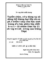
Carotenoid Profile. For carotenoid analyses, we chose three
of the ripest gac fruit that we could find. A typical chromatogram
of carotenoids extracted from gac aril is shown in Figure 1.
Concentrations of the major carotenoids in aril of a single fruit
are given in Table 4, along with relative standard deviation
(RSD) values, which were between 5 and 15%. The ranges of
carotenoid values obtained from three fruit are given in Table
5. The primary carotenoid in aril is lycopene (range, 1546.5-
3053.6 µg/g FW). Of this amount, 2.7-13.2% was present as
cis-lycopene (82.1-204.4 µg/g FW), and 86.8-97.3% was in
the trans-isomeric form (1342.1-2971.5 µg/g FW). The caro-
tenoid having the next highest concentration was β-carotene at
636.2-836.3 µg/g FW, predominately as the trans isomer
(74.7-93.9%; 509.7-701.2 µg/g FW). The cis isomer of
β-carotene comprised 6.1-25.3% (39.1-172.6 µg/g FW) of the
total β-carotene. Of the major carotenoids in aril, R-carotene
was present at the lowest concentration (67.0-106.8 µg/g FW).
Table 1. Weight Distribution of Gac (Momordica cochinchinensis,
Spreng) Fruit, % Total Fresh Weight (n ) 2)
fruit part
fresh weight
(g) % dry wt % total fresh wt
whole fruit 772.0 100.0
aril 190.0 21.7 24.6
seed
a
130.0 16.8
skin 55.0 7.1
mesocarp 373.7 I ) 8.0, O ) 6.9
b
48.4
connective tissue 22.6 10.71 2.9
a
No. of seeds per fruit ) 28, average seed weight ) 4.67 g.
b
I ) inner
mesocarp, O ) outer mesocarp.
Table 2. FAME Composition of Gac Aril, % Total FAMEs (n ) 2)
FAME fruit no. 1 fruitno. 2
avg
%
myristic (14:0) 0.5 0.5 0.5
palmitic (16:0) 32.1 26.4 29.2
palmitoleic (16: 1 ∆
9
) 0.2 0.3 0.3
stearic (18:0) 3.2 12.2 7.7
oleic (18:1 ∆
9)
33.7 30.8 32.3
cis-vaccenic (18:1 ∆
11
) 0.9 0.7 0.8
linoleic (18:2 ∆
9,12
) 28.7 27.5 28.1
R-linolenic (18:3 ∆
9,12,15
) 0.3 0.8 0.5
arachidic (20:0) 0.1 0.5 0.5
eicosa-11-enoic (20:1 ∆
11
) 0.5 0.3 0.4
Table 3. FAME Composition of Gac Seeds, % Total FAMEs (n ) 3)
FAME fruit no. 1 fruit no. 2 fruit no. 3 avg
palmitic (16:0) 6.2 5.2 5.3 5.6
palmitoleic (16:1 ∆
9
) 0.1 n.d.
a
n.d. 0.1
stearic (18:0) 71.7 55.2 54.5 60.5
oleic (18:1 ∆
9
) 4.8 11.2 11.0 9.0
cis-vaccenic (18:1 ∆
11
) 0.4 n.d. 0.7 0.5
linoleic (18:2 ∆
9,12
) 11.2 24.8 25.0 20.3
R-linolenic (18:3 ∆
9,12,15
) 0.5 0.6 0.4 0.5
arachidic (20:0) 1.3 2.0 1.7 1.6
eicosa-11-enoic (20:1 ∆
11
) 0.8 1.0 1.4 1.1
eisoa-13-enoic (20:1 ∆
13
) 3.0 n.d. n.d. 3.0
a
n.d. ) not detected.
Table 4. Carotenoid Composition of Gac Fruit (µG/g FW)
a
carotenoid gac aril gac mesocarp
b
trans lycopene 1902.9 0.0
SD
c
122.2
RSD
d
6.4
cis-lycopene isomers 117.0 0.0
SD 17.3
RSD 14.8
trans β-carotene 641.0 43.7
SD 70.7 6.0
RSD 11.0 13.7
cis β-carotene 128.7 14.6
SD 7.5 1.6
RSD 5.8 11.0
R-carotene 84.3 13.3
SD 9.7 1.0
RSD 11.5 7.5
a
Mean values of three samples from a single fruit.
b
Samples were at first
divided into inner, outer, top, middle, and bottom to detect gradients, if any, along
the thickness and axis of the fruit. No gradients were found, but variations from
one location to another occurred.
c
SD ) standard deviation, %.
d
RSD ) relative
standard deviation, %.
276 J. Agric. Food Chem., Vol. 52, No. 2, 2004 Ishida et al.
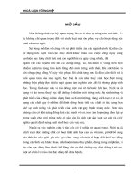
Our data (Tables 4 and 5, Figure 2) on gac mesocarp show no
lycopene, substantial amounts of the trans isomer of β-carotene
(range, 11.3-43.7 µg/g FW) and smaller amounts of cis-β-
carotene. (5.0-14.6 µg/g FW; 25-30.7% of the total), giving
a total β-carotene concentration of 16.3-58.3 µg/g FW. Smaller
amounts of R-carotene (6-13.3 µg/g FW) were found in gac
mesocarp. We also detected phytofluene, phytoene, and trace
amounts of zeaxanthin and β-cryptoxanthin, but no lutein in
either aril and mesocarp tissues.
In contrast, Aoki et al. (23) reported 380 ( 71 µg/g of
lycopene in gac mesocarp, compared to our findings of none
detectable. The authors also reported 101 ( 38 and 22.1 ( 15.2
µg β-carotene/g FW in extracted samples of aril and mesocarp,
respectively, and 16 and 9 µg/g of zeaxanthin and 35 and 2
µg/g of β-cryptoxanthin in saponified samples of mesocarp and
aril tissues, respectively. We suggest that the presence of
lycopene in gac mesocarp samples was probably a result of
contamination of samples with oil from gac aril. In preparing
samples for carotenoid analysis, care must be taken to use
mesocarp tissues that have not been in direct contact with aril.
This is somewhat difficult, because oil from the aril tends to
spread over surfaces when the fruit is first cut open.
Carotenoid composition is especially noteworthy, because the
aril is such a good source of lycopene and β-carotene, providing
Figure 1. A typical chromatogram of carotenoids obtained after extraction from gac aril and separation by HPLC.
Table 5. Carotenoid Concentrations in Gac Aril Fruit Tissue, Range (µg/gFW)
a
lycopene β-carotene R-carotene
tissue
trans- cis- total % cis trans- cis- total % cis total
aril 1342.1−2971.5 82.1−204.4 1546.5−3053.6 2.7−13.2 509.7−701.2 39.1−172.6 636.2−836.3 6.1−25.3 67.0−106.8
mesocarp 0 0 0 0 11.3−43.7 5.0−14.6 16.3−58.3 25−30.7 6−13.3
a
Samples from three fruits analyzed in triplicate.
Figure 2. A typical chromatogram of carotenoids obtained after extraction from gac mesocarp and separation by HPLC.
Fatty Acid and Carotenoids in Gac Fruit J. Agric. Food Chem., Vol. 52, No. 2, 2004 277
concentrations that exceed most other sources. Our data on
lycopene concentration show that the fruit are capable of forming
concentrations that are more than 76 times the concentration
found in commercial tomato fruit. The concentration of total
lycopene in the ripest of the three fruit samples was 3053 µg/g
FW, compared to 40-50 µg/g FW in commercially available
tomato. Its total β-carotene concentration was 682.3 µg/g FW
or 22.3% of the total lycopene concentration in the aril (this
ratio of β-carotene/lycopene varied among the three sampled
fruit, ranging between 22.3 and 41.1%). β-Carotene concentra-
tions in gac mesocarp were also high, but much lower than those
in aril. Our data show higher concentrations of both lycopene
and β-carotene extracted from gac fruit tissues than those of
others (1, 3, 23). This might reflect variability of carotenoid
concentrations among individual fruits, depending on factors
such as degree of ripeness and conditions of culture. In addition,
our modified extraction procedure was designed specifically to
avoid the loss of cis-lycopene isomers, which are of interest
because of evidence that shows that the cis-isomers of lycopene
and β-carotene are more bioavailable (more readily absorbed)
than the trans forms (11, 31). We also evaluated carotenoid
components after HPLC separation of stereoisomers.
The coexistence of both high concentrations of unsaturated
fatty acids and carotenoids in gac aril serves to enhance the
bioavailability of these carotenoids. Studies show that co-
ingestion of lycopene with fat increases the intestinal uptake of
both β-carotene and lycopene (32, 33). Thus, it is evident that
gac fruit is a valuable source of lycopene and β-carotene, two
carotenoids that have been shown to have protective antioxidant
effects against the deleterious consequences of various major
degenerative diseases.
ACKNOWLEDGMENT
We thank Le Thuy Vuong for introducing us to the gac fruit,
providing us with a sample of dried aril and oil, and supporting
us with her enthusiasm and encouragement in this project. We
also thank Glenn E. Bartley, Jiann-Tsyh Lin, and Gary Takeoka
for reviewing our manuscript and Karen Phung for her interest
in the research and her generous donations of gac seed and
frozen fruit.
LITERATURE CITED
(1) Vuong, L. T.; Dueker, S. R.; Murphy, S. P. Plasma β-carotene
and retinol concentrations of children increase after a 30-d
supplementation with the fruit Momordica cochinchinensis (gac).
Am. J. Clin. Nutr. 2002, 75, 872-879.
(2) Guichard, F.; Bui, D. S. La matiere colorante du fruite du
Momordica cochinchinensis Spr. Annales de l′ecole Superieure
de Medecine et de Pharmacie de l ‘Indochine 1941, 141, 42.
(3) West, C. E.; Poortvliet, E. J. The carotenoid content of foods
with special reference to developing countries. USAID-VITAL,
Washington, DC, 1993.
(4) Thompson, S. E.; Tomes, M. L.; Wann, E. V.; McCollum, J. P.;
Characterization of crimson tomato fruit color. Proc. Am. Soc.
Hortic. Sci.1965, 86, 610-616.
(5) Tomes, M. L. Temperature inhibition of carotene synthesis in
tomato. Bot. Gaz. 1963, 124, 180-185.
(6) Giovannucci, E. Tomatoes, tomato-based products, lycopene, and
cancer: Review of the epidemiologic literature. J. Nat. Cancer
Inst. 1999, 91, 317-331.
(7) Giovannucci, E.; Ascherio, A.; Rimm, E. B.; Stampfer, M. J.;
Colditz, G. A.; Willett, W. C. A prospective study of tomato
products, lycopene, and prostate cancer risk. J. Nat. Cancer Inst.
1995, 94, 391-398.
(8) Gerster, H. The potential role of lycopene for human health. J.
Am. Coll. Nutr. 1997, 176, 109-126.
(9) Michaud, D. S.; Feskanich, D.; Rimm, E. B.; Colditz, G. A.;
Speizer, F. E.; Willett, W. C.; Giovannucci, E. Intake of specific
carotenoids and risk of lung cancer in 2 prospective US cohorts.
Am. J. Clin. Nutr. 2000, 72, 990-997.
(10) Di Mascio, P.; Kaiser, S.; Sies, H. Lycopene is the most efficient
biological carotenoid singlet oxygen quencher. Arch. Biochem.
Biophys. 2000, 274, 532-538.
(11) Stahl, W.; Sies, H. Uptake of lycopene and its geometrical
isomers is greater from heat-processed than from unprocessed
tomato juice in humans. J. Nutr. 1992, 122, 2161-2166.
(12) Chen, L.; Stacewicz-Sapuntzakis, M.; Duncan, C.; Sharifi, T.;
Ghosh, L., van Breemen, R.; Ashton, D.; Bowen, P. E. Oxidative
DNA damage in prostate cancer patients consuming tomato
sauce-based entrees as a whole-food intervention. J. Nat. Cancer
Inst. 2001, 93, 1872-1879.
(13) Klipstein-Grobusch, K.; Launer, L. J.p; Geleijnse, J. M.; Boeing,
H.; Hofman, A.; Witteman, J. C. Serum carotenoids and
atherosclerosis. The Rotterdam Study. Atherosclerosis 2000, 148,
49-56.
(14) Polidori, M. C.; Savino, K.; Alunni, G.; Freddio, M.; Senin, U.;
Sies, H.; Stahl, W.; Mecocci, P. Plasma lipophilic antioxidants
and malondialdehyde in congestive heart failure patients: rela-
tionship to disease severity. Free Radicals Biol. Med. 2002, 32,
148-152.
(15) Rissanen, T. H.; Voutilainen, S.; Nyyssonen, K.; Lakka, T. A.;
Sivenius, J.; Salonen, R.; Kaplan, G. A.; Salonen, J. T. Low
serum lycopene concentration is associated with an excess
incidence of acute coronary events and stroke: the Kuopio
ischaemic heart disease risk factor study. Brit. J. Nutr. 2001,
85, 749-754.
(16) Kristenson, M.; Zieden, B.; Kucinskiene, Z.; Elinder, L. S.;
Bergdahl, B.; Elwing, B.; Abaravicius, A.; Razinkoviene, L.;
Calkauskas, H.; Olsson, A. G. Antioxidant state and mortality
from coronary heart disease in Lithuanian and Swedish men:
concomitant cross sectional study of men aged 50. Brit. Med. J.
1997, 314, 629-633.
(17) Mares-Perlman, J. A.; Brady, W. E.; Klein, R.; Klein, B. E.;
Bowen, P. Stacewicz-Sapuntzakis, M.; Palta, M. Serum anti-
oxidants and age-related macular degeneration in a population-
based case-control study. Arch. Ophthalmol. 1995, 113, 1518-
1523.
(18) Pollack, A.; Oren, P.; Stark, A. H.; Eisner, Z.; Nyska, A.; Madar,
Z. Cataract development in sand and galactosemic rats fed a
natural tomato extract. J. Agric. Food Chem. 1999, 47, 5122-
5126.
(19) Gale, C. R.; Hall, N. F.; Phillips, D. I.; Martyn, C. N. Plasma
antioxidant vitamins and carotenoids and age-related cataract.
Ophthalmology 2001, 108, 1992-1998.
(20) Simonelli, F.; Zarrilli, F.; Mazzeo, S.; Verde, V.; Romano, N.;
Savoia, M.; Testa, F.; Vitake, D. F.; Rinaldi, M.; Sacchetti, L.
Serum oxidative and antioxidant parameters in a group of Italian
patients with age-related maculopathy. Clin. Chim. Acta 2002,
320, 111-115.
(21) Vuong, L. T.; King, J. C. A method for preserving gac fruit oil,
a rich source of beta-carotene and essential fatty acids in North
Vietnam, submitted for publication.
(22) De Shan, M.; Hu, L. H.; Chen. Z. L. A new multiflorane
triterpenoid ester from Momordica cochinchinenensis Spreng.
Nat. Prod. Lett. 2001, 15, 139-145.
(23) Aoki, H.; Nguyen, T. M. K.; Kuze, N.; Tomisaka, K.; Chuyen,
N. V. Carotenoid pigments in GAC fruit (Momordica cochinchin-
ensis SPRENG). Biosci. Biotechnol. Biochem. 2002, 66, 2479-
2482.
(24) Christie, W. W. The preparation of derivatives of fatty acids.
Chapter 4. In Gas chromatography and lipids: A practical guide;
Christie, W. W., Ed.; Oily Press Ltd: Dundee, Scotland 1989;
pp 66-84.
(25) Ishida, B. K.; Ma, J. C.; Chan, B. G.; Bartley, G. E.; Grossman,
J. N. A modified method for simple, rapid HPLC analysis of
lycopene isomers. Acta Hort. 2001, 542, 235-242.
278 J. Agric. Food Chem., Vol. 52, No. 2, 2004 Ishida et al.
(26) Ishida, B. K.; Ma, J.; Chan, B. A simple, rapid method for HPLC
analysis of lycopene isomers. Phytochem. Anal. 2001, 12, 194-
198.
(27) Hilditch, T. P. and Williams, P. N. The Chemical Constitution
of Natural Fats, 4th ed.; John Wiley & Sons Inc.: New York,
1964; pp 187-202.
(28) Hilditch, T. P. and Williams, P. N. The Chemical Constitution
of Natural Fats, 4th ed.; John Wiley & Sons Inc.: New York,
1964; pp 319-331.
(29) Hilditch, T. P. and Williams, P. N. The Chemical Constitution
of Natural Fats, 4th ed.; John Wiley & Sons Inc.: New York,
1964; p 269.
(30) Hawkins, D. J.; Kridl, J. C. Characterization of acyl-ACP
thioesterases of mangosteen (Garcinia mangostana) seed and
high levels of stearate production in transgenic canola. Plant J.
1998, 13, 743-752.
(31) Britton, G. Structure and properties of carotenoids in relation to
function. FASEB J. 1995. 9, 1551-1558.
(32) Erdman, J. The physiologic chemistry of carotenes in man. Clin.
Nutr. 1988, 7, 101-106.
(33) Bohm, V. Bitsch, R. Intestinal absorption of lycopene from
different matrixes and interactions to other carotenoids, the lipid
status and the antioxidant capacity of human plasma. Eur. J.
Nutr. 1999, 38, 118-125.
Received for review August 20, 2003. Revised manuscript received
November 12, 2003. Accepted November 12, 2003.
JF030616I
Fatty Acid and Carotenoids in Gac Fruit J. Agric. Food Chem., Vol. 52, No. 2, 2004 279