Tài liệu Guidelines for the Management of Inflammatory Bowel Disease (IBD) in Children in the United Kingdom pdf
Bạn đang xem bản rút gọn của tài liệu. Xem và tải ngay bản đầy đủ của tài liệu tại đây (569.03 KB, 36 trang )
This publication has been produced by the IBD Working Group of
BSPGHAN with the financial support of
CICRA – Crohn’s in Childhood Research Association and
NACC – National Association for Colitis & Crohn’s Disease
County Print & Design ຜ 01622 605368 13416/BS
registered charity
number 278212
registered charity
number 1117148
Guidelines for the Management of
Inflammatory Bowel Disease (IBD)
in Children in the United Kingdom
UK IBD Working Group on behalf of the British Society of Paediatric
Gastroenterology Hepatology and Nutrition (BSPGHAN)
October 2008
1
Guidelines for the Management of
Inflammatory Bowel Disease (IBD)
in Children in the United Kingdom
Authors
≥ B K Sandhu, J M E Fell, R M Beattie, S G Mitton
Authors’ affiliations
≥ Prof. Bhupinder K Sandhu. Department of Paediatric Gastroenterology,
Bristol Royal Hospital for Children; and Centre for Child and Adolescent Health Bristol
University and the University of West of England (UWE).
≥ Dr John ME Fell. Department of Paediatric Gastroenterology,
Chelsea and Westminster Hospital, London.
≥ Dr R Mark Beattie. Department of Paediatric Gastroenterology,
Southampton General Hospital, Southampton.
≥ Dr Sally G Mitton. Department of Paediatric Gastroenterology,
St. Georges University London, Cranmer Terrace, London.
Correspondence to Chair of UK Paediatric IBD Working Group
≥ Dr Sally G Mitton MD FRCPCH
Consultant Paediatric Gastroenterologist
Dept Child Health
St. Georges University London, Cranmer Terrace, London SW17 0RE
Guidelines for the Management of Inflammatory Bowel Disease (IBD) in Children in the United Kingdom
2
1. Introduction
2. Inflammatory Bowel Disease
3. Management of Crohn’s Disease
4. Management of Ulcerative Colitis
5. Associated aspects of IBD
6. Service Delivery
Index
Guidelines for the Management of Inflammatory Bowel Disease (IBD) in Children in the United Kingdom
3
Inflammatory bowel disease (IBD) encompasses two related but distinct disorders
of as yet unknown aetiology. Crohn’s disease (CD) is a chronic, idiopathic
transmural inflammation which can affect one or several segments of the digestive
tract. Ulcerative colitis (UC) is a chronic idiopathic inflammation of the rectum
extending continuously over a variable length of the colon from the distal to the
proximal end. Indeterminate colitis (IC) is reserved for cases of colitis in which
findings are not sufficient to allow differentiation between CD and UC
[1].
1.1 Development of guidelines [2-4]
These guidelines are the work of the IBD working group of the British Society of Paediatric
Gastroenterology Hepatology and Nutrition (BSPGHAN) and are for use by clinicians and allied
professionals caring for children with IBD in the United Kingdom (UK). There is paucity of paediatric
trials of high methodological quality to provide a comprehensive evidence based document. Thus
these clinical guidelines have had to be consensus based, informed by the best available evidence
from the paediatric literature and high quality data from adult IBD literature, together with the clinical
expertise and multidisciplinary experience of IBD experts comprising paediatric gastroenterologists
represented by BSPGHAN. They provide an evidence and consensus based document describing good
clinical practice for the investigation and treatment of IBD in children which will promote consistency
of the management of such conditions. Individual cases must be managed on the basis of all clinical
data available for that child. Parent and patient preferences must be sought and joint decisions made.
These guidelines will be published on the BSPGHAN web site which will allow simple and regular
updating in the future and easy access for Society members and others.
The IBD working group of BSPGHAN performed a comprehensive literature search of treatment
modalities in paediatric IBD intervention studies using electronic databases (Medline, Pub med,
Cochrane and Ovid). Evidence was graded using the Scottish Intercollegiate Guidelines Network
‘SIGN’ [2]. Methodology and detailed evaluation of evidence will be published as a separate paper.
The British Society of Gastroenterology (BSG) produced evidence based guidelines for the
management of IBD in adults [3] for which a comprehensive literature search was also performed
using electronic databases (Medline, Pub Med, and Ovid; keywords: “inflammatory bowel disease”,
“ulcerative colitis”, and “Crohn’s disease”). The format of the paediatric guidelines is based on the
BSG guidelines but uses, where available, paediatric data and practice. Where there is no or very
little paediatric data or there is controversy, the evidence based evaluation by the authors of the
BSG guidelines for adults with IBD has been used together with the ECCO consensus document [4].
1 Introduction [1]
Guidelines for the Management of Inflammatory Bowel Disease (IBD) in Children in the United Kingdom
4
2.1 Definitions [1, 4-5]
UC is characterised by diffuse mucosal inflammation limited to the colon. Disease extent can be
divided into distal or more extensive disease. ‘‘Distal’’ disease refers to colitis confined to the
rectum (proctitis) or rectum and sigmoid colon (proctosigmoiditis). More extensive disease includes
‘‘left sided colitis’’ (up to the splenic flexure), ‘‘extensive colitis’’ (up to the hepatic flexure), and
pancolitis (affecting the whole colon).
CD is characterised by patchy, transmural inflammation, which may affect any part of the
gastrointestinal tract. It may be defined by location (terminal ileal, colonic, ileocolic, upper
gastrointestinal), or by pattern of disease (inflammatory, fistulating, or stricturing). These variables
have been combined in the Montreal classification [5]. About 10% of children with IBD affecting the
colon are unclassifiable after considering clinical, radiological, endoscopic, and pathological criteria,
because they have some features of both conditions. This is termed indeterminate colitis (IC).
2.2 Epidemiology [6-12]
The only prospective national survey of IBD in children aged <16 years in the UK [6] showed the
incidence to be 5.2 per 100,000 individuals per year (60% CD, 28% UC and 12% IC). It is slightly
more common in boys and there is a slightly higher rate of UC in Asian children than in other ethnic
groups. The mean age at diagnosis was 11.9 years. For CD there were approximately equal
proportions of ileitis, colitis and ileo-colitis, and for UC almost 90% of children had a pancolitis [7].
A systematic review of the epidemiological studies in North American cohorts estimates the
incidence at 3–4 per 100,000 individuals per year [8]. UC and CD are diseases of young people with
a peak incidence between the ages of 10 and 40 years. Data from Scotland and Wales suggests that
the incidence has risen over the last twenty years [9-10] with 25% of all cases presenting in
children and young people. However the incidence of CD may now have plateaued and that of UC
may be increasing [11] so there is a need to determine current incidence trends again across the
UK. IBD can affect any age; of the children presenting with IBD 5% are below 5 years [7] and only
15% of adults are over 60 years, at diagnosis. Projected estimates suggest that up to 240,000
people are affected by IBD in the UK [12].
2 Inflammatory Bowel Disease
Guidelines for the Management of Inflammatory Bowel Disease (IBD) in Children in the United Kingdom
5
2.3 Pathogenesis [13]
The etiologies of both UC and CD remain unknown. The consensus is that both diseases are
probably a response to environmental triggers (infection, drugs, or other agents) in genetically
susceptible individuals. The genetic component is stronger in CD than in UC. Smoking increases the
risk of CD, but decreases the risk of UC through unknown mechanisms [13].
Theories and evidence for pathogenetic mechanisms are too complex to be considered in this
document. The broad areas examined are epidemiology, the gut/environmental interface, the
inflammatory process, and genetics of each disease. Epidemiological studies have considered diet,
drug, and vaccination history, seasonal variation, water supply, and social circumstances. The
gut/environmental interface includes work on luminal bacteria, biofilms, the epithelial glycocalyx
and mucus, epithelial barrier function, epithelial remodelling and immune/epithelial interactions.
The inflammatory process has been examined through cell signalling pathways, cytokine profiles,
eicosanoid and other inflammatory mediators, lymphocyte trafficking, cell surface molecules,
interactions between stromal and immune cells, and neuroimmune communication. Researchers in
genetic susceptibility to IBD have adopted a candidate gene approach, genome wide screening
through micro satellite markers and, most recently, both genome wide association scans and
studies on functional gene expression. Mutations of one gene (CARD15/ NOD2), located on
chromosome (Chr) 16, have been associated with small intestinal CD in white (but not oriental)
populations and link innate immunity and the bacterial population of the gut. Recent genome wide
association scans have implicated two new pathways; T-cell regulation by the IL23 pathway via the
gene IL23R, and the process of autophagy, which controls intracellular bacteria, by the genes
ATG16L1 and IRGM. Other genes have yet to be identified, although their existence is strongly
suggested by replicated linkage to a number of chromosomes.
2.4 Clinical features and pattern of disease [7, 14-20]
In children with UC blood loss [84%], diarrhoea [74%] and abdominal pain [62%] are all common
[7]. Weight loss is less common in UC [35%] than CD [58%]. Other symptoms include lethargy and
anorexia. The most common reported extra intestinal symptom is arthropathy [10%]. Skin
manifestations are rare. Children with IC have predominantly colitic symptoms. With modern
medical and surgical management, the disease now has a slight excess of mortality in the first two
years after diagnosis, but little subsequent difference from the normal population [14, 15]. A
severe attack of UC is still a potentially life threatening illness. The clinical course of UC is marked
by exacerbation and remission. About 50% of patients with UC have a relapse in any year. An
appreciable minority has frequently relapsing or chronic, continuous disease. In children with
moderate to severe disease at diagnosis colectomy rate is around 25% at 5 years. Disease
severity at diagnosis is predictive of long-term outcome. Symptoms of CD are more
heterogeneous and the nonspecific symptoms in children with CD may delay diagnosis. Abdominal
pain, diarrhoea and weight loss were considered to be the “classic triad” of CD but now only a
minority present in this way. The clinical presentation of childhood CD over the last two decades
has changed. Data from the Hospital for Sick Children, Toronto during 1980-1989, showed that
80% of CD children presented with the classical triad [16] but a more recent very large population
based survey of childhood IBD in the UK during 1998-1999, found only 25% presented in this way
[7]. 44% of patients with CD have no diarrhoea but the majority [72%] complain of abdominal pain.
Many children with CD present with vague complaints such as lethargy, anorexia and abdominal
discomfort or with isolated growth failure. A significant minority have markedly impaired final adult
height [17, 18]. Neglect to record growth parameters, particularly for those not presenting to a
paediatrician, has been identified [7, 17, 20]. Other symptoms may include fever, nausea, vomiting,
delayed puberty, psychiatric disturbance and erythema nodosum [7]. The clinical course of CD is
characterised by exacerbations and remission. CD tends to cause greater disability than UC.
Table 1
Presenting symptoms and signs of children in UK with CD; data from the national study [7]
Guidelines for the Management of Inflammatory Bowel Disease (IBD) in Children in the United Kingdom
6
Patients CD (n = 379) IC (n = 72) UC (n = 172)
Common symptoms
Abdominal pain 274 (72%) 54 (75%) 106 (62%)
Diarrhoea 214 (56%) 56 (78%) 127 (74%)
Bleeding 84 (22%) 49 (68%) 145 (84%)
Weight loss 220 (58%) 25 (35%) 53 (31%)
Lethargy 103 (27%) 10 (14%) 20 (12%)
Anorexia 94 (25%) 9 (13%) 11 (6%)
Other symptoms
Arthropathy 28 3 11
Nausea/vomiting 22 1 1
Constipation/soiling 4
Psychiatric symptoms 3
Secondary amenorrhoea 11
Signs
Anal fistula 17
Growth failure/delayed puberty 14 1
Anal abscess, ulcer 8
Erythema nodosum/rash 61
Liver disease 325
Appendicectomy 2
Toxic megacolon 1
Guidelines for the Management of Inflammatory Bowel Disease (IBD) in Children in the United Kingdom
7
2.5 Diagnosis and investigations [1, 21-24]
The need to diagnose children with IBD in a systematic way to provide tissue diagnoses and disease
distribution was recognised over 25 years ago [22]. To ensure all children receive optimal care,
members of the IBD working group of the European Society of Paediatric Gastroenterology,
Hepatology and Nutrition (ESPGHAN) have developed a consensus protocol for investigation of
these children [1]. The diagnosis of IBD is confirmed by clinical evaluation and a combination of
biochemical, endoscopic, radiological, histological, or nuclear medicine investigations. The diagnosis
of UC is made on clinical suspicion supported by appropriate macroscopic findings on colonoscopy,
typical histological findings on biopsy, and negative stool examinations for infectious agents. For
CD, the diagnosis depends on demonstrating focal lesions with transmural inflammation and
granuloma in at most, 40-60%.
2.5.1 History and examination
A full history should include recent travel, medication, dietary and family history and a detailed
bowel history with stool frequency, consistency, urgency, and presence of blood, mucus or pus per
rectum. Abdominal pain, malaise, fever, weight loss, and symptoms of extra intestinal
manifestations of IBD (joint, cutaneous, and eye) should be sought. General examination includes
wellbeing, weight and height centiles, pubertal status using Tanner staging, pulse rate, blood
pressure, temperature, abdominal examination for tenderness, distension, masses including
inspection of perianal area for skin tags, fissures, ulcers and/or oedema suggesting CD.
2.5.2 Initial investigations [1]
Laboratory investigations should include Full Blood Count (FBC), C Reactive Protein (CRP),
Erythrocyte Sedimentation Rate (ESR), liver function tests (esp. albumin). Reduced levels of
haemoglobin, raised inflammatory markers (CRP, ESR and platelets) and reduced serum albumin
are suggestive of IBD. In some UC patients, however they may be normal. Stool cultures should be
carried out to exclude infectious diarrhoea and stool tested for Clostridium difficile toxins A and B.
Additional tests may be needed for patients who have travelled abroad. Identification of the
pathogen however does not necessarily exclude a diagnosis of IBD, as a first episode of IBD may
present after documented enteric infection. In children from populations at risk of tuberculosis
(TB), this should be excluded.
Perinuclear antineutrophil cytoplasmic antibody (pANCA) is positively associated with UC and
Anti-Saccharomyces Cerevisiae Antibody (ASCA) with CD but the diagnostic sensitivity of these
serological markers only ranges between 60% and 80% so are of limited clinical use. The non-
invasive stool tests of faecal calprotectin and lactoferrin may become increasingly important both
for screening and also monitoring disease activity in order to avoid more invasive investigations.
Abdominal radiography is essential for assessment of patients with suspected severe colitis to
exclude colonic dilatation and silent perforation.
Guidelines for the Management of Inflammatory Bowel Disease (IBD) in Children in the United Kingdom
8
2.5.3 Upper GI endoscopy and colonoscopy [1]
Ideally all children suspected of having IBD should have upper and lower GI endoscopy preferably
with intubation of terminal ileum and multiple biopsies from all segments in the upper (oesophagus,
stomach, duodenum) and lower intestinal tract (ileum, caecum, ascending colon, transverse colon,
descending colon, sigmoid and rectum) for histological diagnosis [1]. A barium meal and follow
through should be performed in all children who might have CD to evaluate the involvement of
small bowel. Disease distribution may be important to aid diagnosis when pathognomic histological
features are not present. Histological evidence of CD in the upper GI tract can be present in up to
30% of cases even in the absence of upper GI symptoms. Unlike adults, over 90% of children with
UC have a pancolitis making full colonoscopy advisable. Sigmoidoscopy does not have a role except
in severe UC where the risk of bowel perforation is higher, making flexible sigmoidoscopy a safer
option. It may be appropriate to defer investigations until the clinical condition improves. The
majority of IC behaves like UC but a few are later diagnosed as CD. Once tissue diagnosis and
disease distribution are documented, appropriate treatment can be chosen. Histology of terminal
ileal biopsies may help to exclude other diagnoses (e.g. TB, Behcet’s syndrome, lymphoma,
vasculitis) as well as assessing the extent of IBD and in children from a population at high risk of
TB, tissue should be sent for TB culture.
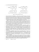
Ileocolonoscopy and upper GI endoscopy with histology
of multiple biopsies from all segments
CD UC
if diagnosis of UC
is uncertain or is IC
Radiolo
g
y – small bowel follow throu
g
h (SBFT)
Figure 1
Porto criteria for diagnosis of IBD in children [1]
Guidelines for the Management of Inflammatory Bowel Disease (IBD) in Children in the United Kingdom
9
2.5.4 Other investigations
Technetium white cell scanning documents areas of inflammation and is undertaken in several centres
and is a safe, non-invasive investigation which may lack specificity but can be helpful to define disease
extent. It may however give a false negative result if the child is on steroids and also may not show
oesophageal or pelvic inflammation. Ultrasound in skilled hands is a sensitive and non-invasive way of
identifying thickened small bowel loops in CD and may identify abscesses or free fluid in the
peritoneum. Computed tomography and increasingly, magnetic resonance imaging (MRI) e.g. of the
pelvis, may help evaluate activity and complications of disease e.g. fistula. Due to decreased radiation
exposure, small bowel MRI is replacing small bowel follow through in some centres. Laparoscopy may
be helpful in selected patients, for example if intestinal TB is possible. Capsule endoscopy is not widely
used in children at present but may become increasingly valuable in the diagnosis of small intestinal
disease. Capsule endoscopy cannot be used in the presence of strictures as it may be retained.
2.6 Histopathology [23]
Histopathological examination of biopsy specimens should be carried out according to the principles
outlined in the BSG document “Guidelines for the initial biopsy diagnosis of suspected chronic
idiopathic inflammatory bowel disease” [23]. The type of IBD should be clearly defined along with
other co-existent diagnoses or complications and the presence or absence of dysplasia recorded.
2.7 Imaging [24]
It is desirable that clinicians discuss imaging with an appropriate radiologist, to avoid unnecessary
exposure to ionizing radiation. A multidisciplinary forum is best to review the results of imaging in
the context of the clinical history so that appropriate management can be planned.
2.8 Treatment of IBD [7, 25]
This consists of bringing active disease into remission followed by prevention of relapse (figure 2 & 3).
Choice of treatment is influenced by disease type, distribution and associated presenting features
such as weight loss, short stature and pubertal status. Recent data [7] suggests that, in CD,
involvement of the gastrointestinal tract is much more widespread with only 9% of children having
isolated small bowel disease and 7%, isolated colonic disease. The majority have both colonic and
small bowel involvement, nearly half have gastro-duodenal and 20%, jejunal disease. Not only is
paediatric-onset IBD characterised by extensive intestinal involvement at diagnosis, but the majority
of children show rapid progression of disease [25]. Evaluation of treatment efficacy includes
assessment of symptomatic improvement, weight gain and later, improved height velocity,
biochemical remission e.g. resolution of abnormal blood inflammatory markers and, in some cases,
re-evaluation of disease activity by endoscopy to confirm mucosal healing. There are few
randomised controlled drug trials in children. Many medications are unlicensed for use in children
and are unavailable in “child friendly” form i.e. available as large tablets rather than liquid form.
Choice of medication depends on the child’s co-operation and the parents’ willingness to administer
treatment; for example, a child with distal colitis may not accept treatment with enemas.
Therapy for IBD is a rapidly evolving field, with many new biological agents under investigation that
are likely to change therapeutic strategies radically in the next decade.
Guidelines for the Management of Inflammatory Bowel Disease (IBD) in Children in the United Kingdom
10
Benefits and risks of any treatment should be discussed openly with patient and
their family particularly in relation to steroids and immunomodulators. Factors
such as the potential risk of immuno-suppression, bone marrow suppression and
malignancy must be discussed and the discussion recorded in the notes. Disease
activity can be expressed using a disease activity index such as the PCDAI
[26].
3.1 Induction of Remission at diagnosis or disease relapse [27-72]
The choice of treatment in most cases is between exclusive enteral nutrition and oral
corticosteroids. This is concordant with the BSG guidelines which also state that there is insufficient
evidence to recommend the use of other agents outside trials/specialist centres. Recently some
centres have started using azathioprine at diagnosis for those with severe disease. Azathioprine
prevents relapse but is not fully effective until at least three months after starting the drug.
3.1.1 Exclusive Enteral Nutrition [27-35] Evidence Levels (EL) 1+ to1-, 2-, 3 & 4
≥ Exclusive enteral nutrition is effective first line therapy for small and large bowel disease,
inducing remission in 60-80% of cases.
≥ Factors that influence its use include patient and parent choice, compliance, palatability,
lack of corticosteroid toxicity, potential benefits in terms of improved nutritional status
and growth.
≥ The choice is between polymeric (e.g. Modulen IBD
1
, Alicalm
2
) or elemental (e.g. EO28
2
)
feeds. There appears to be no significant difference in efficacy between the two. Both feeds
are available in different flavours and it has been suggested that polymeric feeds may be
more palatable. Administration via a naso-gastric tube or gastrostomy is an option.
≥ Duration of exclusive enteral nutrition is usually 6 weeks. Most children need approximately
120% of Reference Nutrient Intake (RNI). This however needs to be tapered according to
individual needs and dietetic support is essential. Food may be reintroduced cautiously over
1-3 weeks, dependant on patient symptoms whilst weaning the enteral feed.
1
Néstle Clinical Nutrition
2
SHS International Ltd
3 Management of Crohn’s Disease [26]
3.1.2 Corticosteroids [35-40] EL 1-, 2-, 3 & 4
≥ Prednisolone 1-2 mg per/kg/d (maximum 40mg a day) is effective first line therapy for
small and large bowel disease.
≥ Treatment should be at full dose for 2-4 weeks until remission achieved (with review at
least every 2 weeks in clinic or via telephone, until clinical remission) and thereafter
gradual reduction of the dose over 4-8 weeks depending on the response.
≥ Ensure adequate dietary intake of calcium and vitamin D and if insufficient consider
supplement (e.g. Calcichew D
3
tablet daily).
≥ Gastric acid suppression with proton pump inhibitors (e.g. Omeprazole) may be required in
the presence of gastritis.
3.1.3 Other management strategies at induction [41- 50]
≥ Antibiotics (EL 3) – metronidazole (7.5mg/kg/dose tds) +/- Ciprofloxacin (5mg/kg/dose
bd) for peri-anal disease.
≥ Aminosalicylates (EL 1-, 3) in high dose (mesalazine 50-100mg/kg/d, maximum 3-4g/d
or Sulphasalazine 40-60mg/kg/d (maximum 3g/d, can increase to 100mg/kg/d if
tolerated) may be effective in mild disease. Topical Mesalazine is effective in mild to
moderate left sided colitis. Regular 6 monthly blood monitoring of liver and renal function
is essential.
≥ Budesonide 9mg/d (EL 1-, 3) is less effective than prednisolone as first line therapy for
isolated ileocaecal disease but has fewer side effects.
≥ Intravenous (iv) steroids (EL3) In children with severe disease at presentation iv steroids
should be given: hydrocortisone 2mg/kg qds (maximum 100mg qds) or
methylprednisolone 2mg/kg od (60mg/d maximum).
≥ Azathioprine (EL3) may be introduced immediately (after checking TPMT levels are
satisfactory) in those with severe disease but takes at least three months to be fully
effective.
≥ Surgery for complication e.g. abscess/fistula after MRI pelvis to assess extent of perianal
disease.
≥ Parenteral nutrition (EL3) may be required as nutritional support for patients with
severe complicated disease.
Guidelines for the Management of Inflammatory Bowel Disease (IBD) in Children in the United Kingdom
11
3.2 Refractory or non-responsive CD [51-78]
Patients in whom standard induction therapy including high dose iv steroids has failed to induce
remission either at diagnosis or during subsequent relapse are defined as having non-responsive
CD. Steroid refractory CD may be defined as active disease despite an adequate dose (1-2 mg/kg/d;
minimum 20mg/d) and duration (at least 2 weeks) of steroid therapy. Such patients should be
considered for treatment with immuno-modulators if surgery is not an immediate consideration.
≥ Azathioprine (2-2.5mg/kg/d) or 6-mercaptopurine (1–1.25mg/kg/d) (EL3) after checking
TPMT levels are satisfactory. Of patients intolerant to azathioprine, up to 50% will
tolerate 6-mercaptopurine.
≥ Methotrexate 15mg/m
2
(EL 3), once weekly given subcutaneously (s/c). Remission
usually within 4 weeks but further improvement may be seen after 16 weeks. Parenteral
weekly administration is of benefit if non-adherence to oral medications is a major issue. If
it is not an issue, patients can switch to oral Methotrexate providing there is not
significant small bowel disease which might interfere with absorption.
≥ Infliximab 5mg/kg/dose at weeks 0, 2 & 6 (EL2-, 3), can be effective in patients who are
refractory or intolerant to steroids in combination with immuno-modulators and in whom
surgery is inappropriate. There should be a plan at the outset for using Infliximab, with the
length of course clearly defined e.g. 3 doses and then reassessment. Prior to starting
Infliximab, sepsis should be excluded including TB (CXR/Mantoux and molecular
quantification tests). Patients already on immunosuppression may have a false negative
Mantoux. Prior to starting treatment, the patient and their family should be counselled
about Infliximab including a discussion about the risks of malignancy and written consent
should be obtained. Guidelines for Infliximab use in adults have been produced by NICE.
≥ Surgery should be considered especially for isolated ileocaecal disease, strictures or fistulae
and for those in whom medical treatment has failed. It is essential that there is close
collaboration between gastroenterologists and a surgeon experienced in paediatric IBD.
≥ In CD surgery is not curative and management is directed at minimising the impact of
disease. At least 30% of patients require surgery in the first 10 years of disease and
approximately 70–80% will have surgery in their lifetime.
3.3 Other disease sites
≥ Oral: CD can be managed with exclusive enteral nutrition, exclusion diet (benzoate and
cinnamon free), topical steroids and/or intra-lesional steroid injections. Azathioprine,
Infliximab and Thalidomide may be considered for resistant disease (EL3).
≥ Gastro duodenal disease: proton pump inhibitors, used with standard therapy, may
reduce symptoms
≥ Fistulising and peri-anal disease:
≥ Metronidazole (7.5mg/kg/dose tds) (EL4) for at least six weeks and/or Ciprofloxacin
(5mg/kg/dose bd) is appropriate treatment for simple perianal disease.
Guidelines for the Management of Inflammatory Bowel Disease (IBD) in Children in the United Kingdom
12
Guidelines for the Management of Inflammatory Bowel Disease (IBD) in Children in the United Kingdom
13
≥ Azathioprine (2-2.5mg/kg/d) or 6MP (1-1.25mg/kg/d) (EL3) may be effective
treatment for perianal and enterocutaneous fistulae (Check TPMT prior to treatment)
but there is a delay in onset of action.
≥ Infliximab iv; 3 infusions of 5mg/kg each at 0, 2 and 6 wks (EL2-, 3) may be effective
treatment for perianal and enterocutaneous fistulae but should be reserved for
patients’ refractory to other treatments. A pelvic MRI scan should be carried out to
exclude any abscess and to diagnose fistulae before starting Infliximab.
≥ Surgery – abscess drainage, fistulotomy and seton insertion may be appropriate
particularly prior to Infliximab treatment. Image with pelvic MRI.
Induction of Remission
+/- aminosalicylates
Maintenance
(Following relapse or treatment resistance)
Azathioprine
or 6-mercaptopurine (6MP)
(Check TPMT level first)
Intolerance or resistance
Exclusive enteral liquid feeds
for 6 weeks
Corticosteroids
(reducing course)
consider Methotrexate
(s/c) if fail to respond
to Azathioprine
or 6-mercaptopurine
Infliximab
if fails, Adalimumab,
cyclos
p
orine, thalidomide
Surgery
if localised disease
or s
p
ecific indications
Second line treatment
Third line treatment
Figure 2
Crohn’s Disease Treatment Flow Chart
3.4 Maintenance of remission in CD [77-95]
≥ There is no role for maintenance steroids for patients with CD in remission. For patients
who are steroid dependent every effort must be made to find other effective treatment.
≥ Azathioprine (2-2.5mg/kg/d) or 6-mercaptopurine (1-1.25mg/kg/d) (EL3) should be
initiated as maintenance therapy in cases that relapse in less than 6 months, relapse two
or more times per year following initial successful therapy and in all that are steroid
dependent. Also post operatively, for complex, fistulating or extensive disease. TPMT
should be checked prior to initiating treatment and is probably best done at diagnosis. In
Azathioprine non responders it may be useful to check serum Thioguanine nucleotides
levels to see if noncompliant or not absorbing. When to stop Azathioprine is controversial.
There is some evidence that over half of all adults will relapse within 3 years of stopping
Azathioprine and hence the usual practice of stopping at 4 years may not be valid. This
should be discussed with the patient and parents and also adult gastroenterology
colleagues as part of the transition plan. Certainly it should not be discontinued at key
times during pubertal growth and/or education and most continue until the time of
transfer to the adult GI physicians.
≥ Methotrexate 15mg/m
2
once weekly s/c (EL1-, 3), if azathioprine or 6-mercaptopurine is
ineffective or poorly tolerated, with folic acid 5mg 24hrs after each dose to ameliorate
any GI side effects. FBC and LFTs must be monitored; every 2 wks for first 4 wks,
thereafter once a month.
≥ Enteral nutrition (EL 2-) Supplementary therapy may reduce the risk of relapse and may
improve growth and nutritional status.
≥ 5ASA, Mesalazine (EL4) little role in maintaining remission but may be of limited benefit
in high dose (50-100mg/kg/d as tolerated) for mild disease.
≥ Infliximab (EL3) If remission is induced with Infliximab, maintenance with Infliximab may
be necessary (5mg/kg IV, 8 weekly). It may be necessary to escalate to a higher dose
(10mg/kg) for loss of responsiveness and if successful, should revert to lower dose for
subsequent infusions. Consider reinvestigating first to exclude ongoing sepsis, stricture
and bacterial overgrowth. Stopping co-existing immunosuppression after six months
should be considered (there is emerging data of lymphoma risk with Infliximab which may
or may not be related to concomitant administration of Azathioprine or 6-mercaptopurine
with Infliximab). Assess at least annually to consider if Infliximab can be discontinued. If
patient develops hypersensitivity to Infliximab these symptoms may be abolished or
ameliorated with a dose if iv hydrocortisone +/- antihistamine prior to Infliximab infusion.
≥ Other anti-TNF therapy (EL3) In patients initially responsive to Infliximab who become
resistant or intolerant, alternative anti-TNF agents can be considered e.g. Adalimumab
(s/c) 80mg stat followed by 40mg every other week. Reassess endoscopically and if
necessary radiologically, before starting second-line biologic therapy.
≥ Other Agents (EL4) There is little evidence for a beneficial effect of probiotics, fish oil
and Trichuris (worm) therapy to maintain remission in CD.
Guidelines for the Management of Inflammatory Bowel Disease (IBD) in Children in the United Kingdom
14
Guidelines for the Management of Inflammatory Bowel Disease (IBD) in Children in the United Kingdom
15
Treatment depends on disease activity and distribution. Disease activity can be
expressed using a clinical activity index
[98-100]. If on evaluation disease is severe
the patient needs admission to a paediatric gastroenterology unit for intensive
intravenous therapy. If disease is fulminant, patient needs urgent resuscitation, an
abdominal x-ray to exclude perforation and joint medico-surgical assessment and
management (see later). The majority (90%) of children with UC have pancolitis,
less than 10% have left sided colitis, 4% have disease confined to the rectum
alone and 4% have rectal sparing
[7]. Half of those without pancolitis at
presentation will rapidly progress to pancolitis
[25]. Infective aetiology should be
sought as this may co-exist with active disease but in severe disease, immediate
treatment with corticosteroids should not be delayed.
4.1 Induction of Remission at diagnosis or disease relapse [101-123]
4.1.1 Mild or left sided UC [101-107]
≥ Topical Mesalazine (EL 1-,3) or to a slightly lesser extent, steroids in liquid, foam or
suppositories are effective therapy for mild to moderate left sided colitis or isolated
rectal disease (1-2g daily). However, single therapy with topical mesalazine or steroids
for distal disease is less effective than a combination of oral and topical therapy.
≥ Oral Mesalazine (50-100mg/kg/d, maximum 3-4g daily) or Sulphasalazine 40-
60mg/kg/d (maximum 3g/d, can increase to 100mg/kg/d if tolerated) (EL1-,3).
Sulphasalazine is better tolerated if introduced over 10 days to attain full dosage and is
particularly effective for UC or IC and for arthropathy. Only Sulphasalazine is available in
liquid form. Olsalazine and Balsalazide are alternatives if intolerant to those above and
newer, once daily preparations are becoming available. Monitor liver and renal function six
monthly.
≥ Oral steroids (EL1-,3) (Prednisolone 1-2mg/kg/d, maximum 40mg/d) for patients in
whom 5-ASA preparations (+/- topical agents) are ineffective.
4.1.2 Moderate to severe UC [108-123]
≥ Corticosteroids (EL3) usually Prednisolone 1-2mg/kg/d (maximum 40mg/d).
≥ Treat at full dose for 2-4 weeks until remission (review at least 2 weekly in clinic or via
telephone, until clinical remission).
≥ Then gradually taper over 4-8 weeks.
4 Management of Ulcerative Colitis [7, 25, 98-100]
≥ If the child relapses during weaning consider ‘back a step’.
4.1.3 Acute Severe Colitis/Toxic Megacolon [108- 123]
Children with severe colitis should be admitted to hospital for intravenous therapy and close
monitoring of temperature, pulse rate, stool frequency, CRP, FBC & a plain abdominal X-ray as a
baseline to look for colonic dilatation. Regular reassessment is essential.
≥ Early surgical opinion is essential and patient should be managed jointly between
physician and surgeon.
≥ Intravenous (iv) fluids/blood transfusion if required.
≥ Steroids iv (EL4) Hydrocortisone 2mg/kg qds (maximum dose 100mg qds) or methyl
prednisolone 2mg/kg/d (maximum dose 60mg/d). Failure to respond by 72 hours suggests
the need for escalation of therapy or colectomy [98].
≥ At least daily plain abdominal X-ray if toxic/unwell.
≥ Antibiotics iv (EL4) only if infection is suspected or sometimes prior to surgery e.g.
cefotaxime (50mg/kg/dose tds) and Metronidazole (7.5mg/kg/dose tds).
≥ Urgent surgical review is also indicated with a view to early colectomy if there is evidence
of toxic megacolon (diagnosed if diameter >5.5cm transverse colon and/or >9cm in
caecum, based on adult data) and in cases which are deteriorating.
≥ Cyclosporine iv (EL3) 2-4mg/kg/d, aiming for trough levels of 100-200ng/ml, can be
considered in cases not responding to steroids as a temporary measure to delay/avoid
colectomy allowing recovery and initiation of second line immunosuppressant. Tacrolimus
may be an alternative.
≥ Infliximab iv (EL3) – there is some evidence that Infliximab could be considered in non
responding acute severe UC.
Guidelines for the Management of Inflammatory Bowel Disease (IBD) in Children in the United Kingdom
16
Guidelines for the Management of Inflammatory Bowel Disease (IBD) in Children in the United Kingdom
17
Induction of Remission
Maintenance
ASA/Sulphasalazine
(mild disease)
Corticosteroids
(moderate to severe disease)
Azathioprine or
6-mercaptopurine
Second line treatment
Aminosalicylates
Recurrent relapse or treatment resistance
Surgery
(consider cyclosporine)
Specific Conditions
Acute toxic megacolon
Resistance to medical therapy
Oral aminosalicylates
+/- topical treatment i.e. enemas
(aminosalicylates or steroid)
Left sided/distal colitis (UC)
Figure 3
Ulcerative Colitis Treatment Flow Chart
4.2 Maintenance of Remission [124-135]
≥ Aminosalicylates. (EL4) Maintenance therapy with aminosalicylates is recommended for
all patients but can consider stopping medication in distal and mild disease in remission
for > 2 years.
≥ Oral Mesalazine 50-100mg/kg/d, maximum 3g/d (EL4) should be continued as first line
maintenance therapy (monitor liver and renal function 6 monthly). Once daily mesalazine
preparations are licensed in the UK for use in adults.
≥ Sulphasalazine 30-60mg/kg/d (EL4) is an alternative but may have greater side effects. It
is the only formulation available in liquid. It may be helpful in cases with associated
arthropathy.
≥ Steroids (EL3,4) There is no role for steroid therapy in maintenance of remission.
≥ Azathioprine (2-2.5mg/kg/d) or 6-mercaptopurine (1-1.25mg/kg/d) (EL3) should be
initiated as maintenance therapy in cases who fail to wean off steroids, or relapse in
less than 6 months, or relapse two or more times per year despite adequate maintenance
therapy with 5 ASA. 3 monthly monitoring of FBC and LFTs is necessary as bone
marrow suppression or autoimmune liver disease can develop. In Azathioprine non
responders, serum thioguanine nucleotide levels can be measured to see if noncompliant
or not absorbing.
≥ Generally continue aminosalicylates with Azathioprine, for cancer-protective effect.
≥ When to stop Azathioprine is controversial. There is some evidence in adult patients that
over half will relapse within 3 years of stopping Azathioprine and hence the usual practice
of stopping at 4 years may not be valid. This issue should be discussed with the patient
and parents and also adult gastroenterology colleagues as part of the transition plan.
Certainly it should not be discontinued at key times during pubertal growth and/or
education and most continue until the time of transfer to the adult GI physicians.
4.3 Indeterminate Colitis
Treat as UC. Re-evaluate as histological picture and/or disease distribution may change to CD or UC.
Guidelines for the Management of Inflammatory Bowel Disease (IBD) in Children in the United Kingdom
18
Guidelines for the Management of Inflammatory Bowel Disease (IBD) in Children in the United Kingdom
19
5.1 Nutrition [27-34, 136-146]
≥ Nutrition is an integral part of management of children with IBD and nutritional status
should be assessed at presentation and at follow up.
≥ Exclusive enteral nutritional therapy is disease modifying in children with CD.
≥ Nutritional support should be considered as adjunctive therapy for any patient with CD
or UC who has malnutrition. Nasogastric/gastrostomy tube feeding can be considered.
5.2 Growth [17, 18, 21, 143-151]
≥ Growth is an important marker of well-being in children with chronic disease.
≥ Routine assessment of growth (height & weight) and pubertal status (Tanner staging) are
required at presentation and 3–6 monthly throughout disease course. Patients may prefer
pubertal self-assessment.
≥ Growth suppression in inflammatory bowel disease may be related more to poor disease
control than corticosteroid use.
≥ In children with inflammatory bowel disease it may also be influenced by treatments used
and thus is one of the parameters that may influence which therapy is chosen.
≥ Children with CD have improved short term growth when enteral feeds were used to
induce remission, compared to those given corticosteroids.
≥ Supplemental enteral feeding or cyclical enteral nutrition for children with CD in remission
may improve growth and help maintain remission.
≥ In CD children with localised disease and poor growth, who are in early puberty,
impressive catch up growth has been documented post resection of the diseased segment.
≥ Close collaboration with an endocrinologist (preferably in a joint clinic) is important for
managing children who have growth failure.
5.3 Bone health [152-157]
≥ In children with CD, osteopenia may be present at diagnosis.
≥ DEXA scans can be used to document bone density but there is no indication for routine use.
≥ Improved nutritional state may improve bone health.
5 Associated Aspects of IBD
Guidelines for the Management of Inflammatory Bowel Disease (IBD) in Children in the United Kingdom
20
≥ The role of routine calcium and vitamin D supplementation is unclear. Calcium and vitamin
D supplementation should be considered in children with significant nutritional
impairment, during the pubertal growth spurt and during steroid treatment.
≥ In severe osteopenia the opinion of an endocrinologist/rheumatologist should be sought.
5.4 Pain management
≥ Very few patients require long term analgesia and persistent severe pain may indicate
poor disease control or complications which need to be identified e.g. pending perforation.
≥ Adequate management of pain however is important and some children require regular
analgesia. Care must be taken with Opiate use in the acute phase.
≥ Consider pain management team if control is difficult.
≥ There may be an element of functional pain coexisting with that due to their disease.
5.5 Routine monitoring
On Azathioprine
≥ Monitor 2 weekly bloods for the first 4 weeks, once monthly next 2 months, then 3
monthly – FBC, LFTs, amylase (and CRP for disease activity). Aim to keep lymphocyte
count 1000-1500 as indicator of drug efficacy.
≥ Thiopurine Methyltransferase (TPMT) level should be checked prior to starting
Azathioprine. Start at lower dose of Azathioprine if TPMT is borderline low and monitor
carefully. If very low, i.e. homozygous for deficient gene, avoid Azathioprine. In
Azathioprine non responders, serum thioguanine nucleotide levels may indicate
noncompliance or lack of absorption.
≥ Vaccines: Record vaccination history and any previous chicken pox infection. If negative,
check varicella status and if possible vaccinate before start of treatment. If not
vaccinated, consider immunoglobulin post exposure. Advise no live vaccines to be given
while on immunosuppressives.
5.6 Morbidity and mortality [14, 15, 158-161]
≥ It is not known if there is an increased mortality in children in the first few years after
diagnosis.
≥ In adults there is a morbidity rate but there is also a small increase in mortality for both
UC (hazard ratio 1.44, 95% CI 1.31 to 1.58) and CD (HR 1.73, CI 1.54 to 1.96). This is
largely dependent on age and distribution of disease.
≥ Adult data have shown UC and to a much lesser extent, Crohn’s colitis is associated with
an increased risk of colonic carcinoma.
Guidelines for the Management of Inflammatory Bowel Disease (IBD) in Children in the United Kingdom
21
6.1 Impact of IBD on patients and society
≥ 25% of IBD cases present before the age of 18 years and diagnosis is commonly made in
the second and third decades.
≥ IBD in children can result in growth failure, delayed sexual development and loss of
education.
≥ Nutrition and growth are important issues in paediatric IBD, particularly CD, the aim of
treatment being to induce and then maintain disease remission with minimal side effects
on growth and puberty.
≥ The treatment priority is thus slightly different to adult practice, with not only symptom
control and quality of life being priorities but also ensuring that disease control is
sufficient to facilitate normal growth and pubertal development.
≥ Patients find symptoms of UC or CD embarrassing and humiliating and can develop
significant psychological problems.
≥ Medical treatments such as corticosteroids or immunosuppressive drugs can cause
secondary health problems.
≥ Side effects such as weight gain and acne can be significant issues with adolescents.
≥ Procedures such as unprepared sigmoidoscopy in clinic are poorly tolerated by children
and teenagers, and may damage their trust in IBD clinicians.
≥ Surgery may be complicated by future reduced fertility, impotence or even intestinal
failure and these should be discussed with the patient and parents.
≥ The impact of IBD in children is disproportionately high on the patient, their family or
other carers and society, as presentation is at a young age with the potential to cause
lifelong ill health.
≥ Initial investigation and treatment should ideally be managed by a specialist paediatric
gastroenterologist and follow up shared with the referring district hospital and
Paediatrician as part of a regional clinical network. For the physically mature who has
completed growth, is emotional mature and without ongoing psychological or educational
problems, investigations may be possible locally with an adult GI physician experienced in
the management of adolescents with IBD provided the care is shared with the local
paediatrician with a GI interest and standards are within the National Strategic
Framework (NSF) for children. These patients should at least, be discussed with the ‘lead’
paediatric gastroenterology centre.
≥ Guidelines for a National IBD Service Standards for all ages are due to be finalised in
November 2008.
6 Service Delivery
6.2 Approach to care
The complexity of cases means that facilities and expertise are necessary beyond those normally
provided in district hospitals. Shared care pathways are essential between specialist paediatric
gastroenterology units and district general hospitals particularly if the child is pre-pubertal and
service specific and clinical standards are vital (NACC/BSG/BSPGHAN Standards, Welsh Govt
Standards doc for Gastro/Hep/Nutr).
Any specialty service must be arranged around the needs of the child and family with the child
receiving the highest quality care but as close to home as possible (e.g. outreach clinics) as part of
a managed clinical network.
It is clear that the following are important elements in any clinical network:
≥ Shared care pathways between specialist paediatric gastroenterology units and district
general hospitals are crucial for optimising care. It is important to have within district
general hospitals a designated paediatrician with an interest in gastroenterology
(especially IBD), an adult GI physician with an interest in young people with IBD, a
paediatric dietician and ideally, a nurse specialist with whom the specialist team and
others can liaise, as part of a shared care clinical network. Investigations should be done
in a setting appropriate for, and experienced in, the treatment of children with IBD.
Younger patients should be managed in the tertiary gastroenterology centres.
≥ At the regional ‘Lead Centre’ there must be a specialist multidisciplinary team including
paediatric gastroenterologists, paediatric surgeons, IBD nurse specialist, dietician with
knowledge of IBD, histopathologist, anaesthetist, radiologist, and psychologist.
≥ Access to upper and lower GI endoscopy including urgent access in a child friendly
environment for all children with symptoms of IBD.
≥ Rapid access to advice and clinic or day case unit appointments in the event of a relapse
or complications.
≥ Adequate time and space in outpatients and wards to meet the unpredictable pattern of
disease to allow discussion, explanation and counselling, and to provide information and
education material.
≥ Easy access to private, clean toilet facilities for patients both as in and outpatients and at
school.
≥ Administrative and clinical support including a specialist IBD nurse for supporting shared
care pathways.
≥ Participation in local and national specialist audit.
≥ Transition care arrangements must be an integral part of any service, preferably with joint
clinics held between adult and paediatric gastro teams which the young people can attend
for as long as is appropriate. Transition guidelines endorsed by BSPGHAN are now available
from Crohn’s in Childhood Research Association (CICRA) and National Association of
Crohn’s and Colitis (NACC) with separate sections for patient, parent and professional.
Guidelines for the Management of Inflammatory Bowel Disease (IBD) in Children in the United Kingdom
22