Tài liệu YALE OBSTETRICAL AND GYNECOLOGICAL SOCIETY YOGS doc
Bạn đang xem bản rút gọn của tài liệu. Xem và tải ngay bản đầy đủ của tài liệu tại đây (10.71 MB, 84 trang )
Spring 2011 Volume 4
THE JOURNAL FOR ALUMNI AND FRIENDS OF YALE OB/GYN
YALE OBSTETRICAL AND
GYNECOLOGICAL SOCIETY
YOGS
the journal for alumni and friends of yale oB/Gyn
I
Contributors
Editor-In-Chief – Mary Jane Minkin, MD
Managing Editor – Dianna Malvey
The YOGS Journal is published yearly by the Yale University Department of Obstetrics, Gynecology and
Reproductive Sciences, PO Box 208063, FMB 337, New Haven, Connecticut 06520-8063.
Tel: 203-737-4593; Fax: 203-737-1883
On the Web: />Copyright © 2011 Yale University School of Medicine. All Rights Reserved.
Cover Photo:
Nathan Smith, First Professor of Surgery & Obstetrics at Yale Medical School.
From the portrait in the Rotunda of Yale Medical School.
Rights: Yale University, Carl Kaufman & William Sacco, Yale Photo & Design.
Copyright 2011 Yale University School of Medicine. All Rights Reserved.
the journal for alumni and friends of yale oB/Gyn
1
TABLE OF CONTENTS
Editor’s Note 2
Historical Note 5
Residents’ Research Day Visiting Professor Grand Rounds 17
Other Selected Grand Rounds Presentations 20
Residents’ Research Day - Abstracts of Resident Presentations 42
Abstracts from Recent Scientific Meetings 50
The Year in Review 59
Photo Highlights 66
News Items 70
Forms 79
ya le oBstetrical and GynecoloGical society
2
EDITOR’S NOTE
Welcome to our 2011
edition of the YOGS
Journal. Although the
classical Luddite, I do
realize that moderniza-
tion of the publishing
world has occurred, which
allows for more informa-
tion sharing with lower
costs. Last year, we were
able to share with you some exciting Gyn Oncol-
ogy videos of robotic surgery on the web. This
year, we are going to bring you the full texts of
two excellent historical talks by Dr. Kohorn and
Dr. Gross. After all, we are celebrating the 200th
anniversary of the medical school and our depart-
ment as well!
Dr. Kohorn’s history of the department is printed
in full in our electronic version.
Former resident Dr. Gary Gross has written
a wonderfully thorough article on Griswold v.
Connecticut, the Supreme Court decision that
provided, as Dr. Gross describes it, “women the
freedom to control their reproductive futures and
to achieve entry to education, professions, ca-
reers and self-realization beyond that promised by
‘biology is destiny.’”
Dr. Gross’s article is also printed in full in our
electronic version; to bring you some of the high-
lights of what you will find there, here is a bit of a
preview:
Griswold v. Connecticut overturned the Comstock
Laws, the 1870s legislation which barred dis-
semination of information about reproduction and
birth control even to married couples. Connecti-
cut’s version of these statutes was crafted by P.T.
Barnum! In New York, Margaret Sanger raised
the first major challenge to the Comstock Laws
in 1914, opening her first birth control clinic in
Brooklyn in 1916. The Connecticut Birth Control
League (CBCL), founded by actress Katharine
Hepburn’s mother and her friends, started lobby-
ing the legislature in Connecticut in 1923 to re-
peal P.T. Barnum’s laws. Dr. Gross outlines all the
legislative adventures that occurred in the years
through 1961 when the CBCL was renamed
Planned Parenthood of Connecticut and they
hired Estelle Griswold as their Executive Direc-
tor. She worked closely with our then chairman,
Dr. C. Lee Buxton, and Dr. Virginia Stuermer, who
also saw patients at Planned Parenthood. They
were arrested for distributing condoms to mar-
ried couples, and the case ultimately reached the
U.S. Supreme Court.
Dr. Gross then describes and analyzes the legal
issues surrounding the medical highpoints from
1961 through 1965, including Dr. Buxton’s asser-
tion, when let out on $100 bond, “I thought I was
worth more than $100.” As Dr. Gross concludes,
“Those of us who have never lived through a
world where contraception was deemed illegal
can scarcely envision a world where the right
to privacy in all its permutations is not taken for
granted. We must be wary. Recent events do
not portend all that well.” This important article
gives us a thorough history of a remarkable time
in our department, state and nation. Remember,
as George Santayana said, “Those who cannot
remember the past are condemned to repeat it.”
Of course, we also want to share with you news
of exciting additions to our department and of our
latest accomplishments.
In trying to keep everyone up on the latest de-
velopments locally and in our specialty, we have
selected five Grand Rounds from the past year to
share with you. Dr. Haywood Brown came from
Duke to educate and entertain our residents on
Research Day in June, and he shared a compre-
hensive view of preconception evaluations at the
attendant Grand Rounds. Our chair, Dr. Charles
Lockwood, reviewed the current state of inves-
the journal for alumni and friends of yale oB/Gyn
3
tigation for recurrent pregnancy loss. Dr. Lubna
Pal, one of our former residents and now director
of our Polycystic Ovarian Syndrome Clinic, up-
dated us on the current state of the art in PCOS.
Dr. Gil Mor, whom I always advertise as the only
person on earth who can make apoptosis fun and
understandable, educated us on his research on
ovarian stem cells. Dr. Elizabeth Erekson shared
her passion for prolapse work with a review of
mesh interventions in surgical approaches to
vaginal vault suspensions.
We are also hoping that many of you will be in
attendance at our annual YOGS reunion in New
Haven on April 2, honoring Dr. Peter Schwartz. In
addition to our afternoon scientific talks and our
dinner at the Peabody with open mike, we will
have an after-dinner (non-scientific!) speaker, Dr.
Alan DeCherney. We are looking forward to see-
ing everyone there.
And of course, you know that I’ll make my usual
appeal: If you’re not a YOGS member already,
why not? If you’re reading this, you are a member
of the family – and it’s a pretty respectable one
at that! So send in your dues, and support your
alumni association.
Mary Jane
ya le oBstetrical and GynecoloGical society
4
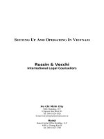
Le to Right: Dr. Paul Rekers, Dr. Gervase Connors, Dr. Spiers, Dr. Orvan Hess, Dr. John Homans, Dr. Arthur Morse, Dr.
Herbert oms, Dr. Irving Friedman
2010
1914
the journal for alumni and friends of yale oB/Gyn
5
HISTORICAL NOTE
Ernest I. Kohorn, Professor Emeritus,
Section of Gynecologic Oncology and Urogy-
necology, Department of Obstetrics, Gynecol-
ogy and Reproductive Sciences, Yale University
School of Medicine, New Haven, Connecticut
Ernest I. Kohorn, MA (Cantab), MA (Yale),
MChir (Cantab), FRCS (England), FRCOG, FACOG
A History of the Department of Gynecology
and Obstetrics at the 200th Anniversary of
Yale Medical School
Presented at Grand Rounds, Department of
Gynecology & Obstetrics, January 2011. The por-
tion of this history from 1800 to 1965 has been
reproduced with permission of the Yale Journal
of Biology and Medicine (copyright 1993). It has
been abridged and revised. The text since that
time is original.
We are currently celebrating the 200th anniver-
sary of the Yale School of Medicine’s Department
of Obstetrics, Gynecology and Reproductive Sci-
ences. In 1993, I described the Department’s first
150 years, “from Nathan Smith to Lee Buxton”
(1). Today I will recapitulate those 150 years (2)
but then will concentrate on the Department’s
last 50 years, try to place these recent times into
some perspective, and discuss their significance
in relation to the present state of medical practice
and specifically to obstetrics and gynecology.
Many current and distinguished members and
graduates of this program may not be mentioned
in this account. That needs to await a detailed and
more comprehensive future history.
The Yale School of Medicine was the brainchild
of President Ezra Stiles (Figure 1), the seventh
president of the University and a noted educator,
author, Congregationalist minister and theolo-
gian. He felt that Yale College should expand to
have both a law school and a medical school (2).
The founding of the Connecticut Medical Soci-
ety in 1792 appears to have been a prerequisite
for the establishment of the medical school (3).
This Society was given the authority to appoint
examining committees, to issue medical licenses
to those found qualified, and to confer honorary
degrees in medicine. It took another 30 years for
the Yale Medical School to begin its activities, in
part due to the fact that the Medical Society only
met formally once a year.
Figure 1.
Ezra Stiles, 1727 – 1795.
Seventh President of Yale College.
Lawyer, Pastor at Newport,
Rhode Island and New Haven.
Portrait by Moulthrop.
Ezra Stiles died in 1795 and was succeeded by
another noted Congregationalist minister, Timo-
thy Dwight (Figure 2), who incidentally was the
grandson of the Rev. Jonathan Edwards, one
of the greatest early American theologians and
famous fiery preacher (1703-1758).
Mason Fitch Cogswell (Figure 3) and Eli Ives
(Figure 4), both members of the Connecticut
Medical Society, were instrumental in support-
ing the founding of the medical school at Yale. In
1802 a professorship of chemistry was instituted
in Yale College, and Benjamin Silliman (Figure 5)
was appointed. He was then studying law at Yale.
To prepare himself for this task, Silliman went
to Philadelphia, then the center of scientific and
medical learning in North America, to study with
noted physicians Caspar Wistar, Benjamin Smith
Barton and James Woodhouse at the University
of Pennsylvania. The first appointment to the
!
ya le oBstetrical and GynecoloGical society
6
clinical faculty was Mason Cogswell, who was
appointed professor of surgery and anatomy, fol-
lowed by Jonathan Knight (Figure 6) who was ap-
pointed assistant professor. Knight was president
of the National Medical Convention that in 1846
evolved into the American Medical Association
(AMA). Knight also served as president of the
AMA from 1853 to 1854.
Cogswell was the leading surgeon in Connecticut
and was prominent in civic affairs. He established
the first institution in the United States for the treat-
ment of the “deaf and dumb” (his daughter was
hearing impaired) and was also the founder of the
Hartford Retreat for the Insane. However, Cogswell
preferred to stay in Hartford. Eneas Munson (Figure
7), also from Hartford and a founder of the Con-
necticut Medical Society, was appointed professor
of Materia Medica and botany. However, he felt
that at age 75, he was too old to lecture to students
and, although he maintained his professorship, the
actual teaching was performed by Eli Ives, who
also became the first lecturer and then professor of
Materia Medica. Ives also studied at the University
of Pennsylvania under the great Benjamin Rush,
Caspar Wistar and Benjamin Smith Barton.
Because Cogswell and Munson did not take up
their designated duties, appointing an active
teacher and clinician at the new medical school
became a matter of urgency. The Yale Corpora-
tion finally and successfully invited Nathan Smith
(Figure 8) to be the first professor of surgery and
obstetrics. We need to note that the portraits of
all these individuals are prominently displayed
on the upper floor of the rotunda of the Yale
Medical School Library right outside the Beau-
mont Room. Before he came to Yale, Smith had
founded three other medical schools, those at
Dartmouth College, Bowdoin College and the
University of Vermont. At that time, Smith was
spending most of his time at Dartmouth where
he lectured on anatomy, surgery, chemistry and
the theory and practice of physic. Oliver Wendell
Holmes later commented that Smith occupied
not one chair but a settee of professorships.
His income derived from student fees, as each
student paid $133 for the required courses, and
from his private practice. President Wheelock of
Dartmouth, coming from one of Nathan Smith’s
lectures, was so inspired that he led the evening
prayers: “Oh Lord, we thank Thee for the oxygen
gas. We thank Thee for the hydrogen gas and
all the gases. We thank Thee for the cerebrum
and the cerebellum and the medulla oblongata.”
Smith traveled widely across New Hampshire and
Vermont, always on horseback and usually with
his apprentices. Clinical teaching and discussion
went on throughout their journey.
Smith’s appointment at Yale College was initially
opposed by President Timothy Dwight, who
thought he might be an infidel, a free thinker in
the pattern of Voltaire and Rousseau, and to have
been influenced by the writings of Tom Paine.
After long correspondence between Cogswell
and Silliman and Nathan Smith, the Yale College
authorities were finally reassured about Smith’s
religious orthodoxy, and his appointment as the
first professor of the theory and practice of physic,
Figure 2.
Timothy Dwight,
Eighth President of Yale College,
1795 – 1817.
Grandson of Jonathan Edwards.
Figure 3.
Mason Fitch Cogswell.
Figure 4.
Eli Ives, Professor of Materia Medica,
Lecturer in Pediatrics.
From the portrait in the rotunda of the
Yale Medical School Library.
!
!
!
the journal for alumni and friends of yale oB/Gyn
7
surgery and obstetrics was confirmed. His was
the sixth such appointment in North America.
Smith had a wide repertoire of achievement. He
was the second person to operate for an ovarian
tumor – July 5, 1821. He did not know of Ephraim
McDowell’s feat in Danville, Kentucky, eight years
earlier. Smith had performed an autopsy on a pa-
tient with this diagnosis previously and confirmed
that the pedicle could be ligated without difficulty.
Unlike McDowell, he allowed the ligated pedicle
to fall back into the abdomen. He realized that
typhoid fever was associated with dehydration
and recommended fluids and support rather than
purging. He treated osteomyelitis conservatively
and not by amputation as was the recommended
practice at the time. Joseph Smith, who later
founded the Mormon religion, developed typhoid
osteomyelitis of the tibia at the age of 18. Nathan
Smith treated the lad conservatively by draining
the pus and removing dead bone fragments, thus
avoiding amputation. It is doubtful that an ampu-
tee could have gone “West.”
While at Yale, Smith continued his teaching and
practice activities at Dartmouth and also Vermont
where his second son, Ryno Smith, was profes-
sor of anatomy and physiology. Ryno Smith later
moved to Philadelphia and helped found Jef-
ferson Medical College. David Paige Smith was
appointed to the Ives Chair of the Theory and
Practice of Medicine at Yale in 1873. All of Nathan
Smith’s four sons, nine grandsons and six great-
grandsons entered medicine. Smith died quite
suddenly of a “febrile illness” on January 26,
1829, aged 66. Those of you who wish for more
detail may consult the article in the Yale Journal
of Biology and Medicine from 1993 (1).
The time from then to the beginning of the 20th
century is known as a “silent century.” Little
academic record has survived. Thomas Hubbard
(Figure 9) succeeded Smith to the chair of obstet-
rics. He was a successful and conscientious sur-
geon from Pomfret, Connecticut, and remained
in the professorship until 1838. Timothy Phelps
Beers was the next professor. Beers had received
his MD degree from Yale and, although he had a
large practice of some 5000 patients, he was a
painfully diffident teacher. His lectures in obstet-
rics, it was said, were illustrative of a “difficult
and protracted delivery.”
From the beginning, Yale medical students were
required to write a thesis for the MD degree. In
1836 the subject of one of these was “ausculta-
tion in pregnancy,” 17 years after Laennec had
described the stethoscope; clearly this was the
beginning of fetal monitoring.
Pliny Adams Jewett (Figure 10) succeeded Beers.
He, however, was appointed surgeon in chief to
the Knights Hospital in New Haven during the Civil
War. Because of this he resigned his professorship
and was succeeded by Thomas Hubbard in 1864.
In 1830, Jonathan Knight had suggested to the Yale
Corporation that obstetrics and diseases of children
merited a separate professorship. Only in 1867 was
the professorship changed from “Obstetrics” to
“Obstetrics and Diseases of Women and Children.”
Figure 5.
Benjamin Silliman, 1779 – 1864.
Professor of Chemistry and Geology.
From the portrait in the rotunda of the
Yale Medical School Library.
Figure 6.
Jonathan Knight, Professor of
Anatomy and Physiology, 1813 – 1838.
Professor of Surgery, 1838 – 1864.
From the portrait by Nathaniel Jocelyn
in the rotunda of the Yale Medical School
Library.
Figure 7.
Eneas Munson. Appointed rst
Professor of Materia Medica and
Botany at the Medical Institution
of Yale College but stayed in Hartford.
He was aged 79 years.
!
!
!
ya le oBstetrical and GynecoloGical society
8
However, Hubbard attended only 32 deliveries in 15
years. He was a “difficult and peppery individual.”
His appointment marked the first serious contro-
versy in the history of the medical school during
its first half-century. In protest, Jonathan Knight
resigned his professorship. Finally Hubbard was
forced to resign. His successor, Frank Beckwith,
had to resign in 1885 because he could not “afford
his professorship on the salary he was paid.” The
professorial salary was so small that he had to use
the wards of the hospital as his private clinic.
In 1871 the New Haven Dispensary had opened
on Crown Street and moved to York Street in
1878. A training school for nurses, the second
in the United States, opened in 1873 and was
housed in what was to become known as the
Hope Building. At this time the medical school
severed its association with the Connecticut
Medical Society and became incorporated as a
graduate school of Yale University. The Medical
Society provided medical licensure and the Uni-
versity the academic degree of MD. The hospital
moved to Congress Avenue in 1873. During the
last decade of the 19th century and the first
decade of the 20th, the obstetric wards were not
used for teaching because the “clinical material”
was insufficient, so most senior students took
additional courses at New York Lying-In Hospital
(now New York Hospital).
Yale was one of the medical schools rated by the
1910 Flexner Report as being “worthy of continu-
ation.” The Department of Obstetrics was the first
clinical department at Yale where faculty members
were hired on a full-time basis. In 1914, Josiah
Morris Slemons, a Hopkins graduate and formerly
professor of obstetrics and gynecology at the Uni-
versity of California, was charged with the organiza-
tion of the formal department. The assistant profes-
sor was Arthur H. Morse, also a Hopkins graduate.
Herbert Thoms was laboratory assistant. Six years
later Slemons resigned to return to his practice in
Los Angeles, and Morse (Figure 11) was appointed
to the chair that he held until 1945.
Morse was a charter member of the American
Board of Obstetrics and Gynecology. It was
Morse who invited Gertrude Van Wagenen (Figure
12) to come to Yale to initiate the macaque mon-
key colony that eventually led to the definitive
description of the reproductive physiology of both
the female and male macaque. That work also al-
lowed the subsequent discovery of the “morning-
after pill.” During his 28 years as chair, there were
only 15 publications, all in obstetrics. However,
Morse was an “unsparing and fine teacher with
insight and deep interest and unfailing kindness…
He was always impeccably dressed in a white
coat with a fresh flower in his buttonhole.”
The next chairman was Herbert Thoms (Figure
13). He was born in Waterbury, Connecticut, in
1885 and came to Yale Medical School directly
from high school. He interned at Backus Hospital
in Norwich and Memorial Hospital in New Lon-
don and did residency training at Sloane Hospital
for Women in New York, the first gynecological
hospital in the United States, founded by Marion
Sims in 1854. Thoms then went to Johns Hop-
Figure 8.
Nathan Smith, First Professor of Surgery and
Obstetrics at Yale Medical School.
Arrived om Dartmouth 1813.
Died 1829, aged 66.
From the portrait in the rotunda of the Yale
Medical School Library.
Figure 9.
omas Hubbard, 1776 – 1838.
Professor of Surgery, 1829 – 1838,
Professor of Obstetrics 1829 – 1830.
From the portrait in the rotunda of the Yale
Medical School Library.
Figure 10.
Pliny Adams Jewett,
Professor of Obstetrics and
Diseases of Children,
1856 – 1863.
From the portrait in the second oor corridor of
the Yale Medical School Library.
!
!
!
the journal for alumni and friends of yale oB/Gyn
9
kins and joined the Yale faculty in 1915. His major
scientific contribution was the introduction and
refinements of x-ray pelvimetry. Thom’s view of
the pelvis set the standards of the time. It was
not until 1967 that the practice of performing
full pelvimetry on all primipara at Yale was aban-
doned. It is remarkable that there appeared to be
no increase in leukemia among the offspring of
all these mothers. Thoms was not only an expert
clinical and academic obstetrician with several
inventions of instruments, but also a medical
historian and an accomplished artist, lithographer
and engraver.
During the early 1950s little gynecologic surgery
was practiced or taught at Yale. In 1952, there-
fore, Dean Hugh Long and Gustave Lindskog,
professor of surgery, invited John McLean Morris
(Figure 14) to New Haven to remedy this short-
coming. Morris was then in Dr. Meigs’ Depart-
ment of Gynecologic Surgery at Massachusetts
General Hospital in Boston, where he trained
together with Drs. Ullfelder, Ingersoll, Langdon
Parsons and Summers Sturgis. He spent a year
with Hans Kottmeier at the Radiumhemmet in
Stockholm and learned that radiation was an
alternative to surgery, particularly in the manage-
ment of cancer of the cervix. For Morris, coming
to Yale was an abrupt change from Harvard where
gynecology was a separate department related to
surgery rather than to obstetrics, and where staff
members had full surgical training.
Morris established gynecologic surgery at Yale and
created a close link with radiation therapy, a symbi-
osis that lasted through his lifetime. The standards
of excellence and accountability he established are
recalled by generations of still-trembling former
Yale Ob/Gyn residents. He was responsible, with
Chu Chang, for developing the radiation system
used at Yale for treating cancer of the cervix. With
Meigs he described the distinction between resec-
table and non-resectable cancer of the cervix that
was later included in the FIGO classification of that
disease. With Robert Scully he described testicular
feminization and, based on the original work of
Gertrude Van Wagenen, he helped to develop the
“morning-after pill,” so fulfilling a deep interest in
population control. John Morris became emeritus
in 1985 and died in 1993. A more detailed descrip-
tion of his life and contribution to gynecology is
available in the October 2009 issue of Connecticut
Medicine (3).
Charles Lee Buxton (Figure 15) succeeded Thoms
as chairman in 1954. He was an undergraduate
at Princeton, obtained his MD from Columbia in
1932, and in 1940 obtained the MedScD degree.
Following an internship in Cooperstown and
research at Harvard from 1933 to 1934, he did his
residency at the Sloane Hospital, New York, and
at Columbia. He was invited to the chair at Yale in
1953 at a salary of $22,000 a year. Buxton was
what would now be called a reproductive surgeon.
Some of the endocrinologists nurtured by Buxton
include Walter Herrmann, who trained in Swit-
zerland, came as an endocrinologist to Yale and
went on to become chairman, first in Seattle and
then in Geneva, and Raymond Van de Wiele, who
Figure 11.
Arthur Henry Morse, 1880 – 1950.
Chairman, 1920 – 1945.
ree of his trainees became chairmen.
He brought Gertrude Van Wagenen to Yale.
Charter member of the American
Board of Obstetrics and Gynecology.
Figure 12.
Gertrude Van Wagenen,
1893 – 1975.
Pioneer of macaque ovarian and
testicular physiology and co-discoerer of
“morning-aer” pill.
!
!
ya le oBstetrical and GynecoloGical society
10
trained in Belgium and went on to become the
endocrinologist at Columbia. Both were pioneers
in the investigation of steroid physiology of the
ovary. Luigi Mastroianni grew up in New Haven,
where both his parents were physicians, went to
Yale College for his MD and to Boston University
for his residency. He worked with John Rock at
Harvard and came to Yale as assistant professor
in 1954. Subsequently he became an endocrinol-
ogist at the University of Pennsylvania, and then
its chair for over 25 years.
Buxton’s greatest contribution was as a visionary
who recognized good ideas that had the potential
to be realized. He then sought persons with ex-
pertise to develop these ideas and thus attracted
individuals who initiated research programs in
endocrinology, fetal monitoring and diagnostic
ultrasound. This was the beginning of subspecial-
ty disciplines in the United States. First Buxton
invited Nathan Kase to initiate a Section of Endo-
crinology. Kase was a graduate of Columbia and,
following residency at Mount Sinai in New York,
he did a fellowship in steroid biochemistry at the
Worcester Foundation. He was a charismatic and
exciting teacher of molecular and clinical endocri-
nology and “could bring the steroid nucleus to life
and make it dance.” His Saturday morning lec-
tures were crowded with faculty, residents and
students. These lectures resulted in the publica-
tion of the now-standard textbook he produced,
together with Robert Glass and Leon Speroff.
The second field that Buxton nurtured was fetal
electrocardiography. This research had been initi-
ated at Yale by Orvan Hess, but it was Edward
Hon (Figure 16) who brought it to fruition and
made it into a tool for routine clinical practice.
Diagnostic obstetric ultrasound was the other
technique that Buxton thought would provide in-
novative information. He had visited Ian Donald’s
ultrasound unit in Glasgow and had close con-
tact with William Nixon, chairman at University
College Hospital in London. Like Thoms before
him, Buxton was deeply interested in the natural
childbirth program that was active in Nixon’s unit,
the first for a clinic population. Nixon and Buxton
arranged for Ernest Kohorn to go to Glasgow to
learn this new technique and bring it to Yale dur-
ing a fellowship. In those early days a patient with
a retained placenta was taken to the operating
room by Ian Donald so that the exact position of
the placenta in the uterus could be determined
manually while at the same time performing an
ultrasound scan to confirm its location by that
technique (Figure 17).
Buxton was also a social activist and was called
“the gentle crusader.” It is interesting that a phy-
sician concerned with reproductive failure should
also concern himself with reproductive control,
and it is this connection that made Buxton into
the complete and caring doctor that he exempli-
fied. The issue of contraception had been brought
to the Connecticut Supreme Court on two oc-
casions, first in 1940 when there was a criminal
suit and the court decided that the wording of the
law was clear in not permitting doctors to pre-
scribe contraception. The second case, in 1942,
Figure 13. Herbert oms,
Professor and Chair,
Obstetrics and Gynecology,
1945 – 1952.
Figure 14. John McLean Morris,
1915 – 1993.
Princeton graduate, MD om Harvard.
Trained at Massachusetts General
Hospital. Came to Yale in 1952 and
established gynecologic surgery.
Figure 15. Charles Lee Buxton,
1904 – 1969.
Chairman 1954 – 1967.
MD and MedScD om Columbia.
Initiated subspecialization at Yale.
Helped legalize contraception in
the State of Connecticut.
!
!
!
the journal for alumni and friends of yale oB/Gyn
11
was initiated by Professor Wilder Tileston of the
Yale Medical School, and the court again upheld
the constitutionality of the law. The issue was
brought to a head when Dr. Buxton and Estelle
Griswold, executive director of Planned Parent-
hood of Connecticut, opened a birth control clinic
in November 1961. Both were arrested. Buxton
later remarked that he thought he was worth
more than the hundred-dollar bail demanded.
Both were fined. The appeal reached the Su-
preme Court of the United States in October
1965, and the law was overturned. Justice Doug-
las delivered the majority opinion of the Court
with Justices Goldberg and Brandon and Chief
Justice Warren concurring.
During Dr. Buxton’s tenure as chair, there were
several individuals who came as residents, usually
stayed as junior faculty members and then went
on to have distinguished careers. Dr. David Ger-
shenson (Figure 18) went to a fellowship with Dr.
Felix Rutledge at M.D. Anderson Hospital, stayed
on the faculty in gynecological oncology and even-
tually became chair of that institution. Phillip DiSaia
(Figure 19) did his residency at Yale and also went
on as a fellow to M.D. Anderson Hospital, becom-
ing director of the division of gynecological oncol-
ogy at the University of California at Irvine in 1989.
He subsequently became chair of the department
and Associate Vice Chancellor for Health Sciences,
associate dean, and is presently the director of the
Gynecological Oncology Group.
Two other notable residents arrived at Yale during
the Buxton era. One was Leon Speroff (Figure 20)
who graduated from Denison University, Ohio,
and went to Case Western Reserve for his MD.
While a second-year medical student, he decided
he “wanted to be America’s Grantly Dick-Read”
(a British obstetrician regarded as the father of
the natural childbirth movement). At that time
Yale had the only academic department in the
United States that promoted natural childbirth.
Speroff did a summer clerkship. Subsequently
he received a telegram from Dr. Buxton, invit-
ing him to become a resident at Yale. When he
arrived, Nathan Kase had just become chair and
persuaded him to change and become an endo-
crinologist. While a resident, he persuaded the
hospital to increase the salary for residents from
$150 a month to $300 a month, which was still
$200 short of the cost of living at that time. Like
Kase before him, he went on to the Worcester
Foundation and then came back to Yale as assis-
tant professor, subsequently becoming associate
professor. He left to become chairman at Case
Western Reserve and later professor of obstet-
rics and gynecology at Oregon Health Sciences
University, where he had an exciting and distin-
guished career.
The second person was Philip Sarrel (Figure 21),
another resident during the Buxton era. He went
to college at Dartmouth and did a medical intern-
ship at Mount Sinai Hospital in New York. During
his residency at Yale, he organized a special clinic
for unwed mothers that became a national mod-
el. His research interests remained in sexuality,
contraception and menopause. He was founder
of the Yale Menopause Program and the Yale Sex
Figure 16. Edward Hon, 1917 – 2009.
Came to work in endocrinology but
turned to study fetal electrocardiography.
In the 1960s, with Orvan Hess, developed
electronic fetal heart rate monitoring.
Figure 17. Ultrasound scan of placenta
performed by Ian Donald in Glasgow,
while Ernest Kohorn explored the
uterus manually to conrm the position
of the placenta.
Figure 18. David Gershenson,
Yale resident, gynecologic
oncology fellow at M.D. Anderson
Hospital, where he became sta
surgeon and chairman.
!
!
!
ya le oBstetrical and GynecoloGical society
12
Counseling Service. He was on the faculty of
Gynecology and Obstetrics as well as Psychiatry.
He became emeritus in 2009.
Buxton had great personal charm and was a
genial and attentive host. Many of us remember
with affection the Sunday morning brunches he
gave, attended by all members of the staff. He
left the Department prepared for subspecializa-
tion and ready to absorb the knowledge explosion
of the last quarter of the 20th century.
Following a national search, Edward Quilligan
(Figure 22) was appointed to the chair of gyne-
cology and obstetrics in 1967. Quilligan obtained
his bachelor’s degree and MD from Ohio State
University and performed his residency at Case
Western Reserve in Cleveland, where he re-
mained on the faculty. When he came to New
Haven he worked with Edward Hon, who was at
that time establishing fetal electrocardiography as
a monitoring methodology during labor. For this
research to be statistically valid, a greater number
of patients were required than were available in
New Haven. Quilligan and Hon therefore moved
to the University of Southern California (USC) in
Los Angeles, where Quilligan served as chair.
Subsequently Quilligan went as chair to the Uni-
versity of Wisconsin and then to the University
of California at Davis. He also served as associ-
ate vice president of health sciences at USC and
then dean at UC Irvine. In 1989 he returned to
teaching at UC Irvine Medical Center in Orange.
He contributed substantially to the development
of the practice of fetal monitoring during labor
and showed that fetal distress could be identified
at a much earlier stage. The goal was to reduce
complications and infant mortality. Subsequently
he focused on uterine function in pregnancy, the
role of abnormal oxygen levels in fetal brain dam-
age, fetal breathing and fetal sleep states.
When Quilligan left for Los Angeles in 1969, Na-
than Kase (Figure 23) was promoted to the chair.
As described previously, Kase had been recruited
by Dr. Buxton to initiate the section of endocri-
nology. Kase brought Alan DeCherney (Figure
24) and Donald Coustan to the Department. The
initial intent was to initiate a general practice of
obstetrics and gynecology within the depart-
ments. It soon became clear that this was not an
academically profitable venture. DeCherney went
on to create the first in vitro fertilization unit in the
Eastern United States. He was aided in this ven-
ture by Neri Laufer, a graduate of Hadassah Medi-
cal School in Jerusalem, who was then working
in the Biology Department at Yale with Professor
Clement Markert. This unit has since become one
of the leading departments of in vitro fertilization
in the country. DeCherney went on to become
professor and chair of obstetrics and gynecology
at Tufts University School of Medicine in Boston.
Then he became chair and director of reproductive
endocrinology at UCLA and subsequently moved
to Washington to become head of the Program in
Reproductive Endocrinology at the Eunice Ken-
nedy Shriver National Institute of Child Health and
Human Development at the National Institutes of
Health. He was elected a member of the Institute
of Medicine of the National Academies in 2004.
Figure 19. Phillip DiSaia, Brown graduate.
Also went to M.D. Anderson for fellowship.
Chief of Gynecologic Oncology at UC
Irvine in 1989, then Chair and
Vice-Chancellor. Now Director of
Gynecologic Oncology Group.
Figure 20. Leon Spero, BA, Denison
College, MD at Case Western,
Yale residency, then Assistant and
Associate Professor, then Chair at
Case Western and ultimately joined
the faculty at Oregon Health
Sciences University in Portland.
Figure 21. Philip Sarrel. Residency at Yale,
stayed and became Professor of Gynecology
and Obstetrics and of Psychiatry. Founded
Unwed Mothers’ Program, the Menopause
Program and the Yale Sex Counseling Program.
!
!
!
the journal for alumni and friends of yale oB/Gyn
13
Donald Coustan (Figure 25) became a perinatolo-
gist, particularly interested in diabetic pregnancy.
He went on to become chair at Brown Medical
School and became a national authority on dia-
betic pregnancy.
In the fifth Lee Buxton Memorial Lecture in 1990,
Nathan Kase described how he saw the Depart-
ment during the previous 20 years. He came to
Yale in 1962 with a salary of $8,000 a year. This
was raised to $11,000 when he received his first
research grant. “At that time,” he said, “it was
possible to combine research and teaching with a
defined subspecialty clinical practice. Obstetrics
and gynecology was in the midst of a transition
from one being a professor ‘of all things’ to an
emphasis on the subspecialty in perinatology or
oncology or endocrinology. There was an explo-
sion of knowledge; in 1962 pregnancy tests were
done by the rabbit assay. Two years later there
was immunoassay. Obstetrics and gynecology
had become reproductive science. We could fol-
low clinical and research interests as sharehold-
ers and partners in the intellectual enterprise. We
could develop new areas of expertise. There were
really no bosses to tell us what to do. The sal-
ary was that of civil servants, but there was little
constraint on our intellectual effort. Those were
the 1960s and 1970s.”
John Hobbins (Figure 26) returned to the faculty
soon after Kase became chair. He had graduated
from Hamilton College and New York Medical
College and completed his residency at Yale prior
to military service. He was recruited to initiate the
division of perinatology. Besides fetal ultrasound
diagnosis, Dr. Hobbins developed fetoscopy and
the in-utero diagnosis of haemoglobinopathies. He
made possible the prenatal diagnosis of Ellis-van
Creveld syndrome and of Duchenne muscular dys-
trophy. He established intraperitoneal and subse-
quently perfected intravascular fetal transfusions.
Dr. Hobbins is regarded as a “formidable teacher”
and trained many of today’s leaders in the field,
including Roberto Romero, E. Albert Reece and
Joshua Copel, among many others. He moved to
the University of Colorado in Denver in 1992.
Peter E. Schwartz (Figure 27) was recruited to
Yale to initiate a section of Gynecologic Oncology.
He had graduated from Union College and Albert
Einstein College of Medicine, did his residency
at Yale and was advised to go to M.D. Anderson
Hospital for fellowship in gynecologic oncology.
Figure 22. Edward Quilligan,
Chairman 1967 – 1969.
Came om Cleveland, Ohio, and
departed for the University of California,
Los Angeles.
Figure 23. Nathan Kase,
Chairman 1969 – 1978.
Came om Mount Sinai, New York.
Returned there as Chairman and
subsequently Dean.
Figure 24. Alan DeCherney.
Initiated in vitro fertilization at Yale
with Neri Laufer. Went to be Chair at
Tus, then at University of California,
Los Angeles, and is now Director of
Reproductive Medicine at NIH.
!
!
!
Figure 25. Donald Coustan,
Resident, Assistant and Associate
Professor at Yale. Chair, Depart-
ment of Obstetrics and Gynecology
at Brown University. Expert on
diabetes in pregnancy.
Figure 26. John Hobbins
as chief resident, 1965. Gradu-
ated om Hamilton College and
New York Medical College and did
residency at Yale. He took over ultra-
sound om Kohorn in 1972.
Developed the Perinatology Division
and trained numerous specialists in
that eld. He is now at the Univer-
sity of Colorado, Dener.
!
!
ya le oBstetrical and GynecoloGical society
14
When Dr. Morris stepped down as chief of gyne-
cology, Schwartz established the oncology sec-
tion and the training fellowship. His major interest
has been the early diagnosis of ovarian cancer
and the use of “prophylactic” chemotherapy in
the initial management of advanced ovarian can-
cer. His introduction of chemotherapy for germ
cell tumors has preserved the fertility of many
generations of affected young women.
Kase also brought Harold Behrman (Figure 28)
and Richard Hochberg (Figure 29) to Yale, both
reproductive scientists who established labora-
tories that have made major significant contribu-
tions to that science.
But all was not perfect. Robert Glass was remark-
able in that he went to Yale College and Yale School
of Medicine and then remained on the faculty for 10
years. When he was to be promoted to full profes-
sor, the medical school had financial problems and
there were no funds for promotion in any depart-
ment in the medical school. This was because of
the University rule that any tenured professor had to
have set aside sufficient funds in the endowment of
the University to pay for the length of the professor-
ship. To solve this problem, the University created a
class of “clinical” professors. Such persons did not
have tenure in the traditional sense of the word but
had a “continuing appointment” that could not be
terminated except if the whole class of such profes-
sors was terminated. Glass felt betrayed and left Yale
to become a professor at the University of California,
San Francisco.
Nathan Kase left Yale after eight years as chair
in 1977 to become chairman at his alma mater,
Mount Sinai Medical School in New York, where
he subsequently became dean and had a further
distinguished career.
Frederick Naftolin (Figure 30) was appointed
Kase’s successor in 1978 and remained as chair
for 23 years, nearly rivaling the 28-year tenure
of Dr. Morse. Naftolin had graduated from the
University of California at San Francisco and had
obtained a D. Phil. degree from Oxford University,
working with Geoffrey Harris, the discoverer of
the pituitary portal system. He had spent time at
Harvard and at the time of his move was chair-
man of the Department of Obstetrics and Gyne-
cology at McGill in Canada. A member of his fac-
ulty recently said, “Naftolin’s greatest contribution
was his passion for research of any sort. He al-
lowed academic freedom wherever it would lead,
even outside the traditional gynecology obstetrics
field. That’s how we ended up in neuroscience
in our department. We could pursue our dreams
and excellence wherever they would lead.” After
retirement, he left Yale to become director of
biologic research in the Department of Obstet-
rics and Gynecology at the New York University
School of Medicine where he is at present.
Dr. Charles Lockwood (Figure 31) became chair-
man in 2002. He had received his undergraduate
education at Brown University, his medical train-
ing at the University of Pennsylvania, served
Figure 27. Peter E. Schwartz,
MD, om Albert Einstein College of
Medicine, Residency at Yale. Gynecologic
Oncology Fellowship at M.D. Anderson.
Recruited to Yale in 1979 to organize
Section of Gynecologic Oncology.
Figure 28. Harold Behrman,
1939 – 2008. BSc and MSc University of
Manitoba, PhD University of North Carolina.
Postdoctoral fellowship at Harvard.
In 1971 became Chair of Reproductive
Biology at the Merck Institute. Came to
Yale as Director, Reproductive Biology,
in 1975.
Figure 29. Richard Hochberg,
PhD om Hahnemann Medical College, 1967.
Discoered “lipoidal derivatives” of steroids,
including estrogen. Developed rst in vitro
assay for estrogens.
!
!
!
the journal for alumni and friends of yale oB/Gyn
15
a residency at Pennsylvania Hospital and a Mater-
nal-Fetal Medicine fellowship at Yale under John
Hobbins. After a two-year sojourn at Tufts, he
completed a postdoctoral fellowship under Yale
Nemerson at Mount Sinai in molecular hemosta-
sis, where he stayed on the faculty until 1995.
He became chairman at New York University that
year. During his present tenure, the faculty num-
ber has increased to 59 persons, and the Depart-
ment is thriving under his tutelage.
It is noteworthy that many of the residents who
came to Yale with academic ambition decided to
go into clinical practice rather than pursue a pro-
fessorial career. Conversely, many who entered
residency with a view toward clinical practice
were so stimulated by the enthusiastic atmo-
sphere encouraging research that they became
academicians. Frequently this was quite a dra-
matic transformation. It should also be noted that
many of the graduates of the residency program
went into private practice in New Haven and sur-
rounding towns and played a significant role in
resident and medical student teaching.
looKIng baCKward, looKIng forward
What has changed in obstetrics and gynecology
in the last 40 years? The clinician scholar profes-
sor track has become routine for all clinicians,
and most clinical departments reserve tenure-
track professorships for their research faculty.
Soon after his appointment, Dr. Naftolin tried to
obtain a clinical professorship for himself, but it
appeared he would lose status and the respect of
other chairpersons. He therefore had to retain his
tenure-track professorship. A clinician educator
track has been instituted also, to try and encour-
age good teaching in the medical school.
There is no doubt that the enthusiasm for re-
search and teaching continues to fulfill the ambi-
tion of many of those who rise through the ranks
of professorships and infuse that spirit into the
medical students. Research grants 50 years ago
were not too difficult to obtain. In spite of the
present national fiscal problems and tight NIH pay
lines, the Department’s research operation has
grown markedly in size and international stature
in the last nine years and is academically very
successful. In 1964, Dr. Buxton had a faculty of
eight and one administrative secretary. In the
past eight years the Department has grown to 59
clinical and research faculty, 25 postdoctoral re-
search fellows, 16 clinical fellows and 97 clerical,
technical and managerial staff. Research funding
in FY2011 is projected to exceed $16 million with
nearly $11 million in total NIH dollars.
What is of concern is that the teaching of medi-
cal students and even residents has become
more challenging and difficult. Patients spend
little time in the hospital as inpatients, and there
is less time for them to meet medical students.
The leisure of outpatient teaching has largely
disappeared so that the opportunity to learn by
example has become a luxury. I do not believe
that one can teach clinical medicine by computer
modeling. The emphasis on throughput, patient
safety, patient satisfaction, expense reductions
and revenue generation, so prevalent in medicine
today, makes good clinical teaching a challenge.
This is the most urgent problem of medical
schools in the United States at the present time.
Our physicians are so busy, both in academic and
in private practice, that there is even little time to
come to Grand Rounds. That surely can be fixed!
However, the future is bright! There are many
star players in our Department at the present
time, and their achievements will surpass and
certainly rival those of all previous generations.
Figure 30. Frederick Naolin,
MD, DPhil.
Chairman 1978 – 2001.
Came om McGill University,
Canada. Departed for
New York University, New York.
He has more than
700 publications.
Figure 31. Charles Lockwood,
MD, MHCM. e Anita O’Keee
Young Professor of Women’s Health
and Chair, Department of Obstet-
rics, Gynecology and Reproductive
Sciences, Yale University School
of Medicine. Chief of Obstetrics
& Gynecology, Yale-New Haven
Hospital. Appointed 2002.
!
!
ya le oBstetrical and GynecoloGical society
16
REFERENCES
1. Kohorn E.I., The Department of Gynecology and Obstetrics at Yale; the First One Hundred Fifty
Years, from Nathan Smith to Lee Buxton. Yale Journal of Biology and Medicine, 66: 85–105, 1993.
2. Medicine at Yale, 1810–1910, www.med.yale.edu/library/historical/bicentennial/1810. Much of the
information for the “first 150 years,” cited in reference #1,was gleaned from the then unpublished
manuscript by E. H. Thompson, “The early years of the Medical Institution of Yale College,” in the
Yale Medical Historical Library.
3. Kohorn E.I., John McLean Morris, a career in surgery, gynecology and reproductive physiology. Con-
necticut Medicine, 73: 223-227, 2009.
To view Dr. Kohorn’s article in its entirety, please visit:
*
To view Dr. Gross’s article on Griswold v. Connecticut, please visit:
**
* Presented at Grand Rounds, Department of Gynecology & Obstetrics, January 2011. The portion of this history from 1800 to 1965 has
been reproduced with permission of the Yale Journal of Biology and Medicine (copyright 1993). It has been abridged and revised. The
text since that time is original.
** The original version of this article was first given as a speech at the ABCD-sponsored 40th anniversary celebration of the Griswold v. Con-
necticut decision held at the Massachusetts State House in March 2005. It was modified for presentation at the June 7, 2005 celebration
held at the Senate Office Building. The article was further modified for presentation at Yale Ob/Gyn Grand Rounds in 2005 and presented
at the Beaumont Society on March 17, 2006. We are publishing this article in honor of Yale’s 200th anniversary and the 50th birthday of
the approval of oral contraceptives for contraceptive purpose in the United States.
the journal for alumni and friends of yale oB/Gyn
17
Preconception/Interconception
Counseling and Care
Prenatal care should begin in the preconception
period with risk assessment being the primary
objective for preconception education (1). Precon-
ception education is important because evidence
suggests that women who plan pregnancy are
more likely to have a healthy birth outcome.
This is particularly relevant because the leading
causes for infant mortality in the United States
are congenital anomalies, preterm birth, low birth
weight and chronic medical disease morbidities
complicating pregnancy. Unfortunately, only 50%
of pregnancies in the United States are planned,
which is directly related to high infant mortality
and racial disparity in infant mortality compared to
other developed countries.
The definition posed by the Center for Disease
Control (CDC) for preconception care is interven-
tion that aims to identify and modify biomedical,
behavioral and social risks to a woman’s health
through prevention and management. Intercon-
ception care is the time period between pregnan-
cies, which is generally about 18 to 24 months
postpartum, where the woman can direct her
attention to healthier lifestyle goals to improve
upon pregnancy outcomes.
The important elements to effective preconcep-
tion care include screening for medical and social
risk factors, providing appropriate immunizations,
counseling based on medical and genetic history,
age and ethnic risk, health education and inter-
ventions such as weight loss and control of
diabetes and blood pressure, known to improve
pregnancy outcome and overall adult health.
Immunization status should be evaluated for
rubella, varicella, hepatitis B and diphtheria,
tetanus, pertussis (Tdap vaccine). Infections with
potential risk to the fetus include cytomegalovi-
rus (CMV), toxoplasmosis, parvovirus and HIV.
For some women in high-risk situations, the
immunization for CMV and parvovirus may be ap-
propriate. HIV testing is a routine component of
prenatal laboratory testing. Women with preexist-
ing medical conditions should receive counsel-
ing prior to pregnancy to understand the risk of
those conditions on their health and the health
of the fetus. For example, in the U.S., obesity is
the leading chronic disease of reproductive-age
women; chronic hypertension occurs in 22% and
diabetes in 7%.
COUNSELING ON SPECIFIC CONDITIONS
DIABETES
Uncontrolled pregestational diabetes is associ-
ated with increased risk for congenital anomalies,
specifically heart and neural tube defects, still-
birth and birth trauma. The pregestational diabetic
should aim to optimize diabetes control prior to
conception. The goal should be to have a hemo-
globin A1C level <6.5%. A hemoglobin A1C level
>6% is associated with a 15% to 20% increased
risk for miscarriage and a 5% to 10% risk for birth
defects. Also, renal function should be assessed
because of maternal risk for preeclampsia, and
an ophthalmological examination should be
performed to evaluate for retinopathy so that
appropriate treatment can occur for this condi-
RESIDENTS’ RESEARCH DAY VISITING PROFESSOR GRAND ROUNDS
Haywood L. Brown, MD
Roy T. Parker Professor and Chairman
Department of Obstetrics and Gynecology
Duke University Medical Center, Durham, North Carolina
ya le oBstetrical and GynecoloGical society
18
tion prior to pregnancy. The patient should be
educated with the goal of maintaining euglycemia
with whatever regimen she is using to control
diabetes. The objective is a fasting blood sugar
level <100 mg/dl and two-hour postprandial blood
sugars <120 mg/dl. These patients should also be
on a vitamin with folic acid supplementation prior
to conception and maintain a folate-rich diet.
HYPERTENSION
Women with chronic (essential) hypertension are
at increased risk for stroke, renal and cardiovas-
cular compromise and preeclampsia. Pregnancy
complications associated with chronic hyperten-
sion are placental abruption, fetal growth restric-
tion and stillbirth. Women with chronic hyper-
tension should have a baseline renal function
evaluation and review of medication. ACE inhibi-
tors should be avoided during pregnancy because
of the risk for congenital renal tubular dysplasia.
The goal for blood pressure control is <140/90
mmHg with a single medication. Commonly used
medications for control of hypertension are beta
blockers and calcium channel blockers.
SEIZURE DISORDER (EPILEPSY)
Women with seizure disorders controlled with
medication should be counseled on medication
risk for birth defects. All medications used for
seizure control have some risk for causing birth de-
fects, and the benefits for seizure control must be
weighed against the risk for the medications. The
patient should be counseled that medications may
need to be adjusted upward in order to maintain
seizure control. The goal is to adjust or reevaluate
the need for medications to those with the lowest
risk. Valproic acid, in particular, is associated with
increased risk for neural tube defects and cardiac
defects. All women with a history of seizures
should be on a vitamin containing folic acid.
OBESITY
Women who are overweight or obese should
be aware of the increased risk for birth defects,
medical complications of pregnancy including
preeclampsia and gestational diabetes, and the
risk for cesarean delivery. It is recommended that
women aim to establish and maintain a normal
body mass index (BMI) prior to pregnancy and
follow the Institute of Medicine (IOM) guidelines
for weight gain during pregnancy (2). A derivative
study of the FASTER Trial, evaluating the link be-
tween obesity and cesarean delivery, noted that
women classified as morbidly obese had a risk
for cesarean delivery of 47.4% (3).
Interconceptionally, obese women should aim to
create a healthy lifestyle with diet and exercise
to achieve a healthier weight prior to the next
pregnancy. They should recognize the benefits of
breastfeeding to themselves and their infant for
long-term health. Breastfed infants have a lower
risk for adult cardiovascular disease and obesity.
HEART DISEASE
Adult heart disease puts the patient at risk for
pregnancy morbidity and mortality. Specifically,
women with corrected congenital heart disease
have a risk for recurrence of congenital heart
defects in offspring. Uncorrected adult congenital
heart disease can result in decompensation due
to physiological increase in plasma volume during
pregnancy. Women with artificial heart valves,
specifically mechanical valves, typically require
Coumadin to prevent thromboembolism.
However, Coumadin is teratogenic, and women
with mechanical valves contemplating pregnancy
must be apprised of the risk for thromboem-
bolism if Coumadin is discontinued in favor of
heparin during early embryogenesis. Prior cardio-
myopathy should be considered a contraindica-
tion to pregnancy because of the increased risk
for mortality.
OTHER CONDITIONS
Women with collagen disease and thyroid condi-
tions should also be counseled for potential mor-
bidity and should be advised of any medication
risk prior to conception.
THE INFERTILE COUPLE
Approximately 15% to 20% of couples of repro-
ductive age have difficulty conceiving. The patient
and her partner must appreciate any preexisting
medical conditions that pose a risk to mother or
fetus. All women should be aware of the in-
creased risk for multiple gestations with ovulation
the journal for alumni and friends of yale oB/Gyn
19
induction or in vitro fertilization. Advanced mater-
nal age is often a factor for the infertile female,
and the risk for aneuploidy should be discussed
prior to conception. All women undergoing infer-
tility treatment should be on a vitamin containing
folic acid prior to conception.
FOLIC ACID RECOMMENDATIONS
In 1992 the U.S. Public Health Service advised a
vitamin containing folic acid for all reproductive-
age women to reduce the risk for neural tube
defects and for improvement of overall pregnancy
outcome. In 2004, only 40% of reproductive-age
women reported taking a vitamin with folic acid.
Preconception recommendations are for at least
400ug of folic acid daily beginning four weeks
prior to conception and continuing for the first
three months of pregnancy. The women should
also maintain a folic-rich diet prior to conception
and throughout pregnancy.
DRUGS AND MEDICATIONS
A number of drugs that are taken for medical
conditions are known to pose a teratogenic risk
to the developing embryo. Some specific drugs
include valproic acid, Coumadin, isotretinoin and
lithium. The patient should be advised of the risk
of alcohol and to avoid cigarette smoking and il-
licit drug use.
There are a number of teratogen information
services and computer databases to consult that
provide appropriate counseling to women, includ-
ing MICROMEDEX, REPROTOX (Reproductive
Toxicology Center) and TERIS (Teratogen Informa-
tion Service).
There is no evidence that caffeine or aspartame
(Nutrasweet) is teratogenic. One study showed
that heavy use (>300 mg/day; >8 cups of coffee)
increased risk for stillbirth (OR 3.0, CI 1.5-5.9) (4).
PREGNANCY AFTER PREGNANCY LOSS
There are no good data on appropriate timing to
optimize pregnancy outcome after pregnancy
loss. Each patient should be evaluated about grief
response and should begin trying for the next
pregnancy when she is ready.
THE EXPECTANT FATHER (PARTNER)
The partner should be involved in preconcep-
tion counseling. Partner involvement leads to
a healthier birth outcome. The partner should
also appreciate the long-term health benefits of
breastfeeding for mother and child. A study by
Arora and associates (5) indicated that 40% to
75% of women reported that their partners’ opin-
ion or preference impacted their decision about
breastfeeding.
SUMMARY
Prenatal care should begin in the preconception
period to counsel and address health concerns
that can impact mother and child. All women con-
templating pregnancy should begin a multivitamin
supplemented with folic acid at least four weeks
prior to conception.
REFERENCES:
1. Center for Disease Control and Prevention. MMWR. 2006; 55(6):1-21.
2. Institute of Medicine of the National Academies. Weight Gain During Pregnancy. Washington, DC.
National Academies Press; 2009.
3. Weiss JL, et al. Obesity, obstetric complications and cesarean delivery rate – a population-based
screening study. Am J Obstet Gynecol. 2004; 190:1091-7.
4. Wisborg K, Kesmodel U, Bech BH, Hedegaard M, Henriksen TB. Maternal consumption of coffee during
pregnancy and stillbirth and infant death in first year of life: prospective study. BMJ. 2003; 326:420.
5. Arora S, McJunkin C, Wehrer J, Kuhn P. Major factors influencing breastfeeding rates: Mother’s
perception of father’s attitude and milk supply. Pediatrics. 2000; 106:E67.
ya le oBstetrical and GynecoloGical society
20
Etiology and Management of Recurrent
Spontaneous Abortion
INTRODUCTION
Management of recurrent spontaneous abortion
(SAB) is quite challenging. Affected patients are
often offered non-evidence based or anecdotal
treatments, there is no consensus on definitions,
and prevalence estimates are confounded by the
high background rate of pregnancy wastage. It is
generally accepted that 1% of couples suffer two
or more consecutive pregnancy losses prior to
the third trimester (1).
At least half of sporadic SABs have aneuploid
karyotypes, most commonly trisomies, followed
by polyploidy and monosomy X (2). Maternal age
is strongly associated with the risk of both SAB
and aneuploidy. One prospective cohort study of
over 36,000 women examined relative miscarriage
and aneuploidy rates in three age groups: less
than 35 years, 35–39 years, and 40 years or older
(3). Multivariate logistic regression adjusting for po-
tential confounders determined that, compared to
women <35 years, those 35–39 years old had an
increased risk for SAB with an adjusted odds ratio
(adjOR) of 2.0 (95% confidence intervals, 1.5–2.6)
while those ≥40 years of age had an adjOR of 2.4
(95% CI, 1.6–3.6) for SAB. Moreover, the associa-
tion of embryonic chromosomal abnormalities
with these two age groups produced adjORs of
4.0 (95% CI, 2.5–6.3) and 9.9 (95% CI, 5.8–17.0),
respectively. A second larger Scandinavian pro-
spective cohort study of 634,272 women having
1.2 million pregnancies found progressively higher
SAB rates with increasing maternal age: <12% for
women 20–29 years, 15% for those 30–34 years,
24.6% for those 35–39 years, 51% for ages 40–44
and 93.4% for women ≥45 years (4).
While there is no universally accepted expla-
nation for why aneuploidy is associated with
advanced maternal age, former Yale Ob/Gyn
resident and fellow, David Keefe, now chair of
Ob/Gyn at New York University, has posited that
progressive shortening of oocyte telomere length
due to the cumulative effects of oxidative stress
may be the culprit (5). Such shortening of
telomere can lead to abnormal chiasma formation
and, hence, nondisjunction.
GENETIC CAUSES
Between 25% and 57% of patients with recurrent
SAB have recurrent aneuploid conceptuses (6, 7).
Patients with recurrent miscarriage undergoing in
vitro fertilization (IVF) with preimplantation genetic
testing have far higher rates of abnormal embryos
compared with controls (70.7% vs. 45.1%; P
<0.0001) (8). Given the link between advanced ma-
ternal age and aneuploidy, it is not surprising that
recurrent SAB patients are significantly older than
the general obstetric population (9). Thus, as with
sporadic SABs, oxidative stress-induced reduc-
tions in oocyte telomere length may be a causative
factor (5). Another hypothesis put forth has been
skewed inactivation of the X chromosome, causing
either nondisjunction or unmasking of an X-linked
dominant or germ line developmentally lethal
mutation on the X chromosome. However, recent
studies have refuted such an association (10, 11).
Based on a chapter in Management of High-Risk Pregnancy: An
Evidence-based Approach, John T. Queenan, Catherine Y. Spong
and Charles J. Lockwood editors, Blackwell Publishing, Malden,
MA 2010.
OTHER SELECTED GRAND ROUNDS PRESENTATIONS
Charles J. Lockwood, MD, MHCM
e Anita O’Keee Young Professor of Women’s Health and Chair
Department of Obstetrics, Gynecology and Reproductive Sciences
Yale University School of Medicine
Chief of Obstetrics & Gynecology
Yale-New Haven Hospital
the journal for alumni and friends of yale oB/Gyn
21
Potential treatments for recurrent aneuploidy are
speculative at best. Low folate levels have been
linked to miscarriage when the fetal karyotype is
abnormal (OR of 1.95; 95% CI, 1.09–3.48) but not
when the fetal karyotype is normal (OR 1.11; 95%
CI, 0.55–2.24) (12). Thus, it would seem prudent
to treat patients experiencing recurrent SAB with
periconceptional folate supplementation. A sec-
ond strategy proposed for patients with recurrent
miscarriage resulting from advanced maternal
age-related aneuploidy is IVF with preimplanta-
tion genetic screening (PGS) for trisomies com-
monly found in abortus specimens. However,
randomized controlled trials examining outcomes
of IVF with PGS for common aneuploidies in
women of advanced reproductive age have not
demonstrated any benefit (13, 14).
A 30-fold increased occurrence of balanced
translocations has been found among couples
with recurrent miscarriage with a prevalence of
3.6% (15). Affected couples experience up to a
29% SAB rate, with 36% of the abortuses found
to have an unbalanced translocation (16). For
this reason high-resolution parental karyotyping
should be performed in couples with unexplained
recurrent early SAB. It is unclear whether IVF
with PGS reduces loss rates in couples with bal-
anced translocations and recurrent loss (17).
Mendelian or single gene defects may also
contribute to recurrent SAB, including X-linked
and autosomal recessive disorders or germ line
mutations involving loss of heterozygosity. Ad-
vances in whole genomic sequencing now permit
the sequencing of SAB samples to discover
putative single gene causes. At Yale, the cost of
such screening is $2,000, nearly comparable to
the cost of karyotyping the products of concep-
tion. This approach will likely identify mutations in
developmentally relevant genes such as those in
the Tbx, HOX, SOX and FOX gene families. Alter-
natively, future studies may find that methylation
defects in the promoter regions of these genes
are common causes of aberrant development.
For couples without such financial resources or
when no fresh abortus specimen is available for
sequencing, evaluation of the placental histology
may provide clues as to the presence of devel-
opmental abnormalities, including the presence
of trophoblast inclusions, abnormal invaginations
of the villous surface which on section appear as
inverted islands of trophoblast (18). This service
is provided at Yale by Dr. Harvey Kliman in our
Department.
INFECTIOUS DISEASES
While acute severe bacterial, parasitic and viral
infections can cause sporadic SABs, there are
no unequivocal data establishing an association
between chronic genital tract carriage of bacteria
and recurrent miscarriage. Moreover, there is no
evidence that the presence of Chlamydia tracho-
matis, Ureaplasma urealyticum, Mycoplasma
hominis, human cytomegalovirus (HCMV), adeno-
associated virus (AAV) and human papillomavi-
ruses (HPV) is associated with even isolated first
trimester SAB (19). There is also no significant
association between recovery of genital tract
Chlamydia trachomatis or the presence of an-
tichlamydial antibodies and recurrent SAB (20).
Furthermore, while bacterial vaginosis (BV) has
been associated with SAB (adjOR 2.67; 95% CI,
1.26–5.63) (21), this association appears more
robust with second rather than first trimester
pregnancy loss (22).
CELIAC DISEASE
There is growing evidence of a link between
clinically apparent celiac disease and recurrent
SAB. Kotze reported a higher prevalence of
SABs among 76 adult celiac patients vs. 84 adult
controls with irritable bowel syndrome (24.4%
vs. 11.6%) (p = 0.003) (23). Furthermore, he
observed that pregnancy outcomes improved in
12 celiac patients after treatment with a decrease
in SABs from 38.9% to 5.6%) (p = 0.045). Other
investigators have made similar observations
(24, 25). Therefore, symptomatic celiac disease
appears to be associated with multiple SABs, and
treatment appears to improve live birth rates.
ENDOCRINOPATHIES
Poorly controlled diabetes is a well-known cause
of recurrent SAB. However, there is no evidence
that subclinical diabetes causes recurrent miscar-
riage (26). However, patients with recurrent SAB
more commonly display antithyroid peroxidase
and anti-thyroglobulin antibodies (27). Moreover,
non-randomized studies have suggested that
ya le oBstetrical and GynecoloGical society
22
levothyroxine therapy may decrease SAB rates in
euthyroid antibody positive women (27). In con-
trast, recent studies have found no link between
polycystic ovarian syndrome (PCOS) and recur-
rent SAB (28, 29). In addition, Legro and associ-
ates randomized 626 infertile PCOS patients to
receive clomiphene citrate plus placebo, extend-
ed-release metformin plus placebo, or a combina-
tion of metformin and clomiphene for up to six
months, and observed live birth rates of 22.5%,
7.2% and 26.8%, respectively; the rate of SABs
was not different among the groups (30). Thus,
screening for PCOS and treating affected patients
with metformin do not seem appropriate in the
management of patients with recurrent SAB.
Progesterone plays a crucial role in the mainte-
nance of endometrial hemostasis while the anti-
progestin RU 486 can induce menstruation and
early abortion by inhibiting these salutary effects
of progesterone (31-33). These studies provide
biological plausibility for the theory that luteal
phase defects could promote early pregnancy
loss. However, recurrent SAB patients with docu-
mented luteal phase defects actually have lower
recurrent SAB rates than those without such a
defect (34). Moreover, meta-analysis of trials of
progesterone therapy for recurrent miscarriage
has not demonstrated a benefit (35).
In contrast, patients with recurrent SAB and
hyperprolactinemia have improved live birth rates
following treatment with bromocriptine (36).
Thus, it may be useful to obtain prolactin levels
in such patients, and a trial of therapy in hyper-
prolactinemic women with recurrent SAB may
improve live birth rates.
UTERINE ABNORMALITIES
The link between uterine structural abnormali-
ties and recurrent loss has been suggested by
small case-control studies subject to enormous
ascertainment and selection biases. A number of
theories have been suggested to account for a
putative association between uterine anomalies
and recurrent SAB, including decreased vascular-
ity in the septum, increased inflammation and a
reduction in sensitivity to steroid hormones (37).
However, there are also no controlled random-
ized clinical trials of pregnancy outcome following
resection of the uterine septum. Moreover, open
metroplasty is rarely recommended for bicornu-
ate or didelphys uteri due to the attendant risks
of infertility and uterine rupture during pregnancy,
as well as the more favorable associated preg-
nancy outcomes in patients with these defects.
Submucous myomas that distort the uterine
cavity have been posited as causes of recurrent
miscarriage and reduced IVF success rates, and
hysteroscopic resection may improve fertility
and live birth rates (38, 39). Asherman syndrome
and polyps have also been posited as causes of
recurrent SAB, and descriptive series suggest
improvements in pregnancy outcomes following
hysteroscopic resection (40). Thus, patients with
recurrent SAB should be screened for uterine
defects by sonohysterography. Subsequent 3-D
ultrasound, available at our Long Wharf site, can
allow differentiation of bicornuate from septate
uteri without resorting to expensive MRI imaging.
INHERITED THROMBOPHILIAS
The association between inherited thrombo-
philias and recurrent SABs has been suggested
by small case-control studies. Meta-analysis of
31 studies reported a modest link between factor
V Leiden (FVL) and first trimester SAB with OR
of 2.01 (95% CI, 1.13–3.58) but a stronger asso-
ciation with late (>19 weeks) non-recurrent fetal
loss (OR 3.26; 95% CI, 1.82–5.83) (41). A sec-
ond meta-analysis of the link between FVL and
adverse pregnancy events noted no association
with first trimester losses but a strong associa-
tion with two or more second or third trimester
fetal losses (OR 10.7; 95% CI, 4.0–28.5) (42).
Similarly, a large European retrospective cohort
study compared pregnancy outcomes among 571
women with thrombophilias having 1524 preg-
nancies, compared with 395 controls having 1019
pregnancies, and reported an association be-
tween inherited thrombophilias and stillbirth (OR
3.6; 95% CI, 1.4–9.4) but not with SAB (OR 1.27;
95% CI, 0.94–1.71) (43). However, more recent
prospective studies have not shown an associa-
tion between FVL and other common inherited
thrombophilias with SAB, stillbirth and other
adverse pregnancy outcomes (44-48). Thus, retro-
spective studies do not demonstrate an associa-
the journal for alumni and friends of yale oB/Gyn
23
tion between inherited thrombophilias and early
(<10 weeks) pregnancy loss, and prospective
studies in low-risk populations do not suggest an
association between inherited thrombophilias and
later losses.
In addition, it is unclear that anticoagulation
therapy prevents recurrent fetal loss among such
patients. Kaandorp and associates conducted a
randomized clinical trial among 364 women with
a history of unexplained recurrent SAB, com-
paring 80 mg of aspirin plus open-label LMWH
(nadroparin), 80 mg of aspirin alone, or placebo,
observed no difference in live birth rates among
the three study groups (54.5%, 50.8% and
57.0%, respectively) and found no significant ben-
efits among the 16% of women with inherited
thrombophilia (49).
Given these findings, there is no apparent value
to establishing the diagnosis of inherited throm-
bophilia in patients with recurrent early pregnancy
loss. There is also no consensus on the utility
of such evaluations among patients with later
pregnancy losses and other adverse pregnancy
outcomes. Finally, there is no clear evidence that
treatment with anticoagulation drugs improves
pregnancy outcomes among such patients.
ANTIPHOSPHOLIPID ANTIBODIES
Antiphospholipid antibody (APA) syndrome is de-
fined by the combination of a prior deep venous
or arterial thrombosis, characteristic obstetric
complications, or thrombocytopenia coupled with
laboratory confirmation of APA (50). The latter cri-
teria include: medium to high titer IgG or IgM an-
ticardiolipin antibodies (ACA), IgG or IgM anti- β
2
-
glycoprotein-I (a β
2
GPI) antibodies at levels ≥99th
percentile, or the presence of a lupus anticoagu-
lant (LAC). These APAs must be found on two or
more occasions at least 12 weeks apart. Obstet-
ric complications include at least one fetal death
at 10 weeks’ or more gestation, at least one
preterm birth before 35 weeks, or at least three
consecutive SABs before the 10th week. All other
causes of pregnancy morbidity must be excluded.
The APAs are immunoglobulins directed against
proteins bound to negatively charged (anionic)
phospholipids. They can be detected by screening
for antibodies binding directly to protein epitopes
(e.g., β
2
glycoprotein-1, prothrombin, annexin
V), by indirectly detecting antibodies reacting to
proteins present in an anionic phospholipid matrix
(e.g., cardiolipin and phosphatidylserine) or by
evaluating the “downstream” coagulation effects
of these antibodies on in vitro prothrombin activa-
tion (i.e., lupus anticoagulants) (51).
Five to 15% of women with recurrent SAB have
documented APA compared with 2% to 5% of
the general obstetrical population (52). The pres-
ence of LAC is associated with ORs of 3.0–4.8 for
fetal loss while the presence of ACA has ORs of
0.86–20.0 for fetal loss (53). These antibodies are
more strongly associated with fetal rather than
embryonic loss. Indeed, compared with patients
having unexplained first trimester losses without
APA, those with antibodies more often have doc-
umented fetal cardiac activity prior to a loss (86%
vs. 43%; p <0.01) (54). In addition, meta-analysis
of seven studies reported no significant associa-
tion between APA and either clinical pregnancy
(OR 0.99; 95% CI, 0.64–1.53) or live birth rates
(OR 1.07; 95% CI, 0.66–1.75) in patients undergo-
ing IVF (55).
Treatment of affected patients requires both low
molecular weight heparin (LMWH) and low dose
aspirin. Mak and associates performed a meta-
analysis of randomized clinical trials, comparing
the efficacy of unfractionated heparin or LMWH
plus aspirin to aspirin alone in patients with APA
and recurrent pregnancy loss (56). Five trials
involving 334 patients were available for analysis,
and live birth rates between the two treatment
groups were 74.3% and 55.8%, respectively.
IMMUNOLOGIC CAUSES
A link between elevated circulating natural killer
(NK) cell activity and recurrent SAB has been
suggested by several small studies. The underly-
ing theory is that excess decidual NK cell activity
may damage the implanting blastocyst or de-
range early placentation to promote miscarriage.
Yamada and colleagues reported that elevated
peripheral blood preconception NK cell activity
(>46%; relative risk [RR] 3.6; 95% CI, 1.6–8.0)
and percentages of circulating NK cells (>16.4%;