Tài liệu EFFECTS OF ANTIDEPRESSANTS Edited by Ru-Band Lu pdf
Bạn đang xem bản rút gọn của tài liệu. Xem và tải ngay bản đầy đủ của tài liệu tại đây (8.49 MB, 204 trang )
EFFECTS OF
ANTIDEPRESSANTS
Edited by Ru-Band Lu
EFFECTS OF
ANTIDEPRESSANTS
Edited by Ru-Band Lu
Effects of Antidepressants
Edited by Ru-Band Lu
Published by InTech
Janeza Trdine 9, 51000 Rijeka, Croatia
Copyright © 2012 InTech
All chapters are Open Access distributed under the Creative Commons Attribution 3.0
license, which allows users to download, copy and build upon published articles even for
commercial purposes, as long as the author and publisher are properly credited, which
ensures maximum dissemination and a wider impact of our publications. After this work
has been published by InTech, authors have the right to republish it, in whole or part, in
any publication of which they are the author, and to make other personal use of the
work. Any republication, referencing or personal use of the work must explicitly identify
the original source.
As for readers, this license allows users to download, copy and build upon published
chapters even for commercial purposes, as long as the author and publisher are properly
credited, which ensures maximum dissemination and a wider impact of our publications.
Notice
Statements and opinions expressed in the chapters are these of the individual contributors
and not necessarily those of the editors or publisher. No responsibility is accepted for the
accuracy of information contained in the published chapters. The publisher assumes no
responsibility for any damage or injury to persons or property arising out of the use of any
materials, instructions, methods or ideas contained in the book.
Publishing Process Manager Romina Skomersic
Technical Editor Teodora Smiljanic
Cover Designer InTech Design Team
First published June, 2012
Printed in Croatia
A free online edition of this book is available at www.intechopen.com
Additional hard copies can be obtained from
Effects of Antidepressants, Edited by Ru-Band Lu
p. cm.
ISBN 978-953-51-0663-0
Contents
Preface IX
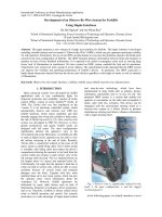
Chapter 1 Evaluation of the Humoral Immune
Response of Wistar Rats Submitted to
Forced Swimming and Treated with Fluoxetine 1
Eduardo Vignoto Fernandes,
Emerson José Venancio and Célio Estanislau
Chapter 2 Effects of Antidepressants on
Inhibitory Avoidance in Mice: A Review 23
Concepción Vinader-Caerols,
Andrés Parra and Santiago Monleón
Chapter 3 Participation of the Monoaminergic System in
the Antidepressant-Like Actions of Estrogens:
A Review in Preclinical Studies 47
Carolina López-Rubalcava, Nelly Maritza Vega-Rivera,
Nayeli Páez-Martínez and Erika Estrada-Camarena
Chapter 4 Antidepressants and Morphological
Plasticity of Monoamine Neurons 73
Shoji Nakamura
Chapter 5 Serotonin Noradrenaline
Reuptake Inhibitors (SNRIs) 91
Ipek Komsuoglu Celikyurt,
Oguz Mutlu and Guner Ulak
Chapter 6 Antidepressants Self-Poisoning in Suicide and
Suicide Attempt: Acute Toxicity and Treatment 109
Sara Santos Bernardes, Danielle Ruiz Miyazawa,
Rodrigo Felipe Gongora e Silva, Danielle Camelo Cardoso,
Estefânia Gastaldello Moreira and Conceição Aparecida Turini
Chapter 7 Rational Polypharmacy in
the Acute Therapy of Major Depression 131
Per Bech and Claudio Csillag
VI Contents
Chapter 8 Antidepressant Drugs and Pain 143
Blanca Lorena Cobo-Realpe, Cristina Alba-Delgado,
Lidia Bravo, Juan Antonio Mico and Esther Berrocoso
Chapter 9 Antidepressant Drug Use
in Patients with Diabetes Mellitus
Type 1 – The Effect of Medication on
Mental Problems and Glycemic Control 163
Jana Komorousová and Zdeněk Jankovec
Chapter 10 Effects of Fluoxetine and Venlafaxine on
the Salivary Gland – Experimental Study 181
Silvana da Silva,
Luciana Reis de Azevedo, Antônio Adilson Soares de Lima,
Beatriz Helena Sottile França, Maria Ângela Naval Machado,
Aline Cristina Batista Rodrigues Johann and
Ana Maria Trindade Grégio
Preface
Depression could be called the black death of the twenty-first century due to its high
prevalence (life time prevalence could be 10-15% or higher). It often occurs in people
during their middle age, 30-50 years old, and costs much because of the medical
resources used to treat it and the higher suicide and rate of recurrence. In addition,
people with depression are often comorbid with anxiety disorders and lack of efficient
treatment. Even for the patients with anxiety disorders, the most useful medications
are antidepressants.
From 1970 to 1990, antidepressants drug delivery has developed rapidly, including
monoamine oxidase inhibitors (MAOIs), tricyclic antidepressants (TCAs),
tetracyclic antidepressants (TCAs), selective serotonin reuptake inhibitors (SSRIs)
and serotonin-norepinephrine reuptake inhibitors (SNRIs), being the most
commonly used. These medications are among the most commonly prescribed
by psychiatrists and other physicians, and their effectiveness and adverse effects
are the subject of many studies and competing claims. As more studies are
carried out more evidence of the other effects of antidepressants have been
reported; antidepressants are no longer anti-depressant/mood only, but provide
other effects.
The editor tried to integrate various aspects of treatment for depression and the
effects of antidepressants. In recent years, more and more researchers are exploring
the mechanisms in psychiatry and psychopharmacology of treating psychiatric
illnesses. Some hypotheses have been challenged through various points of view,
but, the hypothesis on monoamine still plays an important role in treating
depression. From the viewpoint of traditional psychopharmacology, animal models
to clinical trials in humans, a comprehensive review was carried out to understand
the possible pathology of depression. In addition, the other therapeutic effects of
antidepressants, as well as side effects, are also reported in this book. Moreover,
psychotherapy has also been reported to have similar effects, especially
cognitive-behavioural therapy; these treatments are also reported to work for
depression. On the fundamental understanding of pharmacological effects and the
relationship with depression, the therapeutic effect of psychotherapy could be more
applicable.
X Preface
The editor tried to help the readers who are beginners in this field to have a
comprehensive and basic knowledge of antidepressants and further, have inspiration
for their future studies.
Ru-Band Lu
Department of Psychiatry, National Cheng Kung University & Hospital, Tainan,
Taiwan
1
Evaluation of the Humoral Immune
Response of Wistar Rats Submitted to
Forced Swimming and Treated with Fluoxetine
Eduardo Vignoto Fernandes,
Emerson José Venancio and Célio Estanislau
State University of Londrina,
Brazil
1. Introduction
The term stress was introduced into the biomedical field by Hans Selye (1936) in reference to
a General Adaptation Syndrome which would consist of all non-specific systemic reactions
that occur during an intense and chronic exposure to a stressor (e.g., pressure at work and
poor diet). This syndrome would be different from the specific adaptive reactions (such as
muscle hypertrophy caused by exercise performed on a regular basis) and immune
responses (Selye, 1936).
A study evaluating occupational stress in nurses presented the most common symptoms
involved: a feeling of fatigue, headache or muscle pain due to tension (neck and shoulders),
decreased sexual interest, a feeling of discouragement in the morning, sleep difficulties,
upset stomach or stomach pain, muscle tremors, feeling short of breath or shortness of
breath, decreased appetite, tachycardia when under pressure, sweating and flushing
(Stacciarini & Tróccoli, 2004). The main psychological symptoms present in people with
stress are anxiety, tension, insomnia, alienation, interpersonal difficulties, self-doubt,
excessive worry, inability to concentrate, difficulty relaxing, anger and emotional
hypersensitivity (Lipp, 1994).
Stress has been considered one of the biggest causes of depression. After a situation of great
stress, approximately 60% of individuals develop depression. Psychosocial problems (work
pressure, job loss and debt) can also be preconditions for its emergence (Kendler et al. 1995;
Post, 1992).
Major depression is a mood disorder whose prevalence throughout life, depending on the
population, is estimated at between 0.9 to 18% and involves a significant risk of death
(Waraich et al., 2004). It is estimated that men and women with depression are 20.9 and 27
times, respectively, more likely to commit suicide than those without depression (Briley &
Lépine, 2011).
Multiple environmental factors have been associated with the etiology of depression.
Adverse events during childhood and everyday stress are described as important factors for
Effects of Antidepressants
2
the development of depression (Kessler, 1997). Children with a history of sexual abuse, living
in troubled homes or who receive little attention from parents have a high risk of becoming
depressed adults (Kessler, 1997). Stressful events such as the loss of a loved one, job loss, or
partner separation are factors associated with the onset of depression (Kessler, 1997).
Individual personality is also a predisposing factor to depression, as evidenced by the higher
frequency of depression in people with a tendency to be sad when they experience a stressful
event (Fava & Kendler, 2000). Gender is strongly associated with depression. Studies have
shown that depression is on average twice as common in women as in men (Bromet et al.,
2011). Interestingly, a decrease in the female/male proportion of depression has been observed
in young adults (18 to 24 years), possibly due to greater gender equality in today’s society
(Seedat et al., 2009). Besides environmental factors, individual genetic characteristics also
contribute to susceptibility to depression (Jabber et al., 2008).
In addition to the psychological changes associated with depression, immune system
changes are often found in depressed individuals (Altenburg et al., 2002). Several studies
have indicated that stress and depression involve the individual in a chronic process that
results in host defense failure against microorganisms and a higher likelihood of developing
certain cancers. These alterations are probably associated with profound changes in the
functioning of the immune system of individuals suffering from depression (Reiche et al.,
2004; Irwin et al., 2011). Epidemiological and experimental evidence shows that changes in
the defense capability of the individual are related to decreased proliferative capacity of
peripheral blood lymphocytes stimulated with mitogens in vitro (Schleifer et al., 1985;
Schleifer et al., 1996), a decrease in the cytotoxic activity of natural killer cells (NK) (Schleifer
et al., 1996; Calabrese et al., 1987; Nunes et al., 2002), the suppression of T-cell activity due to
increased apoptosis and decreased cell proliferation in response to antigens (Szuster-
Ciesielski et al., 2008; Schleifer et al., 1984). Moreover, imbalance in cytokine levels is often
observed, such as increased levels of interleukin 2 (IL-2), interleukin 6 (IL-6) and interferon-
alpha (IFN-α) (Seidel et al. 1995; Vismari et al., 2008). The results have been conflicting
regarding humoral immune response and immunoglobulin levels in the blood. A significant
increase in IgM levels in patients with depression was observed by Kronfol (1989) and Song
et al. (1994), although other studies have been unable to detect significant changes in
immunoglobulin levels in the peripheral blood of patients with depression (Bauer et al.,
1995; Nunes et al., 2002). These changes in the immune system probably directly and/or
indirectly compromise host immunity against microorganisms (Miller, 2010). On the other
hand, the immune system changes observed in individuals with depression may not be
caused by changes in the central nervous system of these individuals but instead may be
directly related to the origin of such changes, including the development of a pro-
inflammation state directly related to the onset of a depressive state, which is suggested by
the hypothesis that macrophages act as a cause of depression (Miller, 2010). This hypothesis
is related to an increased secretion of proinflammatory cytokines such as interleukin 1 (IL-1),
IFN-α, and the resulting change in production of corticotrophin-releasing factor (CRF) and
adenocorticotrophic hormone (ACTH) (Smith, 1991).
Importantly, animal models of stress and depression have shown immune system changes,
including increased production of IL-1, the number of circulating neutrophils and lowered
resistance to infection by bacteria. Mice that had been transgenically modified to exhibit a
depressive type of behavior (catalepsy) and were inoculated with sheep red blood cells
Evaluation of the Humoral Immune Response of Wistar
Rats Submitted to Forced Swimming and Treated with Fluoxetine
3
(SRBC) had lower amounts of platelet-forming cells and antigen-specific T lymphocytes
than their parents without this disorder. In rats with high levels of anxiety, lower
concentrations of specific T lymphocytes were also found five days after inoculation with
SRBC (Kubera et al., 1996; Pedersen & Hoffman-Goetz, 2000; Altenburg et al., 2002; Robles et
al., 2005; Alperin et al., 2007; Loskutov et al., 2007; Miller, 2010).
Because this disorder severely compromises the functioning of individuals, several
alternative treatments for depression have been proposed, including psychotherapy and
pharmacotherapy, as well as a combination of both types. The use of antidepressant drugs
for treating patients with depression began in the late 1950s. Since then, many drugs with
potential antidepressants have been made available and significant advances have been
made in understanding their possible mechanisms of action (Stahl, 1997). Only two classes
of antidepressants were known until the 80's: tricyclic antidepressants and monoamine
oxidase inhibitors. Both, although effective, were nonspecific and caused numerous side
effects (Lichtman et al., 2009). Over the past 20 years, new classes of antidepressants have
been discovered: selective serotonin reuptake inhibitors, selective serotonin/norepinephrine
reuptake inhibitors, serotonin reuptake inhibitors and alpha-2 antagonists, serotonin
reuptake stimulants, selective norepinephrine reuptake inhibitors, selective dopamine
reuptake inhibitors and alpha-2 adrenoceptor antagonists (Bezchlibnyk-Butler & Jeffries,
1999). Serotonin reuptake inhibitors belong to this new generation of antidepressant drugs;
fluoxetine is the most commonly prescribed drug for treating depression and anxiety
because of its efficacy, safety and tolerability (Egeland et al., 2010).
Despite the current extensive use of antidepressant drugs, few studies have investigated the
effects of antidepressant drugs on the immune system (Janssen et al., 2010). Experimental
and clinical evidence suggests that changes in the immune system in patients with
depression can be reversed by the use of antidepressant drugs (Leonard, 2001).
In animal models the use of fluoxetine has been associated with significant changes in
immunity. Laudenslager & Clarke (2000) inoculated rhesus monkeys (Macaca mulatta) with
tetanus toxoid and found increased levels of IgG anti-tetanus. When analyzing the effect of
the antidepressant desipramine and fluoxetine, it was observed that animals treated with
these antibodies showed higher plasma levels than those treated with saline.
Some studies with mice have showed the effects of fluoxetine on humoral immune response.
Kubera et al. (2000) observed that continuous administration of fluoxetine in C57BL/6 mice
for four weeks results in decreased IL-4 production and in increased IL-6 and IL-10
production. Genaro et al. (2000) found that fluoxetine has an inhibitory action on the
proliferation of B lymphocytes induced by lipopolysaccharide (LPS) or anti-IgM. On the
other hand, fluoxetine increases the proliferative action of B lymphocytes, being stimulated
by suboptimal concentrations of anti-IgM. In an experimental model of depression in
BALB/c, Edgar et al. (2002) observed a decrease in lymphoproliferative response induced by
mitogens (phytohemagglutinin and concavalina A), an increase in the proliferative response
of B lymphocytes to lipopolysaccharide (LPS) and that the chronic administration of
fluoxetine reverses these immune changes.
The experimental investigation of depression in humans is largely ethically unfeasible.
Thus, animal models of depression have been developed for this purpose, such as the
Effects of Antidepressants
4
olfactory bulbectomy, learned helplessness, restraint stress and forced swimming (Willner,
1990). Forced swimming is a widely used model for preclinical evaluation of the possible
effects of antidepressant drugs (Porsolt et al., 1977). Its widespread use is mainly due to its
ease of implementation, the reliability of its results confirmed in various laboratories and its
ability to detect the action of almost all classes of currently available antidepressants (Borsini
& Meli, 1988).
In this study we evaluated the humoral immune response of rats chronically submitted to a
model of stress/depression, i.e., forced swimming for twenty-five days and daily treatment
with fluoxetine. Antibody production was assessed five days after the rats were inoculated
with sheep red blood cells and, after the last day of forced swimming, the animals were
euthanized and the adrenal glands, thymus and spleen were removed and weighed.
A growing number of people are diagnosed with stress and depression, for which
antidepressant drugs are increasingly prescribed. Although many of their effects on
individuals are known, there have been few studies reporting the effects of antidepressants
on human and/or animal immune systems, especially regarding humoral immunity.
Although experimental, this study has great social significance principally due to the large
number of people vaccinated annually who are also undergoing regular treatment with
antidepressants. The objective of this study was to evaluate the humoral immune response
of Wistar rats submitted to forced swimming and treated with fluoxetine.
2. Methodology
2.1 Animals and experimental groups
A sample of 72 male Wistar rats with a body mass of about 300 grams was obtained from
the Central Vivarium of the State University of Londrina’s Center of Biological Sciences for
use in the experiment.
The experiment was conducted at the vivarium of the Department of General Psychology
and the Behavior Analysis Center of Biological Sciences of the State University of Londrina.
The rats were housed in polypropylene cages (40 cm x 34 cm x 17 cm) with up to six animals
per cage. Water and feed were provided ad libitum throughout the experiment, the
vivarium temperature was maintained at approximately 25°C and a 12 hour light/dark
cycle was established (light from 7:00 am). The animals’ body weight was measured daily
before the forced swimming session.
In order to study the effects of chronic forced swimming, chronic fluoxetine treatment and
an immunization protocol, roughly half of the animals were submitted to chronic forced
swimming sessions and the rest were kept in the vivarium. Each of these groups was
subdivided and treated chronically with fluoxetine or saline. Again, each of the four groups
was subdivided with part of the animals submitted to the immunization protocol and the
other part not. Thus, the following eight groups were involved in the procedure: control
saline not immunized (Ctl-Sal-n-Im, n=10); control saline immunized (Ctl-Sal-Im, n=10);
control fluoxetine not immunized (Ctl-Fxt-n-Im, n=9); control fluoxetine immunized (Ctl-
Fxt-Im, n=9); swimming saline not immunized (Swm-Sal-n-Im, n=10); swimming saline
immunized (Swm-Sal-Im, n=10); swimming fluoxetine not immunized (Swm-Fxt-n-Im,
n=7); swimming fluoxetine immunized (Swm-Fxt-Im, n=7).
Evaluation of the Humoral Immune Response of Wistar
Rats Submitted to Forced Swimming and Treated with Fluoxetine
5
The experimental procedures were approved by the Ethics Committee on Animal
Experimentation of the State University of Londrina, Project No. 6977, Case No.
16828/2010.
2.2 Protocol of forced swimming
The forced swimming model was performed in accordance with Lucki (1997) to evaluate the
acute effect. In the current study, forced swimming sessions were performed daily for
twenty-five days and the behavior of the animals was rated on the first and last day. Forced
swimming was performed in a black plastic cylinder (50 cm high and 22 cm in diameter) in
which the water was 30 cm deep and kept at 25 ± 2°C. The sessionss were performed
individually for 15 minutes between 12 and 2 pm. At the end of the session, each animal was
removed from the cylinder and dried. The cylinder was cleaned and the water replaced
between use by different groups.
2.3 Fluoxetine: Dilution and application
We used the drug Daforin® (fluoxetine hydrochloride 20mg/ml) diluted 1:2 in saline
solution for the experiment. Thirty minutes after the end of each forced swimming session,
the animals received 10 mg/kg/day of fluoxetine or saline intraperitoneally (i.p.). The
injections began at the first session (pretest) and finished on the penultimate day of the
experiment (the 24th day).
2.4 Behavioral evaluation
For behavioral analysis, the animals were filmed during the first five minutes of the 1st and
the 25th session of forced swimming. After the tests, the videos were stored on a computer
for further analysis.
The amount of time the animals spent in the following behaviors was recorded: floating
(complete immobility or faint movements, i.e., the minimum necessary to keep the
nose/head above the surface), climbing (vigorous movements with forepaws above the
surface or against the cylinder wall) and swimming (horizontal movement without the front
legs breaking the surface of the water). The behavioral data were recorded by a trained
observer (minimal intra-observer agreement: 0.85).
2.5 Blood collection and immunization
On days 5, 10 and 25 of the study at the end of the forced swimming session, all animals
were sedated by non-lethal inhalation of ethyl ether and approximately 1 mL of blood was
collected by cardiac puncture. The collected blood was stored in 1.5 ml plastic tubes
containing 50 μL of 5% EDTA. On days 5 and 20 the animals belonging to subgroups Ctl-
Sal-Im, Ctl-Fxt-Im, Swm-Sal-Im and Swm-Fxt-Im, were inoculated i.p. with a 250 μl solution
of 2.5% SRBC.
2.6 Preparation of antigen
The following protocol was used to extract proteins from sheep erythrocytes: the sheep red
blood cells were centrifuged in test tubes at a speed of 1000g for 15 minutes. The cell pellet
Effects of Antidepressants
6
was then suspended in saline, centrifuged at 1000g for 15 minutes and the leukocyte layer
was removed (this process was repeated twice more). After the third wash, the supernatant
was removed and 30 ml of Tris-EDTA [5 mM buffer 2-Amino-2-hydroxymethyl-propane-
1,3-diol/hydrochloric acid (Tris-HCl), pH 7.6, containing 1 mM Ethylenediamine tetraacetic
acid (EDTA)] was added. The tubes were subjected to centrifugation at 25000g for 30
minutes (this process was repeated until the supernatant had turned pink). The contents of
the tubes were then filtered through cheesecloth and underwent a final wash with Tris-
EDTA. The pellet obtained was suspended in 0.1% Sodium Dodecyl Sulfate (SDS) in
Phosphate Buffered Saline (PBS) at a volume three times that of the pellet. The suspension
was dialyzed for 24 hours at room temperature and the PBS/SDS solution was changed at
least twice. Aliquots of the suspension were stored at -20°C. The protein suspension dosage
followed Bradford (1976).
2.7 Sacrifice
On the 25th day of study, after finishing the forced swimming test, the animals were again
non-lethally sedated by inhalation of ethyl ether for blood collection, after which the animals
were sacrificed by lethal ethyl ether inhalation. The spleen, thymus and adrenal glands of
each rat were subsequently removed to assess the relative weight.
2.8 ELISA
To assess the production of antibodies (IgM, IgG1 and IgG2a), an enzyme-linked
immunosorbent assay (ELISA) containing 100 µl of a solution of 2.5 mg/ml sheep
erythrocyte proteins obtained in the above-described manner was added to each well. The
plasma was diluted 1:100. The dilutions of peroxidase conjugated anti-IgM, anti-IgG1
(Zymed) and anti-IgG2a (BETHYL) were 1:10000, 1:20000 and 1:5000, respectively.
ELISA was conducted according to the following protocol: first, the 96-well plates were
coated with 100 µl of the antigen diluted in carbonate-bicarbonate pH 9.6 and incubated
overnight at 4°C. The plates were then washed 3 times with PBS-Tween 0.05% and blocked
with 150 µl of PBS with skim milk (PBS-milk) 5% in each well for 1 h at 25°C. After 3 washes
with PBS-Tween 0.05%, plasma samples diluted in PBS-milk 1% (100 µl of 1:100 diluted
sample per well) were incubated for 1 h at 25°C. The plates were then washed 3 times with
PBS-Tween 0.05% and the conjugate (100 µl of conjugate diluted in PBS-milk 1% per well)
anti-IgM, anti-IgG1, or anti-IgG2a was incubated for 1 h at 25ºC, washed 3 times with PBS-
Tween 0.05%, and then the substrate (sodium acetate buffer 0.1 M pH 5, containing TMBZ –
tetramethylbenzidine of 1% and H
2
O
2
- hydrogen peroxide 0.005%) was added (100 µl of
substrate/well). After incubation in the dark for 15 minutes at 25°C, 50 µl of 1N H
2
SO
4
was
added per well. Reading was performed in a microplate reader at 450 nm.
2.9 Statistical analysis
Statistical analysis was performed with Statistica 5.0®. To evaluate homogeneity and
normality, the Levene and Kolmogorov-Smirnov tests were used. To evaluate antibody
production (IgM, IgG1 IgG2a), four-way repeated-measures ANOVA was performed
including the effects of the swimming sessions (Ctl X Swm), fluoxetine treatment (sal X fxt),
immunization (n-Im X Im) and repeated measurement factor of blood sampling time
Evaluation of the Humoral Immune Response of Wistar
Rats Submitted to Forced Swimming and Treated with Fluoxetine
7
(preImmunization X after the 1st immunization X after the 2nd immunization). Behavioral
comparisons were also performed by means of four-way ANOVAs, but with a different
repeated-measures factor (Session 1 x Session 25). Repeated-measures comparisons of the
following masses were conducted: body (fluctuation), spleen, adrenal gland and thymus.
Therefore, the above described remaining factors were analyzed in three-way ANOVAs run
for this purpose. When interactions of main effects were found to be significant, Tukey post
hoc tests were applied. The significance level was set at P <0.05.
3. Results
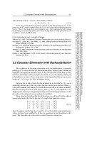
Figure 1a shows the production values of IgM antibody groups. The results show no effects
for stress (F [1.64] = 0.348, P> 0.05), but effects for immunization (F [1.64] = 20.050, P <0.001),
drug (F [1.64] = 6.673, P <0.05), time (F [2.128] = 32.208, P <0.001), interaction between
immunization and time (F [2.128] = 21.710, P <0.001), drug and time (F [2.128] = 7.383, P
<0.001) and immunization, drug and time (F [2.128] = 9.268, P <0.001). Comparing the pre,
post1 and post2 immunization periods, the Tukey test showed that there was an increase in
IgM production only for the Ctl-Sal-Im and Swm-Sal-Im groups. We observed that only
animals treated with saline responded to inoculation with SRBC, while fluoxetine inhibited
the production of antibodies.
The production of IgG2a antibody (Figure 1b) appeared to be similar to the values observed
for IgM. Four-way ANOVA showed no stress effect (F [1.64] = 1.188, P> 0.05), but effects for
immunization (F [1.64] = 26.326, P <0.001), drug (F [1.64] = 7.139, P <0.05), time (F [2.128] =
25.483, P <0.001) , immunization and drug interaction (F [1.64] = 7.814, P <0.01),
immunization and time (F [2.128] = 25.734, P <0.001), drug and time (F [2.128] = 6.578, P <
0001) and immunization, drug and time (F [2.128] = 6.630, P <0.01). In the pre, post1 and
post2 immunization periods, the Tukey test showed increased production of IgG2a only in
Ctl-Sal-Im and Swm-Sal-Im. Only non-stressed animals treated with saline responded to
inoculation with sheep red blood cells, while fluoxetine inhibited the production of
antibodies.
Figure 1c shows the production values for IgG1 antibody. There were no effects for stress (F
[1.64] = 0.404, P> 0.05) drug (F [1.64] = 0.001, P> 0.05), but effects for immunization (F [1.64]
= 48.908, P <0.001), time (F [2.128] = 81.116, P <0.001), interaction between stress and drug (F
[1.64] = 9.370, P <0.01), immunization and time (F [2.128] = 67.428, P <0.001), stress,
immunization and drug (F [1.64] = 11.223, P <0.01), stress, drug and time (F [2.128] =
18.953, P <0.001) and stress, immunization, drug and time (F [2.128] = 20.187, P <0.001).
Comparing the pre, post1 and post2 immunization periods, an increase in IgG1
production was observed only for the Ctl-Sal-Im and Swm-Fxt-Im groups. It was observed
that stress and fluoxetine in isolation inhibit the production of IgG1, but that stress and
drugs together interacted to cause antibody production similar to that of the control
group (Ctl-Sal-Im).
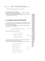
The variation in rat body mass was not altered by immunization (F [1.64] = 0.34, P> 0.05),
although stress (F [1.64] = 19.948, P <0.001) and drug effects (F [1.64] = 111.595, P <0.001)
were observed. There was no significant interaction between variables. Intergroup
comparison revealed that fluoxetine was responsible for reducing body mass (Figure 2).
Effects of Antidepressants
8
Fig. 1. Variation (mean ± SEM) in the production of antibody. We analyzed the variation in
the production of antibodies (IgM, IgG2a and IgG1) at three different points in time (pre-
immunization, five days after the first immunization and 5 days after the second
immunization). Fluoxetine was responsible for suppressing the production of IgM (a) and
IgG2a (b). In relation to IgG1 (c), the administration of only stress and fluoxetine impaired
antibody production. However, the interaction between these variables did not impair
production. * Different the pre-immunization and 5 days after the first immunization (P
<0.001); #Different from Ctl-Sal-Im 5 days after the second immunization (P <0.001); º
Different Swm-Sal-Im 5 days after the second immunization (P <0.002).
Evaluation of the Humoral Immune Response of Wistar
Rats Submitted to Forced Swimming and Treated with Fluoxetine
9
Fig. 2. Variation (mean ± SEM) in body mass. It was observed that both fluoxetine and
swimming resulted in reduced body mass. @ Different the saline group that underwent the
same treatment (P < 0.05); § Different from the control group that underwent the same
treatment (P < 0.05).
There was no stress (F [1.64] = 2.660, P> 0.05) or immunization effect (F [1.64] = 0.373, P>
0.05) on the relative mass of the adrenal glands. There was a significant effect for drug (F
[1.64] = 38.558, P <0.001) and interaction between drugs and immunization (F [1.64] = 2.479,
P <0.05). The Tukey test showed an increase in relative mass of the adrenal group Swm Fxt-
n-Im compared to its control Swm-Salt-n-Im (Table 1).
There was no stress effect on the relative mass of the spleen (F [1.64] = 0.728, P> 0.05), but
there was a drug effect (F [1.64] = 19.534, P <0.001, Table 1). Nevertheless, there was no
significant difference between groups in post hoc comparisons.
There was no stress (F [1.64] = 0.276, P> 0.05) or immunization effect (F [1.64] = 0.704, P>
0.05) on relative thymus mass, but a drug effect (F [1.64] = 32.504, P <0.001) and an
interaction between stress and drug (F [1.64] = 7.535, P <0.05) was detected. It was observed
that the drug reduced the relative mass of the thymus in unstressed animals treated with
fluoxetine (Table 1).
Control Swim
Organ Saline Fluoxetine Saline Fluoxetine
n-Im Im n-Im Im n-Im Im n-Im Im
Adrenals 6.6
±
0.6 7.6
±
0.3 9.7
±
0.7 10.0
±
0.8 6.9
±
0.8 7.7
±
0.8 13.1
±
1.6 9.8
±
0.5
Spleen 158.4
±
3.4 150.4
±
5.6 222.2
±
27.7 206.8
±
26.8 143.4
±
5.2 164.0
±
7.6 202.0
±
20.5 189.5
±
18.0
Thymus 66.2
±
4.3 71.5
±
3.8 36.0
±
7.3 36.1
±
5.0 54.9
±
5.1 57.4
±
5.7 42.1
±
7.9 47.2
±
3.4
Table 1. Relative mass of the adrenal glands, spleen and thymus of rats at the end of the
experiment. It was observed that fluoxetine was responsible for changing the relative mass
of the three organs analyzed, with the adrenal glands and thymus increased and the spleen
reduced (P <0.05). Measure (1 = 0.001% of the body mass).
Figure 3a shows the duration of floating behavior. Statistical analysis showed no effects for
immunization (F [1.30] = 0.078, P> 0.05) or drug (F [1.30] = 1.099, P> 0.05) but effects for time
Effects of Antidepressants
10
Fig. 3. Variation in the time of analyzed behaviors. Fluoxetine treatment increased floating
(a) and reduced climbing (c) behavior between Session 1 and 25; no alteration was found in
swimming behavior (b). The animals treated with saline did not show significant alterations
in behavior between sessions. *, significant difference compared to Session 1 (P <0.01). @,
significant difference in the same session compared to the saline group that had been
otherwise submitted to the same treatment (P <0.05).
Evaluation of the Humoral Immune Response of Wistar
Rats Submitted to Forced Swimming and Treated with Fluoxetine
11
(F [1. 30] = 30.010, P <0.001). An interaction between factors occurred only with drug and
time (F [1.30] = 5.989, P <0.05). Comparing the 1st and the 25th session, a reduction was
observed only in the nonimmunized, drug treated group. There was also a distinction
observed between the Fxt-n-Im and Sal-n-Im groups at the 25th session.
For swimming, statistical analysis revealed no effects for immunization (F [1.30] = 0.208, P>
0.05), drug (F [1.30] = 0.861, P> 0.05), time (F [1.30] = 0.563, P> 0.05) or interaction of factors
(Figure 3b).
Figure 3c shows the time of analyzed behaviors. There were no effects for immunization (F
[1.30] = 0.081, P> 0.05) or drug (F [1.30] = 0.091, P> 0.05) and effects for time (F [1. 30] =
32.243, P <0.001). There was an interaction between drug and time (F [1.30] = 5.338, P <0.05).
Comparing the 1st and 25th sessions, an increase in climbing time was detected in the Fxt-
Im group.
4. Discussion
The current study investigated the effects of chronic stress and the administration of the
drug fluoxetine on humoral immune response. It assessed primary and secondary immune
response against sheep red blood cells, variation in body mass and the relative mass of the
adrenal glands, thymus and spleen, as well as the behavior of rats subjected to a daily forced
swimming protocol, which is an model used to assess depression-like behavior in rodents.
In general, stress is considered to be an immunosuppressant. Elenkov & Chrousos (1999)
conducted an extensive review on the influence of stress on the immune system and found
that acute stress produced subacute or chronic immunosuppressive activity on cellular
immune response. On the other hand, stress also was found to have an immunostimulating
effect on humoral immune response. Another literature review Segerstrom & Miller (2004) that
included research from the last 30 years on the effects of stress on immune function in men
and women found no relationship between acute and subacute stress regarding modulation of
humoral immune response. Nevertheless, it was observed that stress is associated with chronic
immunosuppression in that it lowered antibody capacity against an influenza virus.
The ability of stress to inhibit cellular immune response (Th1) is probably related
glucocorticoid and catecholamine suppression of pro-inflammatory cytokines, IL-12, IFN-γ
and TNF-α (Elenkov & Chrousos, 1999). Regarding the suppression of cellular immune
response, several studies have shown that stress can cause a predisposition to autoimmune
diseases (rheumatoid arthritis and type 1 diabetes), allergies (asthma, food allergies and
emphysema), and some types of cancer, including Kaposi's sarcoma and Epstein-Barr virus
associated B-cell lymphomas (Reiche et al., 2004).
On the other hand, the modulation of humoral immune response by stress is a controversial
topic in the literature because studies differ regarding the possible modulation. Baldwin et
al. (1995) submitted rats to a stress regime that can be considered subchronic (forced
swimming for 3-5 days, 60 minutes each session) and found no differences in the production
of anti-sheep red blood cells between stressed and unstressed rats. Besides studies that have
found no increase, others have observed a decrease. Kennedy et al. (2005) submitted rats to
acute restraint stress and found that it did not alter the production of IgG1 (Th2) but
suppressed the production of IgM and IgG2a (Th1) antibodies. Stanojevic et al. (2003)
Effects of Antidepressants
12
verified the effects of shock stress for five days in rats and, after immunization with bovine
serum albumin (BSA), found that there was suppressed production of IgG anti-BSA
compared to controls upon second exposure to the antigen. Hawley et al. (2006) showed that
stress caused by high social competition in birds involves a lower production of anti-sheep
erythrocytes. Rammal et al. (2010) found that anxious mice produced fewer IgA and IgE
antibodies than their nonanxious counterparts, and when both groups were subjected to
restraint stress, it appears that all studied antibodies (IgA, IgE and IgG) were suppressed in
both groups. On the other hand, Guéguinou et al. (2011) analyzed the natural antibodies of
mice subjected to a rotational velocity model (2 and 3 G-force) for 21 days and found an
increase in IgG levels of animals subjected to 2Gs. Thus, it can be inferred that the type and
length of exposure to the stressor has a direct relationship with the modulated production or
elimination of certain antibodies.
In our study, the chronic stress of forced swimming did not interfere in the production of
antibody classes IgM and IgG2a, although the production of IgG1 was suppressed. These
results are similar to those of Kennedy et al. (2005). The modulation of IgG1 antibody
production in mice suggests a suppression of the Th2-type response, which in rats is
associated with the production of antibodies to this class of immunoglobulins. On the other
hand, the results suggest that the Th1 immune response is not affected by forced swimming
since we did not observe a change in the levels of IgG2a antibodies. It is important to note
that the production of antibodies in response to an antigen derived from a complex network
of cellular interactions that involve the production of molecules with opposite effects, such
as cytokine IFN-γ in mice, which has a stimulating effect on cellular immune response and
IgG2a antibody production as well as an inhibiting effect on humoral immune response and
the production of IgG1 antibody, whereas IL-4 has the opposite effect. The fact that the
forced swimming model results in the removal of IgG1 antibodies from production suggests
that, by mechanisms not yet understood, stress results in the modulation of signals involved
in Th2 response without changing the Th1 response. Whereas there is an antagonistic
relationship between IFN-γ and IL-4, these results suggest that the stress-modulated
molecular mechanism does not directly involve the main molecules responsible for
modulation of antibody production. Recent studies have shown that the role of
neurotransmitters in immune system function may be more important than previously
considered (Rosas-Ballina et al., 2011).
Besides the relationship between stress and humoral immune response, we investigated the
action of fluoxetine on this relationship. Although the 25 days of forced swimming in the
present study did not affect the normal production of IgM or IgG2a but inhibited IgG1, we
can speculate that the chronic use of this model may stimulate cellular immune response.
The administration of fluoxetine inhibited the production of all immunoglobulin classes
studied, which shows its general immunosuppressive effect, both for Th1 and Th2.
However, the interaction of forced swimming x fluoxetine normalized the production of
IgG1. This suggests that stress alone diverts the immune response to Th1-type, while
fluoxetine alone has an immunosuppressive effect on humoral immune response. On the
other hand, administration of fluoxetine in animals subjected to forced swimming can
modulate the immune response to a Th2 pattern. A study about the effects of fluoxetine on
humoral immune response showed that mice with rheumatoid arthritis that were treated
with fluoxetine (10 or 25 mg/kg/day) for seven days had no changes in the levels of anti-
collagen antibodies (IgG1 and IgG2a) (Sacre et al., 2010). This result is at odds with the
Evaluation of the Humoral Immune Response of Wistar
Rats Submitted to Forced Swimming and Treated with Fluoxetine
13
findings of this study since the time/effect analysis of fluoxetine showed immunosuppression
of all studied classes of antibodies after twenty-four days of treatment. These results suggest
that the effect of fluoxetine depends on the physiological state of the animal. It is important to
note that fluoxetine administered concomitantly with stress can have an immunostimulatory
effect. Frick et al. (2009) observed that chronic restraint stress in rats causes decreases in CD4 +
T lymphocytes and no change in CD8 + T lymphocyte but when treated with fluoxetine, initial
values of CD4 + T cells were restored. According to Freire-Garabal et al. (1997), stressed rats
treated with fluoxetine had a higher number of circulating lymphocytes than their control
counterparts (stressed and not treated with fluoxetine).
The reduction in specific antibody levels observed in our study is probably related to the
action of fluoxetine on the production of cytokines and B lymphocytes, the cells responsible
for producing antibodies, as has been observed in other studies. Kubera et al. (2000)
demonstrated that the administration of fluoxetine for more than four weeks suppresses the
production of IL-4, the main stimulus for differentiating T helper cells into Th2 cells. The
decrease in Th2 production may influence isotype synthesis or immunoglobulin levels.
Regarding the plasma level of antibodies, Laudenslager & Clarke (2000) observed an
increase in immunoglobulin class IgM and IgG and a decrease in the levels of specific IgG
antibodies against the tetanus toxoid immunogen in monkeys (Macaca mulatta). Therefore,
fluoxetine can induce an increased level of total Ig and a decreased level of specific
antibodies. However, Sluzewska et al. (1995), studying depressed patients treated with
fluoxetine, showed a decrease in IL-6, the cytokine responsible for the growth of B
lymphocytes, which differentiate into antibody producers. Moreover, Genaro et al. (2000)
observed that fluoxetine had an inhibitory effect on the proliferation of B lymphocytes that
had been stimulated by LPS.
The immunosuppressive action of fluoxetine cannot be restricted to the production of
antibodies. Pellegrino & Bayer (2002) observed that the in vitro proliferation of lymphocytes
from rats that had received fluoxetine via i.p. (5 mg/kg) was lower than their respective
controls, suggesting that the antidepressant has an immunosuppressive role for
lymphocytes. Fazzini et al. (2009) found that three weeks of continued fluoxetine use in rats
triggered an increase in CD8 + T lymphocytes and reduced CD4 + T cells.
The immunomodulatory action of fluoxetine probably involves the participation of
cytokines. Patients with major depression have high levels of IL-6, and treatment with
fluoxetine for 8 weeks leads to normalization of the cytokine levels (Nishida et al., 2002).
Frick et al. (2008), studying cancerous rats, observed that fluoxetine treatment has a direct
relationship with increased production of anti-tumor cytokines (IFN-γ and TNF-α), which
resulted in lower rates of tumor growth and, therefore, longer survival time. On the other
hand, Roumestan et al. (2007) found that fluoxetine had an anti-inflammatory effect (5, 10,
15 and 20 mg/kg) when rats were treated thirty minutes prior to inoculation with LPS and
reported reductions of 60% in TNF-α levels and 50% in mortality compared to controls.
Sacre et al. (2010) also observed that fluoxetine had an anti-inflammatory effect in rats with
rheumatoid arthritis that were treated with 25 mg/kg for seven days, as reflected in reduced
levels of IL-12 and joint damage. On the other hand, some studies have failed to show a
relationship between fluoxetine and the modulation of cytokine production (Kubera et al.,
2004; Maes et al. 1995; Jazayeri et al., 2010). Grundmann et al. (2010) treated rats orally with
10 mg/kg/day of fluoxetine for 21 days and observed no changes in the production of
proinflammatory cytokines (IL-6 and TNF-α).
Effects of Antidepressants
14
The production of pro- and anti-inflammatory cytokines due to stress plus fluoxetine is
dependent on the type of stress and route of drug administration. Sprague-Dawley strain
rats, after 21 days of restraint stress and chronic oral treatment with fluoxetine (10 mg/kg),
showed lower production of IL-6 than stressed-only animals, although TNF-α levels
increased, reaching values similar to those of untreated stressed animals (Grundmann et al.,
2010). On the other hand, Kubera et al. (2006) pre-treated rats with imipramine (5 mg/kg) 1,
5 and 24 hours before forced swimming and found that the splenocytes of treated animals
produced more IL-10 than controls (stressed and treated with vehicle), with no IFN-γ
differences observed in any group. Rogoz et al. (2009) treated rats 1, 5 and 24 hours before
forced swimming with 10 mg/kg of fluoxetine i.p. and observed that the interaction
between stress and fluoxetine did not alter the splenocyte production of IL-10 or IFN-γ.
There are reports that the chronic administration of fluoxetine either causes weight loss
(Wellman et al., 2003) or prevents weight gain (Gutierrez et al., 2002). In the present study,
the chronic administration of fluoxetine led to a reduction in body mass when compared
with saline treatment; this reduction was more pronounced when the animals were treated
with the drug and subjected to forced swimming. First et al. (2011) treated rats for five
weeks with fluoxetine (5 mg/kg) and observed reduced body mass. However, when they
were treated with the drug and submitted to chronic stress with multiple stressors,
fluoxetine prevented weight loss due to this protocol. Zafir & Banu, (2007) also observed
weight maintenance by chronic administration of fluoxetine in animals subjected to restraint
stress. It is important to point out that the above-mentioned studies differed in the degree of
stress generated. In this study 15 min/day of forced swimming did not prevent the animals
from gaining weight, but First et al. (2011), using various types of stressors for five weeks
and Banu & Zafir (2007), using four hours of restraint stress, observed reduced body mass.
Thus, combined multiple stressors or prolonged restraint seem to be more stressful than
forced swimming. Considered jointly, these studies indicate two seemingly opposite effects
of fluoxetine: in the presence of severe stressors known to induce mass reduction, the drug
prevents such losses, while in the presence of mild stressors, the drug leads to weight loss,
which suggests an anorexic effect. Human studies have confirmed the anorectic effect of
fluoxetine in that reductions in body mass from the chronic administration of fluoxetine
were observed in obese individuals (Wise, 1992).
Stress affects the mass of the adrenal glands and lymphoid organs such as the thymus and
spleen. Baldwin et al. (1995) investigated the effects of forced swimming (3 to 5 days, sixty
minutes per session) and found that the number of rats housed together (one or five)
influenced the relative masses of the adrenal glands, spleen and thymus, the production of
corticosterone and body mass. They observed that forced swimming, regardless of the type
of accommodation, reduced the spleen, thymus and body mass of animals, but did not alter
the production of corticosterone or the relative mass of the adrenal glands. When the
animals were subjected to social isolation and forced swimming, however, there was
increased corticosterone production and adrenal mass in addition to the above-mentioned
effects, showing that these two models administered separately do not lead to stress, but
together are stressful. Regarding the chronic effect of forced swimming, Zivkovic et al.
(2005a) found that after submitting rats to 21 days of this protocol, the thymus weight of
stressed animals was lower than that of non-stressed animals. In another study by the same
authors (2005b), blood was collected from rats after their final swimming session for
analysis of circulating corticosterone levels and it was observed that, even after 21 days of
chronic forced swimming, corticosterone values remained high.
Evaluation of the Humoral Immune Response of Wistar
Rats Submitted to Forced Swimming and Treated with Fluoxetine
15
In our study, the adrenal gland mass of Wistar rats submitted to swimming (15 min daily for
25 days) did not change, which was a further similarity with the findings of Baldwin et al.
(1995), i.e., body mass reduction in animals submitted to swimming. Our study differed
from the above-mentioned studies in that our stress model did not lead to changes in spleen
or thymus mass. Moreover, Connor et al. (1998) observed no changes in Sprague-Dawley
spleen weight after acute forced swimming, which shows that, depending on the strain and
stress time, body mass values may or may not vary.
Fluoxetine is also responsible for changing the mass of the adrenal glands, spleen and
thymus of rodents. Garabal-Freire et al. (1997) submitted mice to a sound stressor (100 dB, 1
to 3 hours per day, four to twelve days) and observed a decrease in the number of thymic
and spleen cells; this stressor also contributed to a reduction in relative thymus weight, a
condition reversed by treatment with fluoxetine (5 mg/kg). Kubera et al. (2006) treated rats
with three doses of imipramine 1, 5 and 24 hours before forced swimming and found that
acute treatment with this drug did not alter the relative thymus weight, but did reduce
spleen weight. In the present study, 24 days of fluoxetine treatment (10 mg/kg) reduced the
relative thymus weight and body mass of rats and increased spleen and adrenal gland mass.
Thus, either chronic treatment with fluoxetine stressed the animals or the change in relative
adrenal mass is merely a reflection of the change in body mass since the adrenal glands did
not necessarily increase. However, the sudden loss of body mass would have led to this
apparent increase.
The currently-used antidepressants have specific compounds that act on different regions of
the central nervous system, so it is expected that their use in rats or mice would lead to
improvement in depression symptoms, i.e., reduced time floating (passive behavior) and
increased time climbing and/or swimming (active behavior) during a forced swimming
stressor (Piras et al., 2010). However, increases in climbing and/or swimming are dependent
on the type of drug administered (Cryan & Lucki, 2000). Page et al. (1999) observed a
reduction in floating time and an increase in swimming time in rats treated with fluoxetine.
Carr et al. (2010) used fluoxetine in rats (20 mg/kg) three times before forced swimming
yielded similar results. Cryan & Lucki (2000) compared fluoxetine and reboxetine in a rat
forced swimming model and found that both drugs led to reduced flotation time, although
the former increased swimming time and the latter increased climbing time.
To investigate the effects of chronic treatment with fluoxetine (10 mg/kg), Hansen et al.
(2011) treated Wistar rats for 48 days with subcutaneous injections, after which the animals
were subjected to forced swimming. The results showed that the floating, swimming and
climbing times of treated rats were similar to those observed when fluoxetine was
administered 24 hours before the test (acute effect). Pedreañez et al. (2011), studying the
effects of forced swimming as a chronic stressor, carried out fifteen 30-min forced swimming
sessions and found that the animals’ active behavior dropped by 84 percent between the
first and last session.
In our experiment, forced swimming was performed over a chronic period (twenty-five
days). The results were expected to be similar to those in the literature (with test-retest
separated by 24 h) or to those of Pedreañez et al. (2011) for rats subjected to chronic
swimming. We found that, after the chronic treatment period, the time values were different
from those observed when the test and retest were separated by 24 h. Stressed animals
treated with saline had no alterations in floating, swimming or climbing times, indicating