Tài liệu Molecular Neurobiology of Alzheimer Disease and Related Disorders doc
Bạn đang xem bản rút gọn của tài liệu. Xem và tải ngay bản đầy đủ của tài liệu tại đây (3.42 MB, 312 trang )
Molecular Neurobiology of Alzheimer Disease and
Related Disorders
To Megumi Takeda (September 12, 1957 – February 4, 2002)
Molecular Neurobiology
of Alzheimer Disease and
Related Disorders
Editors
Masatoshi Takeda Osaka
Toshihisa Tanaka
Osaka
Ramón Cacabelos Corun
~
a
140 figures, 72 in color, and 18 tables, 2004
Basel · Freiburg · Paris · London · New York ·
Bangalore · Bangkok · Singapore · Tokyo · Sydney
Bibliographic Indices. This publication is listed in bibliographic services, including Current Contents
®
and
Index Medicus.
Drug Dosage. The authors and the publisher have exerted every effort to ensure that drug selection and
dosage set forth in this text are in accord with current recommendations and practice at the time of publication.
However, in view of ongoing research, changes in government regulations, and the constant flow of information
relating to drug therapy and drug reactions, the reader is urged to check the package insert for each drug for
any change in indications and dosage and for added warnings and precautions. This is particularly important
when the recommended agent is a new and/or infrequently employed drug.
All rights reserved. No part of this publication may be translated into other languages, reproduced or
utilized in any form or by any means electronic or mechanical, including photocopying, recording, microcopying,
or by any information storage and retrieval system, without permission in writing from the publisher.
© Copyright 2004 by S. Karger AG, P.O. Box, CH–4009 Basel (Switzerland)
www.karger.com
Printed in Switzerland on acid-free paper by Reinhardt Druck, Basel
ISBN 3–8055–7603–X
Library of Congress Cataloging-in-Publication Data
Molecular neurobiology of Alzheimer disease and related disorders / editors, Masatoshi Takeda,
Toshihisa Tanaka, Ramón Cacabelos
p. ; cm.
Includes bibliographical references and index.
ISBN 3–8055–7603–X (hard cover)
1. Alzheimer’s disease–Molecular aspects. 2. Molecular neurobiology. I. Takeda,
Masatoshi, 1949 II. Tanaka, Toshihisa. III. Cacabelos, Ramón.
[DNLM: 1. Alzheimer Disease–metabolism. 2. Alzheimer Disease–physiopathology. 3.
Alzheimer Disease–genetics. 4. Neurobiology. WT 155 M7183 2003]
RC523.M663 2003
616.8Ј3107–dc22
2003055886
Prof. Masatoshi Takeda
Department of Psychiatry and
Behavioral Proteomics
Osaka University
Graduate School of Medicine
Osaka, Japan
Prof. Ramón Cacabelos
EuroEspes Biomedical Research Center
Institute for CNS Disorders
Bergondo, Coruña
Dr. Toshihisa Tanaka
Department of Psychiatry and
Behavioral Proteomics
Osaka University
Graduate School of Medicine
Osaka, Japan
Contents
VIII Foreword
Nishimura, T. (Osaka)
X Preface
Takeda, M.; Tanaka, T. (Osaka); Cacabelos, R. (Coruña)
1 Methods of Regulating Alzheimer Pathogenesis: Diet, Oxidative
Damage and Inflammation
Cole, G.M.; Morihara, T.; Lim, G.P.; Calon, F.; Teter, B.; Yang, F.; Frautschy, S.A.
(Sepulveda, Calif.)
17 The RNA-Binding Protein Causes Aberrant Splicing of Presenilin-2
Pre-mRNA in Sporadic Alzheimer’s Disease
Katayama, T.; Manabe, T. (Osaka); Imaizumi, K. (Takayama); Sato, N.; Hitomi, J.;
Kudo, T.; Yanagita, T.; Matsuzaki, S. (Osaka); Mayeda, A. (Miami, Fla.);
Tohyama, M. (Osaka)
31 Alzheimer’s ␥-Secretase Mechanism Produces Amyloid--Protein Like
Peptides Simultaneously with Release of Intracellular Signaling
Fragments
Okochi, M.; Fukumori, A.; Satoh, Y.; Aidaralieva, N.; Tanii, H.; Kamino, K.;
Tanaka, T.; Kudo, T.; Takeda, M. (Osaka)
42 Pivotal Role of Neurofibrillary Degeneration in Alzheimer Disease and
Therapeutic Targets
Iqbal, K.; Alonso, A. del C.; El-Akkad, E.; Gong, C X.; Haque, N.; Khatoon, S.;
Tanimukai, H.; Tsujio, I.; Grundke-Iqbal, I. (Staten Island, N.Y.)
V
52 Tau Pathology of Sporadic Tauopathies
Arai, T.; Akiyama, H.; Tsuchiya, K.; Iritani, S.; Ishiguro, K. (Tokyo); Yagishita, S.
(Kanagawa); Oda, T. (Chiba); Odawara, T.; Iseki, E. (Yokohama); Ikeda, K. (Tokyo)
62 Deregulation of GSK-3 and JNK in a Mouse Model of Tauopathy:
A Kinase Combination That Induces Alzheimer-Type Tau
Hyperphosphorylation
Tatebayashi, Y.; Sato, S.; Akagi, T.; Chui, D H.; Miyasaka, T.; Planel, E.;
Murayama, M.; Takashima, A. (Saitama)
71 Clinical Assessment of the Genetic Risk Functions in Alzheimer’s Disease
Kamino, K.; Kida, T.; Takeda, M. (Osaka)
79 Hydrogen Sulfide Is Severely Decreased in Alzheimer Disease Brains
Kimura, H. (Tokyo)
84 Functional Analysis of the Presenilin Complex and ␥-Secretase Activity
Tomita, T.; Takasugi, N.; Tsuruoka, M.; Niimura, M.; Hayashi, I.; Takahashi, Y.;
Morohashi, Y.; Isoo, N.; Tanaka, S.; Sato, C.; Iwatsubo, T. (Tokyo)
94 Pharmacogenomic Studies with a Combination Therapy in Alzheimer’s
Disease
Cacabelos, R.; Fernández-Novoa, L.; Pichel, V.; Lombardi, V.; Kubota, Y. (Coruña);
Takeda, M. (Osaka)
108 Nicotinic Receptor Stimulation Blocks Neurotoxicity Induced by
Amyloid-

via the Phosphatidylinositol-3-Kinase Cascade
Kihara, T.; Shimohama, S. (Kyoto)
123 Involvement of Unfolded Protein Responses in Alzheimer’s Disease
Kudo, T.; Katayama, T. (Osaka); Imaizumi, K. (Takayama); Kanayama, D.; Sowa, M.;
Okochi, M.; Tohyama, M.; Takeda, M. (Osaka)
134 Advances in the Development of Biomarkers for Alzheimer’s Disease –
From CSF Total Tau and Amyloid-(1–42) Proteins to Phosphorylated
Tau and Amyloid--Antibodies
Hampel, H.; Teipel, S.; Faltraco, F.; Brettschneider, S.; Goernitz, A.; Buerger, K.;
Moeller, H J. (Munich)
157 Genetic Analysis of Familial Alzheimer’s Disease in a Japanese
Population
Wakutani, Y.; Adachi, Y.; Wada-Isoe, K.; Yamagata, K.; Urakami, K.;
Nakashima, K. (Yonago)
Contents VI
164 Oxidative Stress in Alzheimer Disease: The Earliest Cytological and
Biochemical Feature
Nunomura, A.; Chiba, S. (Asahikawa); Takeda, A. (Sendai); Smith, M.A.;
Perry, G. (Cleveland, Ohio)
172 Neurogenesis: A Promising Therapeutic Target for Alzheimer Disease and
Related Disorders
Grundke-Iqbal, I. (Staten Island, N.Y.); Tatebayashi, Y. (Saitama); Lee, M.H.;
Li, L. (Beijing); Iqbal, K. (Staten Island, N.Y.)
183 Learning Deficits in N279K Tau Transgenic Mice and an Assembly Model
of Tau Protein
Taniguchi, T. (Himeji); Matsuyama, S. (Kobe); Minoura, K. (Takatsuki);
Iso, H. (Nishinomiya); Sasaki, M. (Himeji); Tomoo, K.; Ishida, T. (Takatsuki);
Mori, H. (Osaka); Tanaka, C. (Himeji)
195 Animal Models of Tauopathies
Ishihara, T.; Nakashima, H. (Okayama)
205 Aberrant Splicing of Tau Transcripts in Frontotemporal Dementia with
Parkinsonism Linked to Chromosome 17
Yamamoto, N.; Kondo, S.; Yoshino, S.; Okumura, M.; Imaizumi, K. (Ikoma)
215 Tau Filament Formation and Associative Memory Deficit in Aged Mice
Expressing Mutant (R406W) Human Tau
Miyasaka, T.; Tatebayashi, Y.; Chui, D H.; Akagi, T. (Saitama); Mishima, K I.;
Iwasaki, K.; Fujiwara, M. (Jonan-Ku); Tanemura, K.; Murayama, M. (Saitama);
Ishiguro, K. (Machida); Planel, E.; Sato, S.; Hashikawa, T.; Takashima, A. (Saitama)
225 Activated Protein Kinases and Phosphorylated Tau Protein in Alzheimer
Disease
Tanaka, T.; Yamamori, H. (Osaka); Wada-Isoe, K. (Tottori); Tsujio, I.; Takeda, M.
(Osaka)
236 A Functional Genomics Approach to the Analysis of Biological Markers in
Alzheimer Disease
Cacabelos, R. (Coruña/Madrid); Lombardi, V.; Fernández-Novoa, L.; Kubota, Y.;
Corzo, L.; Pichel, V. (Coruña); Takeda, M. (Osaka)
286 Epilogue
Cacabelos, R. (Madrid)
289 Author Index
291 Subject Index
Contents VII
Foreword
The dawn of psychogeriatrics in Japan was celebrated with the symposium
entitled ‘Psychiatry for the Elderly’ in the frame of the annual meeting of the
Japanese Society of Psychiatry and Neurology in 1954, on which occasion
Professor Ziro Kaneko (Osaka University), Professor Tadashi Inose (Yokohama
City University), and Professor Naotake Shinfuku (Tottori University) deliv-
ered their lectures on the psychological process of aging, neuropathology of
aging and psychopathology of aging, respectively. The proceedings of the sym-
posium entitled The Psychiatric Aspects of Senility (Igagu-shoin, Tokyo, 1956)
were published as a monograph in Japanese; this was an epoch-making achieve-
ment in Japanese psychiatry because the interest in psychogeriatrics had been
so sparse until then.
In the 1960s, dementia in Japanese elderly people was mainly regarded to
be cerebrovascular dementia. Most Alzheimer’s disease patients were unrecog-
nized and there were only a few case reports of early-onset Alzheimer’s disease.
In those days, basic research in Alzheimer’s disease was confined to neuro-
pathology or histochemistry. Electron microscopy, however, revealed the
unique structure of paired helical filaments in Alzheimer brains, which trig-
gered biochemical research aimed at elucidating the mechanism of paired heli-
cal filament formation. My colleagues at the Department of Neuropsychiatry,
Osaka University and I found that soluble proteins were insolubilized in
Alzheimer brains, which was reported at the International Meeting of
Neuropathology in Budapest in 1974. This report, which attracted considerable
interest and stimulated neurochemical research on the dementia brain in several
VIII
leading institutes, implied that neurochemical or biochemical research could be
successfully applied to elucidate the pathogenesis of Alzheimer’s disease. I am
proud of this contribution of the Department of Neuropsychiatry, Osaka
University which I chaired at that time and I am happy to observe the strong
trend of psychogeriatric research launched by Professor J. Kaneko, as men-
tioned above, and pursued under the leadership of the present chairman,
Professor M. Takeda.
Most of us would agree with the recognition that research activity in the
Department of Neuropsychiatry, Osaka University, has played an important role
in Alzheimer research and the Department achieved a solid reputation as one of
the leading research institutes in psychogeriatrics.
The success of the 21
st
Annual Meeting of the Japanese Dementia Study
Society and the International Symposium on Neurobiology of Alzheimer’s
Disease and Related Disorders in October 2002 appears to be additional evi-
dence for this. The International Symposium, especially, had an impact on
Alzheimer research in this country, gathering many scientists from major
research institutes in Japan and abroad to exchange their research findings. I
would say the program of the symposium was well suited to stimulate young
researchers in this field.
This monograph contains selected papers presented at the symposium and,
just like the monograph The Psychiatric Aspects of Senility published half a
century ago, will certainly contribute to promote scientific research in this
field. It is my pleasure to write a foreword to this book and I would like to con-
gratulate this collaborative achievement of Professors Masatoshi Takeda,
Toshihisa Tanaka, and Ramón Cacabelos, who dedicated their time to this
monograph, cultivating the long tradition of research of the Department of
Neuropsychiatry, Osaka University.
Prof. emeritus of Osaka University Tsuyoshi Nishimura, Osaka
Foreword IX
X
Preface
The average life expectancy of human beings had remained essentially
unchanged since ancient times until the end of the 18th century, and it was only
in those two centuries that our life expectancy increased. Since then, from an
aging society (the elderly exceeding 7%), our society has become an aged
society (the 65-year-olds and over exceeding 14% of the total population). In
Japan this has happened in 2000; thus Japan has transited from an aging to an
aged society in only 24 years, which is the most rapid transition in the world –
almost four times faster than many European countries. The last national census
of Japan reported that the average life expectancy of the Japanese was 85.23
years for females and 78.32 years for males in the year 2002. The Japanese now
enjoy the longest average life expectancy, whereas in 1947 it was only 53.96
years for females and 50.06 years for males. Due to this rapid extension of life
expectancy, Japanese society is now facing strains and problems related to its
high proportion of elderly people (17%), and its very high percentage (7%) of
very old people (above 75 years old).
In many European countries, the increase in the elderly population has
already brought about some changes and modifications in the social life system,
but there are still many things to be implemented to build a new society in
which people can lead mutually cooperative lives regardless of their biological
age. In the 21st century, the elderly population will increase all over the world
because developing countries are showing a more rapid increase in the elderly
population at the present time. By the year 2025, 70% of the elderly will live in
developing countries, and by the year 2050, 80% of the elderly will be found in
present developing countries. These facts indicate that the rapid increase in the
elderly population is a global social problem to be solved by taking advantage
of information and experience from all countries. In a sense, Japan is the top
runner in terms of society aging, and the Japanese experience may serve as an
example to younger societies in other countries.
Alzheimer’s disease is the most malignant disease in aged societies.
It affects 6–10% of the elderly population, causing impairment in cognitive
functions and significant disability in daily living for more than 10 years.
In Japan it affects 750,000 individuals, and by the year 2035, this number will
have increased to 1.5 million.
Neurofibrillary tangles, amyloid deposits and neuronal loss are the three
hallmarks of Alzheimer’s disease. Neurofibrillary tangles and amyloid plaques
are insoluble depositions with unique structural characteristics, abundantly
observed in Alzheimer brains and to some extent in normal aged brains. Due to
the insolubility of these unique structures in Alzheimer brain tissues, they were
difficult to study by usual biochemical methods in the past. In 1980s, owing to
the use of a solubilization method with formic acid or perchloric acid, the neu-
robiological study of Alzheimer’s disease made significant progress. The major
neurobiological findings include partial identification of the amino acid
sequence of amyloid precursor protein (APP) (1984), identification of amyloid
precursor protein gene on chromosome 21 (1987), detection of mutations in APP
with familial Alzheimer’s disease (1991), identification of apolipoprotein E4 as
a significant risk (1993), discovery of presenilin-1 and presenilin-2 (1994).
Some neurobiological research outcomes have been applied in the clinical treat-
ment of patients with Alzheimer’s disease. Acetylcholine esterase inhibitors are
now widely used to treat Alzheimer patients. Tau and beta-amyloid protein lev-
els can be useful as biological diagnostic markers of Alzheimer’s disease.
Active research is going on, aiming to elucidate the pathogenesis of
Alzheimer’s disease. Major topics of neurobiological study of Alzheimer’s
disease include the unraveling of the molecular mechanisms of neurofibrillary
tangle formation in neuronal and glial cells; the molecular processing of
amyloid precursor protein in intracellular organelles and in extracellular space,
and the molecular mechanism of neuronal loss. In this book, these major topics
are covered by leading scientists in the field of neurobiology of Alzheimer’s
disease.
Alzheimer’s disease attracted researchers from diverse academic fields,
including clinical, basic and social sciences. It is essential to promote the
understanding of this formidable disease and to share new findings among
researchers in the field.
Clinical and basic research of Alzheimer’s disease has been the main
interest of the Department of Psychiatry and Behavioral Proteomics,
Preface XI
Osaka University Graduate School of Medicine, since the time of Professor Jiro
Kaneko and Professor Tsuyoshi Nishimura, and it has been our great pleasure
to compile this book as a mile stone of the activity in our Department.
In October 2002, the Department of Psychiatry and Behavioral Proteomics,
Osaka University Graduate School of Medicine organized the 21st Annual
Meeting of the Dementia Study Academy of Japan, in which more than 400
researchers in this field got together to discuss their research progress in clinical
and basic fields of dementia study. The three-day meeting program included
two official symposia entitled ‘Neurobiology of Amyloid and Presenilins’ and
‘Neurodegeneration Mechanism with Tau, Syneclein and Neurofilaments’, two
satellite symposia entitled ‘Early Diagnosis of Alzheimer’s Disease’ and
‘Treatment of Alzheimer’s Disease’, four luncheon seminars of ‘Strategy for
BPSD’, ‘Neuroimaging of Dementia’, ‘Normal Pressure Hydrocephalus’, and
‘Treatment of Vascular Dementia’, in addition to 85 general presentations.
In conjunction with the 21st Annual Meeting of the Dementia Study
Academy of Japan, we organized an International Symposium on the ‘Molecular
Neurobiology of Alzheimer Disease and Related Disorders’. The articles in
this book were selected from papers presented at this two-day International
Symposium, which was very successful, with the participation of eight leading
scientists from the USA, Canada and Europe. They are: Dr. Greg M. Cole
(University of California), Dr. Khalid Iqbal (New York State Institute for Basic
Research), Dr. Peter St. George-Hyslop (University of Toronto), Dr. Konrad
Beyreuther (University of Heidelberg), Dr. Ramon Cacabelos (EuroEspes
Biomedical Research Center), Dr. Harold Hampel (University of Munich),
Dr. Inge Grundke-Iqbal (New York State Institute for Basic Research), and
Dr. Roger M. Nitsch (University of Zurich).
We were very happy to host leading scientists from all over the world and
are thankful to the speakers, and especially to the authors of the articles of this
book, which, we believe, will be useful not only to basic scientists but also to
clinicians interested in Alzheimer’s disease and related disorders.
Masatoshi Takeda, MD, PhD
Toshihisa Tanaka, MD, PhD
Ramón Cacabelos, MD, PhD
Preface XII
October 5, 2002 First day at Hotel Osaka Sun Palace
October 5, 2002
(First row) from left to right
Ramón Cacabelos, Khalid Iqbal, Roger Nitsch, Inge Grundke-Iqbal, Masatoshi Takeda, Konrad Bayreuther, Greg Cole, Peter St. George-Hyslop, Harald Hampel
(Second row)
Takashi Kudo, Tetsuaki Arai, Katsuya Urakami, Katsuhiko Yanagisawa, Nobuo Yanagusawa, Takeshi Tabira, Hiroshi Mori, Tomohiro Miyasaka, Yoshitaka
Tatebayashi, Toshihisa Tanaka
(Third row)
Kouzin Kamino, Masaki Nishimura, Taisuke Tomita, Taiichi Katayama, Kazunori Imaizumi, Hideo Kimura , Shun Shimohama, Hisahi Tanii, Akihiko Nunomura,
Masayasu Okochi
Preface XIII
Preface XIV
October 6, 2002 Second day at Hotel Osaka Sun Palace
October 6, 2002
(First row) from left to right
Ramón Cacabelos, Harald Hampel, Khalid Iqbal, Inge Grundke-Iqbal, Konrad Bayreuther, Yasuo Ihara, Masatoshi Takeda, Roger Nitsch, Greg Cole
(Second row)
Takashi Kudo, Toshihisa Tanaka, Katsuhiko Yanagisawa, Akihiko Takashima, Takeshi Ishihara, Takeshi Tabira, Akihiko Nunomura, Tetsuaki Arai
(Third row)
Yoshitaka Tatebayashi, Taiichi Katayama, Hiroshi Mori, Masaki Nishimura, Tomohiro Miyasaka,Taizo Taniguchi, Kazunori Imaizumi, Masayasu Okochi
Takeda M, Tanaka T, Cacabelos R (eds): Molecular Neurobiology of Alzheimer Disease and
Related Disorders. Basel, Karger, 2004, pp 1–16
Methods of Regulating Alzheimer
Pathogenesis: Diet, Oxidative Damage
and Inflammation
Greg M. Cole, Takashi Morihara, Giselle P. Lim, Frederic Calon,
Bruce Teter, Fusheng Yang, Sally A. Frautschy
VA Medical Center, GRECC, Greater Los Angeles Healthcare System,
Sepulveda, Calif., USA
The Amyloid Cascade in Alzheimer’s Disease – Inflammation,
Oxidative Damage and Synapse Loss
While the most obvious lesions diagnostic of Alzheimer’s disease (AD)
are extracellular -amyloid plaques containing large deposits of aggregated,
fibrillar amyloid- protein (A) and neurofibrillary tangles of filamentous
‘hyperphosphorylated’ -protein, synapse loss appears to be a better and more
proximal correlate of cognitive deficits. AD brain has significant but very
selective neuron loss with a 20–40% loss in cortical synaptophysin or presy-
naptic terminals that progresses throughout the disease and correlates with the
clinical decline. Postsynaptic defects have been less well studied, even though
the loss of the postsynaptic markers neurogranin and drebrin has been reported
to be a much larger 70–80% [1].
While amyloid deposits are widely distributed in AD, neuron loss is much
more limited and better correlated with neurofibrillary tangles in selected
cortical and hippocampal layers; this has led to a longstanding debate on the
significance of tangles versus amyloid. The view that tangles and tau pathology
are the critical events has received new support from the discovery of tau muta-
tions in a subset of frontal temporal dementia (FTD) cases [2]. However, data
from transgenics expressing normal human tau genes have not resolved the
controversy since they lack both neuron loss and tangles, even when
co-expressed with amyloid- precursor protein (APP) mutations. Instead, in
animal models, only the mutant human tau (FTD mutations) is linked to tangle
formation [3, 4], and it is still unresolved whether these mice have neurodegen-
eration equivalent to AD brain.
Because A deposits occur early in AD and Down’s syndrome and increased
A42 amino acid peptide production is a direct consequence of numerous muta-
tions known to cause AD, amyloid peptides are reasonably hypothesized to play
a causal role in the disease [5]. Additional support for this hypothesis has come
from evidence that amyloid peptides under aggregating conditions can be acutely
toxic to cultured neurons from rodents or humans [6]. In essence, the amyloid
hypothesis suggests that elevated A42 leads to excessive accumulation of amy-
loid fibrils or oligomer intermediates that in turn cause neurotoxicity, both
directly as well as via effects on inflammation, oxidative damage and secondary
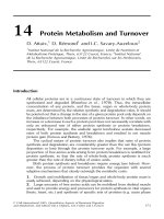
accumulation of intraneuronal phosphotau and synuclein (fig. 1).
Cole/Morihara/Lim/Calon/Teter/Yang/Frautschy 2
Amyloid peptide production
Curcumin lowers cholesterol
Oligomers/fibrils
Curcumin inhibits A aggregation
Amyloid clearance
Curcumin increases CD11c
in phagocytes and
faciliates clearance
Synapse and neuron dysfunction and loss
Curcumin suppresses
A-induced loss of NR2B and PSD-95,
synaptic markers essential for memory
Dementia
Curcumin inhibits
A-induced
memory deficits
Oxidative damage
Curcumin inhibits lipid peroxidation,
scavenges NO-based radicals and
suppresses iNOS mRNA
Intracellular aggregates
ptau, synuclein, A?
Curcumin may potentiate
HSP induction
Inflammation
Curcumin inhibits induction of
pro-inflammatory cytokines
CD11b, iNOS and COX-2 mRNA
Fig. 1. The amyloid cascade and curcumin intervention.
A challenge for the amyloid cascade hypothesis has been that transgenic
mice that develop abundant amyloid pathology, including neuritic plaques, fail
to show tangles or significant neuron loss [7, 8]. At least some of the APP
transgenic mice have ϳ20–30% focal synaptophysin loss, but this has not
been clearly related to memory deficits which typically appear early in the
pathogenesis [9–11].
APP transgenic mice clearly model some amyloid cascade events, particu-
larly amyloid deposition and neuritic plaque formation. Importantly, they have
a microglial/inflammatory response [12], elevated oxidative damage [13, 14],
clear but limited synaptotoxicity [7, 8, 11] and cognitive deficits related to the
A [15–18]. They are therefore a useful model for testing the impact of poten-
tially disease-modifying environmental risk factors and drugs directed at the
amyloid cascade.
Protective Factors Reducing the Risk of Alzheimer’s Disease in
Epidemiological Studies
The identification of autosomal dominant genetic risk factors has been
critical in producing reasonable causal pathways and a hope for new drugs for
treatment. The majority of AD cases are not early onset and autosomal domi-
nant, but are late onset, probably related, in part, to incompletely penetrant
genes, for example, the relatively potent risk conferred by APOE4. That is,
these weak genetic risk factors, do not invariably cause AD by themselves but
require the presence of other risk factors. The most certain of these is aging,
which appears to result in limited AD-like pathology even in nondemented
humans, dogs, some primates and various other mammals, but not in rodents.
While aging is one of the most important risk factors, aging alone is not suffi-
cient, nor is amyloid pathology. AD likely arises from the interaction between
genetic and environmental risk factors, which may offer immediately available
opportunities to reduce disease risk. For minimum risk of toxicity and maxi-
mum public health impact, prevention should focus on evaluation of inexpen-
sive agents with a long history of use.
Nonsteroidal Anti-Inflammatory Drugs
Perhaps the best established protective factor for AD is chronic use of
nonsteroidal anti-inflammatory drugs (NSAIDs) [19, 20], which is consistent
with AD-associated brain inflammation. Amyloid peptide aggregates interact
with multiple microglial receptors and directly cause activation with attendant
increases in toxic cytokines, superoxide and nitric oxide production [21]. A can
also directly activate complement pathways [22, 23]. In addition to A, neuronal
Regulating Alzheimer Pathogenesis 3
injury can activate a secondary inflammatory response. Based on extensive
evidence for an inflammatory response in AD brain involving microglial and
complement activation and cytokine cascades [20, 24], epidemiological methods
were employed to search for evidence of protection afforded by NSAID use that
would support an important causal role for inflammation. Reduced risk was
found not only in arthritis sufferers taking chronic high NSAID doses, but also
in community-based studies where relatively safe and lower, ‘analgesic’doses of
over-the-counter NSAIDs like ibuprofen and naproxen were frequently used
[20, 24, 25]. In a study of twins and sib pairs, NSAID usage, most commonly
ibuprofen, was associated with both reduced risk and delayed onset sufficient to
account for reduced risk [26].
Antioxidants
Increased oxidative damage to proteins, DNA, RNA and lipids occurs in
AD compared to control brains [27] and this damage occurs early [28]. A
aggregates cause oxidative damage to neuronal cultures by elevating hydrogen
peroxide production [29], binding metals [30], forming peptide radicals [31]
or inducing microglial activation [32]. Antioxidants can protect against A
toxicity, suggesting they might slow pathogenesis [33]. In fact, diets rich in
antioxidants [34] and vitamin E in particular [35, 36] appear to reduce AD
risk. Unlike NSAIDs, which appear to affect only disease risk, antioxidants
may also have an impact on progression [36]. However, a clinical trial found
that vitamin E had only a modest (but significant) effect in reducing AD
progression [37].
Statins
Cholesterol has been associated with increased amyloid in the brain [38]
and increased A production [39]. Epidemiological data found increased
dietary fat and cholesterol associated with increased AD risk [40], but follow-up
research on the Rotterdam cohort has not confirmed this result [41].
Consistent with a causal role for cholesterol, several epidemiological studies
associated the use of cholesterol-lowering statins with large reductions in AD
risk [42–44]. Although outside the scope of this review, dietary cholesterol
increases and statins reduce amyloid accumulation in APP transgenic mice
[45, 46].
Dietary Fish and n–3 Fatty Acids
Polyunsaturated fatty acids are important targets for oxidative damage
because they generate lipoxyl radicals as products resulting in autocatalytic lipid
peroxidation with additional aldehyde products that attack macromolecules.
While proteins and nucleic acids are typically considered the ultimate targets,
Cole/Morihara/Lim/Calon/Teter/Yang/Frautschy 4
two of the most peroxidizable fatty acids, arachidonic acid [C20:4 (n–6), AA]
and docosahexaenoic acid [C22:6 (n–3), DHA], play important functional roles.
AA is the major substrate for enzymatic oxidation by the cyclooxygenase and
lipoxygenase pathways. DHA is concentrated in synapses and a potential ligand
for the PPAR and retinoid receptor (RXR, LXR) transcription factors. DHA
also modulates membrane fluidity, membrane enzymes, G protein and channel
activities [47]. Precursors in the n–6 series beginning with linoleic acid are used
to synthesize AA and precursors in the n–3 series beginning with linolenic acid
are used to synthesize DHA. Because of the precursor pathways and their regu-
lation, the absolute dietary levels of AA or DHA are less important than the ratio
of n–6/n–3 fatty acids in regulating inflammatory and cardiovascular pathways.
A dietary intake n–6/n–3 fatty acid ratio of about 4:1 has been considered fairly
optimal. WHO recommends between 3:1 and 4:1 while the Japanese Society for
Lipid Nutrition recommends 2:1 [48]. Elevated n–6/n–3 dietary fat increases AD
risk [49, 50]. Japanese who move to Brazil and eat more meat and less fish have
higher overall dementia and AD rates [51]. High levels of fish consumption have
been associated with reduced risk of age-related cognitive decline [52] and
dementia, including AD [40].
Low DHA has also been associated with AD risk in the US. Those in the
bottom 50% of serum DHA levels in the Framingham study had increased AD
risk of 67%, and in those that also had E4, risk of low scores on the Mini Mental
State Examination rose 400% [53]. Similarly, low dietary intake and low blood
levels of DHA appear to increase risk for AD [54]. However, other studies have
been negative. In our view, it is not only dietary intake deficiency, but focal oxi-
dation leading to local deficiency that is likely to be important in AD. Global
measures of nonenzymatic oxidation of both AA and DHA to isoprostanes are
clearly increased in AD [55–59].
In conclusion, there is both an epidemiological literature and a rationale
for AD risk reduction with NSAIDs, antioxidants, statins and n–3 fatty acids.
Evidence that any of these agents can suppress or delay AD pathology in rodent
models indicates they have a very good chance of working in humans where
they have already been associated with reduced AD risk.
Testing Epidemiological Risk Factors in Animal Models
Amyloid Cascade Interventions – Nonsteroidal Anti-Inflammatory Drugs
Because the strongest epidemiological support for a single protective
NSAID has been for ibuprofen, our first choice was to test the effect of ibupro-
fen in the Tg2576 HuAPPsw line where plaque formation begins at ϳ10 months
and robust plaque-associated microgliosis was evident by 16 months of age [60].
Regulating Alzheimer Pathogenesis 5
Chronic treatment (from 10 to 16 months old) with 375 ppm ibuprofen
reduced plaque burden and levels of sodium dodecyl sulfate-insoluble, formic
acid extracted, total A measured with ELISA by 40–50%. Interleukin-1 and
GFAP protein levels were significantly reduced. Ibuprofen also reduced
microglia-stained area per plaque, a marker of dystrophic neurites (ubiquitin),
and caspase activation per plaque [61], suggesting that it had an impact not only
on amyloid, but on the peri-plaque response to amyloid as well. Other studies
have also found that ibuprofen or other NSAIDs can limit amyloid accumula-
tion [62], raising the issue of the mechanism of amyloid reduction. Ibuprofen
can prevent or reverse age-related cognitive deficits (water maze) in APPsw
mice [Hsiao-Ashe et al., unpubl. obs.]. These studies demonstrate a potential
for ibuprofen as an effective intervention.
Ibuprofen and Amyloid Reduction
In our initial efforts to determine a mechanism for the impact of ibuprofen
on amyloid we ruled out effects on APP levels [61]. However, it is important to
note that because the transgene is driven by the prion promoter, several possi-
ble NSAID effects on APP expression will be absent in the transgenic model
but might be significant in AD patients. Amyloid may be cleared by microglial
phagocytosis via scavenger receptors and ibuprofen could stimulate this by
increasing CD36 receptors via PPAR-␥. Alternatively, amyloid-activated com-
plement pathways, leading to C3b/iC3b opsonization could also enhance clear-
ance through CD11 receptors [63]. However, ibuprofen had no effect on the
level of expression of CD36, C1q or CD11 [Morihara et al., unpubl. obs.].
In summary, we found no evidence to support an effect of ibuprofen on amyloid
clearance.
Evidence was recently discovered, indicating a novel COX-independent
mechanism of action for selected NSAIDs, including ibuprofen, via reducing
A42 production [64]. We confirmed in vitro that A42 production in HEK293
cells expressing APPsw was selectively reduced by ibuprofen, and was also
reduced by profen R-enantiomers (R-ibuprofen and R-flurbiprofen) with weak
COX inhibitory activity [65]. These results are entirely consistent with data
from Koo and Golde [64], suggesting that A42 reduction does not require
COX inhibition. R-flurbiprofen or related compounds with little or no COX
inhibitor activity have the potential to be used at high enough doses to chroni-
cally limit A42 production in the absence of significant side-effects. Whether
ibuprofen itself can be used for this purpose is not yet clear. The in vitro doses
required for this are readily attained in plasma, but may be difficult to achieve
in brain, suggesting that the A42-lowering activity of ibuprofen will be
restricted by dose-limiting toxicity. Nevertheless, in vivo evidence for selective
A42 reduction by NSAIDs [64] argues that an effect on gamma secretase
Cole/Morihara/Lim/Calon/Teter/Yang/Frautschy 6
activity may slow amyloid accumulation. It is also possible that amyloid sup-
pression in vivo may involve other, more traditional NSAID targets related to
COX inhibition and control of the inflammatory response in glia.
One possibility we have examined is suppression of the astrocyte-derived,
pro-amyloidogenic proteins ␣
1
-antichymotrypsin (ACT) and apolipoprotein E
(ApoE). Both of these factors bind A in AD brain and regulate amyloid
formation in vitro and in vivo [66, 67]. We have observed significant reductions
in mRNA for ApoE in ibuprofen-treated transgene-negative animals [Teter et al.,
unpubl. obs.] and in a murine homologue for ACT in ibuprofen-treated
transgene-positive animals [Morihara et al., submitted]. These actions may
require only relatively low, COX-inhibitory doses, and may be shared with
naproxen and other NSAIDs that do not reduce A42 production, but which
appear to reduce AD risk.
Amyloid Cascade Interventions – Antioxidants
Because high dose vitamin E treatment had a modest impact on AD
progression, we sought to test a potentially more potent antioxidant interven-
tion and set up a screen of potential treatments using a rat CNS A-infusion
model [68, 69]. We chose to test curcumin, a well-studied and purified
compound from the turmeric spice that is a potent antioxidant and scavenger
of OH, O
2
Ϫ
and NO radicals. Curcumin inhibits brain lipid peroxidation
(associated with -aggregation) [70] 5–10 times better than ␣ tocopherol
(vitamin E) and is more effective in scavenging NO-based radicals [71] associ-
ated with ␣-synuclein pathology [72]. In addition to curcumin itself, whose
CNS bioavailability is limited by glucoronidation, metabolites including tetra-
hydrocurcumin, ferulic acid and vanillin are also potent antioxidants that likely
contribute to in vivo antioxidant activity [73]. Unlike vitamin E, chronic
administration of the major tetrahydrocurcumin metabolite from 13 months of
age can significantly extend both mean and maximum life span in male
C57Bl/6 mice [74]. Curcumin is a novel anti-inflammatory that controls
inflammation by inhibiting AP-1- and NFB-driven expression of cytokines
[75], iNOS [76] and Cox-2 [77]. It is tolerated well at chronic high doses and
was screened by the US National Toxicology Program prior to making the
short list of compounds under consideration for cancer chemoprevention by
the US National Cancer Institute [78]. Its safety at active doses is indicated by
the long history of use as a turmeric extract for multiple indications in tradi-
tional Indian (Ayurvedic) and Chinese medicine for thousands of years,
notably for promoting wound healing and control of inflammation. Like
aspirin, which was also discovered as a traditional anti-inflammatory medical
extract, curcumin has more than one beneficial effect. These data suggested
curcumin and related species (curcuminoids) and metabolites might afford
Regulating Alzheimer Pathogenesis 7
greater protection than vitamin E by controlling inflammation and oxidative
damage and promoting CNS lesion ‘healing’.
Because of the strong preclinical safety and broad-spectrum efficacy data
and identified structure, we chose to first test curcumin in a rat A infusion
model [69]. This model is also being used to test the efficacy of steroids [79],
vitamin E and other antioxidants and antioxidant cocktails [Frautschy et al.,
unpubl. obs.]. Results indicated that dietary curcumin at 2,000 ppm was a
potent anti-A compound in vivo. More detailed follow-up studies in the
A infusion model showed 500 ppm dietary curcumin reduced lipid peroxida-
tion (F2-isoprostanes), reduced A deposition by 80% and prevented post-
synaptic marker (NR2B, PSD-95) loss and A-induced cognitive deficits in
acquisition in the Morris water maze [69]. While there was a reduction in
plaque-independent microglia, consistent with an anti-inflammatory activity,
we also saw an increase in the microglial response to the diffuse plaques. We
then sought to confirm and extend these results in the Tg2576 APPsw mouse,
using the same 10–16 months of age treatment protocol used for the ibuprofen
study. In this model, 160 ppm dietary curcumin reduced oxidized protein
(measured as carbonyls) by 50–70%, interleukin-1 by 57% and A burden
and A levels (by ELISA) by 43–50% [13]. Similar to the results in the rat
infusion model, microgliosis was also inhibited by 33% in neuron layers, but
microgliosis was stimulated by 250% adjacent to plaques.
Amyloid Cascade Interventions – Mechanism of Curcumin
Inhibition of b-Amyloidosis
The reduction in A accumulation by curcumin observed in both mouse and
rat models might be due to reductions in A production or aggregation, or to an
increase in clearance. Curcumin did not reduce total A or A42 production in
HEK293 cells in vitro, but because curcumin can lower plasma and tissue
cholesterol [80, 81], it may be able to indirectly lower A production in vivo.
Nevertheless, the observation that the drug reduced exogenously infused A
accumulation argues that postproduction effects are likely important. Curcumin
itself can directly bind to plaques in vitro and in vivo and directly inhibit A
aggregation in vitro with an IC
50
below that required for inhibition of lipid
peroxidation in vitro [Yang et al., unpubl. obs.]. This suggests direct targeting
of A aggregation.
Curcumin also reduced total A (by ELISA) in unfixed cryostat
human AD slices incubated in vitro with murine microglia, but had no effect
in the absence of microglia. Immunolabeling for A suggested curcumin
stimulated phagocytosis of A by the murine microglia. Similarly, we used
confocal double-labeling to observe apparent microglial phagocytosis of
plaques in 16-month-old Tg2576 mice fed curcumin (160 ppm), suggesting
Cole/Morihara/Lim/Calon/Teter/Yang/Frautschy 8
that this effect occurred in vivo. Because confocal double-labeling cannot
resolve the intracellular location of microglial amyloid, careful ultrastructural
studies are being conducted to confirm phagocytosis of amyloid. However,
other evidence suggests that curcumin promotes a microglial phagocytic
phenotype.
Microglia display a complex array of phenotypic stages characterized both
morphologically and by stage-specific marker expression. Kloss et al. [82, 83]
have analyzed and staged microglial activation by changes in the pattern of inte-
grins, using the facial nerve transection model in mice to induce microglia acti-
vation without allowing injury-associated monocyte invasion.
They define 5 stages:
•
Stage 0 ‘resting’: ␣M2 (CD11b, complement receptor 3, highly ramified)
•
Stage 1 ‘alert’: ␣M2 and its ligand, ICAM-1 (hypertrophied with reduced
ramification)
•
Stage 2 ‘homing and adhesion’: ␣51, ␣61 (limited MHCI, B7.2,
reduced CD11b, ICAM-1)
•
Stage 3a ‘phagocytosis’: upregulation of stage 2 markers, CD11b and
appearance of CD11c (␣X2)
•
Stage 3b ‘bystander microglia’: ramified, not phagocytic, with very high
␣41 integrin ϩ most stage 3a markers (MHCI, B7.2, ICAM-1) but with-
out CD11c/D18 (␣X2).
‘Bystander activation’ can be induced by diffusible molecules from glial
‘nodules’ at sites of injury and probably by glia at plaques. Most microglia in
APP transgenics are not phagocytic when analyzed at the ultrastructural level
[84] and appear to fit the description of being arrested at the ‘bystander
microglia’ stage. Consistent with that view, we find that the majority of peri-
plaque microglia show little or no CD11c [Yang, unpubl. obs.]. Because
CD11b is on resting microglia, but upregulated in stage 3b, it is a marker that
should be detectable in the resting state, but also a useful index of increasing acti-
vation, while CD11c is a phagocyte-specific marker. We measured the expres-
sion of these markers, using real-time RT-PCR. With this approach, CD11c but
not CD11b mRNA was induced in the cortex of APP transgenics relative
to transgene-negative animals at 16 months of age while both were induced by
22 months. Curcumin (160 ppm) significantly reduced CD11b but increased
CD11c mRNA in the cortex [Morihara et al., unpubl. obs.]. Plaque-associated,
CD11c-labeled microglia that had a nonramified phagocytic morphology were
markedly increased in the curcumin-treated group. Collectively, these data pro-
vide support for mechanisms of curcumin action, involving both direct inhibi-
tion of A aggregation and stimulation of the periplaque microglial phagocytic
phenotype, leading to amyloid clearance, possibly via C3b/iC3b opsonized A
aggregates binding upregulated CD11c.
Regulating Alzheimer Pathogenesis 9
Neurodegeneration in AD vs. APPsw Mice
Like many neurodegenerative disorders, AD has relatively selective neuron
loss in uniquely vulnerable populations including those in the hippocampal
CA1, the entorhinal cortex layer II, and other tangle-vulnerable cortical layers
and subcortical nuclei. There is also a ϳ20–50% loss in synaptophysin or
presynaptic terminals in vulnerable regions like association cortex which cor-
relates with clinical decline. While region-dependent dendritic decline is less
well studied, the loss of the postsynaptic markers neurogranin and drebrin has
been reported to be more profound (70–80% loss) [1, 85]. In contrast, despite
extensive amyloid pathology, human APPsw transgenic mice and even bigenic
mice coexpressing mutant presenilin have not shown comparable levels of
region-dependent neuron and presynaptic marker loss. The most common
explanation has been the lack of tangle formation. In addition to rare neuron
loss, some transgenic models have ϳ20–30% focal synaptophysin loss in parts
of the hippocampus, but this modest loss has not been clearly related to cogni-
tive deficits. Instead, cognitive deficits in the mouse models either precede
most of the pathology or correlate with the ‘maintained’ synaptophysin that is
associated with extensive sprouting [10].
In the Tg2576 APPsw mouse, presynaptic marker loss (using Western
blots), can only be seen in the oldest mice (24–30 months of age) when there is
a ϳ30% drop in synaptophysin [Cole et al., unpubl. obs.]. Consistent with post-
synaptic loss in AD, old (22 month old) APPsw mice show loss of postsynaptic
markers, the most robust of which is a 60% loss of the dendritic spine actin-
binding protein, drebrin, that also shows severe loss in AD.
Fish Oil and n–3 Fatty Acids
As discussed above, DHA is enriched in synapses and DHA is reduced in
diets associated with AD risk. DHA levels in AD brain are down, at least in
part, because oxidized DHA is increased in AD. In addition to the multiple
useful effects of DHA in the brain [47], DHA can protect against apoptotic
neurodegeneration in a neuronal cell line in vitro [86]. A major part of this
protective effect may be due to control of the PI-3 to Akt kinase pathway that
phosphorylates the proapoptotic Bcl-xl/Bcl-2 death promoter proteins and thus
inhibits caspase activation [87].
Based on these observations, we hypothesized that the standard rodent
chow may be neuroprotective because it is enriched in n–3 fatty acids (soy oil
and fish meal) and the ratio of n–6/n–3 is ϳ4:1, optimized for rodent develop-
ment and health. Breeder chow has slightly increased fat and an n–6/n–3 ratio
of about 7:1. On this basis we removed the fish and soy sources of n–3 fatty
acids and added n–6 fatty acid (safflower oil rich) to create an extreme ‘bad
American diet’ or BAD diet depleted of n–3 (n–6/n–3 ratio of about 85:1).
Cole/Morihara/Lim/Calon/Teter/Yang/Frautschy 10