Tài liệu The Encyclop Dia Britannic 1 pdf
Bạn đang xem bản rút gọn của tài liệu. Xem và tải ngay bản đầy đủ của tài liệu tại đây (3.49 MB, 691 trang )
THE ENCYCLOPÆDIA BRITANNICA
A DICTIONARY OF ARTS, SCIENCES, LITERATURE AND GENERAL
INFORMATION
ELEVENTH EDITION
VOLUME III
AUSTRIA LOWER to BISECTRIX
[E-Text Edition of Volume III - Part 1 of 2, Slice 2 of 3 - BACONTHORPE to
BANKRUPTCY]
BACONTHORPE [Bacon, Baco, Bacconius], JOHN (d. 1346), known as "the
Resolute Doctor," a learned Carmelite monk, was born at Baconthorpe in Norfolk. He
seems to have been the grandnephew of Roger Bacon (Brit. Mus. Add. MS. 19. 116).
Brought up in the Carmelite monastery of Blakeney, near Walsingham, he studied at
Oxford and Paris, where he was known as "Princeps" of the Averroists. Renan,
however, says that he merely tried to justify Averroism against the charge of
heterodoxy. In 1329 he was chosen twelfth provincial of the English Carmelites. He
appears to have anticipated Wycliffe in advocating the subordination of the clergy to
the king. In 1333 he was sent for to Rome, where, we are told, he first maintained the
pope's authority in cases of divorce; but this opinion he retracted. He died in London
in 1346. His chief work, Doctoris resoluti Joannis Bacconis Anglici Carmelitae
radiantissimi opus super quattuor sententiarum libris (published 1510), has passed
through several editions. Nearly three centuries later, it was still studied at Padua, the
last home of Averroism, and Lucilio Vanini speaks of him with great veneration.
See Brucker, Hist. Crit. iii. 865; Stöckl, Phil. d. Mittel. ii. 1044-1045; Hauréau, Phil.
Scol. ii. 476; K. Prantl, Ges. d. Logik, iii. 318. For information as to his life, not found
otherwise and of doubtful accuracy, see J. B. de Lezana's Annales Sacri, iv.
BACSANYI, JANOS (1763-1845), Hungarian poet, was born at Tapolcza on the
11th of May 1763. In 1785 he published his first work, a patriotic poem, The Valour
of the Magyars. In the same year he obtained a situation as clerk in the treasury at
Kaschau, and there, in conjunction with other two Hungarian patriots, edited the
Magyar Museum, which was suppressed by the government in 1792. In the following
year he was deprived of his clerkship; and in 1794, having taken part in the conspiracy
of Bishop Martinovich, he was thrown into the state prison of the Spielberg, near
Brünn, where he remained for two years. After his release he took a considerable
share in the Magyar Minerva, a literary review, and then proceeded to Vienna, where
he obtained a post in the bank, and married. In 1809 he translated Napoleon's
proclamation to the Magyars, and, in consequence of this anti-Austrian act, had to take
refuge in Paris. After the fall of Napoleon he was given up to the Austrians, who
allowed him to reside at Linz, on condition of never leaving that town. He published a
collection of poems at Pest, 1827 (2nd ed. Buda, 1835), and also edited the poetical
works of Anyos and Faludi. He died at Linz on the 12th of May 1845.
BACTERIOLOGY. The minute organisms which are commonly called "bacteria"
[1]
are also known popularly under other designations, e.g. "microbes," "micro-
organisms," "microphytes," "bacilli," "micrococci." All these terms, including the
usual one of bacteria, are unsatisfactory; for "bacterium," "bacillus" and
"micrococcus" have narrow technical meanings, and the other terms are too vague to
be scientific. The most satisfactory designation is that proposed by Nägeli in 1857,
namely "schizomycetes," and it is by this term that they are usually known among
botanists; the less exact term, however, is also used and is retained in this article since
the science is commonly known as "bacteriology." The first part of this article deals
with the general scientific aspects of the subject, while a second part is concerned with
the medical aspects.
I. The Study of Bacteria
The general advances which have been made of late years in the study of bacteria are
clearly brought to mind when we reflect that in the middle of the 19th century these
organisms were only known to a few experts and in a few forms as curiosities of the
microscope, chiefly interesting for their minuteness and motility. They were then
known under the name of "animalculae," and were confounded with all kinds of other
small organisms. At that time nothing was known of their life-history, and no one
dreamed of their being of importance to man and other living beings, or of their
capacity to produce the profound chemical changes with which we are now so
familiar. At the present day, however, not only have hundreds of forms or species
been described, but our knowledge of their biology has so extended that we have
entire laboratories equipped for their study, and large libraries devoted solely to this
subject. Furthermore, this branch of science has become so complex that the
bacteriological departments of medicine, of agriculture, of sewage, &c., have become
more or less separate studies.
The schizomycetes or bacteria are minute vegetable organisms Definition. devoid of
chlorophyll and multiplying by repeated bipartitions. They consist of single cells,
which may be spherical, oblong or cylindrical in shape, or of filamentous or other
aggregates of cells. They are characterized by the absence of ordinary sexual
reproduction and by the absence of an ordinary nucleus. In the two last-mentioned
characters and in their manner of division the bacteria resemble Schizophyceae
(Cyanophyceae or blue-green algae), and the two groups of Schizophyceae and
Schizomycetes are usually united in the class Schizophyta, to indicate the generally
received view that most of the typical bacteria have been derived from the
Cyanophyceae. Some forms, however, such as "Sarcina," have their algal analogues in
Palmellaceae among the green algae, while Thaxter's group of Myxobacteriaceae
suggests a relationship with the Myxomycetes. The existence of ciliated micrococci
together with the formation of endospores—structures not known in the
Cyanophyceae—reminds us of the flagellate Protozoa, e.g. Monas, Chromulina.
Resemblances also exist between the endospores and the spore-formations in the
Saccharomycetes, and if Bacillus inflatus, B. ventriculus, &c., really form more than
one spore in the cell, these analogies are strengthened. Schizomycetes such as
Clostridium, Plectridium, &c., where the sporiferous cells enlarge, bear out the same
argument, and we must not forget that there are extremely minute "yeasts," easily
mistaken for Micrococci, and that yeasts occasionally form only one spore in the cell.
Nor must we overlook the possibility that the endospore-formation in non-motile
bacteria more than merely resembles the development of azygospores in the
Conjugatae, and some Ulothricaceae, if reduced in size, would resemble them. Meyer
regards them as chlamydospores, and Klebs as "carpospores" or possibly
chlamydospores similar to the endospores of yeast. [v.03 p.0157]The former also
looks on the ordinary disjointing bacterial cell as an oidium, and it must be admitted
that since Brefeld's discovery of the frequency of minute oidia and chlamydospores
among the fungi, the probability that some so-called bacteria—and this applies
especially to the branching forms accepted by some bacteriologists—are merely
reduced fungi is increased. Even the curious one-sided growth of certain species
which form sheaths and stalks—e.g. Bacterium vermiforme, B. pediculatum—can be
matched by Algae such as Oocardium, Hydrurus, and some Diatoms. It is clear then
that the bacteria are very possibly a heterogeneous group, and in the present state of
our knowledge their phylogeny must be considered as very doubtful.
Nearly all bacteria, owing to the absence of chlorophyll, are saprophytic or parasitic
forms. Most of them are colourless, but a few secrete colouring matters other than
chlorophyll. In size their cells are commonly about 0.001 mm. (1 micromillimetre or 1
µ) in diameter, and from two to five times that length, but smaller ones and a few
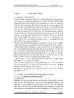
larger ones are known. Some of the shapes assumed by the cells are shown in fig. 1.
and their arrangement.
A. Bacillus subtilis, Cohn, and Spirillum undula, Ehrenb.
B. Planococcus citreus (Menge) Migula.
C. Pseudomonas pyocyanea (Gessard), Migula.
D. P. macroselmis, Migula.
E. P. syncyanea (Ehrenb.), Migula.
F. Bacillus typhi, Gaffky.
G. B. vulgaris (Hauser), Migula.
H. Microspira Comma (Koch), Schroeter.
J, K. Spirillum rubrum, Esmarsch.
L, M. S. undula (Müller), Ehrenb. (All after Migula.)
That bacteria have existed from very early periods is clear from Distribution in Time.
their presence in fossils; and although we cannot accept all the conclusions drawn
from the imperfect records of the rocks, and may dismiss as absurd the statements that
geologically immured forms have been found still living, the researches of Renault
and van Tieghem have shown pretty clearly that large numbers of bacteria existed in
Carboniferous and Devonian times, and probably earlier.
Schizomycetes are ubiquitous as saprophytes in still ponds and Distribution in Space.
ditches, in running streams and rivers, and in the sea, and especially in drains, bogs,
refuse heaps, and in the soil, and wherever organic infusions are allowed to stand for a
short time. Any liquid (blood, urine, milk, beer, &c.) containing organic matter, or any
solid food-stuff (meat preserves, vegetables, &c.), allowed to stand exposed to the air
soon swarms with bacteria, if moisture is present and the temperature not abnormal.
Though they occur all the world over in the space, air and on the surface of exposed
bodies, it is not to be supposed that they are by any means equally distributed, and it is
questionable whether the bacteria suspended in the air ever exist in such enormous
quantities as was once believed. The evidence to hand shows that on heights and in
open country, especially in the north, there may be few or even no Schizomycetes
detected in the air, and even in towns their distribution varies greatly; sometimes they
appear to exist in minute clouds, as it were, with interspaces devoid of any, but in
laboratories and closed spaces where their cultivation has been promoted the air may
be considerably laden with them. Of course the distribution of bodies so light and
small is easily influenced by movements, rain, wind, changes of temperature, &c. As
parasites, certain Schizomycetes inhabit and prey upon the organs of man and animals
in varying degrees, and the conditions for their growth and distribution are then very
complex. Plants appear to be less subject to their attacks—possibly, as has been
suggested, because the acid fluids of the higher vegetable organisms are less suited for
the development of Schizomycetes; nevertheless some are known to be parasitic on
plants. Schizomycetes exist in every part of the alimentary canal of animals, except,
perhaps, where acid secretions prevail; these are by no means necessarily harmful,
though, by destroying the teeth for instance, certain forms may incidentally be the
forerunners of damage which they do not directly cause.
Little was known about these extremely minute organisms History. before 1860. A.
van Leeuwenhoek figured bacteria as far back as the 17th century, and O. F. Müller
knew several important forms in 1773, while Ehrenberg in 1830 had advanced to the
commencement of a scientific separation and grouping of them, and in 1838 had
proposed at least sixteen species, distributing them into four genera. Our modern more
accurate though still fragmentary knowledge of the forms of Schizomycetes, however,
dates from F. J. Conn's brilliant researches, the chief results of which were published
at various periods between 1853 and 1872; Cohn's classification of the bacteria,
published in 1872 and extended in 1875, has in fact dominated the study of these
organisms almost ever since. He proceeded in the main on the assumption that the
forms of bacteria as met with and described by him are practically constant, at any rate
within limits which are not wide: observing that a minute spherical micrococcus or a
rod-like bacillus regularly produced similar micrococci and bacilli respectively, he
based his classification on what may be considered the constancy of forms which he
called species and genera. As to the constancy of form, however, Cohn maintained
certain reservations which have been ignored by some of his followers. The fact that
Schizomycetes produce spores appeals to have been discovered by Cohn in 1857,
though it was expressed dubiously in 1872; these spores had no doubt been observed
previously. In 1876, however, Cohn had seen the spores germinate, and Koch,
Brefeld, Pratzmowski, van Tieghem, de Bary and others confirmed the discovery in
various species.
The supposed constancy of forms in Cohn's species and genera received a shock when
Lankester in 1873 pointed out that his Bacterium rubescens (since named Beggiatoa
roseo-persicina, Zopf) passes through conditions which would have been described by
most observers influenced by the current doctrine as so many separate "species" or
even "genera,"—that in fact forms known as Bacterium, Micrococcus, Bacillus,
Leptothrix, &c., occur as phases in one life-history. Lister put forth similar ideas about
the same time; and Billroth came forward in 1874 with the extravagant view that the
various bacteria are only different states of one and the same organism which he
called Cocco-bacteria septica. From that time the question of the pleomorphism
(mutability of shape) of the bacteria has been hotly discussed: but it is now generally
agreed that, while a [v.03 p.0158]certain number of forms may show different types of
cell during the various phases of the life-history,
[2]
yet the majority of forms are
uniform, showing one type of cell throughout their life-history. The question of
species in the bacteria is essentially the same as in other groups of plants; before a
form can be placed in a satisfactory classificatory position its whole life-history must
be studied, so that all the phases may be known. In the meantime, while various
observers were building up our knowledge of the morphology of bacteria, others were
laying the foundation of what is known of the relations of these organisms to
fermentation and disease—that ancient will-o'-the-wisp "spontaneous generation"
being revived by the way. When Pasteur in 1857 showed that the lactic fermentation
depends on the presence of an organism, it was already known from the researches of
Schwann (1837) and Helmholtz (1843) that fermentation and putrefaction are
intimately connected with the presence of organisms derived from the air, and that the
preservation of putrescible substances depends on this principle. In 1862 Pasteur
placed it beyond reasonable doubt that the ammoniacal fermentation of urea is due to
the action of a minute Schizomycete; in 1864 this was confirmed by van Tieghem, and
in 1874 by Cohn, who named the organism Micrococcus ureae. Pasteur and Cohn also
pointed out that putrefaction is but a special case of fermentation, and before 1872 the
doctrines of Pasteur were established with respect to Schizomycetes. Meanwhile two
branches of inquiry had arisen, so to speak, from the above. In the first place, the
ancient question of "spontaneous generation" received fresh impetus from the
difficulty of keeping such minute organisms as bacteria from reaching and developing
in organic infusions; and, secondly, the long-suspected analogies between the
phenomena of fermentation and those of certain diseases again made themselves felt,
as both became better understood. Needham in 1745 had declared that heated
infusions of organic matter were not deprived of living beings; Spallanzani (1777) had
replied that more careful heating and other precautions prevent the appearance of
organisms in the fluid. Various experiments by Schwann, Helmholtz, Schultz,
Schroeder, Dusch and others led to the refutation, step by step, of the belief that the
more minute organisms, and particularly bacteria, arose de novo in the special cases
quoted. Nevertheless, instances were adduced where the most careful heating of yolk
of egg, milk, hay-infusions, &c., had failed,—the boiled infusions, &c., turning putrid
and swarming with bacteria after a few hours.
In 1862 Pasteur repeated and extended such experiments, and paved the way for a
complete explanation of the anomalies; Cohn in 1872 published confirmatory results;
and it became clear that no putrefaction can take place without bacteria or some other
living organism. In the hands of Brefeld, Burdon-Sanderson, de Bary, Tyndall,
Roberts, Lister and others, the various links in the chain of evidence grew stronger and
stronger, and every case adduced as one of "spontaneous generation" fell to the
ground when examined. No case of so-called "spontaneous generation" has withstood
rigid investigation; but the discussion contributed to more exact ideas as to the
ubiquity, minuteness, and high powers of resistance to physical agents of the spores of
Schizomycetes, and led to more exact ideas of antiseptic treatments. Methods were
also improved, and the application of some of them to surgery at the hands of Lister,
Koch and others has yielded results of the highest value.
Long before any clear ideas as to the relations of Schizomycetes to fermentation and
disease were possible, various thinkers at different times had suggested that
resemblances existed between the phenomena of certain diseases and those of
fermentation, and the idea that a virus or contagium might be something of the nature
of a minute organism capable of spreading and reproducing itself had been
entertained. Such vague notions began to take more definite shape as the ferment
theory of Cagniard de la Tour (1828), Schwann (1837) and Pasteur made way,
especially in the hands of the last-named savant. From about 1870 onwards the "germ
theory of disease" has passed into acceptance. P. F. O. Rayer in 1850 and Davaine had
observed the bacilli in the blood of animals dead of anthrax (splenic fever), and
Pollender discovered them anew in 1855. In 1863, imbued with ideas derived from
Pasteur's researches on fermentation, Davaine reinvestigated the matter, and put forth
the opinion that the anthrax bacilli caused the splenic fever; this was proved to result
from inoculation. Koch in 1876 published his observations on Davaine's bacilli,
placed beyond doubt their causal relation to splenic fever, discovered the spores and
the saprophytic phase in the life-history of the organism, and cleared up important
points in the whole question (figs. 7 and 9). In 1870 Pasteur had proved that a disease
of silkworms was due to an organism of the nature of a bacterium; and in 1871 Oertel
showed that a Micrococcus already known to exist in diphtheria is intimately
concerned in producing that disease. In 1872, therefore, Cohn was already justified in
grouping together a number of "pathogenous" Schizomycetes. Thus arose the
foundations of the modern "germ theory of disease;" and, in the midst of the wildest
conjectures and the worst of logic, a nucleus of facts was won, which has since grown,
and is growing daily. Septicaemia, tuberculosis, glanders, fowl-cholera, relapsing
fever, and other diseases are now brought definitely within the range of biology, and it
is clear that all contagious and infectious diseases are due to the action of bacteria or,
in a few cases, to fungi, or to protozoa or other animals.
as actually observed in hanging drops under very high powers.
A. The spore sown at 11 A.M., as shown at a, had swollen (b) perceptibly by noon,
and had germinated by 3.30 P.M., as shown at c: in d at 6 P.M., and e at 8.30 P.M.;
the resulting filament is segmenting into bacilli as it elongates, and at midnight (f)
consisted of twelve such segments.
B, C. Similar series of phases in the order of the small letters in each case, and with
the times of observation attached. At f and g occurs the breaking up of the filament
into rodlets.
D. Germinating spores in various stages, more highly magnified, and showing the
different ways of escape of the filament from the spore-membrane. (H. M. W.)
Other questions of the highest importance have arisen from the foregoing. About 1880
Pasteur first showed that Bacillus anthracis cultivated in chicken broth, with plenty of
oxygen and at a temperature of 42-43° C., lost its virulence after a few "generations,"
and ceased to kill even the mouse; Toussaint and Chauveau confirmed, and others
have extended the observations. More remarkable still, animals inoculated with such
"attenuated" bacilli proved to be curiously resistant to the deadly effects of subsequent
inoculations of the non-attenuated form. In other words, animals vaccinated with the
cultivated bacillus showed immunity from disease when reinoculated with the deadly
wild form. The questions as to the causes and nature of the changes in the bacillus and
in the host, as to the extent of immunity enjoyed by the latter, &c., are of the greatest
interest and importance. These matters, however, and others such as phagocytosis
(first described by Metchnikoff in 1884), and the epoch-making discovery of the
opsonins of the blood by Wright, do not here concern us (see II. below).
Morphology.—Sizes, Forms, Structure, &c.—The Schizomycetes Form and Structure.
consist of single cells, or of filamentous or other groups of cells, according as the
divisions are completed at once or not. While some unicellular forms are less than 1 µ
(.001 mm.) in diameter, others have cells measuring 4 µ or 5 µ or even 7 µ or 8 µ, in
thickness, while the length may vary from that of the diameter to many times that
measurement. In the filamentous forms the individual cells are often difficult to
observe until reagents are applied (e.g. fig. 14), and the length of the rows of
cylindrical cells may be many hundred times greater than the breadth. Similarly, the
diameters of flat or spheroidal colonies may vary from a few times to many hundred
Cell-wall. times that of the individual cells, the divisions of which have produced the
colony. The shape of the individual cell (fig. 1) varies from that of a minute sphere to
that of a straight, curved, or twisted filament or cylinder, which is not necessarily of
the same diameter throughout, and may have flattened, rounded, or even pointed ends.
The rule is that the cells divide in one direction only—i.e. transverse to the long
axis—and therefore produce aggregates of long cylindrical shape; but in rarer cases
iso-diametric cells divide in two or three directions, producing flat, or spheroidal, or
irregular colonies, the size of which is practically unlimited. The bacterial [v.03
p.0159]cell is always clothed by a definite cell-membrane, as was shown by the
plasmolysing experiments of Fischer and others. Unlike the cell-wall of the higher
plants, it gives usually no reactions of cellulose, nor is chitin present as in the fungi,
but it consists of a proteid substance and is apparently a modification of the general
protoplasm. In some cases, however, as in B. tuberculosis, analysis of the cell shows a
large amount of cellulose. The cell-walls in some forms swell up into a gelatinous
mass so that the cell appears to be surrounded in the unstained condition by a clear,
transparent space. When the swollen wall is dense and regular in appearance the term
"capsule" is applied to the sheath as in Leuconostoc. Secreted pigments (red, yellow,
green and blue) are sometimes deposited in the wall, and some of the iron-bacteria
have deposits of oxide of iron in the membranes.
A. Mixed zoogloea found as a pellicle on the surface of vegetable infusions, &c.; it
consists of various forms, and contains cocci (a) and rodlets, in series (b and c), &c.
B. Egg-shaped mass of zoogloea of Beggiatoa roseo-persicina (Bacterium rubescens
of Lankester); the gelatinous swollen walls of the large crowded cocci are fused into a
common gelatinous envelope.
C. Reticulate zoogloea of the same.
D, E, H. Colonies of Myconostoc enveloped in diffluent matrix.
F. Branched fruticose zoogloea of Cladothrix (slightly magnified).
G. Zoogloea of Bacterium merismopedioides, Zopf, containing cocci arranged in
tablets.
The substance of the bacterial cell when suitably prepared Cell-contents. and stained
shows in the larger forms a mass of homogeneous protoplasm containing irregular
spaces, the vacuoles, which enclose a watery fluid. Scattered in the protoplasm arc
usually one or more deeply-staining granules. The protoplasm itself may be tinged
with colouring matter, bright red, yellow, &c., and may occasionally contain
substances other than the deeply-staining granules. The occurrence of a starch-like
substance which stains deep blue with iodine has been clearly shown in some forms
even where the bacterium is growing on a medium containing no starch, as shown by
Ward and others. In other forms a substance (probably glycogen or amylo-dextrin)
which turns brown with iodine has been observed. Oil and fat drops have also been
shown to occur, and in the sulphur-bacteria numerous fine granules of sulphur.
The question of the existence of a nucleus in the bacteria is Nucleus. one that has led
to much discussion and is a problem of some difficulty. In the majority of forms it has
not hitherto been possible to demonstrate a nucleus of the type which is so
characteristic of the higher plants. Attention has accordingly been directed to the
deeply-staining granules mentioned above, and the term chromatin-granules has been
applied to them, and they have been considered to represent a rudimentary nucleus.
That these granules consist of a material similar to the chromatin of the nucleus of
higher forms is very doubtful, and the comparison with the nucleus of more highly
organized cells rests on a very slender basis. The most recent works (Vejdovsky,
Mencl), however, appear to show that nuclei of a structure and mode of division
almost typical are to be found in some of the largest bacteria. It is possible that a
similar structure has been overlooked or is invisible in other forms owing to their
small size, and that there may be another type of nucleus—the diffuse nucleus—such
as Schaudinn believed to be the case in B. butschlii. Many bacteria when suspended in
a fluid exhibit a power of independent movement which is, of course, quite distinct
from the Brownian movement—a non-vital phenomenon common to all finely-
divided particles suspended in a fluid. Independent movement is effected by special
motile organs, the cilia or flagella. These structures are invisible, with ordinary
illumination in living cells or unstained preparations, and can only be made clearly
visible by special methods of preparation and staining first used by Löffler. By these
methods the cilia are seen to be fine protoplasmic outgrowths of the cell (fig. 1) of the
same nature as those of the zoospores and antherozoids of algae, mosses, &c. Cilia.
These cilia appear to be attached to the cell-wall, being unaffected by plasmolysis, but
Fischer states that they really are derived from the central protoplasm and pass
through minute pores in the wall. The cilia may be present during a short period only
in the life of a Schizomycete, and their number may vary according to the medium on
which the organism is growing. Nevertheless, there is more or less constancy in the
type of distribution, &c., of the cilia for each species when growing at its best. The
chief results may be summed up as follows: some species, e.g. B. anthracis, have no
cilia; others have only one flagellum at one pole (Monotrichous), e.g. Bacillus
pyocyaneus (fig. 1, C, D), or one at each pole; others again have a tuft of several cilia
[v.03 p.0160]at one pole (Lophotrichous), e.g. B. syncyaneus (fig. 1, E), or at each
pole (Amphitrichous) (fig. 1, J, K, L); and, finally, many actively motile forms have
the cilia springing all round (Peritrichous), e.g. B. vulgaris (fig. 1, G). It is found,
however, that strict reliance cannot be placed on the distinction between the
Monotrichous, Lophotrichous and Amphitrichous conditions, since one and the same
species may have one, two or more cilia at one or both poles; nevertheless some stress
may usually be laid on the existence of one or two as opposed to several—e.g. five or
six or more—at one or each pole.
In Beggiatoa, a filamentous form, peculiar, slow, oscillatory Vegetative State.
movements are to be observed, reminding us of the movements of Oscillatoria among
the Cyanophyceae. In these cases no cilia have been observed, and there is a firm cell-
wall, so the movement remains quite unexplained.
Fig. 4.—Types of Spore-formation in Schizomycetes. (After Zopf.)
A. Various stages in the development of the endogenous spores in a Clostridium—the
small letters indicate the order.
B. Endogenous spores of the hay bacillus.
C. A chain of cocci of Leuconostoc mesenterioides, with two "resting spores," i.e.
arthrospores. (After van Tieghem.)
D. A motile rodlet with one cilium and with a spore formed inside.
E. Spore-formation in Vibrio-like (c) and Spirillum-like (a b, a) Schizomycetes.
F. Long rod-like form containing a spore (these are the so-called "Köpfchenbacterien"
of German authors).
G. Vibrio form with spore. (After Prazmowski.)
H. Clostridium—one cell contains two spores. (After Prazmowski.)
I. Spirillum containing many spores (a), which are liberated at b by the breaking up of
the parent cells.
K. Germination of the spore of the hay bacillus (B. subtilis)—the axis of growth of the
germinal rodlet is at right angles to the long axis of the spore.
L. Germination of spore of Clostridium butyricum—the axis of growth coincides with
the long axis of the spore.
While many forms are fixed to the substratum, others are free, being in this condition
either motile or immotile. The chief of these forms are described below.
Fig. 5.—Characteristic groups of Micrococci. (After Cohn.) A. Micrococcus
prodigiosus. B. M. vaccinae. C. Zoogloea stage of a Micrococcus, forming a close
membrane on infusion—Pasteur's Mycoderma. (Very highly magnified.)
Cocci: spherical or spheroidal cells, which, according to their relative (not very well
defined) sizes are spoken of as Micrococci, Macrococci, and perhaps Monas forms.
Rods or rodlets: slightly or more considerably elongated cells which are cylindrical,
biscuit-shaped or somewhat fusiform. The cylindrical forms are short, i.e. only three
or four times as long as broad (Bacterium), or longer (Bacillus); the biscuit-shaped
ones are Bacteria in the early stages of division. Clostridia, &c., are spindle-shaped.
Filaments really consist of elongated cylindrical cells which remain united end to end
after division, and they may break up later into elements such as those described
above. Such filaments are not always of the same diameter throughout, and their
segmentation varies considerably. They may be free or attached at one (the "basal")
end. A distinction is made between simple filaments (e.g. Leptothrix) and such as
exhibit a false branching (e.g. Cladothrix).
Curved and spiral forms. Any of the elongated forms described above may be curved
or sinuous or twisted into a corkscrew-like spiral instead of straight. If the sinuosity is
slight we have the Vibrio form; if pronounced, and the spiral winding well marked, the
forms are known as Spirillum, Spirochaete, &c. These and similar terms have been
applied partly to individual cells, but more often to filaments consisting of several
cells; and much confusion has arisen from the difficulty of defining the terms
themselves.
In addition to the above, however, certain Schizomycetes present aggregates in the
form of plates, or solid or hollow and irregular branched colonies. This may be due to
the successive divisions occurring in two or three planes instead of only across the
long axis (Sarcina), or to displacements of the cells after division.
Growth and Division.—Whatever the shape and size of the Reproduction. individual
cell, cell-filament or cell-colony, the immediate visible results of active nutrition are
elongation of the cell and its division into two equal halves, across the long axis, by
the formation of a septum, which either splits at once or remains intact for a shorter or
longer time. This process is then repeated and so on. In the first case the separated
cells assume the character of the parent-cell whose division gave rise to them; in the
second case they form filaments, or, if the further elongation and divisions of the cells
proceed in different directions, plates or spheroidal or other shaped colonies. It not
unfrequently happens, however, that groups of cells break away from their former
connexion as longer or shorter straight or curved filaments, or as solid masses. In
some filamentous forms this "fragmentation" into multicellular pieces of equal length
or nearly so is a normal phenomenon, each partial filament repeating the growth,
division and fragmentation as before (cf. figs. 2 and 6). By rapid division hundreds of
thousands of cells may be produced in a few hours,
[3]
and, according to the species
and the conditions (the medium, temperature, &c.), enormous collections of isolated
cells may cloud the fluid in which they are cultivated, or form deposits below or films
on its surface; valuable characters are sometimes obtained from these appearances.
When these dense "swarms" of vegetative cells become fixed in a matrix of their own
swollen contiguous cell-walls, they pass over into a sort of resting state as a so-called
zoogloea (fig. 3).
Fig. 6.—Bacillus megaterium. (After de Bary.)
a, a chain of motile rodlets still growing and dividing (bacilli).
b, a pair of bacilli actively growing and dividing.
p, a rodlet in this condition (but divided into four segments) after treatment with
alcoholic iodine solution.
c, d, e, f, successive stages in the development of the spores.
r, a rodlet segmented in four, each segment containing one ripe spore.
g1, g2, g3, early stages in the germination of the spores (after being dried several
days);
h1, h2, k, l and m, successive stages in the germination of the spore.
Fig. 7.—Bacillus anthracis. (After Koch.)
A. Bacilli mingled with blood-corpuscles from the blood of a guinea-pig; some of the
bacilli dividing.
B. The rodlets after three hours' culture in a drop of aqueous humour. They grow out
into long leptothrix-like filaments, which become septate later, and spores are
developed in the segments.
One of the most remarkable Zoogloeae. phenomena in the life-history of the
Schizomycetes is the formation of this zoogloea stage, which corresponds to the
"palmella" condition of the lower Algae. This occurs as a membrane on the surface of
the medium, or as irregular clumps or branched masses (sometimes several inches
across) submerged in it, and consists of more or less gelatinous matrix enclosing
innumerable "cocci," "bacteria," or other elements of the Schizomycete concerned.
Formerly regarded as a distinct genus—the natural fate of all the various [v.03
p.0161]forms—the zoogloea is now known to be a sort of resting condition of the
Schizomycetes, the various elements being glued together, as it were, by their
enormously swollen and diffluent cell-walls becoming contiguous. The zoogloea is
formed by active division of single or of several mother-cells, and the progeny appear
to go on secreting the cell-wall substance, which then absorbs many times its volume
of water, and remains as a consistent matrix, in which the cells come to rest. The
matrix—i.e. the swollen cell-walls—in some cases consists mainly of cellulose, in
others chiefly of a proteid substance; the matrix in some cases is horny and resistant,
in others more like a thick solution of gum. It is intelligible from the mode of
formation that foreign bodies may become entangled in the gelatinous matrix, and
compound zoogloeae may arise by the apposition of several distinct forms, a common
event in macerating troughs (fig. 3, A). Characteristic forms may be assumed by the
young zoogloea of different species,—spherical, ovoid, reticular, filamentous,
fruiticose, lamellar, &c.,—but these vary considerably as the mass increases or comes
in contact with others. Older zoogloeae may precipitate oxide of iron in the matrix, if
that metal exists in small quantities in the medium. Under favourable conditions the
elements in the zoogloea again become active, and move out of the matrix, distribute
themselves in the surrounding medium, to grow and multiply as before. If the
zoogloea is formed on a solid substratum it may become firm and horny; immersion in
water softens it as described above.