Tài liệu CANCER CHEMOPREVENTION WITH DIETARY PHYTOCHEMICALS doc
Bạn đang xem bản rút gọn của tài liệu. Xem và tải ngay bản đầy đủ của tài liệu tại đây (468.22 KB, 13 trang )
Cancer is a growing health problem around the world
— particularly with the steady rise in life expectancy,
increasing urbanization and the subsequent changes in
environmental conditions, including lifestyle. According
to a recent report by the World Health Organization
(WHO), there are now more than 10 million cases of
cancer per year worldwide. In 2003, it is estimated that
approximately 1,300,000 new cases of cancer will be
diagnosed, and more than 550,000 people will die from
cancer in the United States alone.
Although there is no ‘magic bullet’ that can com-
pletely conquer cancer, many types of the disease
might be avoidable. Cancer risk can be reduced by
eliminating the identified carcinogens — or at least
minimizing exposure to them — but, without com-
plete identification of the corresponding risk factors,
such primary prevention might be difficult to imple-
ment. Furthermore, the avoidance of some risk factors
could require large lifestyle changes, which are not easy
to implement.
It has been estimated that more than two-thirds of
human cancers could be prevented through appropriate
lifestyle modification. Richard Doll and Richard Peto
have reported that 10–70% (average 35%) of human
cancer mortality is attributable to diet
1
.Their observa-
tions, which are based on statistical and epidemiological
data, mainly concerned dietary factors that increase risk.
Although the exact percentage is uncertain, there are
several lines of compelling evidence from epidemiologi-
cal, clinical and laboratory studies that link cancer risk
to the nutritional factors.
A wide array of substances derived from the diet
have been found to stimulate the development, growth
and spread of tumours in experimental animals, and to
transform normal cells into malignant ones. These are
regarded as suspected human carcinogens.
So, many dietary constituents can increase the risk of
developing cancer,but there is also accumulating evi-
dence from population as well as laboratory studies to
support an inverse relationship between regular con-
sumption of fruit and vegetables and the risk of specific
cancers. Several organizations — such as the WHO, the
American Cancer Society, the American Institute of
Cancer Research (AICR) and the National Cancer
Institute (NCI) — have established dietary guidelines to
help people reduce the cancer risk (for further informa-
tion, see the 1997 World Cancer Research Fund and
AICR report in online links box).
Many clinical trials on the use of nutritional supple-
ments and modified diets to prevent cancer are ongo-
ing. It is conceivable that in the future people might only
need to take specially formulated pills that contain sub-
stances derived from edible plants to prevent cancer or
delay its onset
2
.However, a precise assessment of the
mechanisms by which the components of fruit and veg-
etables prevent cancer is necessary before they can be
recommended for inclusion in dietary supplements or
before they can be tested in human intervention trials.
Phytochemicals are non-nutritive components in the
plant-based diet (‘phyto’ is from the Greek word mean-
ing plant) that possess substantial anticarcinogenic and
antimutagenic properties. Given the great structural
CANCER CHEMOPREVENTION WITH
DIETARY PHYTOCHEMICALS
Yo ung-Joon Surh
Chemoprevention refers to the use of agents to inhibit, reverse or retard tumorigenesis.
Numerous phytochemicals derived from edible plants have been reported to interfere with a
specific stage of the carcinogenic process. Many mechanisms have been shown to account
for the anticarcinogenic actions of dietary constituents, but attention has recently been
focused on intracellular-signalling cascades as common molecular targets for various
chemopreventive phytochemicals.
College of Pharmacy,
Seoul National University,
Shinlim-dong, Kwanak-ku,
Seoul 151-742, South Korea.
e-mail:
doi:10.1038/nrc1189
768 | OCTOBER 2003 | VOLUME 3 www.nature.com/reviews/cancer
REVIEWS
NATURE REVIEWS | CANCER VOLUME 3 | OCTOBER 2003 | 769
REVIEWS
diversity of phytochemicals, it is not feasible to define
structure–activity relationships to deduce their
underlying molecular mechanisms. A better approach
is to analyse their effects on cancer-associated
signal-transduction pathways.
Importance of plant-derived foods
More than 250 population-based studies, including
case–control and cohort studies, indicate that people
who eat about five servings of fruit and vegetables a day
have approximately half the risk of developing cancer —
particularly cancers of the digestive and respiratory
tracts — of those who eat fewer than two servings. In
the United States, these observations led to the develop-
ment of public-health campaigns such as the ‘Five-a-Day
for Better Health’ programme and a more recent ‘Savor
the Spectrum’ campaign — both were designed to
increase the ingestion of fruit and vegetables by the
population
(BOX 1).Increased consumption of fruit and
vegetables is a global priority in the prevention of cancer
and other chronic disorders. According to the WHO
Report 2002, there are at least 2.7 million deaths globally
per year, which are primarily attributable to low fruit
and vegetable intake.
Ve getables and fruit are excellent sources of cancer-
preventive substances. The NCI has identified about
35 plant-based foods that possess cancer-preventive
Summary
•Many population-based studies have highlighted the ability of macronutrients and
micronutrients in vegetables and fruit to reduce the risk of cancer. Recently,attention
has been focused on phytochemicals — non-nutritive components in the plant-based
diet that possess cancer-preventive properties.
• Despite remarkable progress in our understanding of the carcinogenic process,
the mechanisms of action of most chemopreventive phytochemicals have not been
fully elucidated.
•Chemopreventive phytochemicals can block initiation or reverse the promotion stage
of multistep carcinogenesis. They can also halt or retard the progression of
precancerous cells into malignant ones.
•Many molecular alterations associated with carcinogenesis occur in cell-signalling
pathways that regulate cell proliferation and differentiation. One of the central
components of the intracellular-signalling network that maintains homeostasis is the
family of mitogen-activated protein kinases (MAPKs).
•Numerous intracellular signal-transduction pathways converge with the activation
of the transcription factors NF-κB and AP1. As these factors mediate pleiotropic
effects of both external and internal stimuli in the cellular-signalling cascades, they
are prime targets of diverse classes of chemopreventive phytochemicals.
•Basic helix–loop–helix transcription factors such as NRF2 regulate expression of phase II
enzymes, which detoxify carcinogens and protect against oxidative stress. A number of
phytochemicals have been shown to induce expression of phase II enzymes via NRF2.
• β-Catenin, a multifunctional protein that was originally identified as a component
of cell–cell adhesion machinery, is another important molecular target for
chemoprevention. Several dietary phytochemicals have been shown to target
this molecule.
Box 1 | Chemoprevention initiatives
A number of government programmes have been created in the United States and in Europe to increase vegetable
consumption and decrease cancer incidence. These include the following:
The ‘Five-A-Day for Better Health’ programme
Founded in late 1991, this is the first nationwide health-promotion campaign to encourage people in the United States to
eat fruit and vegetables — at least five servings a day — to reduce the risk of cancer and other chronic diseases. Over the
past decade, there has been a steady increase in both awareness of the health benefits of fruit and vegetables and their
consumption in the United States. The National Cancer Institute (NCI) has recently completed a review of this
programme and reported a series of recommendations for the next round of the initiative (for further information, see
the ‘Five-A-Day for Better Health’report in online links box).
‘Savor the Spectrum’
The NCI’s spring 2002 media promotion, entitled ‘Savor the Spectrum’, urges all Americans to eat five to nine
servings of colourful fruit and vegetables a day for better health. The message of this programme is based on current
research showing that phytonutrients from different colour groups are powerful disease fighters that help our body
fight off cancer and heart disease. NCI has produced a series of guidelines featuring each colour of the‘rainbow’ of
fruit and vegetables.
European Prospective Investigation of Cancer and Nutrition (EPIC)
EPIC is one of the most important multicentre prospective cohort studies ever launched worldwide. Beginning in
1992, EPIC has involved more than half a million (520,000) participants recruited by 20 centres in 10 countries under
the coordination of the International Agency for Research on Cancer (IARC) and partly funded by the ‘Europe
Against Cancer’ programme of the European Commission, as well as by the participating countries. EPIC focuses on
identifying the dietary determinants of cancer, and is aimed at expanding the presently limited knowledge of the role
of nutrition and other lifestyle factors in the aetiology and prevention of cancer and other life-threatening diseases.
Global Strategy on Dietary Prevention of Cancer
For a global extension of the ‘Five-A-Day’ concept of boosting increased consumption of fruit and vegetables, the
WHO organized the third Biennial ‘Five-A-Day’ International Symposium on January 14–15 2003 in Berlin,
Germany.At the meeting, Derek Yach, the WHO Executive Director of Noncommunicable Diseases & Mental
Health, said “Increasing the consumption of fruit and vegetables is a necessary part of the effort to reduce the
growing global burden of chronic diseases including cancer.”The guidelines stated “choose most of the foods you eat
from plant sources”.
770 | OCTOBER 2003 | VOLUME 3 www.nature.com/reviews/cancer
REVIEWS
cancer. Recently, the focus and emphasis have shifted to
the non-nutritive phytochemicals. The NCI has deter-
mined in laboratory studies that more than 1,000 differ-
ent phytochemicals possess cancer-preventive activity. It
is estimated that there could be more than 100 different
phytochemicals in just a single serving of vegetables.
As early as 1980, the NCI’s Chemoprevention
Programme of the Division of Cancer Prevention and
Control began evaluating phytochemicals for safety, effi-
cacy and applicability for cancer prevention. Michael
Sporn coined the term ‘chemoprevention’ in the mid-
1970s to describe the strategy of blocking or slowing the
onset of premalignant tumours with relatively nontoxic
chemical substances. To better define and guide research
in the field of chemoprevention, the NCI Division of
Cancer Prevention started the Chemoprevention
Implementation Group in 1998, and then the Rapid
Access to Preventive Intervention Development pro-
gramme. The NCI has more than 400 potential agents
under investigation and is sponsoring more than 65
Phase I, Phase II and Phase III chemoprevention trials.
These involve various substances or their mixtures,
many of which are foodborne phytochemicals.
Mechanisms of chemoprevention
Carcinogenesis is generally recognized as a multistep
process in which distinct molecular and cellular alter-
ations occur.From the study of experimentally induced
carcinogenesis in rodents, tumour development is con-
sidered to consist of several separate, but closely linked,
stages — tumour initiation, promotion and progression.
Although these divisions are an oversimplification of
carcinogenesis, it is useful to think in these stages when
considering possible opportunities for chemoprevention.
Initiation is a rapid and irreversible process that
involves a chain of extracellular and intracellular
events. These include the initial uptake of or exposure
to a carcinogenic agent, its distribution and transport
to organs and tissues where metabolic activation and
detoxification can occur, and the covalent interaction of
reactive species with target-cell DNA, leading to geno-
toxic damage. In contrast to initiation, tumour promo-
tion is considered to be a relatively lengthy and
reversible process in which actively proliferating pre-
neoplastic cells accumulate. Progression, the final stage
of neoplastic transformation, involves the growth of a
tumour with invasive and metastatic potential.
According to the conventional classification origi-
nally proposed by Lee Wattenberg, chemopreventive
agents are subdivided into two main categories —
blocking agents and suppressing agents
3
.Blocking
agents prevent carcinogens from reaching the target
sites, from undergoing metabolic activation or from
subsequently interacting with crucial cellular macro-
molecules (for example, DNA, RNA and proteins).
Suppressing agents, on the other hand, inhibit
the malignant transformation of initiated cells, in
either the promotion or the progression stage.
Chemopreventive phytochemicals can block or reverse
the premalignant stage (initiation and promotion) of
multistep carcinogenesis. They can also halt or at least
properties. These include garlic, soybeans, ginger,
onion, turmeric, tomatoes and cruciferous vegetables
(for example, broccoli, cabbage, cauliflower and
Brussels sprouts). Numerous cell-culture and animal-
model studies have been conducted to evaluate the
ability of specific edible plants to prevent cancer.
Beyond vitamins to phytochemicals
Many population-based studies have highlighted the
ability of macronutrients (for example, carbohydrate,
proteins, fat and fibre) and micronutrients (for exam-
ple, antioxidant vitamins and trace minerals) that are
contained in vegetables and fruit to reduce the risk of
cancer. The most exciting findings have been achieved
with antioxidant vitamins and their precursors, which
are found in dark, leafy green vegetables and
yellow/orange fruit and vegetables. The NCI has there-
fore sponsored a series of human intervention trials
with individual vitamins and minerals. However,plants
contain numerous chemical substances other than these
micronutrients that might also be useful in preventing
Detoxification
Pro-carcinogen
Ultimate
carcinogen
Metabolic
activation
Cancer-blocking
agents
Ellagic acid
Indole-3-carbinol
Sulphoraphane
Flavonoids
Normal
cell
Initiation
(1–2 days)
Promotion
(>10 years)
Progression
(>1 year)
Initiated cell
Neoplastic
cells
Preneoplastic
cells
Detoxification
Secretion
β-Carotene
Curcumin
EGCG
Genistein
Resveratrol
[6]-Gingerol
Capsaicin
Cancer-suppressing
agents
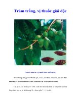
Figure 1 | Dietary phytochemicals that block or suppress multistage carcinogenesis.
Carcinogenesis is initiated with the transformation of the normal cell into a cancer cell (initiated
cell). These cells undergo tumour promotion into preneoplastic cells, which progress to neoplastic
cells. Phytochemicals can interfere with different steps of this process. Some chemopreventive
phytochemicals inhibit metabolic activation of the procarcinogens to their ultimate electrophilic
species, or their subsequent interaction with DNA. These agents therefore block tumour initiation
(blocking agents). Alternatively, dietary blocking agents can stimulate the detoxification of
carcinogens, leading to their secretion from the body. Other phytochemicals suppress the later
steps (promotion and progression) of multistage carcinogenesis (suppressing agents). Some
phytochemicals can act as both blocking and suppressing agents. Adapted from REF. 128.
NATURE REVIEWS | CANCER VOLUME 3 | OCTOBER 2003 | 771
REVIEWS
include carcinogen activation/detoxification by xenobi-
otic metabolizing enzymes; DNA repair; cell-cycle
progression; cell proliferation, differentiation and apop-
tosis; expression and functional activation of oncogenes
or tumour-suppressor genes; angiogenesis and metasta-
sis; and hormonal and growth-factor activity (for
further information, see
ONLINE TABLE 1).
Cellular signalling molecules as targets
During the past two or three decades, there has been
substantial progress in identifying the biochemical
events that are associated with the multistage process
of carcinogenesis, and we are now better aware of how
certain dietary phytochemicals are able to alter this
process
(FIG. 1).Remarkable advances in the cellular
and molecular genetics of carcinogenesis — such as
the identification of numerous oncogenes and
tumour-suppressor genes, specific genes encoding
retard the development and progression of precancer-
ous cells into malignant ones
(FIG. 1).Recent advances
in our understanding of the carcinogenic process at
the cellular and molecular level have shown this block-
ing and suppressing categorization to be an oversim-
plification, and numerous cellular molecules and
events that could be potential targets of chemopreven-
tive agents have been more specifically identified
4–6
.
Therefore, the ability of any single chemopreventive
phytochemical to prevent tumour development
should be recognized as the outcome of the combina-
tion of several distinct sets of intracellular effects,
rather than a single biological response.
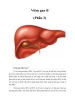
FIGURE 2 illustrates the chemical structures of repre-
sentative dietary phytochemicals that have been known
to possess chemopreventive potential and their dietary
sources. The cellular and molecular events affected or
regulated by these chemopreventive phytochemicals
OO
O
HO OH
O
CH
3
H
3
C
Curcumin
N
H
O
O
HO
H
3
C
Capsaicin
O
O
HO
H
3
C
OH
[6]-Gingerol
O
O
O
OH
OH
OH
OH
OHHO
OH
HO
Epigallocatechin-3-gallate
O
OOH
HO
OH
Genistein
OH
OH
HO
Resveratrol
Lycopene
H
3
C
S
O
N
C
S
Sulphoraphane
O
O
HO
HO
Caffeic acid phenethyl ester
N
H
OH
Indole-3-carbinol
S
Diallyl sulphide
Turmeric
Grapes
Honey
Garlic
Cabbage
Broccoli
Chilli peppers
Ginger
Green tea
Soybeans
Tomatoes
Figure 2 | Representative chemopreventive phytochemicals and their dietary sources.
772 | OCTOBER 2003 | VOLUME 3 www.nature.com/reviews/cancer
REVIEWS
Despite this progress, the identification of molecular
and cellular targets of chemopreventive phytochemicals
is still incomplete. Many of the molecular alterations that
are associated with carcinogenesis occur in cell-signalling
pathways that regulate cell proliferation and differentia-
tion. One of the central components of the intracellular-
signalling network that maintains homeostasis is the
family of proline-directed serine/threonine kinases —
the mitogen-activated protein kinases (MAPKs;
FIG. 3).
Abnormal or improper activation or silencing of the
MAPK pathway or its downstream transcription fac-
tors can result in uncontrolled cell growth, leading to
malignant transformation. Some phytochemicals
‘switch on’ or ‘turn off ’ the specific signalling mole-
cule(s), depending on the nature of the signalling cas-
cade they target, preventing abnormal cell proliferation
and growth
4–12
.Cell-signalling kinases other than
MAPKs, such as protein kinase C (PKC) and phos-
phatidylinositol 3-kinase (PI3K), are also important
targets of certain chemopreventive phytochemicals.
These upstream kinases activate a distinct set of tran-
scription factors, including nuclear factor κB (NF-κB)
and activator protein 1 (AP1;
FIG. 3).
NF-κB and AP1
Numerous intracellular signal-transduction pathways
converge with the activation of the transcription factors
NF-κB and AP1, which act independently or coordinately
to regulate target-gene expression
(FIG. 3).
Aberrant activation of NF-κB has been associated
with protection against apoptosis and stimulation of
proliferation in malignant cells
13,14
, and overexpression
of NF-κB is causally linked to the phenotypic changes
that are characteristic of neoplastic transformation
15
.
Many chemopreventive phytochemicals that are
derived from the diet have been shown to suppress
constitutive NF-κB activation in malignant cells or
NF-κB activation induced by the external tumour pro-
moter phorbol 12-myristate 13-acetate (PMA) or
tumour-necrosis factor-α (TNF-α)
11,16,17
.
AP1 is another transcription factor that regulates
expression of genes that are involved in cellular adapta-
tion, differentiation and proliferation. Functional activa-
tion of AP1 is associated with malignant transformation
as well as tumour promotion
18–21
. AP1 consists of either
homo- or heterodimers between members of the JUN
and FOS families, which interact via a leucine-zipper
domain. This transcription factor is also regulated by
the MAPK-signalling cascade
21–23
.
As NF-κB and AP1 are ubiquitous eukaryotic tran-
scription factors that mediate pleiotropic effects of both
external and internal stimuli in the cellular-signalling
cascades, they are prime targets of diverse classes of
chemopreventive phytochemicals
(FIG. 3).
Phytochemicals targeting NF-κB and AP1
Curcumin, [6]-gingerol and capsaicin.Curcumin — a
yellow pigment that is present in the rhizome of
turmeric (Curcuma longa L.) and related species — is
one of the most extensively investigated phytochemicals,
with regard to chemopreventive potential. Curcumin
carcinogen-metabolizing enzymes, DNA-repair
enzymes and proteins, and regulators of cell cycle and
apoptosis — have given us a better insight into the
process of neoplastic transformation. Advances have
also been made in identifying the factors that mediate
tumour invasion, metastasis and angiogenesis.
NF-κB
NF-κB
NF-κB
IκB
Ub
P
ELK1/SAP1
SRF
SRE
ATF2
TRE
TREκB binding site
c-FOS
c-JUN
c-FOS
c-JUN
c-JUN
Nucleus
Cytoplasm
PI3K
MEKK1
PKC
PDK
AKT
NIK
IKK-α/β/γ
MEK1/2
ERK1/2
MKK4
p38
RAF
JNK
RAS
Curcumin
EGCG
Resveratrol
Curcumin
EGCG
Resveratrol
EGCG
Genistein
EGCG
Curcumin
Curcumin
EGCG
Genistein
Resveratrol
Capsaicin
AP1
Proteasome
26S
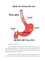
Figure 3 | Effect of phytochemicals on activation of NF-κB and AP1. The NF-κB signalling
pathway converges on the multiprotein complex called the IκB kinase (IKK) signalsome,
leading to IκB phosphorylation (P), ubiquitylation (Ub) and subsequent degradation by the 26S
proteasome. NF-κB is then released and translocated to the nucleus, where it binds to specific
promoter regions of various genes. The IKK signalsome is activated by the NF-κB-inducing
kinase (NIK). Pathways that regulate NIK are likely to involve signalling through a family of
mitogen-activated protein kinases (MAPKs), such as MAPK kinase kinase-1 (MEKK1) — a
kinase that lies upstream of extracellular signal-regulated kinase (ERK) — MAPK/ERK kinase
(MEK1/2) and p38 MAPK. Recent reports showed that NF-κB activation is also regulated by
the AKT signalling pathway
58,59,129
. Phosphatidylinositol 3-kinase (PI3K) activates AKT/protein
kinase B via phosphorylation by 3-phosphoinositide-dependent protein kinase-1 (PDK1).
Genistein specifically inhibits AKT activity and AKT-mediated NF-κB activation
58,59
.
Epigallocatechin gallate (EGCG) can block the activities of PI3K and AKT
49
. There is crosstalk
between the AKT and NF-κB signalling pathways — AKT phosphorylation leads to activation of
NF-κB by stimulating IκB kinase (IKK) activity
129
. IKK is also a target for chemopreventive
phytochemicals, including curcumin
24,28
, resveratrol
71
and EGCG
45,130
. The MAPK family
proteins also regulate expression of AP1 — a heterogenous set of dimeric proteins made up of
members of the c-JUN, c-FOS and ATF families. In this pathway, activation of ERK1/2
phosphorylates ELK1, c-JUN NH
2
-terminal kinase (JNK) phosphorylates c-JUN, and p38
phosphorylates both ELK1 and ATF2. This leads to transcriptional activation of target genes.
External stimuli — including phorbol ester and ultraviolet radiation — activate specific isoforms
of protein kinase C (PKC), which, in turn, leads to stimulation of the p21 RAS–ERK signalling
pathway via RAF and MEK1/2. Activation of p38 and JNK is mediated by MAPK kinase-4
(MKK4), which is under control of the upstream kinase MEKK.
NATURE REVIEWS | CANCER VOLUME 3 | OCTOBER 2003 | 773
REVIEWS
activation of JNK and p38, and deactivation of ERK
41
.
Pharmacological inhibition or dominant-negative
forms of JNK and p38, but not of ERK, abrogated the
capsaicin-induced apoptosis in these cells
41
.
Epigallocatechin gallate (EGCG). EGCG is an antioxi-
dant and chemopreventive polyphenol that is found in
green tea. It has been shown to suppress malignant
transformation in a PMA-stimulated mouse epidermal
JB6 cell line, which seemed to be mediated by blocking
activation of Ap1
(REFS 42,43) or Nf-κb
44
.More rece ntly,
EGCG treatment of human epidermal keratinocytes
resulted in significant inhibition of ultraviolet (UV)-
B-light-induced activation of IKKα,phosphorylation
and subsequent degradation of IκBα and nuclear
translocation of p65
(REF. 45)
.In the Hras-transformed
epidermal JB6 cells, EGCG inhibited Ras-activated Ap1
activity
46,47
.Similar Ap1 inhibition was observed in the
epidermis of transgenic mice that harbour an Ap1-driven
luciferase reporter gene.
Nomura and colleagues
48
have reported the
inhibitory effect of EGCG on UV-light-induced PI3K
activation in mouse epidermal cells. The reduction of
signalling via PI3K–AKT–NF-κB by EGCG was
reported to be mediated through inhibition of ERBB2
(also known as HER2/NEU) receptor tyrosine phos-
phorylation
49
.EGCG also inhibited vascular endothelial
growth factor (VEGF) production by inhibiting consti-
tutive activation of both STAT3 and NF-κB — but not
of ERK or AKT — in human breast and head and neck
cancer cell lines
50
.
EGCG treatment resulted in inhibition of cell
growth, G
0
/G
1
-phase arrest of the cell cycle and induc-
tion of apoptosis in human epidermoid carcinoma
(A431) cells, but not in normal human epidermal ker-
atinocytes (NHEK)
51
. A431 cells were more susceptible
to EGCG-mediated inhibition of constitutive NF-κB
expression and activation than NHEK cells, indicating
that EGCG-caused cell-cycle deregulation and apoptosis
of cancer cells might be mediated through NF-κB inhi-
bition. The roles of EGCG and other tea polyphenols on
cellular signalling have been reviewed recently
52,53
.
Genistein. Genistein — a soy-derived isoflavone — is
believed to contribute to the putative breast- and
prostate-cancer-preventive activity of soya. Genistein
inhibited PMA-induced AP1 activity, expression of
c-FOS and ERK activity in certain human mammary
cell lines
54
.Genistein treatment abrogated NF-κB DNA
binding in human hepatocarcinoma cells stimulated
with hepatocyte growth factor
55
.The downregulation
of c-Jun and c-Fos by genistein was also observed in
UV-light-stimulated skin of SENCAR (sensitivity to
carcinogenesis) mice
56
.
Genistein at the apoptogenic concentration also
inhibited the H
2
O
2
- or TNF-α-induced activation of
NF-κB in both the androgen-sensitive (LNCaP) and -
insensitive (PC3) human prostate cancer cell lines by
reducing phosphorylation of IκBα and the nuclear
translocation of NF-κB
57
.Genistein-mediated inactivation
of NF-κB was associated with downregulation of AKT in
has been shown to suppress tumour promotion in a
mouse model of skin carcinogenesis. Furthermore, pre-
treatment of human colonic epithelial cells with cur-
cumin inhibited TNF-α-induced cyclooxygenase-2
(COX2) gene transcription and NF-κB activation
24
.In
this study, curcumin inhibited IκB degradation by
downregulation of NF-κB-inducing kinase (NIK) and
IκB kinase (IKK)α/β.
When curcumin was applied topically to the dorsal
skin of female ICR mice (a model initially developed at
the Institute of Cancer Research, Fox Chase Cancer
Center), it prevented the PMA-induced activation of
both Nf-κb and Ap1 (
REF. 25
). The inhibition of Nf-κb
was accompanied by blockade of degradation via phos-
phorylation of Iκbα and also by reduced nuclear translo-
cation of the p65 subunit of Nf-κb (
REF. 26; FIG. 3
).
To pically applied curcumin inhibited the catalytic activ-
ity of epidermal extracellular-signal-regulated kinase
(Erk)1/2, which could account for its ability to inactivate
Nf-κb and Cox2
(REF. 26).Curcumin also suppressed the
TNF-α-induced nuclear translocation and DNA binding
of NF-κB in a human myeloid leukaemia cell line by
blocking phosphorylation and subsequent degradation
of IκB
27
. PMA- and hydrogen-peroxide-induced activa-
tion of NF-κB was similarly attenuated by curcumin
treatment. In addition, curcumin inhibited IκBα phos-
phorylation in human multiple myeolma cells
28
and
murine melanoma cells
29
through suppression of IKK
activity, which contributed to its antiproliferative,
proapoptotic and/or antimetastatic activities.
[6]-Gingerol — a phenolic substance that is responsi-
ble for the spicy taste of ginger (Zingiber officinale
Roscoe) — was reported to inhibit tumour promotion
and PMA-induced ornithine decarboxylase (ODC) activ-
ity and Tnf-α production in mouse skin
30
.More recently,
[6]-gingerol has been found to inhibit epidermal growth
factor (Egf)-induced Ap1 activation and neoplastic trans-
formation in mouse epidermal JB6 cells — this was
shown using reduced anchorage-independent formation
of cell colonies in soft agar
31
.
Capsaicin — a pungent component of hot chilli pep-
per (Capsicum annuum L.) — has been suspected to act
as a carcinogen or a co-carcinogen in experimental ani-
mals because of its irritant properties, but other studies
indicate that the compound has chemopreventive and
chemoprotective effects
32–35
.Topical application of cap-
saicin inhibited PMA-induced mouse-skin tumour for-
mation
36
and activation of Nf-κb
37
.This was attributed
to blockade of Iκbα degradation and Nf-κb transloca-
tion into the nucleus. PMA- or Tnf-α-induced Ap1 acti-
vation in mouse skin and cultured human leukaemia
HL-60 cells was also blocked by capsaicin
38
.
Capsaicin inhibited constitutive and induced acti-
vation of NF-κB in human malignant-melanoma cells,
leading to inhibition of melanoma-cell proliferation
39
.
Capsaicin also induced apoptosis in cultured Jurkat
cells through generation of reactive oxygen species
(ROS) and rapid activation of c-JUN NH
2
-terminal
kinase (JNK)
40
.Similarly, capsaicin caused apoptotic
death in HRAS-transformed human mammary
epithelial cells, which was accompanied by marked
774 | OCTOBER 2003 | VOLUME 3 www.nature.com/reviews/cancer
REVIEWS
HeLa cell cultures, which was associated with inhibition
of PKC and protein tyrosine kinase
68
.Similarly, resvera-
trol blocked UV-light-induced activation of NF-κB
through suppression of IKK activation
69
.Resveratrol
suppressed TNF-α-induced phosphorylation and
nuclear translocation of p65, and NF-κB-dependent
reporter-gene transcription in myeloid leukaemia
cells
70
.The suppression of NF-κB coincided with sup-
pression of AP1. Resveratrol also inhibited the TNF-
induced activation of MAPK kinase (MEK) and JNK,
and abrogated TNF-induced caspase activation
70
.
Resveratrol induced apoptosis in fibroblasts after the
induced expression of oncogenic HRAS, possibly
through inhibition of NF-κB activation by blocking
IKK activity
71
.
Miscellaneous phytochemicals. In addition to the
aforementioned phytochemicals, caffeic acid
phenethyl ester (CAPE), sulphoraphane, silymarin,
apigenin, emodin, quercetin and anethole have also
been reported to suppress the activation of NF-κB and
AP1, which might contribute to their chemopreventive
and/or cytostatic effects
16
.
NRF–KEAP1 complex
Other than suppressing tumour promotion or progres-
sion, another important approach to chemoprevention
is to block the DNA damage caused by carcinogenic
insult — the initiation stage of carcinogenesis. Toxic
xenobiotic (‘xeno’, from the Greek word meaning ‘for-
eign’) chemicals, including carcinogens, are detoxified
by
PHASE II ENZYMES —such as glutathione S-transferase
(GST) and NAD(P)H:quinone oxidoreductase (NQO).
The phase II enzyme induction system is an
important component of the cellular stress response
in which a diverse array of electrophilic and oxidative
toxicants can be removed from the cell before they
are able to damage the DNA. Antioxidants exert their
protective effects not only by scavenging ROS, but
also by inducing de novo expression of genes that
encode detoxifying/defensive proteins, including
phase II enzymes. Many xenobiotics activate
stress-response genes in a manner similar to that
achieved by antioxidants. These genes encode
enzymes such as glutathione peroxidase, gamma-glu-
tamylcysteine synthetase (γ-GCS), GST, NQO and
heme oxygenase-1 (HO-1). The 5′-flanking regions
of these genes contain a common cis-element, known
as the
ANTIOXIDANT-RESPONSIVE ELEMENT (ARE) (FIG. 4).
Many basic leucine zipper (bZIP) transcription
factors — including NRF, JUN, FOS, FRA, MAF and
AH receptor — bind to these ARE sequences and
modulate expression of some of the aforementioned
stress-response genes
72
(FIG. 4).
NRF. During oxidative stress or other types of toxic
insult that are induced by xenobiotic chemicals, cer-
tain members of the helix–loop–helix bZIP family of
transcription factors — particularly the nuclear fac-
tor-erythroid 2p45 (NF-E2)-related factors (NRF1
and NRF2) — heterodimerize and bind to the ARE
the prostate cancer
58
and mammary cancer
59
cells. The
same studies also revealed that AKT transfection led to
the activation of NF-κB, which was completely blocked
by genistein treatment, indicating that inhibition of the
crosstalk between AKT and NF-κB could provide a
novel mechanism responsible for pro-apoptotic activity
of genistein.
PMA- or TNF-α-induced NF-κB DNA binding
and NF-κB-derived COX2 promoter activity, as well as
COX2 expression, were inhibited in human alveolar
epithelial carcinoma cells by genistein treatment
60
.In
human U937 monocytes, genistein exerted no sub-
stantial inhibitory effect on DNA binding of NF-κB,
but markedly attenuated its transcriptional activity
61
.
Consistent with this notion, genistein strongly sup-
presses NF-κB transcriptional activity in PMA-stimu-
lated human mammary epithelial cells, as determined
by the
LUCIFERASE-REPORTER-GENE ASSAY but does not inter-
fere with IκB degradation, and subsequent nuclear
translocation and DNA binding of NF-κB (M H.
Chung and Y J.S., unpublished observations).
Genistein might block the phosphorylation of p65
without influencing the IKK activity, thereby hamper-
ing its interaction with co-activators such as cyclic
AMP response element binding protein
(CREB)-binding
protein (CBP/p300), a key element of the transcrip-
tion-initiation complex that bridges DNA-bound
transcription factors to the transcription machinery.
Resveratrol. Resveratrol (3,4′,5-trihydroxy-trans-
stilbene) is a phytoalexin that is present in grapes (Vitis
vinifera) and a key antioxidant ingredient of red wine. It
is believed to be responsible for the so-called ‘French
paradox’, in which consumption of red wine has been
shown to reduce the mortality rates from cardiovascular
diseases and certain cancers. Resveratrol treatment
inhibited PMA-induced COX2 expression and catalytic
activity, via the cyclic-AMP response element (CRE), in
human mammary epithelial cells
62,63
.It also inhibited
PKC activation, AP1 transcriptional activity and the
induction of COX2-promoter activity in PMA-treated
cells. Resveratrol induced apoptosis and reduced the
constitutive activation of NF-κB in both rat and human
pancreatic carcinoma cell lines
64
.Mammary tumours
isolated from rats treated with resveratrol displayed
reduced expression of Cox2 and matrix metallopro-
teinase (Mmp)-9, as well as reduced Nf-
κ
b activation,
compared with controls
65
.Treatment of human breast
cancer MCF-7 cells with resveratrol also suppressed
NF-κB activation and proliferation
65
.
Treatment of androgen-sensitive prostate cancer
cells (LNCaP) with resveratrol caused downregulation
of prostate-specific antigen and p65; these effects were
associated with activation of p53, WAF1, p300/CBP
and APAF1
(REF. 66).Resveratrol-induced apoptosis in
mouse JB6 epidermal cells was associated with phos-
phorylation of p53, which seemed to be mediated
through activation of Erk and p38
(REF. 67).Yu and col-
leagues have shown that resveratrol pretreatment gives
rise to suppression of PMA- and UV-light-induced
activation of AP1 and MAPKs (ERK2, JNK and p38) in
LUCIFERASE-REPORTER-GENE
ASSAY
A recombinant method that is
used to measure transcriptional
activity in which the regulatory
sequence (for example,
promoter or enhancer) of
interest is joined to a firefly
luciferase gene that, following
activation, produces light from
luciferin in the presence of ATP
added to the assay mixture. The
relative intensity of the light
emission is measured with a
luminometer.
CREB
(Cyclic AMP response element
binding protein). CREB is a
leucine zipper transcription
factor that binds to DNA at the
cyclic AMP response element
(CRE) as a homo- or
heterodimer.It has pivotal roles
in the control of cellular
proliferation and differentiation,
apoptosis, intermediary
metabolism, inflammation and
numerous other responses,
particularly in hepatocytes,
adipocytes and haematopoietic
cells.
PHASE II ENZYMES
A group of xenobiotic
metabolizing enzymes that are
mainly involved in the
inactivation and excretion of
carcinogens and other toxic
chemical substances.
ANTIOXIDANT-RESPONSIVE
ELEMENT
(ARE). A specific DNA-
promoter-binding region that
can be transcriptionally
activated by numerous
antioxidants and/or
electrophiles. Many stress-
response genes encoding phase
II detoxification or antioxidant
enzymes such as glutathione
S-transferase, quinone reductase,
and heme oxygenase-1 — which
provide defence against cellular
oxidative stress — have this
element in their 5′-flanking
region to facilitate the
transcription process.
NATURE REVIEWS | CANCER VOLUME 3 | OCTOBER 2003 | 775
REVIEWS
carcinogen benzo[a]pyrene, which was not prevented
by oltipraz, a chemopreventive agent with phase II
enzyme inducing activity
75,84
. Nrf2-null mice also have
defects in detoxifying carcinogens such as aflatoxin B
1
85
.
Stable transfection of L929 cells with a dominant-
negative mutant form of Nrf2 abolished induction of
Ho-1 by several toxicants
86
. Fibroblasts from Nrf2-null
mice were found to express only about 15% as much
Gcs mRNA as wild-type cells
87
.Overexpression of
NRF2 activated ARE-mediated transcription in human
hepatoma (HepG2) cells, and this activation was
further increased by tert-butylhydroquinone
88
.
KEAP1 — a negative regulator of NRF. A cytosolic
actin-binding protein called Kelch-like ECH-associated
protein 1 (KEAP1) has been identified as a docking site
at which the bZIP proteins are sequestered under nor-
mal physiological conditions. For example, KEAP1
suppresses the transcriptional activity of NRF2 by
retaining the transcription factor in the cytoplasm and
hampering its nuclear translocation
(FIG. 4).
The mechanisms by which cells recognize chemo-
preventive antioxidants or phase II enzyme inducers
have not been fully elucidated. The KEAP1–NRF2
complex is an intracellular sensor that recognizes
redox signalling by detecting electrophiles or ROS
89
.
Many phase II gene inducers are able to generate ROS,
or else can be readily converted — nonenzymatically,
via
REDOX CYCLING
— or metabolized to electrophilic
intermediates in the body. Phase II enzyme inducers
mimic pro-oxidants and electrophiles, although most
of them are antioxidants by nature. Therefore, it might
be more appropriate to call ARE an ‘electrophile
response element’ (EpRE). It is plausible that these
reactive species interact with thiol groups of KEAP1
and oxidize or covalently modify the cysteine residues
within KEAP1 and also, possibly, NRF2
(REFS 90–93).
This would cause KEAP1 to release NRF2, so it could
translocate to the nucleus and activate transcription of
phase II enzymes
(FIG. 4).
In accordance with this model, sulphydryl-reactive
agents — such as diethyl maleate —abrogated
KEAP1 repression of NRF2, allowing release of the
transcription factor
89
.In this context, the cysteine
residues in KEAP1 could serve as a molecular sensor
of intracellular redox status, ensuring the proper
and timely expression of genes that are involved in
cellular antioxidant defence or detoxification of
electrophilic toxicants.
Phytochemicals that activate NRF
Exposure of HepG2 cells to the green-tea extract
induces expression of phase II detoxifying enzymes
through ARE
94
.This upregulation was accompanied
by activation of ERK2 and JNK1, as well as immediate-
early genes c-JUN and c-FOS.Subsequent studies have
shown that EGCG transcriptionally activated the
phase II enzyme gene expression in HepG2 cells, as
determined by the ARE reporter-gene assay
95
.In this
experiment, EGCG strongly activated all three MAPKs
(ERK, JNK and p38) and induced caspase-3-mediated
sequence to activate transcription
73
.In human
hepatoma cells that are genetically engineered to
overexpress NRF1 or NRF2,both basal and inducible
transcriptional activities of an ARE reporter gene
were increased.
A role for NRF2 in the regulation of ARE-mediated
gene expression has been shown in studies involving
Nrf2-null mice
73
.These mice fail to induce many of the
genes involved in carcinogen detoxification and protec-
tion against oxidative stress
73–83
.Most notably, the Nrf2-
null mice developed a larger number of tumours in the
forestomach after treatment with the ubiquitous
REDOX CYCLING
A reciprocal transformation
between an oxidant and its
reductive counterpart. An
example is conversion of
catechol to quinone via
semiquinone or vice versa.
PI3K
PKC
ERK
p38
JNK
CCAAT/XRE
ARE
C/EBPβ
C/EBPβ
NRF2
ST
ST
KEAP1
P P
NRF2
ST
KEAP1
NRF2
MAF
SR SH
Phase II enzymes:
GSTA-2
NQO-1
r-GCLC
r-GCLM
HO-1
Curcumin
CAPE
Sulphoraphane
6-HITC
Sulphoraphane
Cell membrane
P P
Figure 4 | Transcriptional activation by NRF2. NRF2 is a transcription factor that regulates
expression of many detoxification or antioxidant enzymes. The Kelch-like-ECH-associated
protein 1 (KEAP1) is a cytoplasmic repressor of NRF2 that inhibits its ability to translocate to
the nucleus. These two proteins interact with each other through the double glycine-rich
domains of KEAP1 and a hydrophilic region in the NEH2 domain of NRF2. KEAP1 contains
many cysteine residues. Phase II enzyme inducers and/or prooxidants can cause oxidation
or covalent modification (R) of these cysteine residues
91
. As a result, NRF2 is released from
KEAP1. In addition, phosphorylation of NRF2 at serine (S) and threonine (T) residues by
kinases such as phosphatidylinositol 3-kinase (PI3K), protein kinase C (PKC)
131
, c-Jun NH
2
-
terminal kinase (JNK) and extracellular-signal-regulated kinase (ERK) is assumed to facilitate
the dissociation of NRF2 from KEAP1 and subsequent translocation to the nucleus. p38 can
both stimulate and inhibit the NRF2 nuclear translocation. In the nucleus, NRF2 associates
with small MAF (the term is derived from musculoaponeurotic-fibrosarcoma virus), forming a
heterodimer that binds to the antioxidant-responsive element (ARE) to stimulate gene
expression. NRF2/MAF target genes encode phase II detoxification or antioxidant enzymes
such as glutathione S-transferase α2 (GSTA2), NAD(P)H:quinone oxidoreductase (NQO1),
γ-glutamate cysteine ligase (γ -GCLC and γ -GCLM) and heme oxygenase-1 (HO-1). PI3K
also phosphorylates the CCAAT/enhancer binding protein-β (C/EBPβ), inducing its
translocation to the nucleus and binding to the CCAAT sequence of C/EBP-β response
element within the xenobiotic response element (XRE), in conjunction with NRF2 binding to
ARE
132
. Transfection of human neuroblastoma cells with PI3K activates ARE, which is
attenuated by a pharmacological inhibitor of PI3K or dominant-negative NRF2
(REF. 133).
Curcumin and caffeic acid phenethyl ester (CAPE) disrupt the NRF2–KEAP1 complex,
leading to increased NRF2 binding to ARE
99,100
. Sulphoraphane directly interacts with KEAP1
by covalent binding to its thiol groups
91
. 6-(Methylsulfinyl)hexyl isothiocyanate (6-HITC) — a
sulphoraphane analogue from Japanese horseradish wasabi — stimulates nuclear
translocation of NRF2, which subsequently activates ARE
98
.
776 | OCTOBER 2003 | VOLUME 3 www.nature.com/reviews/cancer
REVIEWS
(CKI) has been shown to convert β-catenin into a
form that is favoured for phosphorylation by GSK-3β,
and so promotes destabilization of β-catenin
107,108
.Liu
et al. reported a similar function of another isoform of
casein kinase, CKIα
109
.
So, β-catenin needs to be stabilized in the cyto-
plasm to escape the degradation pathway. This occurs
in response to WNT signalling, as well as signalling by
several growth factors, such as platelet-derived
endothelial factor and bacterial lipopolysaccharide.
GSK-3β can be inactivated by phosphorylation of ser-
ine-9, either through WNT signalling or through acti-
vation of the PI3K–AKT pathway
110
. Stabilization of
β-catenin also occurs in the case of either mutation of
APC
111
or axin
112
.In addition, a point mutation at the
phosphorylation site of the amino-terminal domain
of β-catenin turns it into an oncoprotein
103
that is
resistant to phosphorylation by GSK-3β.
Once β-catenin is stabilized, it translocates into
the nucleus and interacts with lymphoid enhancer
factor (LEF)/T-cell factor (TCF) transcription factors,
resulting in transcriptional activation of various
genes. Many of these gene products are involved in
processes such as cell-cycle regulation, cell adhesion
and cellular development
103,113
.Genes that undergo
transactivation mediated by the β-catenin–TCF/LEF
complex include those encoding c-MYC, cyclin-D1,
gastrin, human matrilysin (MMP7), keratin1,
urokinase plasminogen-activated receptor (uPAR),
CD44 and ITF2
(REFS 114,115).Transcription factors
such as c-JUN and FRA1 — two components of AP1
— are reported to be regulated by the transcriptional
activity of the β-catenin–TCF/LEF complex
103
.
Recently, a TCF4-binding element (TBE) has been
identified in the COX2-promoter region, and the
β-catenin–TCF/LEF complex has been shown to
upregulate COX2 gene expression in human colorectal
HT29-APC cells
116
.
Phytochemicals that target β-catenin
Several dietary phytochemicals have been shown to
downregulate the β-catenin-mediated signalling
pathway as part of their molecular mechanism of
chemoprevention. Curcumin and CAPE inhibited
tumorigenesis and decreased β-catenin expression in
the multiple intestinal neoplasia (Min/+) mouse
model
117
.Moreover, curcumin reduced the cellular
leves of β-catenin through caspase-mediated cleavage
of the protein
118
.Downregulation of β-catenin expres-
sion by resveratrol was observed in a human colon
cancer cell line
119
.Expression of a β-catenin–
TCF4-binding reporter construct was reduced in
HEK293 cells by EGCG
120,121
.Indole-3-carbinol
altered the pattern of β-catenin mutation in chemi-
cally-induced rat colon tumours
122
, inhibited adhe-
sion, migration and invasion of cultured human
breast carcinoma cells, and upregulated E-cadherin
and β-catenin
123
.A similar effect was observed with
tangeretin from citrus
124
.COX inhibitors have also
been found to suppress β-catenin signalling and
β-catenin–TCF/LEF transcriptional activity
125–127
.
cell death. Other phytochemicals such as phenethyl
isothiocyanate and sulphoraphane also differentially
regulated the activation of MAPKs and NRF, ARE-
mediated luciferase reporter-gene activity, and phase II
enzyme gene induction
96,97
.
Analysis of gene-expression profiles by an oligonu-
cleotide microarray revealed that sulphoraphane
upregulated expression of Nqo1, Gst and Gcs in the
small intestine of wild-type mice, whereas the Nrf2-null
mice displayed much lower levels of these enzymes
80
.
During extensive screening of vegetable extracts for
GST-inducing activity in cultured rat liver epithelial
RL-34 cells, Morimitsu and colleagues have identified a
sulphoraphane analogue, 6-methylsulphinylhexyl
isothiocyanate (6-HITC), as a key GST-inducer present
in Japanese horseradish, wasabi (Wasabia japonica or
Eutrema wasabi Maxim)
98
.The compound potently
induced both class α Gsta1 and class π Gstp1 isozymes
in RL-34 cells by stimulating nuclear translocation of
Nrf2 and subsequent activation of Are. Oral adminis-
tration of 6-HITC resulted in the induction of hepatic
phase II detoxification enzymes to a greater extent than
sulphoraphane, whereas this induction was abrogated
in Nrf2-null mice
98
.In porcine renal epithelial cells,
both curcumin and CAPE stimulated expression of
Nrf2 by inactivating the Nrf2–Keap1 complex, which
was associated with a significant increase in activity and
expression of Ho-1
(REF. 99; FIG. 4). p38 Mapk, which is
upstream of Nrf2, seems to be involved in curcumin-
induced Ho1 gene induction. In another study, cur-
cumin increased nuclear translocation of Nrf2, Are
DNA binding activity and GCL expression
100
.It is
notable that both curcumin and CAPE bear an α,
β-unsaturated ketone moiety, and can therefore act as
Michael-reaction acceptors that are able to modify cys-
teine thiols located in Keap1. Sulphoraphane also
directly reacts with thiol groups of Keap1
(REF. 91) .
β-Catenin
β-Catenin is another important target of chemopre-
ventive phytochemicals. β-Catenin is a multifunctional
protein that was originally identified as a component
of the cell–cell adhesion machinery. It binds with the
cytosolic tail of E-cadherin and connects actin fila-
ments through α-catenin to form the cytoskele-
ton
101,102
(FIG. 5).It was identified as a component of the
evolutionarily conserved WNT signalling pathway, and
is involved in developmental processes in many organ-
isms, as well as in tumorigenesis. β-Catenin can also
function as a transcription factor, and nuclear translo-
cation of β-catenin has been associated with various
human cancers
103
.
The cytoplasmic β-catenin undergoes rapid
turnover by a large multiprotein complex that consists
of glycogen synthase kinase-3β (GSK-3β), adenoma-
tous polyposis coli (APC), axin and conductin
104,105
.
GSK-3β — either directly or through activation of
APC — phosphorylates β-catenin, leading to ubiquity-
lation followed by proteasomal degradation of
β-catenin
104–106
.Recently, the phosphorylation of the
serine-45 residue of β-catenin by casein kinase Iε
NATURE REVIEWS | CANCER VOLUME 3 | OCTOBER 2003 | 777
REVIEWS
As upregulation of COX2 promotes tumorigenesis,
and β-catenin is found to regulate COX2 expression,
modulation of β-catenin signalling could be another
molecular target for chemoprevention by dietary
phytochemicals.
Future directions
Chemoprevention by edible phytochemicals is now
considered to be an inexpensive, readily applicable,
acceptable and accessible approach to cancer control
and management. With healthcare costs being a key
issue today, it would be cost-effective to promote the
awareness and consumption of phytochemicals as a
cancer-preventive strategy for the general public.
Several nutrients and non-nutritive phytochemi-
cals are being evaluated in intervention trials for their
potential as cancer chemopreventive agents. Despite
significant advances in our understanding of multi-
stage carcinogenesis, little is known about the mecha-
nism of action of most chemopreventive agents. The
chemopreventive effects that most dietary phyto-
chemicals exert are likely to be the sum of several dis-
tinct mechanisms. Disruption or deregulation of
intracellular-signalling cascades often leads to malig-
nant transformation of cells, and it is therefore
important to identify the molecules in the signalling
network that can be affected by individual chemopre-
ventive phytochemicals to allow for better assessment
of their underlying mechanisms.
In many cases, the chemopreventive effects of
dietary chemopreventives in cultured cells or tissues
are only achievable at supraphysiological concentra-
tions — such concentrations might not be attained
when the phytochemicals are administered as part of
diet. Furthermore, phenolic phytochemicals are often
present as glycosides or are converted to other conju-
gated forms after absorption, which might further
lower the bioavailablity. Both pharmacokinetic prop-
erties and bioavailability are key problems in investi-
gating the dietary prevention of cancer and should be
assessed carefully before undertaking intervention
trials with dietary supplements.
The development and use of chemopreventive
agents for intervention trials involve many scientific
disciplines. With the advances in techniques to assess
single nucleotide polymorphisms (SNPs), we are now
more aware of the specific genes that can directly and
indirectly contribute to individual differences in the
susceptibility to carcinogenesis. When high-risk
groups are identified, practitioners might be able to
recommend specific dietary supplements that can
modulate or restore the cellular-signalling events that
are likely to be disrupted in these individuals. The term
‘nutragenomics’ has been coined, and much attention
is being focused on this relatively new area of research.
Tailored supplementation with designer foods that
consist of chemopreventive phytochemicals — each
having their own distinct anticancer mechanisms —
will be available in the near future. These should be
developed in line with advances in the genetic and
molecular epidemiology of carcinogenesis.
PI3K
AKT/PKB
β-cat
β-cat
β-cat
β-cat
β-cat
β-cat
GSK-3β
Axin/conductin
APC
RTKs
Frizzled receptor
WNT ligand
Dishevelled
Ub
Ub
β-cat
Ubiquitylation
E-cadherin
PTEN
Proteasome
26S
Cell growth-
regulatory genes
Activation Repression
TCF/LEF
Growth factors
CBP/p300
P
S
P
T
P
S
P
T
Curcumin
CAPE
Resveratrol
COX inhibitors
?
Indole-3-carbimol
Figure 5 | Effect of phytochemicals on β-catenin signalling. β-Catenin (β-cat) mediates
both growth-factor- and WNT-mediated signalling pathways. The interaction of a WNT-
ligand with its transmembrane receptor — ‘frizzled receptor’ — recruits dishevelled protein,
which inactivates glycogen synthase kinase-3β (GSK-3β) by phosphorylation at serine-9. On
the other hand, interaction of a growth factor with receptor tyrosine kinase (RTK) leads to
the activation of phosphatidylinositol 3-kinase (PI3K), which, in turn, phosphorylates
AKT/protein kinase B (PKB). Phosphorylated AKT also inactivates GSK-3β by serine-9
phosphorylation. A tumour-suppressor protein phosphatase and tensin homologue deleted
on chromosome 10 (PTEN) blocks AKT-mediated inactivation of GSK-3β. GSK-3β — a
component of a multiprotein complex that consists of GSK-3β, adenomatous polyposis coli
(APC), axin and conductin — regulates the intracellular fate of β-catenin, which, in its
membrane-bound form, acts as a component of the cell–cell adhesion machinery and, in its
free cytosolic form, acts as a signalling molecule. In the absence of a growth factor or WNT
signal, GSK-3β phosphorylates cytosolic β-catenin at amino-terminal serine (S) and
threonine (T) residues, which is then targeted for ubiquitylation (Ub) by ubiquitin ligase
followed by proteasomal degradation. In response to the above stimuli, the inactivation of
GSK-3β results in cytosolic stabilization of β-catenin. Besides inactivation of GSK-3β,
mutation of either APC or axin as well as β-catenin causes its stabilization in the cytoplasm.
Stabilized cytosolic β-catenin translocates to the nucleus and binds to T-cell factor
(TCF)/lymphoid enhancing factor (LEF). The β-catenin–TCF/LEF complex acts as a
transcription factor and activates transcription of genes that are involved in the regulation of
cellular growth processes. Some chemopreventive phytochemicals have recently been
reported to target β-catenin-mediated signalling pathways. Curcumin downregulates
β-catenin through caspase-mediated degradation of the protein, resulting in decreased
DNA-promoter-binding activity of the β-catenin–TCF/LEF complex and reduced levels of
c-MYC protein. Caffeic acid phenethyl ester (CAPE) and resveratrol also attenuate
expression of β-catenin. Epigallocatechin gallate (EGCG) inhibits β-catenin–TCF4 reporter
activity and reduces β-catenin protein levels. Indole-3-carbinol shifts the pattern of
β-catenin mutations, thereby hampering its nuclear translocation.
778 | OCTOBER 2003 | VOLUME 3 www.nature.com/reviews/cancer
REVIEWS
1. Doll, R. & Peto, R. The causes of cancer: quantitative
estimates of avoidable risks of cancer in the United States
today. J. Natl Cancer Inst. 66, 1191–1308 (1981).
2. Greenwald, P. Chemoprevention of cancer. Sci. Am. 275,
96–99 (1996).
3. Wattenberg, L. W. Chemoprevention of cancer. Cancer Res.
45, 1–8 (1985).
4. Manson, M. M. Cancer prevention: the potential for diet to
modulate molecular signalling. Trends Mol. Med. 9, 11–18
(2003).
This seminal review (together with reference 11)
discusses the dietary modulation of several key
signalling cascades from mechanistic viewpoints.
5. Milner, J. A., McDonald, S. S., Anderson, D. E. &
Greenwald, P. Molecular targets for nutrients involved with
cancer prevention. Nutr. Cancer 41, 1–16 (2001).
6. Gescher, A., Pastorino, U., Plummer, S. M. & Manson, M. M.
Suppression of tumour development by substances derived
from the diet: mechanisms and clinical implications. Br. J.
Clin. Pharmacol. 45, 1–12 (1998).
7. Ashendel, C. L. Diet, signal transduction and
carcinogenesis. J. Nutr. 125, 686S–691S (1995).
8. Kong, A. N. et al. Signal transduction events elicited by
cancer prevention compounds. Mutat. Res. 480–481,
231–241 (2001).
9. Agarwal, R. Cell signaling and regulators of cell cycle as
molecular targets for prostate cancer prevention by dietary
agents. Biochem. Pharmacol. 60, 1051–1059 (2000).
10. Bode, A. M. & Dong, Z. Signal transduction pathways:
targets for chemoprevention of skin cancer. Lancet Oncol.
1, 181–188 (2000).
11. Manson, M. M. et al. Blocking and suppressing
mechanisms of chemoprevention by dietary constituents.
Toxicol. Lett. 112–113, 499–505 (2000).
12. Owuor, E. D. & Kong, A. N. Antioxidants and oxidants
regulated signal transduction pathways. Biochem.
Pharmacol. 64, 765–770 (2002).
13. Beg, A. A. & Baltimore, D. An essential role for NF-κB in
preventing TNF-α-induced cell death. Science 274,
782–784 (1996).
14. Wang, C. Y., Mayo, M. W., Korneluk, R. G., Goeddel, D. V. &
Baldwin, A. S. Jr. NF-kB antiapoptosis: induction of TRAF1
and TRAF2 and c-IAP1 and c-IAP2 to suppress caspase-8
activation. Science 281, 1680–1683 (1998).
15. Visconti, R. et al. Expression of the neoplastic phenotype by
human thyroid carcinoma cell lines requires NFκB p65
protein expression. Oncogene 15, 1987–1994 (1997).
16. Bharti, A. C. & Aggarwal, B. B. Nuclear factor-κ B and
cancer: its role in prevention and therapy. Biochem.
Pharmacol. 64, 883–888 (2002).
17. Bremner, P. & Heinrich, M. Natural products as targeted
modulators of the nuclear factor-κB pathway. J. Pharm.
Pharmacol. 54, 453–472 (2002).
18. Dong, Z., Birrer, M. J., Watts, R. G., Matrisian, L. M. &
Colburn, N. H. Blocking of tumor promoter-induced AP-1
activity inhibits induced transformation in JB6 mouse
epidermal cells. Proc. Natl Acad. Sci. USA 91, 609–613
(1994).
19. Dong, Z., Lavrovsky, V. & Colburn, N. H. Transformation
reversion induced in JB6 RT101 cells by AP-1 inhibitors.
Carcinogenesis 16, 749–756 (1995).
20. Dong, Z., Huang, C., Brown, R. E. & Ma, W. Y. Inhibition of
activator protein 1 activity and neoplastic transformation by
aspirin. J. Biol. Chem. 272, 9962–9970 (1997).
21. Huang, C., Ma, W. Y., Young, M. R., Colburn, N. & Dong, Z.
Shortage of mitogen-activated protein kinase is responsible
for resistance to AP-1 transactivation and transformation in
mouse JB6 cells. Proc. Natl Acad. Sci. USA 95, 156–161
(1998).
22. Huang, C., Ma, W. Y. & Dong, Z. Requirement for
phosphatidylinositol 3-kinase in epidermal growth factor-
induced AP-1 transactivation and transformation in JB6 P+
cells. Mol. Cell. Biol. 16, 6427–6435 (1996).
23. Watts, R. G. et al. Expression of dominant negative Erk2
inhibits AP-1 transactivation and neoplastic transformation.
Oncogene 17, 3493–3498 (1998).
A crucial role of AP1 in malignant transformation,
especially in the stage of tumour promotion, has been
demonstrated in references 18–23. The signalling
pathways that mediate AP1 activation have also been
proposed in references 21–23.
24. Plummer, S. M. et al. Inhibition of cyclo-oxygenase 2
expression in colon cells by the chemopreventive agent
curcumin involves inhibition of NF-κB activation via the
NIK/IKK signalling complex. Oncogene 18, 6013–6020
(1999).
25. Surh, Y. J., Han, S. S., Keum, Y. S., Seo, H. J. & Lee, S. S.
Inhibitory effects of curcumin and capsaicin on phorbol
ester-induced activation of eukaryotic transcription factors,
NF-κB and AP-1. Biofactors 12, 107–112 (2000).
26. Chun, K. S. et al. Curcumin inhibits phorbol ester-induced
expression of cyclooxygenase-2 in mouse skin through
suppression of extracellular signal-regulated kinase activity
and NF–κB activation. Carcinogenesis 24, 1515–1524
(2003).
27. Singh, S. & Aggarwal, B. B. Activation of transcription factor
NF-κ B is suppressed by curcumin (diferuloylmethane).
J. Biol. Chem. 270, 24995–25000 (1995).
28. Bharti, A. C., Donato, N., Singh, S. & Aggarwal, B. B.
Curcumin (diferuloylmethane) down-regulates the
constitutive activation of nuclear factor-κ B and IκBα kinase
in human multiple myeloma cells, leading to suppression of
proliferation and induction of apoptosis. Blood 101,
1053–1062 (2003).
29. Philip, S. & Kundu, G. C. Osteopontin induces nuclear factor
κB-mediated promatrix metalloproteinase-2 activation
through IκBα/IKK signaling pathways, and curcumin
(diferulolylmethane) down-regulates these pathways. J. Biol.
Chem. 278, 14487–14497 (2003).
30. Park, K. K., Chun, K. S., Lee, J. M., Lee, S. S. & Surh, Y. J.
Inhibitory effects of [6]-gingerol, a major pungent principle of
ginger, on phorbol ester-induced inflammation, epidermal
ornithine decarboxylase activity and skin tumor promotion in
ICR mice. Cancer Lett. 129, 139–144 (1998).
31. Bode, A. M., Ma, W. Y., Surh, Y. J. & Dong, Z. Inhibition of
epidermal growth factor-induced cell transformation and
activator protein 1 activation by [6]-gingerol. Cancer Res. 61,
850–853 (2001).
32. Surh, Y. J. & Lee, S. S. Capsaicin, a double-edged sword:
toxicity, metabolism, and chemopreventive potential. Life
Sci. 56, 1845–1855 (1995).
33. Surh, Y. J. & Lee, S. S. Capsaicin in hot chili pepper:
carcinogen, co-carcinogen or anticarcinogen? Food Chem.
Toxicol. 34, 313–316 (1996).
34. Surh, Y. J. et al. Chemoprotective effects of capsaicin and
diallyl sulfide against mutagenesis or tumorigenesis by vinyl
carbamate and N-nitrosodimethylamine. Carcinogenesis
16, 2467–2471 (1995).
35. Surh, Y. J. More than spice: capsaicin in hot chili peppers
makes tumor cells commit suicide. J. Natl Cancer Instit. 94,
1263–1265 (2002).
36. Park, K. K., Chun, K. S., Yook, J. I. & Surh, Y. J. Lack of
tumor promoting activity of capsaicin, a principal pungent
ingredient of red pepper, in mouse skin carcinogenesis.
Anticancer Res. 18, 4201–4205 (1998).
37. Han, S. S. et al. Capsaicin suppresses phorbol ester-
induced activation of NF-κB/Rel and AP-1 transcription
factors in mouse epidermis. Cancer Lett. 164, 119–126
(2001).
38. Han, S. S., Keum, Y. S., Chun, K. S. & Surh, Y. J.
Suppression of phorbol ester-induced NF-κB activation by
capsaicin in cultured human promyelocytic leukemia cells.
Arch. Pharm. Res. 25, 475–479 (2002).
39. Patel, P. S., Varney, M. L., Dave, B. J. & Singh, R. K.
Regulation of constitutive and induced NF-κB activation in
malignant melanoma cells by capsaicin modulates
interleukin-8 production and cell proliferation. J. Interferon
Cytokine Res. 22, 427–435 (2002).
40. Macho, A., Blazquez, M. V., Navas, P. & Munoz, E. Induction
of apoptosis by vanilloid compounds does not require de
novo gene transcription and activator protein 1 activity. Cell
Growth Differ. 9, 277–286 (1998).
41. Kang, H. J. et al. Roles of JNK-1 and p38 in selective
induction of apoptosis by capsaicin in ras-transformed
human breast epithelial cells. Int. J. Cancer 103, 475–482
(2003).
42. Dong, Z., Ma, W., Huang, C. & Yang, C. S. Inhibition of
tumor promoter-induced activator protein 1 activation and
cell transformation by tea polyphenols, (–)-epigallocatechin
gallate, and theaflavins. Cancer Res. 57, 4414–4419
(1997).
43. Nomura, M. et al. Inhibition of ultraviolet B-induced AP-1
activation by theaflavins from black tea. Mol. Carcinog. 28,
148–155 (2000).
44. Nomura, M., Ma, W., Chen, N., Bode, A. M. & Dong, Z.
Inhibition of 12-O-tetradecanoylphorbol-13-acetate-induced
NF-κB activation by tea polyphenols, (–)-epigallocatechin
gallate and theaflavins. Carcinogenesis 21, 1885–1890
(2000).
45. Afaq, F., Adhami, V. M., Ahmad, N. & Mukhtar, H. Inhibition
of ultraviolet B-mediated activation of nuclear factor κB in
normal human epidermal keratinocytes by green tea
Constituent (–)-epigallocatechin-3-gallate. Oncogene 22,
1035–1044 (2003).
46. Chung, J. Y., Huang, C., Meng, X., Dong, Z. & Yang, C. S.
Inhibition of activator protein 1 activity and cell growth by
purified green tea and black tea polyphenols in H-ras-
transformed cells: structure-activity relationship and
mechanisms involved. Cancer Res. 59, 4610–4617
(1999).
Provides the evidence for specific mechanisms of
inhibition of the Mapk signalling pathway by tea
polyphenols, including EGCG, as the molecular basis
of their antineoplastic effects.
47. Yang, G. Y. et al. Effect of black and green tea polyphenols
on c-jun phosphorylation and H
2
O
2
production in
transformed and non-transformed human bronchial cell
lines: possible mechanisms of cell growth inhibition and
apoptosis induction. Carcinogenesis 21, 2035–2039
(2000).
48. Nomura, M., Kaji, A., Ma, W., Miyamoto, K. & Dong, Z.
Suppression of cell transformation and induction of
apoptosis by caffeic acid phenethyl ester. Mol. Carcinog. 31,
83–89 (2001).
49. Pianetti, S., Guo, S., Kavanagh, K. T. & Sonenshein, G. E.
Green tea polyphenol epigallocatechin-3 gallate inhibits
Her-2/neu signaling, proliferation, and transformed
phenotype of breast cancer cells. Cancer Res. 62,
652–655 (2002).
50. Masuda, M. et al. Epigallocatechin-3-gallate decreases
VEGF production in head and neck and breast
carcinoma cells by inhibiting EGFR-related pathways of
signal transduction. J. Exp. Ther. Oncol. 2, 350–359
(2002).
51. Ahmad, N., Gupta, S. & Mukhtar, H. Green tea polyphenol
epigallocatechin-3-gallate differentially modulates nuclear
factor κB in cancer cells versus normal cells. Arch. Biochem.
Biophys. 376, 338–346 (2000).
52. Lin, J. K., Liang, Y. C. & Lin-Shiau, S. Y. Cancer
chemoprevention by tea polyphenols through mitotic signal
transduction blockade. Biochem. Pharmacol. 58, 911–915
(1999).
53. Lin, J. K. Cancer chemoprevention by tea polyphenols
through modulating signal transduction pathways. Arch.
Pharm. Res. 25, 561–571 (2002).
54. Dampier, K. et al. Differences between human breast cell
lines in susceptibility towards growth inhibition by genistein.
Br. J. Cancer 85, 618–624 (2001).
55. Tacchini, L., Dansi, P., Matteucci, E. & Desiderio, M. A.
Hepatocyte growth factor signal coupling to various
transcription factors depends on triggering of Met receptor
and protein kinase transducers in human hepatoma cells
HepG2. Exp. Cell Res. 256, 272–281 (2000).
56. Wang, Y., Zhang, X., Lebwohl, M., DeLeo, V. & Wei, H.
Inhibition of ultraviolet B (UVB)-induced c-fos and c-jun
expression in vivo by a tyrosine kinase inhibitor genistein.
Carcinogenesis 19, 649–654 (1998).
57. Davis, J. N., Kucuk, O. & Sarkar, F. H. Genistein inhibits
NF-κB activation in prostate cancer cells. Nutr. Cancer 35,
167–174 (1999).
58. Li, Y. & Sarkar, F. H. Inhibition of nuclear factor κB activation
in PC3 cells by genistein is mediated via Akt signaling
pathway. Clin. Cancer Res. 8, 2369–2377 (2002).
59. Gong, L., Li, Y., Nedeljkovic-Kurepa, A. & Sarkar, F. H.
Inactivation of NF-κB by genistein is mediated via Akt
signaling pathway in breast cancer cells. Oncogene 22,
4702–4709 (2003).
This study addresses a possible crosstalk between
NF-κB and AKT signalling pathways, which are
targets of antiproliferative activity exerted by
genistein in human mammary carcinoma cells.
60. Chen, C. C., Sun, Y. T., Chen, J. J. & Chiu, K. T. TNF-α-
induced cyclooxygenase-2 expression in human lung
epithelial cells: involvement of the phospholipase C-γ 2,
protein kinase C-α, tyrosine kinase, NF-κ B-inducing kinase,
and I-κ B kinase 1/2 pathway. J. Immunol. 165, 2719–2728
(2000).
61. Nasuhara, Y., Adcock, I. M., Catley, M., Barnes, P. J. &
Newton, R. Differential IκB kinase activation and IκBα
degradation by interleukin-1β and tumor necrosis factor-α in
human U937 monocytic cells. Evidence for additional
regulatory steps in κB-dependent transcription. J. Biol.
Chem. 274, 19965–19972 (1999).
62. Subbaramaiah, K. et al. Resveratrol inhibits
cyclooxygenase-2 transcription and activity in phorbol ester-
treated human mammary epithelial cells. J. Biol. Chem. 273,
21875–21882 (1998).
This paper provides the first evidence for the
inhibitory effects of resveratrol on the transcription
and activity of COX2 by targeting CRE and PKC.
63. Subbaramaiah, K. et al. Resveratrol inhibits
cyclooxygenase-2 transcription in human mammary
epithelial cells. Ann. NY Acad. Sci. 889, 214–223
(1999).
64. Mouria, M. et al. Food-derived polyphenols inhibit
pancreatic cancer growth through mitochondrial
cytochrome C release and apoptosis. Int. J. Cancer 98,
761–769 (2002).
65. Banerjee, S., Bueso-Ramos, C. & Aggarwal, B. B.
Suppression of 7,12-dimethylbenz(a)anthracene-
NATURE REVIEWS | CANCER VOLUME 3 | OCTOBER 2003 | 779
REVIEWS
induced mammary carcinogenesis in rats by resveratrol:
role of nuclear factor-κB, cyclooxygenase 2, and matrix
metalloprotease 9. Cancer Res. 62, 4945–4954
(2002).
66. Narayanan, B. A., Narayanan, N. K., Re, G. G. & Nixon, D. W.
Differential expression of genes induced by resveratrol in
LNCaP cells: p53-mediated molecular targets. Int. J. Cancer
104, 204–212 (2003).
67. She, Q. B., Bode, A. M., Ma, W. Y., Chen, N. Y. & Dong, Z.
Resveratrol-induced activation of p53 and apoptosis is
mediated by extracellular-signal-regulated protein
kinases and p38 kinase. Cancer Res. 61, 1604–1610
(2001).
68. Yu, R. et al. Resveratrol inhibits phorbol ester and UV-
induced activator protein 1 activation by interfering with
mitogen-activated protein kinase pathways. Mol.
Pharmacol. 60, 217–224 (2001).
69. Adhami, V. M., Afaq, F. & Ahmad, N. Suppression of
ultraviolet B exposure-mediated activation of NF-κB in
normal human keratinocytes by resveratrol. Neoplasia 5,
74–82 (2003).
70. Manna, S. K., Mukhopadhyay, A. & Aggarwal, B. B.
Resveratrol suppresses TNF-induced activation of nuclear
transcription factors NF-κ B, activator protein-1, and
apoptosis: potential role of reactive oxygen intermediates
and lipid peroxidation. J. Immunol. 164, 6509–6519
(2000).
71. Holmes-McNary, M. & Baldwin, A. S. Jr. Chemopreventive
properties of trans-resveratrol are associated with inhibition
of activation of the IκB kinase. Cancer Res. 60, 3477–3483
(2000).
72. Hayes, J. D. & McMahon, M. Molecular basis for the
contribution of the antioxidant responsive element to cancer
chemoprevention. Cancer Lett. 174, 103–113 (2001).
73. Itoh, K. et al. An Nrf2/small Maf heterodimer mediates the
induction of phase II detoxifying enzyme genes through
antioxidant response elements. Biochem. Biophys. Res.
Commun. 236, 313–322 (1997).
A pioneering study elucidating an essential role of
NRF2 in the transcriptional induction of phase II
enzymes. Targeted disruption of the Nrf2 gene
abolished phase II enzyme induction (this paper) and
Nrf2-deficient mice are prone to chemically-induced
carcinogenesis (references 75 and 84).
74. Kwak, M. K. et al. Modulation of gene expression by cancer
chemopreventive dithiolethiones through the Keap1-Nrf2
pathway. Identification of novel gene clusters for cell survival.
J. Biol. Chem. 278, 8135–8145 (2003).
75. Ramos-Gomez, M. et al. Sensitivity to carcinogenesis is
increased and chemoprotective efficacy of enzyme
inducers is lost in nrf2 transcription factor-deficient mice.
Proc. Natl Acad. Sci. USA 98, 3410–3415
(2001).
76. Chan, K., Han, X. D. & Kan, Y. W. An important function of
Nrf2 in combating oxidative stress: detoxification of
acetaminophen. Proc. Natl Acad. Sci. USA 98, 4611–4616
(2001).
77. McMahon, M. et al. The Cap’n’Collar basic leucine zipper
transcription factor Nrf2 (NF-E2 p45-related factor 2)
controls both constitutive and inducible expression of
intestinal detoxification and glutathione biosynthetic
enzymes. Cancer Res. 61, 3299–3307 (2001).
78. Cho, H. Y. et al. Role of NRF2 in protection against
hyperoxic lung injury in mice. Am. J. Respir. Cell Mol. Biol.
26, 175–182 (2002).
79. Chanas, S. A. et al. Loss of the Nrf2 transcription factor
causes a marked reduction in constitutive and inducible
expression of the glutathione S-transferase Gsta1,
Gsta2, Gstm1, Gstm2, Gstm3 and Gstm4 genes in the
livers of male and female mice. Biochem. J. 365,
405–416 (2002).
80. Thimmulappa, R. K. et al. Identification of Nrf2-regulated
genes induced by the chemopreventive agent sulforaphane
by oligonucleotide microarray. Cancer Res. 62, 5196–5203
(2002).
81. Enomoto, A. et al. High sensitivity of Nrf2 knockout mice
to acetaminophen hepatotoxicity associated with
decreased expression of ARE-regulated drug
metabolizing enzymes and antioxidant genes. Toxicol. Sci.
59, 169–177 (2001).
82. Ishii, T. et al. Transcription factor Nrf2 coordinately regulates
a group of oxidative stress-inducible genes in macrophages.
J. Biol. Chem. 275, 16023–16029 (2000).
83. Chan, K. & Kan, Y. W. Nrf2 is essential for protection against
acute pulmonary injury in mice. Proc. Natl Acad. Sci. USA
96, 12731–12736 (1999).
84. Ramos-Gomez, M., Dolan, P. M., Itoh, K., Yamamoto, M. &
Kensler, T. W. Interactive effects of nrf2 genotype and
oltipraz on benzo[a]pyrene-DNA adducts and tumor yield in
mice. Carcinogenesis 24, 461–467 (2003).
85. Kwak, M. K. et al. Role of phase 2 enzyme induction in
chemoprotection by dithiolethiones. Mutat. Res. 480–481,
305–315 (2001).
86. Alam, J. et al. Nrf2, a Cap’n’Collar transcription factor,
regulates induction of the heme oxygenase-1 gene. J. Biol.
Chem. 274, 26071–26078 (1999).
87. Chan, J. Y. & Kwong, M. Impaired expression of glutathione
synthetic enzyme genes in mice with targeted deletion of the
Nrf2 basic-leucine zipper protein. Biochim. Biophys. Acta
1517, 19–26 (2000).
88. Nguyen, T., Huang, H. C. & Pickett, C. B. Transcriptional
regulation of the antioxidant response element. Activation by
Nrf2 and repression by MafK. J. Biol. Chem. 275,
15466–15473 (2000).
89. Itoh, K. et al. Keap1 represses nuclear activation of
antioxidant responsive elements by Nrf2 through binding to
the amino-terminal Neh2 domain. Genes Dev. 13, 76–86
(1999).
90. Dinkova-Kostova, A. T., Massiah, M. A., Bozak, R. E., Hicks, R. J.
& Talalay, P. Potency of Michael reaction acceptors as
inducers of enzymes that protect against carcinogenesis
depends on their reactivity with sulfhydryl groups. Proc. Natl
Acad. Sci. USA 98, 3404–3409 (2001).
91. Dinkova-Kostova, A. T. et al. Direct evidence that sulfhydryl
groups of Keap1 are the sensors regulating induction of
phase 2 enzymes that protect against carcinogens and
oxidants. Proc. Nat. Acad. Sci. USA 99, 11908–11913
(2002).
This work provides the first direct evidence for the
formation of complexes of KEAP1 with the NEH2
domain of NRF2, which is disrupted by phase II
enzyme inducers, such as sulphoraphane.
Sulphoraphane directly reacts with critical cysteine
residues of KEAP1 stoichiometrically.
92. Wolf, C. R. Chemoprevention: increased potential to bear
fruit. Proc. Natl Acad. Sci. USA 98, 2941–2943
(2001).
93. Na, H K. & Surh, Y J. Peroxisome proliferator–activated
receptor γ (PPARγ) ligands as bifunctional regulators of cell
proliferation. Biochem. Pharmacol. 66, 1381–1391 (2003).
94. Yu, R. et al. Activation of mitogen-activated protein kinases
by green tea polyphenols: potential signaling pathways in
the regulation of antioxidant-responsive element-mediated
phase II enzyme gene expression. Carcinogenesis 18,
451–456 (1997).
Here, the molecular basis for induction of phase II
enzymes by green-tea polyphenol was demonstrated.
Green-tea polyphenol stimulates transcription of
phase II enzymes through ARE, which seems to be
regulated by MAPK.
95. Chen, C., Yu, R., Owuor, E. D. & Kong, A. N. Activation of
antioxidant-response element (ARE), mitogen-activated
protein kinases (MAPKs) and caspases by major green tea
polyphenol components during cell survival and death. Arch.
Pharm. Res. 23, 605–612 (2000).
96. Kong, A. N. et al. Induction of xenobiotic enzymes by the
MAP kinase pathway and the antioxidant or electrophile
response element (ARE/EpRE). Drug Metab. Rev. 33,
255–271 (2001).
97. Yu, R. et al. Role of a mitogen-activated protein kinase
pathway in the induction of phase II detoxifying enzymes
by chemicals. J. Biol. Chem. 274, 27545–27552
(1999).
98. Morimitsu, Y. et al. A sulforaphane analogue that potently
activates the Nrf2-dependent detoxification pathway. J. Biol.
Chem. 277, 3456–3463 (2002).
99. Balogun, E. et al. Curcumin activates the haem oxygenase-
1 gene via regulation of Nrf2 and the antioxidant-responsive
element. Biochem. J. 371, 887–895 (2003).
100. Dickinson, D. A., Iles, K. E., Zhang, H., Blank, V. & Forman, H. J.
Curcumin alters EpRE and AP-1 binding complexes and
elevates glutamate-cysteine ligase gene expression. FASEB
J. 17, 473–475 (2003).
101. Kemler, R. From cadherins to catenins: cytoplasmic protein
interactions and regulation of cell adhesion. Trends Genet. 9,
317–321 (1993).
102. Aberle, H., Schwartz, H. & Kemler, R. Cadherin-catenin
complex: protein interactions and their implications for
cadherin function. J. Cell Biochem. 61, 514–523
(1996).
103. Morin, P. J. β-catenin signaling and cancer. Bioessays 21,
1021–1030 (1999).
104. Munemitsu, S., Albert, I., Souza, B., Rubinfeld, B. &
Polakis, P. Regulation of intracellular β-catenin levels by
the adenomatous polyposis coli (APC) tumor-suppressor
protein. Proc. Natl Acad. Sci. USA 92, 3046–3050
(1995).
105. Rubinfeld, B. et al. Binding of GSK3β to the APC-β-catenin
complex and regulation of complex assembly. Science 272,
1023–1026 (1996).
This important paper provides evidence for the role of
GSK-3β as a regulator of APC β-catenin binding. APC
is a good substrate for GSK.
106. Orford, K., Crockett, C., Jensen, J. P., Weissman, A. M. &
Byers, S. W. Serine phosphorylation-regulated ubiquitination
and degradation of β-catenin. J. Biol. Chem. 272,
24735–24738 (1997).
107. Sakanaka, C. Phosphorylation and regulation of β-catenin
by casein kinase I epsilon. J. Biochem. 132, 697–703
(2002).
108. Amit, S. et al. Axin-mediated CKI phosphorylation of
β-catenin at Ser 45: a molecular switch for the Wnt pathway.
Genes Dev. 16, 1066–1076 (2002).
109. Liu, C. et al. Control of β-catenin
phosphorylation/degradation by a dual-kinase mechanism.
Cell 108, 837–847 (2002).
110. Grimes, C. A. & Jope, R. S. The multifaceted roles of
glycogen synthase kinase 3β in cellular signaling. Prog.
Neurobiol. 65, 391–426 (2001).
111. Polakis, P. Wnt signaling and cancer. Genes Dev. 14,
1837–1851 (2000).
112. Satoh, S. et al. AXIN1 mutations in hepatocellular
carcinomas, and growth suppression in cancer cells by
virus-mediated transfer of AXIN1. Nature Genet. 24,
245–250 (2000).
113. Novak, A. & Dedhar, S. Signaling through β-catenin and
Lef/Tcf. Cell Mol. Life Sci. 56, 523–537 (1999).
114. Wong, N. A. & Pignatelli, M. β-catenin: a linchpin in colorectal
carcinogenesis? Am. J. Pathol. 160, 389–401 (2002).
115. Kolligs, F. T. et al. ITF-2, a downstream target of the
Wnt/TCF pathway, is activated in human cancers with
β-catenin defects and promotes neoplastic transformation.
Cancer Cell 1, 145–155 (2002).
116. Araki, Y. et al. Regulation of cyclooxygenase-2 expression
by the Wnt and ras pathways. Cancer Res. 63, 728–734
(2003).
117. Mahmoud, N. N. et al. Plant phenolics decrease intestinal
tumors in an animal model of familial adenomatous
polyposis. Carcinogenesis 21, 921–927 (2000).
118. Jaiswal, A. S., Marlow, B. P., Gupta, N. & Narayan, S.
β-catenin-mediated transactivation and cell–cell adhesion
pathways are important in curcumin (diferuylmethane)-
induced growth arrest and apoptosis in colon cancer cells.
Oncogene 21, 8414–8427 (2002).
119. Joe, A. K. et al. Resveratrol induces growth inhibition,
S-phase arrest, apoptosis, and changes in biomarker
expression in several human cancer cell lines. Clin. Cancer
Res. 8, 893–903 (2002).
120. Dashwood, W. M., Orner, G. A. & Dashwood, R. H.
Inhibition of β-catenin/Tcf activity by white tea, green tea,
and epigallocatechin-3-gallate (EGCG): minor contribution
of H(2)O(2) at physiologically relevant EGCG
concentrations. Biochem. Biophys. Res. Commun. 296,
584–588 (2002).
121. Orner, G. A. et al. Response of Apc
min
and A33
∆Nβ-cat
mutant
mice to treatment with tea, sulindac, and 2-amino-1-methyl-
6-phenylimidazo[4,5-b]pyridine (PhIP). Mutat. Res.
506–507, 121–127 (2002).
122. Blum, C. A. et al. β-Catenin mutation in rat colon tumors
initiated by 1,2-dimethylhydrazine and 2-amino-3-
methylimidazo[4,5-f]quinoline, and the effect of post-
initiation treatment with chlorophyllin and indole-3-carbinol.
Carcinogenesis 22, 315–320 (2001).
123. Meng, Q. et al. Suppression of breast cancer invasion and
migration by indole-3-carbinol: associated with up-
regulation of BRCA1 and E-cadherin/catenin complexes.
J. Mol. Med. 78, 155–165 (2000).
124. Brack, M. E. et al. The citrus methoxyflavone tangeretin
affects human cell–cell interactions. Adv. Exp. Med. Biol.
505, 135–139 (2002).
125. McEntee, M. F., Chiu, C. H. & Whelan, J. Relationship of
β-catenin and Bcl-2 expression to sulindac-induced
regression of intestinal tumors in Min mice. Carcinogenesis
20, 635–640 (1999).
126. Dihlmann, S., Siermann, A. & von Knebel Doeberitz, M. The
nonsteroidal anti-inflammatory drugs aspirin and
indomethacin attenuate β-catenin/TCF-4 signaling.
Oncogene 20, 645–653 (2001).
127. Mori, H. et al. Chemoprevention of large bowel
carcinogenesis; the role of control of cell proliferation and
significance of β-catenin-accumulated crypts as a new
biomarker. Eur. J. Cancer Prev. 11 (Suppl. 2), S71–S75
(2002).
128. Surh, Y. Molecular mechanisms of chemopreventive effects
of selected dietary and medicinal phenolic substances. Mut.
Res. 428, 305–327 (1999).
129. Das, R., Mahabeleshwar, G. H. & Kundu, G. C.
Osteopontin stimulates cell motility and nuclear factor
κB-mediated secretion of urokinase type plasminogen
activator through phosphatidylinositol 3–kinase/Akt
780 | OCTOBER 2003 | VOLUME 3 www.nature.com/reviews/cancer
REVIEWS
signaling pathways in breast cancer cells. J. Biol. Chem.
(in the press).
130. Yang, F. et al. The green tea polyphenol (-)-epigallocatechin-
3-gallate blocks nuclear factor-κB activation by inhibiting Iκ
B kinase activity in the intestinal epithelial cell line IEC-6. Mol.
Pharm. 60, 528–533 (2001).
131. Huang, H. C., Nguyen, T. & Pickett, C. B. Phosphorylation
of Nrf2 at Ser-40 by protein kinase C regulates antioxidant
response element-mediated transcription. J. Biol. Chem.
277, 42769–42774 (2002).
132. Kang, K. W., Park, E. Y. & Kim, S. G. Activation of
CCAAT/enhancer-binding protein β by 2’-amino-3’-
methoxyflavone (PD98059) leads to the induction of
glutathione S-transferase A2. Carcinogenesis 24, 475–482
(2003).
133. Lee, J. M., Hanson, J. M., Chu, W. A. & Johnson, J. A.
Phosphatidylinositol 3-kinase, not extracellular signal-
regulated kinase, regulates activation of the antioxidant-
responsive element in IMR-32 human neuroblastoma
cells. J. Biol. Chem. 276, 20011–20016 (2001).
Acknowledgements
The author thanks the members of his laboratory, especially H.K. Na,
J.K. Kundu, K.S. Chun, J.S. Lee, M.H. Chung, E. Kim and J.M. Lee
(currently at the University of Wisconsin-Madison) for having prepared
the table and illustrations, as well as sorting out the references. Work
in the author’s laboratory is supported by research grants from the
Korea Institute of Science and Technology Evaluation and Planning
(KISTEP) for functional food research and development, Ministry of
Science and Technology.
Online links
DATABASES
The following terms in this article are linked online to:
LocusLink: />β-catenin | AKT | APAF1 | APC | CBP | CKI | COX2 | Egf | ERBB2 |
ERK2 | FOS | GSK-3β | GST | HO-1 | HRAS | IκB | JNK | JUN |
KEAP1 | NF-κB | NRF1 | NRF2 | p300 | p38 | p53 | p65 | PI3K |
PKC | STAT3 | TNF-α | VEGF | WAF1 | γ-GCS
FURTHER INFORMATION
1997 World Cancer Research Fund and AICR report:
/>‘Five A Day for Better Health’ report: />Access to this interactive links box is free online.