Tài liệu Báo cáo khoa học: An anthrax lethal factor mutant that is defective at causing pyroptosis retains proapoptotic activity pdf
Bạn đang xem bản rút gọn của tài liệu. Xem và tải ngay bản đầy đủ của tài liệu tại đây (415.09 KB, 9 trang )
An anthrax lethal factor mutant that is defective at
causing pyroptosis retains proapoptotic activity
Stephanie Ngai, Sarah Batty, Kuo-Chieh Liao and Jeremy Mogridge
Department of Laboratory Medicine and Pathobiology, University of Toronto, Canada
Introduction
Bacillus anthracis lethal toxin (LeTx) is a binary toxin
that is released by the bacterium during an infection.
It consists of a proteolytic component, lethal factor
(LF), and a cell-binding component, protective antigen
(PA), which delivers LF to the mammalian cell cytosol
[1,2]. Injection of purified LeTx into animals causes
death, possibly by inducing vascular leakage that leads
to shock and multiorgan failure [3–6]. The role of
LeTx in anthrax pathogenesis is complex, however,
and probably involves the impairment of the innate
and adaptive immune responses in a number of ways
that aid bacterial survival. In particular, LeTx kills a
subset of immune cell types and impairs function in
others [7–9].
LeTx kills only certain cell types, even though the
known substrates of LF, mitogen-activated protein
kinase kinases (MAPKKs) 1–4, 6 and 7, are ubiqui-
tously expressed and toxin receptors have been found
on all cell types that have been tested [10,11]. Recep-
tor expression level influences the degree of toxin
Keywords
anthrax; lethal toxin; MAPKK; Nlrp1b
Correspondence
J. Mogridge, Department of Laboratory
Medicine and Pathobiology, Medical
Sciences Building, Rm. 6308, 1 King’s
College Circle, University of Toronto,
Toronto, ON, Canada, M5S 1A8
Fax: +1 416 978 5959
Tel: +1 416 946 8095
E-mail:
(Received 31 July 2009, revised 29
September 2009, accepted 23 October
2009)
doi:10.1111/j.1742-4658.2009.07458.x
Anthrax lethal toxin triggers death in some cell types, such as macrophages,
and causes a variety of cellular dysfunctions in others. Collectively, these
effects dampen the innate and adaptive immune systems to allow Bacillus
anthracis to survive and proliferate in the mammalian host. The diverse
effects caused by the toxin have in part been attributed to its interference
with signaling pathways in target cells. Lethal factor (LF) is the proteolytic
component of the toxin, and cleaves six members of the mitogen-activated
protein kinase kinase family after being delivered to the cytosol by the cell-
binding component of the toxin, protective antigen. The effect of cleaving
these mitogen-activated protein kinase kinases is to interfere with extracellu-
lar signal-related kinase (ERK), p38 and c-Jun N-terminal kinase signaling.
Here, we characterized an LF mutant, LF-K518E ⁄ E682G, that was defec-
tive at causing pyroptosis in RAW 264.7 cells and at activating the Nlrp1b
inflammasome in a heterologous expression system. LF-K518E ⁄ E682G did
not exhibit an overall impairment of function, however, because it was able
to downregulate the ERK pathway, but not the p38 or c-Jun N-terminal
kinase pathways. Furthermore, LF-K518E ⁄ E682G efficiently killed mela-
noma cells, which were shown previously to undergo apoptosis in response
to lethal toxin or to pharmacological inhibition of the ERK pathway. Our
results suggest that LF-K518E ⁄ E682G is defective at cleaving a substrate
involved in the activation of the Nlrp1b inflammasome.
Abbreviations
ERK, extracellular signal-related kinase; HA, hemagglutinin; IL, interleukin; JNK, c-Jun N-terminal kinase; LeTx, lethal toxin; LF, lethal factor;
MAPK, mitogen-activated protein kinase; MAPKK, mitogen-activated protein kinase kinase; MTS, 3-(4,5-dimethylthiazol-2-yl)-5-
(3-carboxymethoxyphenyl)-2-(4-sulfophenyl)-2H-tetrazolium; PA, protective antigen.
FEBS Journal 277 (2010) 119–127 ª 2009 The Authors Journal compilation ª 2009 FEBS 119
sensitivity, but it does not determine whether a cell is
inherently susceptible or resistant to killing [12,13].
Cells that require extracellular signal-related kinase
(ERK) activity to proliferate tend to undergo apopto-
sis upon LeTx treatment, whereas intoxicated macro-
phages from certain strains of mice are rapidly killed
by pyroptosis. Pyroptosis differs from apoptosis in that
it is a proinflammatory form of cell death that depends
on caspase-1 activity.
A highly polymorphic gene, Nlrp1b (Nalp1b),
encodes a protein required for the pyroptotic response
to LeTx observed in macrophages derived from some
mouse strains (e.g. BALB ⁄ cJ and C3H ⁄ HeJ) [14].
Nlrp1b detects the activity of LF, and assembles into
an inflammasome complex that activates caspase-1,
which mediates LeTx-induced pyroptosis [14–17].
Other mouse strains (e.g. A ⁄ J and C57BL ⁄ 6J) express
an allele of Nlrp1b that appears to encode a protein
that is nonresponsive to LeTx. Macrophages from
these strains of mice undergo apoptosis after LeTx
treatment, but only if they have been activated by bac-
terial components. One group has suggested that con-
comitant activation of the cells and downregulation of
the p38 mitogen-activated protein kinase (MAPK)
pathway is sufficient to cause apoptosis [18], although
pharmacological inhibition of p38 did not mimic LeTx
activity in another study [19]. The involvement of
MAPK pathway inhibition in the pyroptotic response
to LeTx has not been established.
Some tumor cell lines are susceptible to killing by
LeTx. In many tumor cells, including melanoma cells,
the ERK pathway is constitutively activated, promot-
ing proliferation and survival. Downregulation of this
pathway by LeTx or U0126, a MAPKK1 ⁄ 2 inhibitor,
caused apoptosis in melanoma cells [20]. Furthermore,
treatment of human melanoma tumors in nude mice
with sublethal doses of LeTx led to tumor regression
without any obvious side effects [20], suggesting that
LeTx could potentially be used as a cancer therapeutic
[21].
We performed random mutagenesis on the catalytic
domain of LF, and screened the resulting mutants for
ones that were defective at killing the murine macro-
phage cell line RAW 264.7. We report here the charac-
terization of a double mutant obtained from the
screen, LF-K518E ⁄ E682G. In combination with PA,
LF-K518E ⁄ E682G was defective at killing RAW 264.7
cells and at activating the Nlrp1b inflammasome in a
reconstituted expression system. LF-K518E ⁄ E682G
exhibited wild-type levels of activity towards some, but
not all, of its MAPKK substrates, and consequently
the mutant reduced phosphorylation of ERK, but
not of c-Jun N-terminal kinase (JNK) or p38.
LF-K518E ⁄ E682G also reduced ERK phosphorylation
in a melanoma cell line, but in contrast to what was
observed in RAW 264.7 cells, the mutant was able to
efficiently kill these cells. These data are consistent
with the notion that induction of pyroptosis and apop-
tosis by LF occurs through the cleavage of distinct
substrates.
Results and Discussion
We screened a collection of LF mutants, which were
generated by error-prone PCR, for a mutant that was
defective at killing RAW 264.7 cells (data not shown).
One of the identified mutants contained two substitu-
tion mutations, K518E and E682G (Fig. 1A). Lys518
is within a patch of amino acids that has previously
been implicated in binding MAPKKs [22]. Glu682 is
within an a-helix that also contains the amino acids
A
B
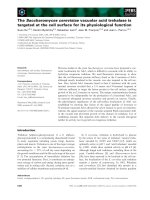
Fig. 1. An LF double mutant, LF-K518E ⁄ E682G. (A) Structure of
the catalytic domain of LF. Amino acids 518 and 682 are shown in
red. Residues of the HExxH motif are shown in green. An opti-
mized peptide substrate is shown in blue. The model was created
using coordinates from Protein Data Bank 1PWW [23] and the com-
puter programs
VMD 1.8.3 [26] and POV-RAY 3.6 (Williamstown,
Victoria, Australia). (B) Limited tryptic digest of wild-type and
mutant LF. LF or LF-K518E ⁄ E682G was incubated with the indi-
cated concentrations of trypsin for 1 h. Protein samples were sub-
jected to SDS ⁄ PAGE and stained with Coomassie blue.
An LF mutant with altered activity S. Ngai et al.
120 FEBS Journal 277 (2010) 119–127 ª 2009 The Authors Journal compilation ª 2009 FEBS
that form the HExxH(686–690) metalloprotease motif
(Fig. 1A) [23]. We performed a limited tryptic
digestion to assess whether the mutations altered the
tertiary structure of LF. Purified wild-type LF or
LF-K518E ⁄ E682G was incubated with various con-
centrations of trypsin, and the mixtures were then
subjected to SDS ⁄ PAGE. Differences between the
patterns of tryptic fragments were observed for
LF-K518E ⁄ E682G and wild-type LF, and the mutant
appeared to be somewhat more sensitive to trypsin
(Fig. 1B). This suggested that although the mutations
altered the tertiary structure of the protein, they did
not cause it to become grossly misfolded and destabi-
lized. As we were interested in characterizing a mutant
with altered catalytic properties, rather than identifying
amino acids that might bind substrates directly, we
decided to study this mutant further.
We first assessed the severity of the cytotoxicity
defect caused by the mutations. PA and various
concentrations of either wild-type LF or LF-K518E ⁄
E682G were incubated with RAW 264.7 cells for 4 h,
and cell viability was estimated using the 3-(4,5-
dimethylthiazol-2-yl)-5-(3-carboxymethoxyphenyl)-2-(4-
sulfophenyl)-2H-tetrazolium (MTS) assay, which
measures mitochondrial function. Whereas the concen-
tration of LF required to kill 50% of the cells (EC50)
was estimated to be 4 · 10
)11
m, LF-K518E ⁄ E682G
did not cause enough cell death under these conditions
for an accurate EC
50
to be determined (Fig. 2A).
Increasing the duration of toxin exposure from 4 h to
24 h did not markedly decrease the EC50 for wild-type
LF or decrease the viability of cells exposed to the
mutant (data not shown). The reduced ability of the
LF mutant to kill RAW 264.7 cells was tested further,
using a trypan blue exclusion assay (Fig. 2B). Cells
were left untreated, or were exposed to a mixture of
10
)8
m PA and 10
)8
m wild-type LF or mutant LF for
either 4 h or 24 h, and the fraction of cells that
excluded trypan blue under each condition was deter-
mined. Similar to what was observed with the MTS
assay, this assay indicated that LF-K518E ⁄ E682G was
less cytotoxic than wild-type LF; increasing the dura-
tion of toxin incubation from 4 h to 24 h did not lead
to an increased level of cell death (Fig. 2B).
To confirm that LF-K518E ⁄ E682G was defective at
activating Nlrp1b, we used an independent approach
that takes advantage of a recently developed heterolo-
gous expression system [24]. HT1080 human fibro-
blasts were transfected with plasmids encoding murine
Nlrp1b, procaspase-1 and pro-interleukin (IL)-1b, and
after 24 h the cells were treated with combinations
of PA, LF, and LF-K518E ⁄ E682G. PA and LF acti-
vated the inflammasome, as determined by the loss of
A
B
C
Fig. 2. LF-K518E ⁄ E682G is defective in killing RAW 264.7 cells and
inducing the Nlrp1b inflammasome. (A) PA and various concentra-
tions of wild-type (WT) LF (m) or LF-K518E ⁄ E682G (s) were incu-
bated with RAW 264.7 cells, and viability was assessed after 4 h,
using the MTS assay. Values represent the mean ± standard error
of the mean for three independent experiments. (B) RAW 264.7
cells were left untreated (black bars) or treated with PA and wild-
type LF (white bars) or PA and LF-K518E ⁄ E682G (gray bars) for 4 h
or 24 h. Viability was assessed as the fraction of cells that
excluded trypan blue. Values represent the mean ± standard error
of the mean for three independent experiments. (C) HT1080 cells
were mock transfected or were transfected with plasmids encod-
ing Nlrp1b, procaspase-1, and pro-IL-1b. After 24 h, cells were trea-
ted with PA and either wild-type LF or LF-K518E ⁄ E682G. IL-1b,
MAPKK1 and b-actin were detected by immunoblotting. The results
shown represent three independent experiments. IB, immunoblot;
IP, immunoprecipitation.
S. Ngai et al. An LF mutant with altered activity
FEBS Journal 277 (2010) 119–127 ª 2009 The Authors Journal compilation ª 2009 FEBS 121
pro-IL-1b in the cytosol and the appearance of IL-1b
in the cell supernatants (Fig. 2C). A lower level of
IL-1b was found in the supernatants of cells treated
with LF-K518E ⁄ E682G, suggesting that the mutant
was defective at activating the inflammasome. LF-
K518E ⁄ E682G entered cells and was catalytically
active, however, because it cleaved MAPKK1
(Fig. 2C).
As it is unclear whether cleavage of MAPKKs by
LF causes pyroptosis of RAW 264.7 cells, we
attempted to correlate cyotoxicity with downregulation
of the MAPK pathways. RAW 264.7 cells were treated
with PA and either wild-type LF or LF-K518E ⁄
E682G, and the cells were then stimulated with lipo-
polysaccharide to activate the signaling pathways.
Cellular lysates were prepared and probed for phos-
phorylated ERK, p38 and JNK by western blotting
(Fig. 3). Exposure of cells to PA and increasing
concentrations of wild-type LF for 1 h resulted in
decreased phosphorylation of the three MAPKs.
Interestingly, increasing the LF concentration from
10
)11
m to 10
)10
m had a considerable effect on cell
viability, but relatively minor effects on the phosphory-
lation of the MAPKs (compare Figs 2A and 3). LF-
K518E ⁄ E682G decreased phosphorylation of ERK
almost as effectively as wild-type LF, but did not
decrease phosphorylation of p38 or JNK below the
level observed in cells treated with lipopolysaccharide
alone. Thus, whereas wild-type LF interfered with sig-
naling in all three MAPK pathways, LF-
K518E ⁄ E682G selectively downregulated the ERK
pathway.
To examine why the mutant demonstrated increased
specificity in downregulating the ERK pathway, we
next compared the abilities of wild-type LF and LF-
K518E ⁄ E682G to cleave MAPKKs (Fig. 4). MAPKK1
and MAPKK2, which phosphorylate ERK, were both
cleaved by wild-type LF as assessed by western blot-
ting. At the highest concentration of LF tested
(10
)8
m), 50% of MAPKK1 and 60% of MAP-
KK2 was cleaved after 1 h. Treatment of cells with PA
and 10
)8
m LF-K518E ⁄ E682G resulted in 50% of
A
B
C
D
Fig. 3. LF-K518E ⁄ E682G inhibits the phosphorylation of ERK, but not of p38 or JNK. RAW 264.7 cells were treated with 10
)8
M PA and the
indicated concentrations of either wild-type (WT) LF or LF-K518E ⁄ E682G for 1 h, and then treated with lipopolysaccharide for 15 min. Cellu-
lar lysates were made and probed for phosphorylated MAPKs or a-tubulin control by western blotting. Representative blots are shown in (A).
(B–D) Results of quantifying the levels of phosphorylated proteins in toxin-treated cells as compared with cells that were not treated with
toxin. Values represent the mean ± standard error of the mean for three independent experiments.
An LF mutant with altered activity S. Ngai et al.
122 FEBS Journal 277 (2010) 119–127 ª 2009 The Authors Journal compilation ª 2009 FEBS
MAPKK1 and 20% of MAPKK2 being cleaved. As
the mutant was able to downregulate the ERK path-
way almost as efficiently as wild-type LF, these results
suggest that MAPKK1 is primarily responsible for
ERK activation under these conditions.
We next sought to determine the cause of
the mutant’s deficiency in downregulating p38 by
examining the cleavage of MAPKK3 and MAPKK6.
LF-K518E ⁄ E682G was modestly defective in cleaving
MAPKK3 as compared with wild-type LF, but was
considerably more defective in cleaving MAPKK6.
The inability of the mutant to prevent phosphorylation
of p38 (Fig. 3) indicated that the level of MAPPK3 ⁄ 6
that remained in the cell was sufficient to support
maximal p38 phosphorylation.
We next probed cellular lysates for MAPKK4 and
MAPKK7, which phosphorylate JNK. LF-K518E ⁄
E682G cleaved similar amounts of MAPKK4 as wild-
type LF. Neither wild-type LF nor the mutant cleaved
appreciable amounts of MAPKK7 after 1 h of toxin
Fig. 4. LF-K518E ⁄ E682G has reduced ability to cleave some MAPKKs. RAW 264.7 cells were treated with 10
)8
M PA and the indicated con-
centrations of either wild-type (WT) LF or LF-K518E ⁄ E682G for 1 h. Cellular lysates were prepared and probed for phosphorylated MAPKKs
by western blotting. The amount of full-length MAPKK remaining after 1 h was quantified. Values represent the mean ± standard error of
the mean for three independent experiments.
S. Ngai et al. An LF mutant with altered activity
FEBS Journal 277 (2010) 119–127 ª 2009 The Authors Journal compilation ª 2009 FEBS 123
treatment. Thus, wild-type LF and LF-K518E ⁄ E682G
exhibited similar activities towards MAPKK4 and
MAPKK7, but only wild-type LF reduced the level of
phosphorylation of JNK to 50% as compared with
the control. There is no evident explanation for these
results; the difference in JNK phosphorylation
observed might be due to an indirect effect of intoxica-
tion.
As downregulation of the ERK pathway has been
shown to be sufficient to cause apoptosis in MALME-
3M cells [20], we next compared the activities of wild-
type LF and LF-K518E ⁄ E682G in a cytotoxicity assay
using this melanoma cell line. PA and either wild-type
or mutant LF were incubated with MALME-3M cells
for 72 h, and viability was estimated using the MTS
assay (Fig. 5A) [20]. The EC50 for wild-type LF was
determined to be 2 · 10
)13
m, and the EC50 for
LF-K518E ⁄ E682G was only about three-fold higher
at 7 · 10
)13
m. These results indicate that LF-K518E ⁄
E682G is markedly more defective, in comparison with
wild-type LF, in killing the murine macrophage cells
than in killing the melanoma cells. We next assessed
the phosphorylation of ERK in MALME-3M cells
treated with either wild-type or mutant LF, and found
that LF-K518E ⁄ E682G downregulated the ERK path-
way almost as effectively as wild-type LF did
(Fig. 5B). This is consistent with previous work indi-
cating the requirement of ERK signaling for survival
of these cells, and suggests that different types of cells
are killed by LF as a result of the cleavage of distinct
substrates.
To summarize, we have isolated an LF mutant that
is impaired in its ability to activate the Nlrp1b inflam-
masome, but remains able to cause apoptosis in a mela-
noma cell line. LF-K518E ⁄ E682G activity prevented
phosphorylation of ERK, but did not prevent phos-
phorylation of JNK or p38. This observation serves to
explain why the mutant retains its ability to kill the
melanoma cells, as it has been shown previously
that inhibition of the ERK pathway is sufficient to
induce apoptosis. It is unclear why the mutant is defec-
tive at causing pyroptosis, but it is presumably because
LF-K518E ⁄ E682G has a diminished capacity to cleave
a substrate that is involved in the activation of Nlrp1b.
Experimental procedures
Reagents
Antibodies raised against the N-terminus of MAPKK1
(catalog no. 07-641) or full-length MAPKK6 (catalog
no. 07-417) were obtained from Upstate (Lake Placid, NY,
USA). Antibody raised against the N-terminus of
MAPKK2 (catalog no. 610235) was obtained from BD Bio-
sciences (San Jose, CA, USA). Antibodies raised against
the N-termini of MAPKK3b (catalog no. 9238), MAPKK4
(catalog no. 9152) and MAPKK7 (catalog no. 4172) were
obtained from Cell Signaling Technologies. Antibodies that
detect phospho-p38 (catalog no. 9215) and phospho-ERK
(catalog no. 9101) were obtained from Cell Signaling Tech-
nologies; and antibody against phospho-JNK was obtained
from Biosource (catalog no. 44-682). A control antibody,
against a-tubulin (T9026), was obtained from Sigma-
Aldrich Canada (Oakville, Canada).
Tryptic digestion of LF
Various amounts of trypsin were incubated with 2 lgof
LF or LF-K518E ⁄ E682G for 1 h at 23 °C in a total volume
of 10 lLof20mm Tris ⁄ HCl (pH 8.0) and 150 mm NaCl.
Digested proteins were subjected to SDS ⁄ PAGE and
stained with Coomassie blue.
Fig. 5. LF-K518E ⁄ E682G causes death of melanoma cells. (A) PA
and various concentrations of wild-type LF (m) or LF-K518E ⁄ E682G
(s) were incubated with MALME-3M cells, and viability was
assessed after 72 h, using the MTS assay. Values represent the
mean ± standard error of the mean for three independent experi-
ments. (B) MALME-3M cells were treated with 10
)8
M PA and
the indicated concentrations of either wild-type (WT) LF or
LF-K518E ⁄ E682G for 2 h, and then treated with 2.5 lgmL
)1
aniso-
mycin for 15 min. Cellular lysates were prepared, and probed for
phosphorylated MAPKs. Values indicate the level of phosphorylated
MAPK in toxin-treated cells as a fraction of the level in cells that
were not treated with toxin. The results shown represent the
mean ± standard error of the mean for three independent
experiments.
An LF mutant with altered activity S. Ngai et al.
124 FEBS Journal 277 (2010) 119–127 ª 2009 The Authors Journal compilation ª 2009 FEBS
Cell lines
Murine macrophage RAW 264.7 cells (ATCC) were cul-
tured in RPMI-1640 supplemented with 5% fetal bovine
serum (HyClone) and 1% penicillin ⁄ streptomycin (Sigma)
at 37 °C in a humidified atmosphere of 5% CO
2
.
MALME-3M cells (ATCC) were cultured in RPMI-1640
supplemented with 10% Nu-Serum (BD Biosciences) and
1% penicillin ⁄ streptomycin.
Protein purification
PA was purified from Escherichia coli as described previ-
ously [25].
The plasmids pWH1520–LF-K518E ⁄ E682G and pWH1520–
LF were transformed into Bacillus megaterium protoplasts
according to the manufacturer’s instructions (MoBiTec). An
overnight culture of B. megaterium expressing either wild-
type LF or LF-K518E ⁄ E682G was used to inoculate 500 mL
of TB containing 10 lgmL
)1
tetracycline. The culture was
grown at 37 °C until a D
600 nm
of 0.8 was reached. At this
time, the expression of LF was induced in the supernatant by
the addition of 20% xylose (Sigma-Aldrich) to a final concen-
tration of 0.5%. The culture was grown for another 4–4.5 h,
and then centrifuged at 7000 g for 30 min in a Sorvall Evolu-
tion RC centrifuge. The supernatant was decanted into
500 mL of autoclaved 40% poly(ethylene glycol) (PEG) 8000
(Sigma), and the resulting solution was rotated overnight at
4 °C. The solution was centrifuged at 9500 g for 30 min, and
the supernatant was decanted. The pellet was resuspended in
10 mL of supernatant, and then centrifuged at 20 000 g for
30 min. The supernatant was decanted, and 10 mL of 20 mm
Tris ⁄ HCl (pH 8.0) was used to dissolve the pellet. The sam-
ple was then centrifuged at 20 000 g for 10 min to remove
undissolved material. The resulting supernatant was filtered
using a 0.2-lm syringe filter (Pall Sciences, Port Washington,
NY, USA) and loaded onto a column containing 1mLof
Q-sepharose (Amersham Pharmacia Biotech, Baie d¢Urfe,
Canada). The column was washed first with 10 mL of 20 mm
Tris ⁄ HCl (pH 8.0) and then with 20 mm Tris ⁄ HCl (pH 8.0)
and 0.15 m NaCl. LF was eluted in 20 mm Tris ⁄ HCl
(pH 8.0) and 0.25 m NaCl.
Cytotoxicity assays
For the MTS assay, RAW 264.7 cells were seeded in 96-
well plates at a density of 1 · 10
5
cells per 100 lL of med-
ium for 24 h, and MALME-3M cells were seeded in 96-well
plates at a density of 3 · 10
4
cells per 100 lL of medium
for 24 h. Cells were washed once with NaCl ⁄ P
i
, and then
incubated in medium with 1 · 10
)8
m PA and various con-
centrations of LF. The viability of RAW 264.7 cells was
assessed after 4 h and 24 h, and that of MALME-3M cells
after 72 h, using the MTS assay according to the manufac-
turer’s instructions (Promega). The EC50 values were deter-
mined using the graphical program graphpad prism 4
(GraphPad, La Jolla, CA, USA).
For the trypan blue exclusion assay, 3 · 10
6
RAW 264.7
cells per well were seeded in six-well plates. Cells were
washed once with NaCl ⁄ P
i
, and then incubated in medium
with 1 · 10
)8
m PA and 1 · 10
)8
m LF for 4 h or 24 h.
The cells were resuspended in medium, stained with trypan
blue, and counted using a hemocytometer.
Nlrp1b reconstitution assay
The Nlrp1b reconstitution assay was performed as described
previously [24]. Briefly, HT1080 cells were transfected with
1 lg each of pNTAP–Nlrp1b, pcDNA3–procaspase-1–T7,
and pcDNA3–pro-IL-1b–hemagglutinin (HA), using 9 lL
of 1 mg mL
)1
polyethyleneimine (pH 7.2). Cells were trea-
ted with 10
)8
m LF and 10
)8
m PA for 3 h. The culture
supernatant was incubated overnight with 1 lL of antibody
against a-HA (H9658; Sigma-Aldrich), and then for 2 h
with 100 lL of protein A Sepharose (GE Healthcare). Pro-
teins were eluted with SDS loading dye and subjected to
immunoblotting using a polyclonal antibody against HA
(sc805; Santa Cruz Biotechnology, Santa Cruz, CA, USA).
Cell pellets were harvested, and then lysed with 300 lL
of EBC buffer (0.5% NP-40, 20 mm Tris, pH 8, 150 mm
NaCl, 1 mm phenylmethanesulfonyl fluoride) for 60 min.
Equivalent amounts of cell lysate protein ( 30 lg) were
subjected to SDS ⁄ PAGE and immunoblotted with a-HA
(sc805) and a-b-actin (A5441; Sigma-Aldrich) antibodies.
Western blot experiments
RAW 64.7 cells were seeded into six-well plates at 10
6
cells
per well. Following overnight incubation, cells were treated
for 1 h with medium alone, or with 1 · 10
)8
m PA and
the indicated concentrations of either wild-type LF or
LF-K518E ⁄ E682G. Lipopolysaccharide (100 ng mL
)1
) was
added to all wells for 15 min. Cells were harvested in
500 lLof1· Cell Lysis Buffer (Cell Signaling Technologies,
Danvers, MA, USA) containing 1 mm phenylmethanesulfo-
nyl fluoride (Sigma), and sonicated four times for a total of
40 s, using a Sonic Dismembrator Model 100 (Fisher Scien-
tific, Pittsburgh, PA, USA). Cell lysates were then clarified
by microcentrifugation at 15 000 g (Eppendorf Centrifuge
5415D) for 10 min at 4 °C. Equal amounts of lysate were
electrophoresed on 10% SDS ⁄ PAGE gels. Proteins were
transferred to nitrocellulose (Pall Life Science), using a
Mini Trans-Blot Electrophoretic Transfer Cell (Bio-Rad,
Mississauga, Canada). Blots were blocked for 1 h in 0.1%
Tween-20 NaCl ⁄ Tris (100 mm Tris ⁄ HCl, pH 8.0, 0.9%
NaCl) containing 5% powdered skimmed milk. Blots were
incubated with primary antibodies diluted according to
the manufacturer’s instructions. Blots were then rinsed
three times in 0.05% Tween-20 NaCl ⁄ Tris and incubated
with either peroxidase-conjugated goat anti-rabbit or goat
S. Ngai et al. An LF mutant with altered activity
FEBS Journal 277 (2010) 119–127 ª 2009 The Authors Journal compilation ª 2009 FEBS 125
anti-mouse IgG secondary antibodies (Pierce, Rockford, IL,
USA) in 0.1% Tween-20 NaCl ⁄ Tris containing 5% pow-
dered skimmed milk. Blots were incubated with SuperSignal
West Dura Extended Duration Substrate (Pierce) for 5 min,
and then visualized using a Kodak Gel-Image Station
2000R.
Acknowledgements
This research was supported by NIH grant RO1
AI067683. J. Mogridge holds the Canada Research
Chair in Bacterial Pathogenesis.
References
1 Abrami L, Reig N & van der Goot FG (2005) Anthrax
toxin: the long and winding road that leads to the kill.
Trends Microbiol 13, 72–78.
2 Young JA & Collier RJ (2007) Anthrax toxin: receptor
binding, internalization, pore formation, and transloca-
tion. Annu Rev Biochem 76, 243–265.
3 Warfel JM, Steele AD & D’Agnillo F (2005) Anthrax
lethal toxin induces endothelial barrier dysfunction.
Am J Pathol 166, 1871–1881.
4 Gozes Y, Moayeri M, Wiggins JF & Leppla SH (2006)
Anthrax lethal toxin induces ketotifen-sensitive intrader-
mal vascular leakage in certain inbred mice. Infect
Immun 74, 1266–1272.
5 Moayeri M, Haines D, Young HA & Leppla SH (2003)
Bacillus anthracis lethal toxin induces TNF-alpha-inde-
pendent hypoxia-mediated toxicity in mice. J Clin Invest
112, 670–682.
6 Cui X, Moayeri M, Li Y, Li X, Haley M, Fitz Y,
Correa-Araujo R, Banks SM, Leppla SH & Eichacker
PQ (2004) Lethality during continuous anthrax lethal
toxin infusion is associated with circulatory shock but
not inflammatory cytokine or nitric oxide release in
rats. Am J Physiol Regul Integr Comp Physiol 286,
R699–R709.
7 Baldari CT, Tonello F, Paccani SR & Montecucco C
(2006) Anthrax toxins: a paradigm of bacterial immune
suppression. Trends Immunol 27, 434–440.
8 Banks DJ, Ward SC & Bradley KA (2006) New insights
into the functions of anthrax toxin. Expert Rev Mol
Med 8, 1–18.
9 Tournier JN, Rossi Paccani S, Quesnel-Hellmann A &
Baldari CT (2009) Anthrax toxins: a weapon to system-
atically dismantle the host immune defenses. Mol
Aspects Med, doi:10.1016/j.mam.2009.06.002.
10 Duesbery NS, Webb CP, Leppla SH, Gordon VM,
Klimpel KR, Copeland TD, Ahn NG, Oskarsson MK,
Fukasawa K, Paull KD et al. (1998) Proteolytic inacti-
vation of MAP-kinase-kinase by anthrax lethal factor.
Science 280, 734–737.
11 Vitale G, Bernardi L, Napolitani G, Mock M &
Montecucco C (2000) Susceptibility of mitogen-
activated protein kinase kinase family members to
proteolysis by anthrax lethal factor. Biochem J 352 Pt
3, 739–745.
12 Abi-Habib RJ, Urieto JO, Liu S, Leppla SH,
Duesbery NS & Frankel AE (2005) BRAF status and
mitogen-activated protein ⁄ extracellular signal-regulated
kinase kinase 1 ⁄ 2 activity indicate sensitivity of mela-
noma cells to anthrax lethal toxin. Mol Cancer Ther 4,
1303–1310.
13 Maldonado-Arocho FJ, Fulcher JA, Lee B & Bradley
KA (2006) Anthrax oedema toxin induces anthrax toxin
receptor expression in monocyte-derived cells. Mol
Microbiol 61, 324–337.
14 Boyden ED & Dietrich WF (2006) Nalp1b controls
mouse macrophage susceptibility to anthrax lethal
toxin. Nat Genet 38, 240–244.
15 Fink SL, Bergsbaken T & Cookson BT (2008)
Anthrax lethal toxin and Salmonella elicit the common
cell death pathway of caspase-1-dependent pyroptosis
via distinct mechanisms. Proc Natl Acad Sci USA 105,
4312–4317.
16 Wickliffe KE, Leppla SH & Moayeri M (2008)
Anthrax lethal toxin-induced inflammasome formation
and caspase-1 activation are late events dependent on
ion fluxes and the proteasome. Cell Microbiol 10,
332–343.
17 Nour AM, Yeung YG, Santambrogio L, Boyden ED,
Stanley ER & Brojatsch J (2009) Anthrax lethal toxin
triggers the formation of a membrane-associated inflam-
masome complex in murine macrophages. Infect Immun
77, 1262–1271.
18 Park JM, Greten FR, Li ZW & Karin M (2002)
Macrophage apoptosis by anthrax lethal factor
through p38 MAP kinase inhibition. Science 297,
2048–2051.
19 Kim SO, Jing Q, Hoebe K, Beutler B, Duesbery NS &
Han J (2003) Sensitizing anthrax lethal toxin-resistant
macrophages to lethal toxin-induced killing by tumor
necrosis factor-alpha. J Biol Chem 278, 7413–7421.
20 Koo HM, VanBrocklin M, McWilliams MJ, Leppla
SH, Duesbery NS & Woude GF (2002) Apoptosis and
melanogenesis in human melanoma cells induced by
anthrax lethal factor inactivation of mitogen-activated
protein kinase kinase. Proc Natl Acad Sci USA 99,
3052–3057.
21 Frankel AE, Koo HM, Leppla SH, Duesbery NS &
Vande Woude GF (2003) Novel protein targeted ther-
apy of metastatic melanoma. Curr Pharm Des 9, 2060–
2066.
22 Liang X, Young JJ, Boone SA, Waugh DS & Duesbery
NS (2004) Involvement of domain II in toxicity of
anthrax lethal factor. J Biol Chem 279, 52473–52478.
An LF mutant with altered activity S. Ngai et al.
126 FEBS Journal 277 (2010) 119–127 ª 2009 The Authors Journal compilation ª 2009 FEBS
23 Turk BE, Wong TY, Schwarzenbacher R, Jarrell ET,
Leppla SH, Collier RJ, Liddington RC & Cantley LC
(2004) The structural basis for substrate and inhibitor
selectivity of the anthrax lethal factor. Nat Struct Mol
Biol 11, 60–66.
24 Liao KC & Mogridge J (2009) Expression of Nlrp1b
inflammasome components in human fibroblasts confers
susceptibility to anthrax lethal toxin. Infect Immun 77,
4455–4462.
25 Miller CJ, Elliott JL & Collier RJ (1999) Anthrax
protective antigen: prepore-to-pore conversion.
Biochemistry 38, 10432–10441.
26 Humphrey W, Dalke A & Schulten K (1996) VMD:
visual molecular dynamics. J Mol Graph 14, 33–38.
S. Ngai et al. An LF mutant with altered activity
FEBS Journal 277 (2010) 119–127 ª 2009 The Authors Journal compilation ª 2009 FEBS 127