Tài liệu Báo cáo khoa học: 3T3-L1 adipocyte apoptosis induced by thiazolidinediones is peroxisome proliferator-activated receptor-c-dependent and mediated by the caspase-3-dependent apoptotic pathway doc
Bạn đang xem bản rút gọn của tài liệu. Xem và tải ngay bản đầy đủ của tài liệu tại đây (772.37 KB, 10 trang )
3T3-L1 adipocyte apoptosis induced by thiazolidinediones
is peroxisome proliferator-activated receptor-c-dependent
and mediated by the caspase-3-dependent apoptotic
pathway
Yuanyuan Xiao, Taichang Yuan, Wenqi Yao and Kan Liao
State Key Laboratory of Molecular Biology, Institute of Biochemistry and Cell Biology, Shanghai Institutes for Biological Sciences, Chinese
Academy of Sciences, Shanghai, China
Introduction
To date, studies of adipocyte physiology have primarily
focused on adipogenesis [1–3]. The interaction between
extracellular signals and the transcriptional cascade dur-
ing adipogenesis has been well studied [4]. However,
owing to the remarkable ability of adipocytes to resist
apoptosis, adipocyte apoptosis is much less studied and
is poorly understood. Recently, evidence from several
in vivo and in vitro studies has indicated that apoptosis
is a significant factor in adipocyte depletion during
weight reduction [5–8]. In addition, an animal model of
adipocyte apoptosis has been developed as a tool for the
study of obesity-related diseases [9]. Adipocyte apopto-
sis is induced by some adipokines, such as leptin and
tumor necrosis factor-a [10–12]. Additionally, natural
Keywords
3T3-L1 adipocyte; adipocyte apoptosis;
Akt-1; PPARc; thiazolidinediones
Correspondence
K. Liao, Institute of Biochemistry and Cell
Biology, 320 Yueyang Road, Shanghai
200031, China
Fax: +86 21 54921011
Tel: +86 21 54921113
E-mail:
(Received 11 March 2009, revised 16
November 2009, accepted 24 November
2009)
doi:10.1111/j.1742-4658.2009.07514.x
Although thiazolidinediones (TZDs) are potent promoters of adipogenesis
in the preadipocyte, they induce apoptosis in several other cell types,
such as cancer cells, endothelial cells and T-lymphocytes. In this study,
we investigated the proapoptotic effect of TZDs in mature 3T3-L1
adipocytes, which express high levels of the peroxisome proliferator-acti-
vated receptor-c (PPARc) protein. Apoptosis was induced in mature
3T3-L1 adipocytes by treatment with troglitazone, pioglitazone or prosta-
glandin J2, and could be blocked by the PPARc antagonist GW9662.
Treatment with PPARc agonists also decreased Akt-1 protein and phos-
phorylation levels without affecting phosphoinositide 3-kinase and PTEN.
Further analysis indicated that in troglitazone-treated 3T3-L1 adipocytes,
Bad phosphorylation and Bcl-2 protein levels were reduced, and Bax
translocation to the mitochondria was increased. Subsequently, cyto-
chrome c release and caspase-3 cleavage were observed. TZD-induced
adipocyte apoptosis could be blocked by the caspase-3 inhibitor
Ac-DEVD-CHO or by overexpression of Bcl2. In cultured rat primary
adipocytes, similar apoptosis-inducing effects of troglitazone were also
observed. Thus, TZDs promote apoptosis in adipocytes through a
PPARc-dependent pathway. This apoptosis is mediated by the inhibition
of Akt-1, which decreases Bad phosphorylation and activates the mito-
chondrial apoptotic pathway.
Abbreviations
AO, acridine orange; EB, ethidium bromide; FACS, fluorescence-activated cell sorting; pBad, phosphorylated Bad; pAkt-1, phosphorylated
Akt-1; PGJ2, 15-deoxy-D12,14-prostaglandin J; 2PI, propidium iodide; PI3K, phosphoinositide 3-kinase; PPARc, peroxisome proliferator-
activated receptor-c; SD, standard deviation; TUNEL, terminal deoxynucleotidyl transferase dUTP nick end labeling; TZD, thiazolidinedione.
FEBS Journal 277 (2010) 687–696 ª 2009 The Authors Journal compilation ª 2009 FEBS 687
compounds can be screened for their ability to induce
adipocyte apoptosis, allowing the identification of
potential obesity drugs [13–15]. Although the regulation
of adipocyte apoptosis is not fully understood, it pro-
vides a potential target for treatment of obesity and its
related diseases.
Thiazolidinediones (TZDs), such as troglitazone,
rosiglitazone and pioglitazone, are drugs used clinically
for type II diabetes. TZDs activate the adipogenic
transcription factor peroxisome proliferator-activated
receptor-c (PPARc) and induce metabolic changes in
adipose tissue [16]. The mechanism of insulin sensitiza-
tion by TZDs has been explored, but is not fully
understood. In vivo studies have indicated that TZDs
may increase the number of small adipocytes by pro-
moting adipocyte differentiation, and decrease the
number of large adipocytes by inducing adipocyte
apoptosis [17,18].
PPARc is highly expressed in adipose tissue, and is
considered to be the ‘master switch’ of adipocyte dif-
ferentiation. Upon ligand binding, PPARc heterodi-
merizes with the retinoid X receptor, translocates into
the nucleus, and activates multiple genes, especially
adipogenic genes [19]. In the mature adipocyte, PPARc
regulates the expression of genes involved in lipid
metabolism [19]. Besides its function in adipogenesis,
PPARc is also implicated in leptin-induced adipocyte
apoptosis [10]. During leptin-induced adipocyte apop-
tosis, the expression of PPARc is increased by 80%
[10]. Furthermore, the activation of PPARc stimulates
apoptosis in a variety of cell types, including cancer
cells, endothelial cells and T-lymphocytes [20–25].
These observations provided the impetus for us to
study the effect of TZDs and PPARc on adipocyte
apoptosis.
In comparison with other cell types, adipocytes are
quite resistant to apoptosis. The expression of high
level of PPARc protein in adipocytes, however, should
make them more susceptible to PPARc agonist-
induced apoptosis [19–25]. Although TZDs are potent
PPARc agonists and inducers of adipocyte differentia-
tion, their function in inducing adipocyte apoptosis
has not been fully explored. 3T3-L1 adipocytes consti-
tute a good model system for studying PPARc ago-
nist-induced apoptosis [26,27]. In the present study, we
investigated PPARc agonist-induced adipocyte apopto-
sis by using 3T3-L1 adipocytes and rat primary adipo-
cytes. Adipocyte apoptosis could be induced by
troglitazone, pioglitazone or 15-deoxy-D12,14-prosta-
glandin J2 (PGJ2), and reversed by GW9662, a
PPARc antagonist. Further analysis suggests that
TZDs induce the downregulation of Akt-1 and lead to
the activation of caspase-3-dependent apoptosis.
Results
TZD-induced apoptosis in 3T3-L1 adipocytes
After 3T3-L1 adipocyte differentiation, the expression
of PPARc was greatly increased (Fig. 1A). Troglitaz-
one, an agonist of PPARc, induced apoptosis in
mature 3T3-L1 adipocytes (Fig. 1B,C). Troglitazone-
induced adipocyte apoptosis could be detected by acri-
dine orange (AO) ⁄ ethidium bromide (EB) staining or
Hoechst 33258 staining (Fig. 1B,C). Quantitative anal-
ysis by flow cytometry indicated that adipocyte apop-
tosis increased with prolonged troglitazone treatment
(Fig. 1D). Propidium iodide (PI) staining and terminal
deoxynucleotidyl transferase dUTP nick end labeling
(TUNEL) assay produced similar results (Fig. 1E,F).
Representative fluorescence-activated cell sorting
(FACS) plots of PI staining are shown in Fig. S1.
GW9662 is an antagonist of PPARc, and irrevers-
ibly inhibits PPARc activity [28]. During induction of
3T3-L1 adipocyte differentiation, the presence of
GW9662 completely blocked differentiation (Fig. 2A).
As troglitazone-induced adipocyte apoptosis is proba-
bly mediated by PPARc, GW9662 should have an
inhibitory effect on adipocyte apoptosis. As shown in
Fig. 2B,C, troglitazone-induced adipocyte apoptosis
was, indeed, inhibited in the presence of GW9662. In
addition, pioglitazone, another TZD, and PGJ2, which
directly binds to and activates PPARc [29], also induce
adipocyte apoptosis, and GW9662 treatment blocked
this induction (Fig. 2D–F). Taken together, these
results suggested that TZD-induced adipocyte apopto-
sis is PPAR c-dependent.
Inhibition of Akt-1 in adipocyte apoptosis
The phosphoinositide 3-kinase (PI3K)–Akt-1 signaling
cascade is essential for 3T3-L1 adipocyte differentiation
[30]. In many cells, the PI3K–Akt-1 signaling cascade is
also one of the important signals for cell survival [31].
To investigate the function of the PI3K–Akt-1 signaling
pathway in troglitazone-induced adipocyte apoptosis,
the components of the PI3K–Akt-1 signaling cascade
were analyzed. As shown in Fig. 3A,B, treatment of
3T3-L1 adipocytes with troglitazone reduced the levels
of Akt-1 protein and inhibited its phosphorylation. The
densitometer scanning data of Akt-1 western blot are
shown in Fig. S1. However, other components of the
PI3K–Akt-1 signaling cascade, such as PI3K and
PTEN, were not affected by troglitazone (Fig. 3C).
Troglitazone activates PPARc, a transcription factor
responsible for the expression of many adipogenic
genes [19]. Akt-1 did not appear to be the target gene
Apoptosis induced by TZDs in 3T3-L1 adipocyte Y. Xiao et al.
688 FEBS Journal 277 (2010) 687–696 ª 2009 The Authors Journal compilation ª 2009 FEBS
of PPARc, because the expression of Akt-1 was only
slightly induced by troglitazone, whereas the expres-
sion levels of two PPARc target genes, adipose fatty
acid-binding protein (aP2) and CD36, were greatly
increased in troglitazone-treated 3T3-L1 adipocytes
(Fig. 3D). Thus, the reduction of Akt-1 protein level in
troglitazone-treated adipocytes was most likely due to
decreased Akt-1 protein stability.
GW9662 treatment not only blocked troglitazone-
induced adipocyte apoptosis (Fig. 2B,C), but also
restored the troglitazone-induced decrease in Akt-1
protein level and its phosphorylation (Fig. 3E).
GW9662 treatment also reversed the downregulation
of Akt-1 protein level in pioglitazone-induced or
PGJ2-induced apoptotic adipocytes (Fig. 3F,G).
Downregulation of Akt-1 protein level and inhibition
of its phosphorylation appeared to be common effects
in TZD-induced adipocyte apoptosis.
Troglitazone induces adipocyte apoptosis
through the mitochondrial pathway
Bad is one of the prodeath proteins in the Bcl-2 fam-
ily, and interacts with prosurvival Bcl-2 family mem-
bers to inactivate them [32]. Akt-1 phosphorylates Bad
at Ser136, inactivating Bad and thereby promoting cell
survival [33]. As shown in Fig. 4A, the basal phos-
phorylation level of Bad gradually decreased when the
cells were treated with troglitazone. The decreased
Akt-1 activity in TZD-treated adipocytes might regu-
late adipocyte apoptosis through Bad activity. In addi-
tion, troglitazone treatment decreased the protein level
of prosurvival Bcl-2, but not of prodeath Bax
(Fig. 4A). Consequently, the Bax ⁄ Bcl-2 ratio was
increased, promoting apoptosis.
In the Bcl-2 family, the disequilibrium of prodeath
and prosurvival proteins leads to the translocation of
A
B
C
D
EF
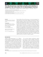
Fig. 1. Troglitazone-induced 3T3-L1 adipocyte apoptosis (A) Oil Red O-stained 3T3-L1 adipocytes 8 days after differentiation induction.
PPARc protein was detected by western blot with an antibody against PPARc. Pre, 3T3-L1 preadipocytes; Adi, differentiated 3T3-L1 adipo-
cytes. (B) AO ⁄ EB staining for apoptotic 3T3-L1 adipocytes. Scale bar: 50 lm. 3T3-L1 adipocytes were treated with 12.5 l
M troglitazone (Tro)
(12.5 l
M) or not (Control) for 96 h, stained with AO ⁄ EB, and visualized by confocal microscopy. (C) Hoechst 33258 staining for apoptotic
3T3-L1 adipocytes. Scale bar: 50 l m. Hoechst, stained with Hoechst 33258; B. field, bright field. (D) Troglitazone-induced 3T3-L1 adipocyte
apoptosis. 3T3-L1 adipocytes treated with troglitazone for the indicated times (0, 24, 48, 72, 96 or 120 h) were stained with PI and analyzed
by FACS. The percentage of cells with sub-G
1
DNA content was counted as apoptotic adipocytes. The results are means ± SDs of three
experiments. *P < 0.05; **P < 0.01. (E) 3T3-L1 adipocyte apoptosis induced by troglitazone at different concentrations. Cells were treated
with troglitazone for 96 h, and analyzed as indicated in (D). (F) TUNEL assay for apoptotic 3T3-L1 adipocytes. Cells were treated with troglit-
azone at the indicated concentrations for 96 h. Adipocyte apoptosis was measured by TUNEL assay.
Y. Xiao et al. Apoptosis induced by TZDs in 3T3-L1 adipocyte
FEBS Journal 277 (2010) 687–696 ª 2009 The Authors Journal compilation ª 2009 FEBS 689
prodeath proteins to the mitochondria, causing cyto-
chrome c release and caspase-3 activation [34,35]. In
troglitazone-treated apoptotic adipocytes, the translo-
cation of Bax from the cytoplasm into the mitochon-
dria, cytochrome c release and cleavage of caspase-3
were all observed (Fig. 4B–D).
To confirm the involvement of Bcl-2 and caspase-3
in troglitazone-induced adipocyte apoptosis, Bcl-2 was
overexpressed in 3T3-L1 cells that were then differenti-
ated into adipocytes (Fig. 5A,B). The overexpression
of Bcl-2 in 3T3-L1 adipocytes greatly inhibited troglit-
azone-induced apoptosis (Fig. 5C,D). In addition,
Ac-DEVD-CHO, a caspase-3 inhibitor, inhibited trog-
litazone-induced adipocyte apoptosis and caspase-3
cleavage (Fig. 5E–G).
The Bcl-2-dependent and caspase-3-dependent apop-
tosis induced by troglitazone in 3T3-L1 adipocytes was
verified in cultured rat primary adipocytes. Cells iso-
lated from rat fat pads were differentiated into adipo-
cytes in vitro (Fig. 6A). In rat primary adipocytes,
troglitazone treatment induced apoptosis, Akt-1
decrease and caspase-3 cleavage (Fig. 6B–D).
Discussion
The function of PPARc in adipogenesis is well estab-
lished [19]. However, in many other types of cell, the
activation of PPARc is linked to the induction of
apoptosis. It has been suggested that PPARc is
involved in the suppression of cell proliferation and
promotion of apoptosis in endothelial and tumor cells
[20–22]. PPARc is also implicated in leptin-induced
adipocyte apoptosis [10]. In the present study, we
found that PPAR c agonists induce adipocyte apoptosis
in a PPARc-dependent manner (Figs 1 and 2). In prea-
dipocytes or small adipocytes, adipogenesis may be the
dominant effect of PPARc agonists, whereas in mature
adipocytes, apoptosis may be induced by PPARc
agonists. These effects may change the balance of adi-
pocyte populations in adipose tissue. The insensitive
A
B
CF
E
D
Fig. 2. GW9662 blocks TZD (troglitazone
and pioglitazone)-induced adipocyte apopto-
sis. (A) Inhibition of 3T3-L1 adipocyte differ-
entiation by GW9662 (GW). GW9662 was
added to the cells during differentiation
induction, and the cells were stained with
Oil Red O at day 8. (B) Inhibition of troglitaz-
one-induced adipocyte apoptosis by
GW9662. Scale bar: 50 lm. 3T3-L1 adipo-
cytes were treated with troglitazone (Tro)
(12.5 l
M) and GW9662 (1–10 lM) for 96 h.
Apoptosis was analyzed by AO ⁄ EB or Hoe-
chst 33258 (Hoe ⁄ B.F) staining. (C) Quantita-
tive analysis of GW9662 inhibition of
adipocyte apoptosis. AO ⁄ EB, apoptosis
measured by AO ⁄ EB staining; Hoe ⁄ B.F,
apoptosis measured by Hoechst 33258
staining. Results are means ± SDs of three
independent experiments. **P < 0.01.
(D) Pioglitazone (Pio)-induced 3T3-L1 adipo-
cyte apoptosis. 3T3-L1 adipocytes were
treated with 1 l
M pioglitazone (1 lM Pio) for
96 h in the presence or absence of 10 l
M
GW9662 (GW) (10 lM). Apoptosis was mea-
sured by AO ⁄ EB staining. (E) Pioglitazone-
induced 3T3-L1 adipocyte apoptosis mea-
sured by Hoechst 33258 staining (hoechst).
(F) PGJ2-induced 3T3-L1 adipocyte apopto-
sis. 3T3-L1 adipocytes were treated with 10
or 25 l
M PGJ2 for 96 h in the presence or
absence of 10 l
M GW9662. Apoptosis was
measured by AO ⁄ EB staining. Results are
means ± SDs of three independent
experiments. *P < 0.05.
Apoptosis induced by TZDs in 3T3-L1 adipocyte Y. Xiao et al.
690 FEBS Journal 277 (2010) 687–696 ª 2009 The Authors Journal compilation ª 2009 FEBS
mature adipocytes may be replaced by sensitive new
adipocytes after treatment with TZDs. This is consis-
tent with the ability of troglitazone to reduce the num-
ber of large adipocytes and improve insulin sensitivity
in obese Zucker rats [17,18].
Akt-1 is one of the key regulators of cell survival,
and many studies have demonstrated that activated
Akt-1 blocks cellular apoptosis [36]. In human lung
carcinoma cells, rosiglitazone suppresses cellular
proliferation via a PPARc-dependent Akt-1 signaling
pathway, as well as by a PPARc-independent AMP-
activated protein kinase pathway [28]. Only the
inhibitory effect through the PPARc-dependent Akt-1
signaling pathway is reversed by GW9662 [28]. Simi-
larly, adipocyte apoptosis is also induced by troglitaz-
one through a PPARc-dependent Akt-1 signaling
pathway (Fig. 3A,B). However, the mechanism by
which PPARc agonists reduce Akt-1 levels is not fully
understood. It is not by suppression of Akt-1 tran-
scription, as Akt-1 transcription is independent of
PPARc (Fig. 3D). The inhibition or reduction of
Akt-1 activity by PPARc agonists has been observed
in many cell types [28,37,38]. It is possible that the
expression of some PPARc target genes affects the
protein degradation, destabilizing the Akt-1 protein. In
addition, the AMP-activated protein kinase pathway
can affect Akt-1 protein levels in the cell [39].
It has been reported that troglitazone induces apopto-
sis in prostate cancer cells by directly binding to Bcl-2
and Bcl-xL [40]. Owing to its inability to bind to Bcl-2
and Bcl-xL, pioglitazone cannot induce apoptosis in
these prostate cancer cells [40]. In adipocytes, troglitaz-
one, pioglitazone and PGJ2 all induce apoptosis
(Fig. 2). However, TZDs do not induce adipocyte apop-
tosis by directly binding to Bcl-2; rather, they do so by
reducing Bcl-2 levels. Bcl-2 overexpression blocks trog-
litazone-induced adipocyte apoptosis (Fig. 5C,D). It is
possible that a similar mechanism is involved in the
reduction of Akt-1 and Bcl-2 protein levels (Figs 3 and
4). Furthermore, the decrease in Akt-1 activity in apop-
totic adipocytes reduces Bad phosphorylation [33]
(Fig. 4A). The decrease of Bcl-2 protein level and
A
D
F
G
E
BC
Fig. 3. Akt-1 reduction in troglitazone (Tro)-induced adipocyte apoptosis. (A) Akt-1 and pAkt-1 in troglitazone-induced 3T3-L1 adipocytes. Cells
were treated with troglitazone at the indicated concentrations (0, 3, 6, 12.5, 25 50 l
M) for 96 h. Akt-1 and pAkt-1 were detected by western
blot. (B) Time course of Akt-1 decrease in troglitazone-induced 3T3-L1 adipocytes. (C) 3T3-L1 adipocytes were treated with troglitazone
(0, 12.5, 25 or 50 l
M) for 96 h, and PI3K p85, PTEN and PPARc were detected by western blot. (D) Expression of aP2, CD36 and Akt-1 in
troglitazone-treated 3T3-L1 adipocytes. 3T3-L1 adipocytes were treated with troglitazone (3, 6 or 12.5 l
M) for 48 h, and mRNA levels were
determined by real-time PCR. Results are means ± SDs of three independent experiments. *P < 0.05; **P < 0.01. (E) Akt-1 activation by
GW6992. 3T3-L1 adipocytes were treated with 12.5 l
M troglitazone for 96 h in the presence or absence of GW9662 (5 or 10 lM). (F) Akt-1
in pioglitazone (Pio)-induced 3T3-L1 adipocytes. Cells were treated with pioglitazone at the indicated concentrations (0, 0.5, 1 or 5 l
M) for
96 h in the presence or absence of 10 l
M GW9662. (G) Akt-1 in PGJ2-induced 3T3-L1 adipocytes. Cells were treated with PGJ2 at the indi-
cated concentrations (0, 10 or 25 l
M) for 96 h in the presence or absence of GW9662 (10 lM GW9662).
Y. Xiao et al. Apoptosis induced by TZDs in 3T3-L1 adipocyte
FEBS Journal 277 (2010) 687–696 ª 2009 The Authors Journal compilation ª 2009 FEBS 691
binding by nonphosphorylated Bad causes the release of
Bax, which is translocated into the mitochondria, where
it activates the mitochondrial pathway (Fig. 4A,B).
Thus, the intrinsic apoptotic pathway (regulation of
Bcl-2 family, cytochrome c release, and caspase-3 cleav-
age) is involved in TZD-induced adipocyte apoptosis.
Our current results suggest that the decreases in lev-
els of several important apoptosis proteins, e.g. Akt-1
and Bcl-2, is the important event in TZD-induced adi-
pocyte apoptosis. The correlation between PPARc
activation and protein degradation requires further
investigation. In light of the apoptotic effect of PPARc
in adipocytes, we conclude that PPARc is one of the
key regulators involved in the physiology of the adipo-
cyte, from its birth to its death.
Experimental procedures
Materials
GW9662, troglitazone, antibodies against PTEN and actin,
horseradish peroxidase-conjugated secondary antibodies,
dexamethasone, 1-methyl-3-isobutylxanthine, DMEM ⁄ F12
and insulin were purchased from Sigma (St Louis, MO,
USA). Pioglitazone, PGJ2 and Ac-DEVD-CHO were from
Cayman Chemical Company (Ann Arbor, MI, USA). Anti-
bodies against Akt-1, phosphorylated Akt-1 (pAkt-1)
(Ser473), Bcl2, Bax and Hsp90 were from Santa Cruz Bio-
technology, Inc. (Santa Cruz, CA, USA). Antibodies
against PPARc, caspase-3 and PI3K p85 were from Cell
Signaling Technology, Inc. (Beverly, MA, USA). APO-
DIRECT kit and antibody against caspase-8 were from BD
Pharmingen (San Jose, CA, USA). Antibodies against Bad
and phosphorylated Bad (pBad) (Ser136) were from Assay
Designs (Ann Arbor, MI, USA). Antibody against cyto-
chrome c was from BioVision, Inc. (Palo Alto, CA, USA).
Hoechst 33258 and antibody against OxPhos complex V
subunit a were from Molecular Probes (Invitrogen, Carls-
bad, CA, USA). SYBR Green real-time PCR master mix
was from Toyobo (Shanghai, China). TRIzol reagent, Lipo-
fectamine 2000 and DMEM were from Invitrogen. Collage-
nase type I was from Worthington Biochemical Corp.
(Lakewood, NJ, USA). Hygromycin B was from Amresco
Inc. (Solon, OH, USA).
Cell culture and Oil Red O staining
3T3-L1 preadipocytes were cultured and induced to differ-
entiate as described previously [41]. On day 8, 3T3-L1
adipocytes were fixed and stained with Oil Red O to reveal
triglyceride droplets [41]. For inhibition of adipocyte differ-
entiation by GW9662, GW9662 was added to 3T3-L1 prea-
dipocytes with the differentiation inducers.
3T3-L1 adipocyte apoptosis
The mature 3T3-L1 adipocytes were treated with troglitaz-
one at the indicated concentrations for 96 h or as indicated
in the figure legends. For the evaluation of apoptosis by
AO ⁄ EB staining, the treated 3T3-L1 adipocytes were
stained with 10 lgÆmL
)1
AO ⁄ EB in culture medium and
visualized by confocal microscope (Leica TCS SP2 Confo-
cal Microscope System, Leica microsystems, Wetzlar,
Germany). Ten microscopic fields were captured for each
sample by fluorescence microscopy, and the average apop-
totic rate was determined using totallab software v2.01
(Nonlinear Dynamics, Newcastle, UK). For Hoechst 33258
staining, the cells were stained with 10 lg ÆmL
)1
Hoe-
chst 33258 for 30 min. The stained cells were visualized and
analyzed as described for AO ⁄ EB staining.
For evaluation of apoptotic cells by flow cytometry [42],
PI staining or TUNEL assay was conducted. For the PI
staining, cells were washed with NaCl ⁄ P
i
and fixed in 70%
(v ⁄ v) ice-cold ethanol overnight. The fixed cells were then
washed with phosphate ⁄ citrate buffer (4 mm citric acid,
pH 7.8, 192 mm Na
2
HPO
4
) for 1 h, treated with RNase
(200 lgÆmL
)1
) for 1 h, stained with 30 lgÆmL
)1
PI in
AB
C
D
Fig. 4. Troglitazone (Tro) induces adipocyte apoptosis through the
mitochondrial pathway. (A) 3T3-L1 adipocytes were treated with
12.5 l
M troglitazone for the indicated time (0, 12, 24, 48, 72 and
96 h). Proteins were detected by western blot with the appropriate
antibodies. (B) Translocation of Bax into mitochondria. Bax in cyto-
solic (Cyto.) and mitochondrial (Mito.) fractions from 3T3-L1 adipo-
cytes treated with troglitazone (0, 3, 6, 12.5, 25 and 50 l
M) for
96 h were detected by western blot. Total, whole cell extract.
(C) Cytochrome c (Cyto C) release in troglitazone-treated 3T3-L1
adipocytes. The cells were treated as described in (B). Hsp90
(Hsp90), OxPhos complex V subunit a (Cox Va) and cytochrome c
were detected by western blot. (D) Caspase-3 (Casp3) cleavage in
troglitazone-treated 3T3-L1 adipocytes. The cells were treated with
troglitazone for 96 h, and the cleaved (clved) caspase-3 was
detected by western blot.
Apoptosis induced by TZDs in 3T3-L1 adipocyte Y. Xiao et al.
692 FEBS Journal 277 (2010) 687–696 ª 2009 The Authors Journal compilation ª 2009 FEBS
NaCl ⁄ P
i
, and then analyzed with a FACScan flow cytome-
ter (Becton Dickinson FACS Calibur, BD Biosciences, San
Jose, CA, USA). For the TUNEL assay, the fixed cells
were stained with an APO-DIRECT Kit (BD Pharmingen),
following the manufacturer’s protocol. Fluorescein isothio-
cyanate-labeled cells were counted as apoptotic cells.
Western blot
3T3-L1 adipocytes were washed with ice-cold NaCl ⁄ P
i
, and
lysed directly in boiling 1 · Laemmli SDS sample buffer
with 20 mm dithiothreitol. The cell extracts were heated to
100 °C for 10 min, and then subjected to SDS ⁄ PAGE and
western blot [41]. Mitochondria and cytosol of 3T3-L1 adi-
pocyte were isolated following the protocol developed by
Piper et al. [43].
Real-time PCR
Total RNA was extracted with TRIzol reagent (Invitrogen),
following the protocol provided by the manufacturer. RNA
(2 lg) was reverse-transcribed with oligodT primer. The
cDNA samples were then diluted to appropriate concentra-
tions for real-time PCR analysis (MJ Opticon 2; Bio-Rad
Laboratories, Hercules, CA, USA). Actin, a constitutively
expressed gene, was used as an internal control. The target
mRNA was normalized against actin in the same sample.
The PCR primers were as follows: actin forward, 5¢-GA
AATCGTGCGTGACATCAAAG-3¢; actin reverse, 5¢-TG
TAGTTTCATGGA TGCCACAG-3¢; Akt-1 forward, 5¢-A
ACGGACTTCGGGCTGTG-3¢; Akt-1 reverse, 5¢-TTGTC
CTCCAGCACCTCAGG-3¢; CD36 forward, 5¢-TCCAGC
CAATGCCTTTGC-3¢; CD36 reverse, 5¢-TGGAGATTAC
A
B
EF
G
CD
Fig. 5. Inhibition of troglitazone (Tro)-induced 3T3-L1 adipocyte apoptosis by Bcl-2 overexpression and the caspase-3 inhibitor Ac-DEVD-
CHO. (A) Adipocyte differentiation of 3T3-L1 cells overexpressing Bcl-2. Vector, control virus-infected cell; Bcl2, Bcl-2 virus-infected cell.
(B) Bcl-2 overexpression in differentiated adipocytes. (C) Troglitazone-induced apoptosis in Bcl-2-overexpressing adipocytes. Cells were
treated with 12.5 l
M troglitazone for 96 h, and apoptosis was measured by AO ⁄ EB staining. (D) Expression of Bcl-2 and Akt-1 in Bcl-2-
overexpressing 3T3-L1 adipocytes. The treatment was the same as in (C). Akt-1 and Bcl-2 were detected by western blot. (E) AO ⁄ EB stain-
ing for apoptotic 3T3-L1 adipocytes. Scale bar: 50 lm. 3T3-L1 adipocytes were treated with 12.5 l
M troglitazone in the presence of
Ac-DEVD-CHO (10 or 20 l
M) for 96 h. (F) Inhibition of apoptosis by Ac-DEVD-CHO. AO ⁄ EB, apoptosis measured by AO ⁄ EB staining; Hoe-
chst, apoptosis measured by Hoechst 33258 staining. Results are means ± SDs of three independent experiments. **P < 0.01. (G) Inhibi-
tion of caspase-3 cleavage by Ac-DEVD-CHO. 3T3-L1 adipocytes were treated with 12.5 l
M troglitazone in the presence or absence of
Ac-DEVD-CHO for 72 or 96 h. Cleaved caspase-3 (Casp3 clved) and total caspase-3 (Casp3) were detected by western blot.
Y. Xiao et al. Apoptosis induced by TZDs in 3T3-L1 adipocyte
FEBS Journal 277 (2010) 687–696 ª 2009 The Authors Journal compilation ª 2009 FEBS 693
TTTTTCAGTGCAGAA-3¢; aP2 forward, 5¢-AAAGACA
GCTCCTCCTCGAAGGTT-3¢; and aP2 reverse, 5¢-TGA
CCAAATCCCCATTTACGC-3¢. Standard curves were gen-
erated with 10-fold serial dilutions ranging from 1 ⁄ 10 to
1 ⁄ 10 000 of the reverse transcription mixture.
Rat primary preadipocyte isolation, culture and
differentiation
Rat (normal male Zucker rat) white adipose tissues from the
epididymal, inguinal, omental and scapular fat pads were
isolated and washed with NaCl ⁄ P
i
. The adipose tissues were
then cut into fine pieces and incubated in collagenase solution
(1 mgÆmL
)1
collagenase type I, 2% BSA in NaCl ⁄ P
i
). The
cells were incubated for 1.5 h, and separated by centrifuga-
tion at 200 g for 10 min. The pellet was resuspended in eryth-
rocyte lysis buffer (155 mm NH
4
Cl, 5.7 mm K
2
HPO
4
, 0.1 mm
EDTA, pH 7.3) and incubated for 10 min. The suspension
was filtered through a size 200 filter and centrifuged at 200 g
for 10 min. The cells were resuspended in DMEM ⁄ F12, and
the medium was changed 3 h after inoculation. Two days
after inoculation (designated as day 0), cells were induced to
differentiate with 1 lgÆmL
)1
insulin, 1 lm dexamethasone,
and 0.5 mm 1-methyl-3-isobutylxanthine. The medium was
replaced with medium containing 1 lgÆmL
)1
insulin after
72 h, and then changed every other day.
Retroviral expression of Bcl-2 in 3T3-L1 cells
The mouse Bcl-2 cDNA was inserted into a pMSCVhyg
vector. 293T cells were cotransfected with pMSCVhyg–Bcl2
and PCL-10A1 to generate the retrovirus. 3T3-L1 preadipo-
cytes were plated, infected with Bcl-2 retrovirus or control
virus, and selected with hygromycin B. The infected 3T3-L1
cells were then differentiated into adipocytes, following the
standard protocol.
Statistical analysis
All experiments were performed at least three times, and
data are expressed as means ± standard deviations (SDs).
Differences were analyzed by Student’s t-test between trea-
ted samples vs. control samples. P < 0.05 was considered
to be statistically significant.
Acknowledgements
This work was supported by grants 30821065 and
30870559 from the China National Nature Sciences
Foundation, 2006CB910703 from the Ministry of
Sciences and Technology of China, and 07dz05907
from the Committee of Sciences and Technology of
Shanghai.
References
1 Wood RJ (2008) Vitamin D and adipogenesis: new
molecular insights. Nutr Rev 66, 40–46.
2 Prestwich TC & Macdougald OA (2007) Wnt ⁄ beta-
catenin signaling in adipogenesis and metabolism. Curr
Opin Cell Biol 19, 612–617.
3 Cousin W, Fontaine C, Dani C & Peraldi P (2007)
Hedgehog and adipogenesis: fat and fiction. Biochimie
89, 1447–1453.
4 Rosen ED & MacDougald OA (2006) Adipocyte differ-
entiation from the inside out. Nat Rev Mol Cell Biol 7,
885–896.
A
C
D
E
B
Fig. 6. Troglitazone (Tro)-induced rat adipocyte apoptosis (A) Differ-
entiation of cultured rat primary fat pad cells. Rat cells from fat
pads, cultured cells isolated from rat fat pads; Rat cells after diff.,
cultured rat fat pad cells after differentiation induction. (B) Apopto-
tic rat adipocytes. Differentiated rat adipocytes were treated with
50 l
M troglitazone for 96 h. (C) AO ⁄ EB staining for apoptotic rat
adipocytes. The adipocytes were treated with troglitazone at the
indicated concentrations for 96 h, stained with AO ⁄ EB, and visual-
ized by confocal microscopy. B. field, bright field. (D) Quantitative
analysis of adipocyte apoptosis. Apoptosis was measured by
AO ⁄ EB staining. Results are means ± SDs of three independent
experiments. **P < 0.01. (E) Rat adipocytes were treated with
troglitazone for 96 h at the indicated concentrations, and proteins
were detected by western blot. Casp3 clved, cleaved caspase-3.
Apoptosis induced by TZDs in 3T3-L1 adipocyte Y. Xiao et al.
694 FEBS Journal 277 (2010) 687–696 ª 2009 The Authors Journal compilation ª 2009 FEBS
5 Prins JB, Walker NI, Winterford CM & Cameron DP
(1994) Human adipocyte apoptosis occurs in malig-
nancy. Biochem Biophys Res Commun 205, 625–630.
6 Prins JB, Walker NI, Winterford CM & Cameron DP
(1994) Apoptosis of human adipocytes in vitro. Biochem
Biophys Res Commun 201, 500–507.
7 Loftus TM, Kuhajda FP & Lane MD (1998) Insulin
depletion leads to adipose-specific cell death in obese
but not lean mice. Proc Natl Acad Sci USA 95,
14168–14172.
8 Papineau D, Gagnon A & Sorisky A (2003) Apoptosis
of human abdominal preadipocytes before and after dif-
ferentiation into adipocytes in culture. Metabolism 52,
987–992.
9 Pajvani UB, Trujillo ME, Combs TP, Iyengar P, Jelicks
L, Roth KA, Kitsis RN & Scherer PE (2005) Fat apop-
tosis through targeted activation of caspase 8: a new
mouse model of inducible and reversible lipoatrophy.
Nat Med 11, 797–803.
10 Della-Fera MA, Qian H & Baile CA (2001) Adipocyte
apoptosis in the regulation of body fat mass by leptin.
Diabetes Obes Metab 3, 299–310.
11 Prins JB, Niesler CU, Winterford CM, Bright NA,
Siddle K, O’Rahilly S, Walker NI & Cameron DP
(1997) Tumor necrosis factor-alpha induces apoptosis of
human adipose cells. Diabetes 46, 1939–1944.
12 Qian H, Azain MJ, Compton MM, Hartzell DL,
Hausman GJ & Baile CA (1998) Brain administration
of leptin causes deletion of adipocytes by apoptosis.
Endocrinology 139, 791–794.
13 Yang JY, Della-Fera MA, Hausman DB & Baile CA
(2007) Enhancement of ajoene-induced apoptosis by
conjugated linoleic acid in 3T3-L1 adipocytes. Apoptosis
12, 1117–1128.
14 Yang JY, Della-Fera MA, Rayalam S & Baile CA
(2007) Effect of xanthohumol and isoxanthohumol on
3T3-L1 cell apoptosis and adipogenesis. Apoptosis 12,
1953–1963.
15 Yang JY, Della-Fera MA & Baile CA (2008) Gugguls-
terone inhibits adipocyte differentiation and induces
apoptosis in 3T3-L1 cells. Obesity 16, 16–22,
doi:10.1038/oby.2007.24.
16 Elte JW & Blickle JF (2007) Thiazolidinediones for the
treatment of type 2 diabetes. Eur J Intern Med 18, 18–25.
17 Okuno A, Tamemoto H, Tobe K, Ueki K, Mori Y,
Iwamoto K, Umesono K, Akanuma Y, Fujiwara T,
Horikoshi H et al. (1998) Troglitazone increases the
number of small adipocytes without the change of white
adipose tissue mass in obese Zucker rats. J Clin Invest
101, 1354–1361.
18 Yamauchi T, Kamon J, Waki H, Murakami K,
Motojima K, Komeda K, Ide T, Kubota N, Terauchi
Y, Tobe K et al. (2001) The mechanisms by which both
heterozygous peroxisome proliferator-activated receptor
gamma (PPARgamma) deficiency and PPARgamma
agonist improve insulin resistance. J Biol Chem 276,
41245–41254.
19 Berger J & Moller DE (2002) The mechanisms of action
of PPARs. Annu Rev Med 53, 409–435.
20 Bishop-Bailey D & Hla T (1999) Endothelial cell apop-
tosis induced by the peroxisome proliferator-activated
receptor (PPAR) ligand 15-deoxy-delta12,14-prosta-
glandin J2. J Biol Chem 274, 17042–17048.
21 Satoh T, Toyoda M, Hoshino H, Monden T, Yamada
M, Shimizu H, Miyamoto K & Mori M (2002) Activa-
tion of peroxisome proliferator-activated receptor-
gamma stimulates the growth arrest and DNA-damage
inducible 153 gene in non-small cell lung carcinoma
cells. Oncogene 21, 2171–2180.
22 Betz MJ, Shapiro I, Fassnacht M, Hahner S, Reincke
M & Beuschlein F (2005) Peroxisome proliferator-
activated receptor-gamma agonists suppress adrenocor-
tical tumor cell proliferation and induce differentiation.
J Clin Endocrinol Metab 90, 3886–3896.
23 Chang TH & Szabo E (2000) Induction of differentia-
tion and apoptosis by ligands of peroxisome prolifera-
tor-activated receptor gamma in non-small cell lung
cancer. Cancer Res 60, 1129–1138.
24 Krishnan A, Nair SA & Pillai MR (2007) Biology of
PPAR gamma in cancer: a critical review on existing
lacunae. Curr Mol Med 7, 532–540.
25 Harris SG & Phipps RP (2001) The nuclear receptor
PPAR gamma is expressed by mouse T lymphocytes
and PPAR gamma agonists induce apoptosis. Eur J
Immunol 31, 1098–1105.
26 Green H & Meuth M (1974) An established pre-adipose
cell line and its differentiation in culture. Cell 3,
127–133.
27 Green H & Kehinde O (1975) An established preadi-
pose cell line and its differentiation in culture. II.
Factors affecting the adipose conversion. Cell 5,
19–27.
28 Han S & Roman J (2006) Rosiglitazone suppresses
human lung carcinoma cell growth through PPAR-
gamma-dependent and PPARgamma-independent signal
pathways. Mol Cancer Ther 5, 430–437.
29 Kliewer SA, Lenhard JM, Willson TM, Patel I, Morris
DC & Lehmann JM (1995) A prostaglandin J2 metabo-
lite binds peroxisome proliferator-activated receptor
gamma and promotes adipocyte differentiation. Cell 83,
813–819.
30 Xu J & Liao K (2004) Protein kinase B ⁄ AKT 1 plays a
pivotal role in insulin-like growth factor-1 receptor
signaling induced 3T3-L1 adipocyte differentiation.
J Biol Chem 279, 35914–35922.
31 Song G, Ouyang G & Bao S (2005) The activation of
Akt ⁄ PKB signaling pathway and cell survival. J Cell
Mol Med 9, 59–71.
32 Reed JC (1998) Bcl-2 family proteins. Oncogene 17,
3225–3236.
Y. Xiao et al. Apoptosis induced by TZDs in 3T3-L1 adipocyte
FEBS Journal 277 (2010) 687–696 ª 2009 The Authors Journal compilation ª 2009 FEBS 695
33 Datta SR, Dudek H, Tao X, Masters S, Fu H, Gotoh
Y & Greenberg ME (1997) Akt phosphorylation of
BAD couples survival signals to the cell-intrinsic death
machinery. Cell 91, 231–241.
34 Murphy KM, Ranganathan V, Farnsworth ML, Kaval-
laris M & Lock RB (2000) Bcl-2 inhibits Bax transloca-
tion from cytosol to mitochondria during drug-induced
apoptosis of human tumor cells. Cell Death Differ 7,
102–111.
35 Adams JM & Cory S (1998) The Bcl-2 protein family:
arbiters of cell survival. Science 281, 1322–1326.
36 Datta SR, Brunet A & Greenberg ME (1999) Cellular
survival: a play in three Akts. Genes Dev 13, 2905–2927.
37 Goetze S, Eilers F, Bungenstock A, Kintscher U,
Stawowy P, Blaschke F, Graf K, Law RE, Fleck E &
Grafe M (2002) PPAR activators inhibit endothelial cell
migration by targeting Akt. Biochem Biophys Res
Commun 293, 1431–1437.
38 Patel L, Pass I, Coxon P, Downes CP, Smith SA &
Macphee CH (2001) Tumor suppressor and anti-
inflammatory actions of PPARgamma agonists are
mediated via upregulation of PTEN. Curr Biol 11,
764–768.
39 Ouchi N, Kobayashi H, Kihara S, Kumada M, Sato K,
Inoue T, Funahashi T & Walsh K (2004) Adiponectin
stimulates angiogenesis by promoting cross-talk between
AMP-activated protein kinase and Akt signaling in
endothelial cells. J Biol Chem 279, 1304–1309.
40 Shiau CW, Yang CC, Kulp SK, Chen KF, Chen CS,
Huang JW & Chen CS (2005) Thiazolidenediones medi-
ate apoptosis in prostate cancer cells in part through
inhibition of Bcl-xL ⁄ Bcl-2 functions independently of
PPARgamma. Cancer Res 65, 1561–1569.
41 Jin S, Zhai B, Qiu Z, Wu J, Lane MD & Liao K (2000)
c-Crk, a substrate of the insulin-like growth factor-1
receptor tyrosine kinase, functions as an early signal
mediator in the adipocyte differentiation process. J Biol
Chem 275, 34344–34352.
42 Nicoletti I, Migliorati G, Pagliacci MC, Grignani F &
Riccardi C (1991) A rapid and simple method for mea-
suring thymocyte apoptosis by propidium iodide stain-
ing and flow cytometry. J Immunol Methods 139,
271–279.
43 Piper RC, Hess LJ & James DE (1991) Differential
sorting of two glucose transporters expressed in insulin-
sensitive cells. Am J Physiol 260, C570–C580.
Supporting information
The following supplementary material is available:
Fig. S1. Densitometer scanning for western blot and
representative FACS plots for PI staining.
This supplementary material can be found in the
online version of this article.
Please note: As a service to our authors and readers,
this journal provides supporting information supplied
by the authors. Such materials are peer-reviewed and
may be re-organized for online delivery, but are not
copy-edited or typeset. Technical support issues arising
from supporting information (other than missing files)
should be addressed to the authors.
Apoptosis induced by TZDs in 3T3-L1 adipocyte Y. Xiao et al.
696 FEBS Journal 277 (2010) 687–696 ª 2009 The Authors Journal compilation ª 2009 FEBS