Tài liệu Báo cáo khoa học: Marine toxins and the cytoskeleton: pectenotoxins, unusual macrolides that disrupt actin pptx
Bạn đang xem bản rút gọn của tài liệu. Xem và tải ngay bản đầy đủ của tài liệu tại đây (642.43 KB, 7 trang )
MINIREVIEW
Marine toxins and the cytoskeleton: pectenotoxins,
unusual macrolides that disrupt actin
Begon
˜
a Espin
˜
a
1
and Juan A. Rubiolo
1,2
1 Departamento de Farmacologia, Facultad de Veterinaria, Universidad de Santiago de Compostela, Lugo, Spain
2 Departamento de Fisiologia Animal, Facultad de Veterinaria, Universidad de Santiago de Compostela, Lugo, Spain
The pectenotoxins (PTXs), macrolactones with multi-
ple polyether ring units that have been shown to con-
taminate shellfish in various parts of the world (Fig. 1)
[1–7], were first isolated from the Japanese scallop
Patinopecten yessoensis [8].
PTX-2 is produced by many species of the dinofla-
gellate genus Dinophysis, and it was initially detected
in Dinophysis fortii [9]. Later, this toxin was isolated
from Dinophysis acuminate, Dinophysis norvegica, Din-
ophysis rotundata and Dinophysis acuta [1–3,6,10–13].
After consumption of the algae by the shellfish, PTX-2
can be metabolized to other PTX derivatives. In the
digestive gland of the scallop P. yessoensis, the C43
methyl group in PTX-2 is oxidized to the alcohol
(PTX-1), aldehyde (PTX-3) and carboxylic acid (PTX-
6) forms [7,13]. Also, PTX-4 and PTX-7 have been
isolated from the digestive glands of scallops collected
in Japan, and these are stereoisomers of PTX-1 and
PTX-6, respectively. After acid treatment of PTX-4
and PTX-7, two isomers, named PTX-8 and PTX-9
respectively, were formed. These last two toxins are
not naturally occurring compounds, but artificial
toxins generated during isolation or acid treatment
[14]. On the other hand, in most bivalve species,
PTX-2 is metabolized to PTX-2 seco acid (PTX-2 SA),
in which the lactone ring of PTX-2 is hydrolyzed to
Keywords
actin cytoskeleton; biotoxins; cell line;
dinoflagellates; hepatotoxicity; macrolide;
mouse bioassay; pectenotoxin; primary
culture; red tide
Correspondence
B. Espin˜ a, Departamento de Farmacologı
´
a,
Facultad de Veterinaria, 27002 Lugo, Spain
Fax: +34 982 252 242
Tel: +34 982 252 242
E-mail:
(Received 7 July 2008, revised 4 September
2008, accepted 8 September 2008)
doi:10.1111/j.1742-4658.2008.06714.x
In recent years, many natural macrolactones have been found that display
toxicity against the actin cytoskeleton. Pectenotoxins are macrolactones
produced by species of the dinoflagellate genus Dinophysis. They were ini-
tially classified within the diarrheic shellfish poisoning group of toxins,
because of their co-occurrence and biological origin, but mice toxicity
assays demonstrated that pectenotoxins do not induce diarrheic symptoms.
Intraperitoneal injection of pectenotoxins into mice produces high hepato-
toxicity as the principal symptom, so the liver seems to be their target
organ. Up to now, 15 pectenotoxin analogs have been discovered, with dif-
ferent toxicological potencies that are related to their structures. Now, it is
generally accepted that the actin cytoskeleton is the principal molecular
target of pectenotoxins. Although recent studies have demonstrated that
pectenotoxins induce actin filament disruption by a capping effect, other
kinds of activity, such as sequestration of actin, cannot be ruled out. All of
the active analogs tested triggered disruption of the actin cytoskeleton and
displayed potencies that correlated with their toxicity in mice. Moreover,
pectenotoxins induce apoptosis to a higher degree in tumor cells than in
normal cells of the same tissue. This fact opens the prospect of studying
new chemotherapy agents and actin cytoskeleton dynamics with potential
clinical applications.
Abbreviations
F-actin, filamentous actin; G-actin, globular actin; OA, okadaic acid; PTX, pectenotoxin; SA, seco acid.
6082 FEBS Journal 275 (2008) 6082–6088 ª 2008 The Authors Journal compilation ª 2008 FEBS
the seco acid form. Epimerization of PTX-2 SA yields
the thermodynamically more stable 7-epi-PTX-2 SA,
and both have been detected in shellfish from Portugal
[6], Ireland [2], New Zealand [4] and Croatia [3].
Besides PTX-2, four other oxidized forms of this
toxin, PTX-11, PTX-12, PTX-13 and PTX-14, have
been isolated from samples of Dinophysis [12,15–17].
PTX-11 is the 34S-hydroxy PTX-2 and is more resis-
tant to enzymatic hydrolysis than PTX-2 when
exposed to mussel hepatopancreas [17]. The PTX-
12 SA ⁄ PTX-12 ratio in naturally contaminated mussel
samples is markedly lower than the PTX-2 SA ⁄ PTX-2
ratio [12], suggesting that PTX-12 is more resistant to
hydrolysis by blue mussel enzymes than PTX-2 but
not as resistant as PTX-11 [17]. PTX-13 is the
32a-hydroxy-PTX-2 whereas PTX-14 is a dehydro
analog of PTX-13 derived by elimination of a water
molecule between the 32- and 36-hydroxyl groups
to form a C32–C36 ether linkage [15]. When these
oxidized derivatives accumulate in scallops instead of
PTX-2 SA, it is an indication that these shellfish lack
the enzymes necessary to hydrolyze PTXs to their SAs,
a process that is considered to be a detoxification
mechanism (Fig. 1).
PTXs were originally classified within the diarrheic
shellfish poisoning group of toxins [8,18]. Earlier stud-
ies indicated that PTX-2 produced severe diarrhea in
mice [19] and that it was toxic after oral administra-
tion [20]. It was also suggested that PTX-2 SA and
7-epi-PTX-2 SA were responsible for outbreaks of
severe diarrheic illness in Australia [21], but it was
later shown that the SAs employed in the dosing
Fig. 1. Chemical structures of pectenotoxins.
B. Espin˜ a and J. A. Rubiolo Actin cytoskeleton disruption by pectenotoxins
FEBS Journal 275 (2008) 6082–6088 ª 2008 The Authors Journal compilation ª 2008 FEBS 6083
experiment were contaminated with okadaic acid
esters, and the latter were considered to be responsible
for the observed effects on the intestine [22]. Recent
oral dosing studies failed to induce diarrhea or toxic-
ity, even at doses as high as 5 mgÆkg
)1
[17,23]. They
also showed that PTXs are much less toxic via the oral
route than via the intraperitoneal route. It is now
accepted that the PTXs do not induce diarrhea [24,25].
PTXs are toxic to the liver when administered intra-
peritoneally in mice, PTX-2 being the most potent.
Oxidation of this toxin to PTX-1, PTX-3 and PTX-6 is
accompanied by a decrease in toxicity [7]. PTX-11 has
been reported to be as toxic as PTX-2, producing the
same symptoms of intoxication in mice [17]. Ito et al.
reported that the liver injuries produced by PTX-6 are
different from those produced by PTX-2; whereas
PTX-2 produced congestion under the liver capsule as
a result of circulatory disorder, PTX-6 caused severe
bleeding in the liver [26].
The first to establish the hepatotoxicity of these
toxins were Terao et al., who showed that PTX-1
produced liver damage after intraperitoneal injection
into mice, inducing necrosis of hepatocytes, principally
in the periportal regions of the hepatic lobules [25].
Hepatocyte death was also observed by Fladmark
et al. [27] in freshly isolated rat and salmon hepato-
cytes, but in this case PTX-1 induced apoptosis rather
than necrosis according to the chromatin hyperconden-
sation, cell shrinkage and lack of Trypan blue uptake
observed [27].
It has been shown that PTX-1, PTX-2, PTX-6 and
PTX-11 disrupt the filamentous actin (F-actin)
cytoskeleton in NRK-52E cells, rabbit enterocytes and
neuroblastoma cells [28–31], and it is proposed that
PTXs exert their toxicity by this mechanism.
PTX structure–activity relationship
Fifteen PTX analogs have been discovered and
described up to now (Fig. 1), although only a few of
them could be included in simultaneous comparative
studies because of their scarcity.
There are two routes of PTX transformation that
are of particular interest, due to their natural occur-
rence and implications: oxidation at C43, and lactone
ring rupture.
PTX-2, considered to be the parental compound,
oxidizes progressively at C43 to PTX-1, PTX-3 and
PTX-6 in the Japanese scallop P. yessoensis [7,13].
These chemical reactions may constitute a detoxifica-
tion route, because toxicity in a mouse bioassay
decreases in the order PTX-2 > PTX-1 > PTX-3 >
PTX-6 [8]. However, oxidation at C34 does not change
intraperitoneal toxicity: PTX-11 has the same LD
50
for
mice as PTX-2 [17].
On the other hand, rupture of the lactone ring inac-
tivates the PTX molecule [23,28]. Although preliminary
studies indicated toxicological and pathological effects
in mice after oral administration of PTX-2 SA and
7-epi-PTX-2 SA at high doses (up to 875 lgÆkg
)1
) [32],
it was suggested that other contaminants from shellfish
were responsible for this toxicity [22]. In fact, more
recent investigations with highly purified PTX-2 SAs
isolated from algae showed these compounds to be
nontoxic to mice by the oral and intraperitoneal routes
at 5000 lgÆkg
)1
[23].
Toxicological studies indicated evident differences
between PTX potencies that can be related to their
particular structures (Fig. 1). Analogs isomerized so as
to contain a six-membered B-ring, PTX-8 and PTX-9,
are more than one order of magnitude less toxic than
those containing a five-membered B-ring. They are
believed to be artefacts produced during the purifica-
tion process [14]. PTX-3 and PTX-6, the 7R-epimers
of PTX-4 and PTX-7, are significantly less toxic than
their corresponding 7S-epimers [7].
Only two in vitro studies have compared the effects
of diverse PTXs on cells, and both agree with the
above cited in vivo toxicological assays. Daiguji et al.
reported that PTX-2 SA was not toxic to KB cells
even at 1.8 lgÆmL
)1
, whereas PTX-2 was cytotoxic at
0.05 lgÆmL
)1
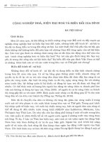
[10]. Ares et al. found that PTX-1,
PTX-2 and PTX-11 modified the actin cytoskeleton
and morphology of BE(2)-M17 neuroblastoma cells.
However, PTX-2 SA did not show any effect on these
cells. Moreover, PTX-2 and PTX-11 were more
potent than PTX-1 (Fig. 2) [28]. This supports the
idea that lactone ring integrity is essential for the
action of PTXs; oxidation on C43 decrease the toxic-
ity of the molecule, and oxidation on C34 does not
affect the potency of PTXs.
Unusual macrolides – antiactin drugs
PTXs are unusual macrolides found in recent years
that, to varying degrees, mimic the activity of endoge-
nous actin-binding proteins [31]. Typically, a macro-
cyclic lactone, a cetonic group and a glycosidic linked
amino-sugar constitute the macrolide structure. Recent
studies have demonstrated that the globular actin
(G-actin)-binding and actin-filament-severing activity
of some typical macrolides, such as kabiramide-C,
reside in the glycosidic amino-sugar region of the
molecule [33]. However, PTXs and the macrolactones
described in the next paragraph lack this amino-sugar
chain.
Actin cytoskeleton disruption by pectenotoxins B. Espin˜ a and J. A. Rubiolo
6084 FEBS Journal 275 (2008) 6082–6088 ª 2008 The Authors Journal compilation ª 2008 FEBS
Although they share a common structure and target,
these antiactin drugs display diverse mechanisms of
action. Latrunculins specifically sequester monomeric
actin, mimicking proteins such as b-thymosins and
inhibiting polymerization of G-actin and promoting
depolymerization of F-actin [31,34]. Mycalolide-B and
aplyronine-A inhibit polymerization of purified actin,
apparently by sequestration of monomeric actin and
actin severing because of the rapid depolymerization
that they induce [31,34]. However, jasplakinolides
induce actin polymerization, stabilizing actin filaments
and actin nucleation [35]. Another group of macrolides,
the halichondramides, exhibit barbed-end capping and
F-actin-severing activity. Moreover, unusual dimeric
macrolides, swinholide-A and misakinolide-A, were,
surprisingly, found to display different effects on actin
dynamics; swinkolide-A severs actin, whereas misakino-
lide-A rapidly caps barbed ends of filaments [31].
The actin cytoskeleton – molecular
target of PTXs
Initial studies performed to elucidate the mechanism of
action of PTXs and their in vitro biochemical effects
demonstrated that PTX-2 inhibited actin polymeriza-
tion in a concentration-dependent manner [36]. PTX-2
also inhibited contractions induced by KCl in the
isolated rat aorta, and formed a 1 : 4 complex with
G-actin [36,37]. Spector et al. reported that PTX-2
sequestered monomeric actin with a K
d
of 20 nm, but
did not exhibit severing or capping activity. Moreover,
PTX-2 disrupted the organization of actin in several
cell types in a time- and concentration-dependent
manner [31].
In a recent study, Allingham et al. obtained the
X-ray structure of the PTX-2–actin complex. In con-
trast to the results of Hori et al., they described a 1 : 1
stoichiometry in a novel site between subdomains 1
and 3 of the actin molecule [38]. In cells, capping pro-
teins such as gelsolin play an important role; capping
of old filaments funnels actin polymerization to sites
where new barbed ends are present, and the generation
of new barbed ends relies on the nucleation mechanism
[39]. But PTXs could also sequester actin monomers
binding to G-actin [36] in a similar way to latrunculin-
A [28,31], avoiding new actin polymerization or nucle-
ation. On the basis of models of the actin filament,
PTX binding would disrupt key lateral contacts
between the PTX-bound actin monomer and the lower
lateral actin monomer within the filament, thereby cap-
ping the barbed end without inducing filament sever-
ing, even though, on the basis of the previous work of
Spector et al., an actin-sequestering effect could not be
ruled out as another possible action (Fig. 3).
Fig. 3. Simplified hypothetical PTX mechanism of action. Two dif-
ferent activities on the actin filaments can be attributed to PTXs:
capping of the growing (+) barbed end of F-actin by binding of the
PTX molecule to the lower lateral actin monomer within the
filament, and G-actin monomer sequestration.
Fig. 2. Effects of diverse PTX analogs on
the actin cytoskeleton of BE(2)-M17 human
neuroblastoma cells. Full projection of Z-ser-
ies captured with confocal microscopy of
cells in which F-actin was labeled with Ore-
gon Green 514 Phalloidin. Control cells (A)
and cells incubated for 4 h with 200 n
M
PTX-11 (B), PTX-2 (C), PTX-1 (D) or PTX-2
SA (E). Figure modified from [28]. Scale bar:
50 lm.
B. Espin˜ a and J. A. Rubiolo Actin cytoskeleton disruption by pectenotoxins
FEBS Journal 275 (2008) 6082–6088 ª 2008 The Authors Journal compilation ª 2008 FEBS 6085
Cytological implications of disruption
of the actin cytoskeleton
Although, according to the histopathological studies,
the liver seems to be the target organ of PTXs, many
cellular models have shown substantial effects of PTX
treatments in the nanomolar range [27,40–42]. The
induction of apoptotic cell death rather than necrosis
by PTX-1 and PTX-2 is well established, on the basis
of morphological changes, reduction of mitochondrial
membrane potential, increases in cytoplasmic cyto-
chrome c and Smac ⁄ DIABLO, and caspase-3 and
caspase-9 activation. The inhibition of PTX-induced
cell death by caspase inhibitors supports the apoptotic
pathway [27,40].
Actin polymerization and dynamics are required for
essential cell processes such as motility, endocytosis,
cytokinesis, and establishing cell–cell and cell–substrate
contacts. In vitro and in vivo studies showed that some
macrolide–actin complexes reduced free G-actin below
the critical concentration and blocked extension of the
(+)-end of the filament, while the free macrolide also
bound to and severed actin filaments [43]. In living
cells, the combined effects of these interactions leads
to a cessation of new filament growth and a disruption
of existing filaments, and is accompanied by a loss of
motility, breakdown of adherens junctions, polyploidy
and, ultimately, apoptosis [43].
Very few studies have compared the effects of PTXs
in diverse cellular models. Chae et al. found that PTX-2
induced apoptosis in p53-deficient cells [40], showing
enhanced cytotoxicity as compared with normal cells.
Recently, Espin
˜
a et al. tested the effect of PTXs on
actin cytoskeleton and cell viability in two hepatic cellu-
lar models [44]. Interesting results point to different
effects of PTXs in Clone 9 cells, a rat hepatocyte cell
line used previously in metabolic and cancer studies
[45], and primary cultured rat hepatocytes (Fig. 4).
Although primary rat hepatocytes, as well as Clone 9
cells, showed a marked change in the pattern of distri-
bution and a decrease in polymerized actin after the
treatment with PTX-2, no morphological effects could
be observed in primary hepatocytes, whereas Clone 9
cells were contracted and rounded. The higher sensitiv-
ity of Clone 9 cells to PTXs in comparison to normal
hepatocytes could be related to the fact that the cell
lines share cytoskeletal and morphological characteris-
tics with cancerous cells. In accordance with this fact,
the tumor cells of the study of Chae et al. were more
sensitive to PTX-2 than normal cells [44].
Fig. 4. Effect of PTX-2 on the actin cyto-
skeleton of two hepatic cellular models.
Clone 9 cells (A, B) and primary cultured rat
hepatocytes (C, D) were incubated for 3 h
with 200 n
M PTX-2 (B, D) or with the toxin
vehicle (control; A, C). Then, they were
stained for F-actin and G-actin with Oregon
Green 514 Phalloidin and Texas Red
DNAse I, and visualized with a confocal
microscope. Scale bar: 50 lm.
Actin cytoskeleton disruption by pectenotoxins B. Espin˜ a and J. A. Rubiolo
6086 FEBS Journal 275 (2008) 6082–6088 ª 2008 The Authors Journal compilation ª 2008 FEBS
Perspectives
The importance of the actin cytoskeleton in pathogenic
cellular process such as angiogenesis, cell adhesion,
cytokinesis and metastasis has made it an attractive
target for the development of anticancer drugs. Drugs
that block the regulation of actin filament dynamics
within tumor cells or in cells infected with pathogens
could be useful in treating cancer and other diseases.
In addition, differential actions of antiactin com-
pounds can reveal interesting data about actin cyto-
skeleton dynamics.
Although the presence in the food chain of PTXs is
being regulated, together with that of diarrhetic shell-
fish poisoning toxins, toxicological tests by oral admin-
istration point to these compounds as nonhazardous
molecules for human health with regard to shellfish
consumption. However, the selective cytotoxicity of
PTXs against certain tumor cell lines and their ability
to induce apoptosis of p53-deficient cell lines, which are
often resistant to other chemotherapeutic agents, makes
them interesting new candidates for cancer therapy.
References
1 Draisci R, Lucentini L, Giannetti L, Boria P & Poletti
R (1996) First report of pectenotoxin-2 (PTX-2) in
algae (Dinophysis fortii) related to seafood poisoning in
Europe. Toxicon 34, 923–935.
2 James KJ, Bishop AG, Draisci R, Palleschi L, Marchiaf-
ava C, Ferretti E, Satake M & Yasumoto T (1999) Liquid
chromatographic methods for the isolation and identifi-
cation of new pectenotoxin-2 analogues from marine
phytoplankton and shellfish. J Chromatogr A 844, 53–65.
3 Pavela-Vrancic M, Mestrovic V, Marasovic I, Gillman
M, Furey A & James KK (2001) The occurrence of
7-epi-pectenotoxin-2 seco acid in the coastal waters of
the central Adriatic (Kastela Bay). Toxicon 39, 771–779.
4 Suzuki T, Mackenzie L, Stirling D & Adamson J (2001)
Pectenotoxin-2 seco acid: a toxin converted from pecte-
notoxin-2 by the New Zealand Greenshell mussel, Perna
canaliculus. Toxicon 39, 507–514.
5 Suzuki T, MacKenzie L, Stirling D & Adamson J
(2001) Conversion of pectenotoxin-2 to pectenotoxin-2
seco acid in the New Zealand scallop, Pecten novaeze-
landiae. Fish Sci 67, 506–510.
6 Vale P & Sampayo MA de M (2002) Pectenotoxin-2 seco
acid, 7-epi-pectenotoxin-2 seco acid and pectenotoxin-2
in shellfish and plankton from Portugal. Toxicon 40,
979–987.
7 Yasumoto T, Murata M & Lee J (1989) Polyether
toxins produced by dinoflagellates. In Mycotoxins and
Phycotoxins ‘88 (Natori S, Hashimoto K & Ueno Y,
eds), pp. 375–382. Elsevier, Amsterdam.
8 Yasumoto T, Murata M, Oshima Y, Sano M, Matsum-
oto Gk & Clardy J (1985) Diarrheic shellfish toxins.
Tetrahedron 41, 1019–1025.
9 Lee JS, Yanagi Y, Kenma R & Yasumoto T (1987)
Fluorometric determination of diarrhetic shellfish toxins
by high performance liquid chromatography. Agricul
Biol Chem 51, 877–881.
10 Daiguji M, Satake M, James KJ, Bishop AG, MacKen-
zie L, Naoki H & Yasumoto T (1998) Structures of new
pectenotoxin analogs, pectenotoxin-2 seco acid and
7-epi-2 seco acid, isolated from a dinoflagelate and
greenshell mussels. Chem Lett 7, 653–654.
11 MacKenzie L, Holland P, McNabb P, Beuzenberg V,
Selwood A & Suzuki T (2002) Complex toxin profiles in
phytoplankton and Greenshell mussels (Perna canalicu-
lus), revealed by LC-MS ⁄ MS analysis. Toxicon 40,
1321–1330.
12 Miles CO, Wilkins AL, Samdal IA, Sandvik M,
Petersen D, Quilliam MA, Naustvoll LJ, Rundberget T,
Torgersen T, Hovgaard P et al. (2004) A novel pecteno-
toxin, PTX-12, in Dinophysis spp. and shellfish from
Norway. Chem Res Toxicol 17, 1423–1433.
13 Suzuki T, Mitsuya T, Matsubara H & Yamasaki M
(1998) Determination of pectenotoxin-2 after solid-
phase extraction from seawater and from the dinoflagel-
late Dinophysis fortii by liquid chromatography with
electrospray mass spectrometry and ultraviolet detec-
tion. Evidence of oxidation of pectenotoxin-2 to pecte-
notoxin-6 in scallops. J Chromatogr A 815, 155–160.
14 Sasaki K, Wright JL & Yasumoto T (1998) Identifica-
tion and characterization of pectenotoxin (PTX) 4 and
PTX7 as spiroketal stereoisomers of two previously
reported pectenotoxins. J Org Chem 63, 2475–2480.
15 Miles CO, Wilkins AL, Hawkes AD, Jensen DJ,
Selwood AI, Beuzenberg V, Mackenzie AL, Cooney JM
& Holland PT (2006) Isolation and identification of
pectenotoxins-13 and -14 from Dinophysis acuta in New
Zealand. Toxicon 48, 152–159.
16 Suzuki T, Beuzenberg V, Mackenzie L & Quilliam MA
(2003) Liquid chromatography–mass spectrometry of
spiroketal stereoisomers of pectenotoxins and the analy-
sis of novel pectenotoxin isomers in the toxic dinoflagel-
late Dinophysis acuta from New Zealand. J Chromatogr
A 992, 141–150.
17 Suzuki T, Walter JA, LeBlanc P, MacKinnon S, Miles
CO, Wilkins AL, Munday R, Beuzenberg V, MacKenzie
AL, Jensen DJ et al. (2006) Identification of pecteno-
toxin-11 as 34S-hydroxypectenotoxin-2, a new pecteno-
toxin analogue in the toxic dinoflagellate Dinophysis
acuta from New Zealand. Chem Res Toxicol 19, 310–318.
18 Yasumoto T & Murata M (1993) Marine toxins. Chem
Rev 93, 1897–1909.
19 Ishige M, Satoh N & Yasumoto T (1988) Pathological
studies on the mice administration with the causative
agent of diarrhetic shellfish poisoning (okadaic acid and
B. Espin˜ a and J. A. Rubiolo Actin cytoskeleton disruption by pectenotoxins
FEBS Journal 275 (2008) 6082–6088 ª 2008 The Authors Journal compilation ª 2008 FEBS 6087
pectenotoxin-2). Hokkaidoritsu Eisei Kenkyushoho 38,
15–18.
20 Ogino H, Kumagai M & Yasumoto T (1997) Toxico-
logic evaluation of yessotoxin. Nat Toxins 5, 255–259.
21 Burgess V & Shaw G (2001) Pectenotoxins – an issue
for public health: a review of their comparative toxicol-
ogy and metabolism. Environ Int 27, 275–283.
22 Burgess V & Shaw G (2003) Investigations into the tox-
icology of pectenotoxin-2-seco acid and 7-epi pecteno-
toxin-2 seco acid to aid in a health risk assessment for
the consumption of shellfish contaminated with these
shellfish toxins in Australia. Report on Project No.
2001/258. National Research Centre for Environmental
Toxicology, Archerfield, Australia.
23 Miles CO, Wilkins AL, Munday R, Dines MH, Hawkes
AD, Briggs LR, Sandvik M, Jensen DJ, Cooney JM,
Holland PT et al. (2004) Isolation of pectenotoxin-2
from Dinophysis acuta and its conversion to pecteno-
toxin-2 seco acid, and preliminary assessment of their
acute toxicities. Toxicon 43, 1–9.
24 Hamano Y, Kinoshita Y & Yasumoto T (1985) Suck-
ling mice assay for diarrhetic shellfish toxins. In Toxic
Dinoflagellates (Anderson DM, White AW & Baden
DB, eds), pp. 383–388. Elsevier, Amsterdam.
25 Terao K, Ito E, Yanagi T & Yasumoto T (1986) Histo-
pathological studies on experimental marine toxin poi-
soning. I. Ultrastructural changes in the small intestine
and liver of suckling mice induced by dinophysistoxin-1
and pectenotoxin-1. Toxicon 24, 1141–1151.
26 Ito E, Suzuki T, Oshima Y & Yasumoto T (2008) Stud-
ies of diarrhetic activity on pectenotoxin-6 in the mouse
and rat. Toxicon 51, 707–716.
27 Fladmark KE, Serres MH, Larsen NL, Yasumoto T,
Aune T & Doskeland SO (1998) Sensitive detection of
apoptogenic toxins in suspension cultures of rat and
salmon hepatocytes. Toxicon 36 , 1101–1114.
28 Ares IR, Louzao MC, Espina B, Vieytes MR, Miles CO,
Yasumoto T & Botana LM (2007) Lactone ring of pecte-
notoxins: a key factor for their activity on cytoskeletal
dynamics. Cell Physiol Biochem 19, 283–292.
29 Ares IR, Louzao MC, Vieytes MR, Yasumoto T &
Botana LM (2005) Actin cytoskeleton of rabbit intes-
tinal cells is a target for potent marine phycotoxins.
J Exp Biol 208, 4345–4354.
30 Leira F, Cabado AG, Vieytes MR, Roman Y, Alfonso
A, Botana LM, Yasumoto T, Malaguti C & Rossini
GP (2002) Characterization of F-actin depolymerization
as a major toxic event induced by pectenotoxin-6 in
neuroblastoma cells. Biochem Pharmacol 63, 1979–1988.
31 Spector I, Braet F, Shochet NR & Bubb MR (1999)
New anti-actin drugs in the study of the organization
and function of the actin cytoskeleton. Microsc Res
Tech 47, 18–37.
32 Burgess VA, Seawright A, Eaglesham G, Shaw G &
Moore M (2002) The acute oral toxicity of the shellfish
toxins pectenotoxin-2 seco acid and 7-epi-pectenotoxin-
2 seco acid in mice. 4th International Conference on
Molluscan Shellfish Safety. Santiago de Compostela,
Spain.
33 Perrins RD, Cecere G, Peterson I & Marriot G (2008)
Synthetic mimetics of actin-binding macrolides: rational
design of actin-targeted drugs. Chem Biol 15, 287–294.
34 Yarmola EG, Somasundaram T, Boring TA, Spector I &
Bubb MR (2000) Actin–latrunculin A structure and
function. Differential modulation of actin-binding
protein function by latrunculin A. J Biol Chem 275,
28120–28127.
35 Bubb MR, Spector I, Beyer BB & Fosen KM (2000)
Effects of jasplakinolide on the kinetics of actin
polymerization. An explanation for certain in vivo
observations. J Biol Chem 275, 5163–5170.
36 Hori M, Matsuura Y, Yoshimoto R, Ozaki H,
Yasumoto T & Karaki H (1999) Actin depolymerizing
action by marine toxin, pectenotoxin-2. Nippon
Yakurigaku Zasshi
114(Suppl. 1), 225P–229P.
37 Karaki H, Matsuura Y, Hori M, Yoshimoto R, Ozaki
H & Yasumoto T (1999) Pectenotoxin-2, a new actin
depolymerizing compound isolated from scallop Patino-
pecten yessoensis. Jpn J Pharmacol 79, 268.
38 Allingham JS, Miles CO & Rayment I (2007) A struc-
tural basis for regulation of actin polymerization by
pectenotoxins. J Mol Biol 371, 959–970.
39 Giganti A & Friederich E (2003) The actin cytoskeleton
as a therapeutic target: state of the art and future direc-
tions. Prog Cell Cycle Res 5, 511–525.
40 Chae HD, Choi TS, Kim BM, Jung JH, Bang YJ &
Shin DY (2005) Oocyte-based screening of cytokinesis
inhibitors and identification of pectenotoxin-2 that
induces Bim ⁄ Bax-mediated apoptosis in p53-deficient
tumors. Oncogene 24, 4813–4819.
41 Jung JH, Sim CJ & Lee CO (1995) Cytotoxic com-
pounds from a two-sponge association. J Nat Prod 58,
1722–1726.
42 Zhou ZH, Komiyama M, Terao K & Shimada Y (1994)
Effects of pectenotoxin-1 on liver cells in vitro. Nat
Toxins 2, 132–135.
43 Tanaka J, Yan Y, Choi J, Bai J, Klenchin VA,
Rayment I & Marriott G (2003) Biomolecular mimicry
in the actin cytoskeleton: mechanisms underlying the
cytotoxicity of kabiramide C and related macrolides.
Proc Natl Acad Sci USA 100, 13851–13856.
44 Espin
˜
a B, Louzao MC, Ares IR, Cagide E, Vieytes
MR, Vega FV, Rubiolo JA, Miles CO, Suzuki T,
Yasumoto T et al. (2008) Cytoskeletal toxicity of
pectenotoxins in hepatic cells. Br J Pharmacol,
doi: 10.1038/bjp.2008.323.
45 Louzao MC, Espina B, Vieytes MR, Vega FV, Rubiolo
JA, Baba O, Terashima T & Botana LM (2007) ‘Fluo-
rescent glycogen’ formation with sensibility for in vivo
and in vitro detection. Glycoconj J 25, 503–510.
Actin cytoskeleton disruption by pectenotoxins B. Espin˜ a and J. A. Rubiolo
6088 FEBS Journal 275 (2008) 6082–6088 ª 2008 The Authors Journal compilation ª 2008 FEBS