Tài liệu Báo cáo khoa học: Membrane targeting and pore formation by the type III secretion system translocon pdf
Bạn đang xem bản rút gọn của tài liệu. Xem và tải ngay bản đầy đủ của tài liệu tại đây (555.81 KB, 13 trang )
REVIEW ARTICLE
Membrane targeting and pore formation by the type III
secretion system translocon
Pierre-Jean Matteı
¨
1
, Eric Faudry
2
, Viviana Job
1
, Thierry Izore
´
1
, Ina Attree
2
and Andre
´
a Dessen
1
1 Bacterial Pathogenesis Group, Institut de Biologie Structurale, UMR 5075 (CNRS ⁄ CEA ⁄ UJF), Grenoble, France
2 Bacterial Pathogenesis and Cellular Responses Team, Centre National de la Recherche Scientifique (CNRS), Universite
´
Joseph Fourier
(UJF), LBBSI, iRTSV, CEA, Grenoble, France
Introduction
Type III secretion systems (T3SS) are complex macro-
molecular machineries employed by a number of bac-
teria to inject toxins and effectors directly into the
cytoplasm of eukaryotic cells. Pathogens carrying this
system, which include Pseudomonas, Yersinia, Salmo-
nella and Shigella spp., as well as clinical Escherichia
coli isolates, can translocate between four and 20 effec-
tors with dramatic effects on the target cell, leading,
for example, to cytoskeleton rearrangement, membrane
disruption or the initiation of apoptosis [1–3].
T3SS are composed of at least twenty distinct pro-
teins that assemble into three major parts. The basal
body of the system, composed of two main ring-like
structures, spans both the inner and outer bacterial
membranes (Fig. 1) [4–7]. This multi-protein structure
is associated with an ATPase, which itself is mem-
brane-associated and faces the bacterial cytoplasm,
and is suggested to be involved in facilitating the entry
of export substrates into the secretion system [8–10].
The basal body of the T3SS is also associated with a
proteinaceous needle that extends outwards from the
bacterial surface and is assumed to act as a conduit
for effector secretion [6,11–13], although direct evi-
dence for this concept is lacking. Because the internal
diameter of the needle is relatively small (2.0–2.5 nm),
effectors probably travel in unfolded ⁄ semi-unfolded
states [11]. Synthesis and assembly of the T3SS itself
are induced once the bacterium is physically associated
Keywords
bacterial infection; injection; membrane;
pore formation; secretion; toxin
Correspondence
A. Dessen, Bacterial Pathogenesis Group,
Institut de Biologie Structurale, UMR 5075
(CNRS ⁄ CEA ⁄ UJF), 41 rue Jules Horowitz,
38027 Grenoble, France
Fax: +33 4 38 78 54 94
Tel: +33 4 38 78 95 90
E-mail:
(Received 21 September 2010, revised 4
November 2010, accepted 26 November
2010)
doi:10.1111/j.1742-4658.2010.07974.x
The type III secretion system (T3SS) is a complex macromolecular machin-
ery employed by a number of Gram-negative species to initiate infection.
Toxins secreted through the system are synthesized in the bacterial cyto-
plasm and utilize the T3SS to pass through both bacterial membranes and
the periplasm, thus being introduced directly into the eukaryotic cytoplasm.
A key element of the T3SS of all bacterial pathogens is the translocon,
which comprises a pore that is inserted into the membrane of the target
cell, allowing toxin injection. Three macromolecular partners associate to
form the translocon: two are hydrophobic and one is hydrophilic, and the
latter also associates with the T3SS needle. In this review, we discuss recent
advances on the biochemical and structural characterization of the proteins
involved in translocon formation, as well as their participation in the modi-
fication of intracellular signalling pathways upon infection. Models of tran-
slocon assembly and regulation are also discussed.
Abbreviations
EHEC, enterohaemorrhagic; EPEC, enteropathogenic; IFN, interferon; SPI, Salmonella pathogenicity island; T3SS, type III secretion system;
TM, transmembrane; TPR, tetratricopeptide.
414 FEBS Journal 278 (2011) 414–426 ª 2010 The Authors Journal compilation ª 2010 FEBS
with an eukaryotic host cell membrane, although the
nature of the cellular signal required and the mecha-
nism of its transduction are still a matter of debate
[14,15].
The third, major part of the T3SS is the ‘translo-
con’, which is generally composed of three proteins
that are exported through the needle upon cell contact
and form a pore on the surface of the eukaryotic cell
that allows toxin entry into the target cytoplasm. Two
T3SS loci-encoded membrane proteins (the hydropho-
bic translocators) and one hydrophilic partner (also
called the V antigen in Pseudomonas aeruginosa and
Yersinia spp.; Figs 1 and 2) comprise the translocon,
and are essential for its formation in all systems stud-
ied to date. Genes that code for translocon members
are encoded within the same operon, which also har-
bours elements that encode chaperones for both the
V antigen and the hydrophobic translocators (i.e. all
molecules required to form the translocon in the well-
studied Yersinia system, for example, are encoded
within the lcrGVHyopBD genetic element).
Translocon components are dispensable for secretion
but are essential for the injection of type III effectors
into the target cytoplasm and therefore are considered
to be the first substrates secreted by the T3SS needle
upon cell contact. In the absence of external secretion
stimuli, all three translocon components remain
associated with their respective chaperones (Fig. 1) and
are stored in the cytoplasm. However, upon cell
contact, the entire cytoplasmic pool of translocator
proteins is released rapidly and concurrently, and
effectors are translocated in an ordered manner
[16,17]. Translocon proteins presumably travel through
the interior of the needle and, once having reached the
outmost extremity of the conduit, all three components
are assumed to associate to form the translocation
pore. The precise order of passage of the individual
translocator proteins to the outside of the system is
unknown (for clarity, the hydrophilic partner is
depicted in Fig. 1 as being the first molecule to be
localized). Within the tripartite organization of the
translocon, the hydrophilic translocator is the only
component that is neither directly, nor indirectly asso-
ciated with the target membrane; rather, it assembles
into a distinct structure at the tip of the T3SS needle,
and potentially plays the role of assembly platform for
the two hydrophobic components [18–23]. The two
others, which carry predicted hydrophobic domains,
have been shown to be directly associated with target
membranes and to exist both in oligomeric and mono-
meric forms [24–26]. In all systems studied to date, the
largest of the hydrophobic translocators displays two
predicted transmembrane (TM) regions (henceforth
termed the major translocator; i.e. YopB in Yersinia
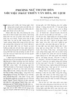
Translocon
Needle
Translocators
Bacterium
Host membrane
AB CD
Fig. 1. Schematic diagram illustrating needle and translocon formation, as well as toxin secretion steps, in the T3SS of P. aeruginosa (a rep-
resentative of the Ysc T3SS family). (A) Upon formation of the base rings (green), PscF is released from its chaperones (PscG and PscE) and
polymerizes to form the T3SS needle. (B) The V antigen PcrV is released from its cytoplasmic partner (PscG) and forms the cap of the PscF
needle. (C) Translocator proteins PopB and PopD release PcrH. (D) Upon formation of the Pop translocon on the eukaryotic membrane, tox-
ins produced in the bacterial cytoplasm release their cognate chaperones and are injected through the translocon pore and into the target
cytoplasm. IM, inner membrane; MO, outer membrane.
P J. Matteı
¨
et al. Membrane targeting and pore formation by the T3SS
FEBS Journal 278 (2011) 414–426 ª 2010 The Authors Journal compilation ª 2010 FEBS 415
spp., PopB in P. aeruginosa, IpaB in Shigella spp. and
EspD in pathogenic E. coli spp.), whereas the smallest
protein (i.e. the minor translocator; YopD, PopD,
IpaC and EspB in the aforementioned organisms) car-
ries a single predicted membrane-association region
(Fig. 2).
Phylogenetic analyses have allowed the classification
of T3SS into seven different families, where macromol-
ecules that compose the base, needle and translocon
display sequence similarities both at the genetic and
locus organizational levels [1]. Thus, the Ysc T3SS of
Yersinia spp. is related to those of P. aeruginosa and
Aeromonas spp., whereas the Inv-Mxi-Spa systems are
found in Shigella, Salmonella, and Burkholderia spp.
In addition, Ssa-Esc systems exist in enteropathogenic
(EPEC) and enterohaemorrhagic (EHEC) Escherichia
coli species (Esc), and also represent the second T3S
system [Salmonella pathogenicity island (SPI)-2] in
intracellular Salmonella spp. (Ssa) [27]. However,
secreted toxins are pathogen-specific, and their
different characteristics and cellular fates influence the
distinct infectious phenotypes of the source microor-
ganism [2]. In this review, only the translocons from
the three aforementioned Ysc, Inv-Mxi-Spa and
Ssa-Esc T3SS families will be discussed.
The hydrophobic translocators
recognize a common chaperone
In the bacterial cytoplasm, the two hydrophobic trans-
locators are associated with a common chaperone that
shares a considerable sequence identity even within dis-
tant species. Recent efforts in the structural character-
ization of T3SS translocator chaperones have revealed
that they adopt a seven-helical tetratricopeptide
(TPR)-like repeat fold [28–30], which is known to be
involved in protein–protein interactions (Fig. 3) [31].
Notably, this fold is also shared by chaperones that
Fig. 2. Diagrammatic analysis of the translocator molecules of the Ysc, Ssa-Esc and Inv-Mxi-Spa systems. TM, predicted transmembrane
region; CC, predicted coiled coil; *, chaperone interaction region; **, region predicted as interacting with the hydrophilic partner; ***, region
predicted as interacting with the hydrophobic partner; a, predicted amphipathic helix. aa, amino acid.
N
N
N
C
C
C
Fig. 3. Chaperones of hydrophobic translocators display a TPR fold. SycD, PcrH and IpgC are shown in yellow, green and magenta, respec-
tively. The peptides located within the concave regions of PcrH and IpgC, corresponding to sections of the N-termini of PopD and IpaB, are
shown as surfaces.
Membrane targeting and pore formation by the T3SS P J. Matteı
¨
et al.
416 FEBS Journal 278 (2011) 414–426 ª 2010 The Authors Journal compilation ª 2010 FEBS
stabilize the building blocks of the T3SS needle [32,33],
suggesting that TPR folds could be specific for chaper-
ones of ‘early’ T3SS substrates, such as translocon and
needle-forming subunits, wheteas other chaperone
folds are employed for effector molecules [30,34]. TPR
folds resemble a ‘cupped hand’, in which target pro-
teins can be recognized either within the ‘palm’ region,
the back of the hand, or both [32]. Notably, TPR
chaperones that recognize translocon hydrophobic
components have been shown to bind to the N-termi-
nal sequences of both major and minor translocator
proteins within the ‘palm’ regions, revealing that one
single chaperone cannot recognize both translocators
concomitantly [30]. It is of note that T3SS transloca-
tors display molten globule characteristics both in the
presence and absence of their respective chaperones
[35,36], which is to be expected for proteins that must
modify their conformations to accomplish a number of
steps essential for their functionality during T3SS toxin
injection. These steps include detachment from their
chaperone, partial unfolding to allow transport
through a thin conduit and, finally, oligomerization in
the presence of lipids (see below). This suggests that
translocator molecules could be partially ‘wrapped’
around their cognate chaperones.
Effector ⁄ translocator-bound chaperones have also
been proposed to interact with the membrane-associ-
ated ATPase located at the base of the T3SS (shown
in orange in Fig. 1). The T3SS ATPase is similar to
the F
1
ATPases [37] and associates into a hexameric
ring, thus being highly reminiscent of the flagellar AT-
Pase FliI [38,39]. The chaperone-ATPase interaction is
suggested to be crucial for complex dissociation and
substrate unfolding in preparation for transport
through the needle [8]. In addition, the detection of
complexes between T3SS ATPases and partner mole-
cules, although challenging as a result of the potential
transient nature of the interactions, has been reported
for needle proteins [40] and a multi-cargo chaperone
[41]. Interestingly, in Salmonella, a small cytoplasmic
protein of the SPI-2 locus (SsaE) was shown to
interact both with translocator protein SseB as well as
with the T3SS ATPase, SsaN [42]. These findings sug-
gest that there is a complex interplay of interactions
between hydrophobic translocators, their cognate
chaperones and the cytosol ⁄ membrane interface of
the T3SS even before their passage through the T3SS
needle.
The major hydrophobic translocator
Major hydrophobic translocators of Shigella (IpaB),
Salmonella (SipB), P. aeruginosa (PopB), Yersinia
(YopB) and pathogenic Escherichia spp. (EspD) all
carry two predicted TM regions, and are predicted to
have a N-terminal coiled-coil region and, occasionally,
a C-terminal amphipathic helix (Fig. 2). It is within
the two TM regions and the intervening loop that
major translocators display the highest level of
sequence identity (Figs 2 and 3), demonstrating the
functional importance of these regions in membrane
association, pore formation and translocation [24,
43–46]. Notably, purified Shigella IpaB remains inti-
mately associated with model membranes, being resis-
tant to extraction with agents that solubilize
superficially-associated proteins. In addition, limited
proteolysis experiments of membrane-imbedded IpaB
confirm that lipids protect the two TM regions, as well
as the intervening sequence from trypsinization [44].
Interestingly, both Shigella IpaB and Salmonella SipB
were shown to form SDS-resistant trimers through
interactions that are formed within their N-terminal
domains [44], although the bilayer-inserted form of
SipB was shown to be hexameric [47].
Intimate association of the major hydrophobic
translocator with target membranes was also shown by
contact haemolysis experiments performed with Shi-
gella, P. aeruginosa and EPEC, which revealed success-
ful membrane insertion of IpaB, PopB and EspD,
respectively, upon T3SS induction [19,44,48]. It is of
note that PopB on its own associates rapidly with arti-
ficial membranes and promotes the efficient release of
small fluorescent molecules from liposomes [49]. How-
ever, infectious Pseudomonas strains in which PopD is
absent can still insert PopB into host membranes but
the strain remains unable to translocate toxins [19],
suggesting that the major hydrophobic translocator
requires a minor translocator for functional translocon
formation.
In some cases, major translocator proteins can show
functional equivalency: DyopB Yersinia strains can
be complemented by plasmids expressing the
pcrGVHpopBD operon, whereas the opposite is also
true for DpopB Pseudomonas strains complemented
with plasmids expressing lcrGVHyopBD . Interestingly,
complementation only occurs if the entire operon is
expressed (and not just the single translocator), sug-
gesting that other partner translocon molecules must
also be present [50]. Conversely, IpaB is not able to
complement either Yersinia or Pseudomonas mutant
strain, suggesting that the bulkier Shigella protein
lacks regions that are conserved in YopB and PopB.
Notably, Shigella ipaB mutants can be complemented
by a plasmid carrying Salmonella sipB, indicating that,
with respect to the hydrophobic translocators of
the Inv-Mxi-Spa system [51], proteins that display
P J. Matteı
¨
et al. Membrane targeting and pore formation by the T3SS
FEBS Journal 278 (2011) 414–426 ª 2010 The Authors Journal compilation ª 2010 FEBS 417
extensive sequence similarities (Fig. 4) also show
comparable functional characteristics.
Recently, it was shown that the extreme C-terminus
of IpaB binds to the T3SS needle, serving as a
‘bridge’ between the eukaryotic membrane and the
Shigella secretion system. IpaB is required for regulat-
ing secretion, and may play the role of host cell sen-
sor. It was proposed that the needle tip, which in
principle contacts all three translocon components,
exists in ‘on’ and ‘off’ states [52], thus suggesting that
all proteins involved in the initial contact with the
target cell may considerably modify their conforma-
tions or oligomerization states during the secretion
process. This proposal is also supported by the sug-
gestion that pH sensing by Salmonella involves modi-
fications in the assembly of the translocon, which
affect the pH gradient within the needle, sending sig-
nals to the base of the T3SS structure [53]. In addi-
tion, Shen et al. [54] identified that distinct IpaB
regions (residues 227–236 and 297–306) are required
for secretion regulation. Further clarifications of this
complex process will thus require the structural
characterization of the translocon, potentially in dif-
ferent states of activation.
The minor hydrophobic translocator
This class of proteins has been studied more exten-
sively, potentially because they carry a single predicted
TM region (Fig. 2) and are thus more biochemically
tractable. Minor translocators are well conserved
amongst different bacterial species, displaying a con-
siderable level of sequence identity levels (i.e. 38% for
Pseudomonas PopD and Yersinia YopD; 29% for Shi-
gella IpaC and Salmonella SipC). Indeed, sections of
IpaC and SipC (as well as YopD and PopD) are inter-
changeable without affecting secretion [55,56]; in the
latter case, however, the proteins can be exchanged
only if the cognate chaperone and translocator part-
ners are present [50]. As is the case for the major
translocator, minor translocators have also been shown
to oligomerize, and this event is essential not only for
pore formation, but also for events that take place
within the eukaryotic cytoplasm [26,57,58]. The two
Fig. 4. Sequence alignments of major trans-
locator proteins that display the highest
level of sequence similarity. Identical resi-
dues are shown in red. Residues in green
and blue display strong and weak similarity,
respectively. The two predicted TM regions
are indicated in boxes.
Membrane targeting and pore formation by the T3SS P J. Matteı
¨
et al.
418 FEBS Journal 278 (2011) 414–426 ª 2010 The Authors Journal compilation ª 2010 FEBS
translocators show clear differences in terms of mem-
brane association, which is evident from the fact that
PopD is less able to release fluorescent dyes from lipo-
somes than PopB (although it readily binds to artificial
membranes) [49], whereas a PcrV knockout mutant
can successfully insert PopB but not PopD into red
blood cell membranes [19]. In addition, in Shigella,
IpaC is required for pore formation but not for mem-
brane insertion of IpaB, suggesting that IpaB may be
the first protein to be inserted in the bilayer, but with-
out IpaC the pore cannot be functional [24].
So far, very limited structural data is available for
any of the translocator molecules. It has been shown
that EspB, IpaC and PopD all possess partly disor-
dered structures, which could potentially be a require-
ment for chaperone release, secretion and the
formation of more complex structures upon attaining
the eukaryotic membrane [35,36,59]. Interestingly,
Costa et al. [60] identified that the C-terminal, coiled
coil amphipathic domain of YopD, whose structure
was solved by NMR by Tengel et al. [61], is essential
for interacting with LcrV and forming oligomers but
does not play a role in YopB recognition. These obser-
vations all point to the multifunctional aspect of the
structures of the translocator proteins, which, in addi-
tion to recognizing chaperones and hydrophobic part-
ners, must also interact with the T3SS needle to permit
toxin translocation.
Minor translocators have been shown, in many
pathogens, to play important roles in the cytoskeletal
rearrangement processes that occur upon T3SS induc-
tion. Salmonella SipC carries two functions: participa-
tion in the formation of the membrane-inserted pore
and acting as an actin nucleation initiator by promot-
ing its own multimerization [57]. In addition, SipC has
been shown to recruit the Exo70 exocyst component,
thus facilitating fusion of exocytic vesicles with the
plasma membrane and increasing Salmonella invasion
efficiency [62]. It is of note that both IpaC and SipC
are essential for Shigella and Salmonella uptake by
macrophages in the early steps of invasion, and have
the ability to induce membrane extensions (filopodia
and lamellipodia) on macrophages [55,63]. Specifically,
IpaC was shown to recruit and activate Src tyrosine
kinase, which is required for actin polymerization, at
specific sites of bacterial entry, in a process where its
63 carboxy-terminal residues play a key role [64].
Interestingly, EspB was shown to be essential for the
development of attaching and effacing (A⁄ E) lesions
by EHEC by recruiting a-catenin, a cytoskeletal pro-
tein that recognizes actin, to the site of bacterial con-
tact [65,66]. In addition, it is also involved in the
inhibition of myosin function, leading to microvillus
effacement [67]. Although the precise sequence of
events that leads to secretion of translocators is not
well understood, it is of note that IpaC has been
shown to localize to the bacterial pole regions upon
T3SS induction in Shigella. This event may be of
importance to locally target all T3SS effectors and effi-
ciently affect cytoskeletal rearrangement processes [68].
Association between hydrophobic
translocators and pore formation
Formation of the translocon potentially requires a
direct association between the two hydrophobic trans-
locators. This possibility has been investigated by
assays ranging from pull-downs to genetic knockouts
and microbiological tests. In E. coli, purified forms of
EspB can recognize EspD found in bacterial lysates
[69], whereas Yersinia pseudotuberculosis YopD recog-
nizes both YopB and the V antigen (LcrV) in pull-
down assays [61].
However, the structural characteristics of the mem-
brane-inserted pore have remained elusive. Neverthe-
less, dye release studies have revealed that the pores
formed by YopB ⁄ YopD and PopB ⁄ PopD have similar
internal diameters, in the range 1.2–3.5 nm [70,71].
In addition, negative staining electron microscopy
images of the PopB or PopD-associated liposomes
structures have suggested an internal diameter of
approximately 25 A
˚
, with an external measurement of
80 A
˚
[26]; atomic force microscopy studies on pores
formed by EPEC indicate an approximate internal
diameter of 2.0 nm [69], whereas the IpaB ⁄ IpaC
Shigella pore has an inner radius of 26 ± 0.4 A
˚
[24].
These measurements are in agreement with the internal
diameter of the T3SS needle [72], which would
facilitate toxin translocation into the host cytoplasm.
However, the exact stoichiometry of the pore remains
a matter of controversy. Ide et al. [69] suggested that
the membrane-inserted structure is composed of six to
eight subunits, which is in agreement with the studies
on SipB from the Salmonella system [47], although the
precise determination of pore stoichiometry in other
species still awaits further study.
The hydrophilic translocator: the
V antigen
The third component of the translocation apparatus is
a hydrophilic protein: PcrV in P. aeruginosa, LcrV in
Yersinia spp, IpaD in Shigella and SipD in Salmonella
spp (Fig. 2). The LcrV protein of Yersinia pestis was
discovered more than 50 years ago as a soluble protec-
tive antigen, and was thus termed the ‘V antigen’ [73].
P J. Matteı
¨
et al. Membrane targeting and pore formation by the T3SS
FEBS Journal 278 (2011) 414–426 ª 2010 The Authors Journal compilation ª 2010 FEBS 419
Indeed, immunization with LcrV or PcrV elicits the
production of antibodies that protect against Yersinia
or Pseudomonas infections in animal models [74–76],
and LcrV was included in the formulation of a vaccine
against plague [77,78]. Although less studied, antibod-
ies directed toward IpaD were also shown to partially
protect erythrocytes and HeLa cells against Shi-
gella flexneri infection [79,80]. Notably, in EPEC and
EHEC, the EspA protein could play a similar role in
translocon assembly, although it displays no sequence
similarity and is structurally distinct from V antigens
from Yersinia and Pseudomonas, forming a filamentous
substructure at the extremity of the E. coli injectisome
needle [81,82].
The hydrophilic translocators are multifunctional
macromolecules that play roles in different processes
such as regulation of secretion, host process hijacking
and toxin translocation; this latter function appears to
be the only one that is common to all bacteria. In Yer-
sinia, the increased synthesis of LcrV triggered by the
activation of the system leads to the titration of LcrG,
which binds LcrV in a 1 : 1 complex. In turn, this
results in a release of the secretion blockade mediated
by LcrG [83,84]. Although PcrV from P. aeruginosa
binds both to PcrG and LcrG, its participation in the
regulation of secretion is still a matter of controversy
[20,85–87]. In addition, LcrV directly affects the host
innate immunity and inflammatory response, which is
not the case for its counterparts in other bacteria. Its
interaction with macrophages induces a decrease in the
production of the pro-inflammatory cytokines tumour
necrosis factor-a and interferon (IFN)-c and an over-
production of interleukin-10, and it has also been
shown to bind to soluble IFN-c in a 1 : 1 complex in
a manner that is independent of the IFN-c receptor
[88–91]; most notably, the N-terminal region of LcrV,
which has been reported to recognize both TLR2 and
CD14 receptors [90]. Furthermore, LcrV also inhibits
the chemotactic migration of polymorphoneutrophiles
[92]. Despite sharing significant amino acid conserva-
tion with LcrV, PcrV from P. aeruginosa does not dis-
play similar activities toward the host immune defence
system [93]. This particular difference in function could
be linked to an additional amino acid stretch present
in LcrV (amino acids 41–59 in LcrV) [90] and may be
related to the differences in virulent behaviours of the
two pathogens.
The role of hydrophilic translocators in toxin trans-
location is closely linked to their localization during
infection. IpaD and LcrV were shown to be present at
the bacterial surface even before contact with the host
cell [94–96]. In addition, the presence of LcrV and
IpaD forming a higher ordered structure at the tip of
the secretion needle was elegantly documented by
electron microscopy [21,79,80]. In Shigella, under con-
ditions that favour infection, the hydrophobic translo-
cators associate with IpaD at the needle tip and may
sense host cell contact and subsequently transmit this
information to the bacterial cytoplasm via the needle
itself [15,23,52,97,98]. On the basis of the crystal struc-
tures of the soluble LcrV and IpaD molecules, which
display dumbbell-like folds [23,99], the hydrophilic
translocator was modelled as a pentamer on top of the
secretion needle [13,23,99]. Indeed, in vitro, PcrV and
LcrV are able to associate into multimers and to form
hollow ring-like structures, with dimensions that are
similar to those observed for PopB and PopD
membrane-associated rings [26,100].
The critical function of the hydrophilic translocator
resides in its participation in toxin translocation.
Knockout mutants prevent the injection of effectors
into the host cell without affecting their secretion
[24,95,101–103]. However, although not required for
pore formation in vitro [49,59,104], the hydrophilic
translocator is essential for the proper insertion of its
hydrophobic counterparts into the host cell membrane
[18,19,22,105]. This is in agreement with findings sug-
gesting that, despite LcrV and PcrV being fairly inter-
changeable, they display a significant specificity toward
their respective hydrophobic translocators [50,102].
Finally, in agreement with the phenotypes associated
with gene deletions, antibodies directed towards PcrV
and LcrV hamper the insertion of the translocation
pore into membranes as well as its functionality [105].
Thus, its position at the tip of the secretion needle and
its importance in the formation of the translocon
strongly suggests that the hydrophilic translocator
could be considered as an assembly platform for the
translocation pore [106].
These collective observations thus allow the proposi-
tion of two distinct models of translocon assembly.
In the first model, both hydrophobic translocators exist
in oligomeric form, with the major partner inserted
stably into the membrane, whereas the minor protein is
the link with the V antigen. In this model, which is in
agreement with the biochemical results obtained for
translocator proteins for most species studied to date,
the minor translocator is only superficially attached to
the membrane. The second, less likely model, involves a
heterooligomer of both hydrophobic translocators,
which themselves contact the V antigen. Although most
evidence points to the first, ‘three-tiered ring’ model,
the scarcity of information with respect to the mode of
assembly of the three proteins suggests that it is still
early to discard the possibility of the translocon being
assembled as a heterooligomer.
Membrane targeting and pore formation by the T3SS P J. Matteı
¨
et al.
420 FEBS Journal 278 (2011) 414–426 ª 2010 The Authors Journal compilation ª 2010 FEBS
Host membrane characteristics and
response to pore formation
The composition of the host cell membrane appears to
be a critical point for the invasion of bacteria, insertion
of translocators and functionality of the pore. Target
membrane cholesterol was shown to be essential for
bacterial adherence, effector translocation, and pedestal
formation by EPEC [107] and for T3SS-induced viru-
lence in both Salmonella and Shigella [46,108,109].
Experiments performed in vitro confirmed that both
hydrophobic translocators of Pseudomonas (PopB
and PopD) could recognize cholesterol-free artificial
bilayers; however, liposomes could only be lysed if
cholesterol were present [26]. Notably, depletion of
cholesterol from cellular membranes by beta-D cyclo-
dextrin diminishes the translocation efficiency of the
Pseudomonas T3SS (F. Cretin & I. Attree, unpublished
data).
Shigella spp. employ their T3SS to induce apoptosis-
like macrophage cell death through phagosome lysis
and subsequent escape into the cytoplasm. This pro-
cess requires the activation of caspase-1, which is spe-
cifically recognized by IpaB. Secreted IpaB associates
not only with the host cell membrane [24], binding to
the hyaluronan receptor CD44 on the cell surface
[110], but also partitions to membrane rafts [111],
which are rich in cholesterol and sphingolipids. Again,
cholesterol is shown to be key for T3SS function
because it is essential for IpaB binding and caspase-1
triggering [46]; notably, both IpaB and SipB bind cho-
lesterol with high affinity [108]. Cholesterol is an ubiq-
uitous component of all eukaryotic membranes,
possibly explaining why T3SS can insert translocon
into a large number of target bilayers.
Negatively-charged phospholipids have also been
shown to be essential for translocation pore insertion
both in a system where protein secretion by live bacteria
was induced in the presence of lipids [104], as well as
in vitro. Purified Pseudomonas proteins PopB and PopD
preferentially recognize phosphatidylserine-containing
liposomes, whereas positively-charged phospholipids
such as phosphoethanolamine prevent introduction of
the molecules on bilayers [26,49]. Of note, however,
most lipid-related effects were observed for the hydro-
phobic components of the pore, with the exception of
the Shigella system, in which deoxycholate and bile salts
were reported as participating actively in recruiting
IpaD, the V antigen ortholog, onto the needle tip, sub-
sequently yielding the complete pore [98,112].
The innate immune response to elements of the
T3SS is highly dependent on translocon formation.
Recently, Auerbuch et al. [113] described the induction
of inflammatory cytokines (nuclear factor jB and type I
interferon) in response to a strain of Y. pseudotubercu-
losis expressing a functional translocation pore but not
after the introduction of T3SS toxins into the cells
independently of pore formation. These results suggest
that, in addition to cytosolic immune sensors that rec-
ognize microbial molecules such as peptidoglycan
[114], eukaryotic cells may also harbour other sensors
recognizing T3SS signals that also affect the immune
response [113]. Interestingly, pH modification was
reported to play a key role in effector translocation
and pore formation by the SPI-2 T3SS of Salmonella
[53]. Finally, modifications in host cell polarity, adhe-
sion and the presence of major eukaryotic signalling
molecules (such as Rac and Rho) at the site of translo-
con assembly on the eukaryotic membrane may influ-
ence pore functionality [115,116]. However, direct
confirmation of the existence of interactions between
translocators and host cell macromolecules is still
lacking.
Conclusions
Despite the large amount of existing data regarding
the characterization of T3SS translocon components of
different bacterial species, many questions remain to
be elucidated with respect to the stoichiometry of pore
formation, membrane targeting and the potential role
that the translocon can play in the regulation of secre-
tion. In addition, little structural information regarding
the hydrophobic components of the translocon is avail-
able. Novel technologies, such as the employment of
lipid nanodiscs [117] or lipidic cubic phase crystalliza-
tion systems [118], both of which allow target proteins
to be stabilized within model bilayer systems, could
promote the formation of homogeneous, lipid-embed-
ded samples. In addition, new methodologies that
combine the use of cryo-electron tomography and 3D
image averaging, and which allow the structural char-
acterization of membrane proteins within their cellular
environment 119], could potentially be employed for
the structural study of the T3SS translocation pore
within the eukaryotic membrane. Given the impor-
tance of T3SS in the infection and invasion processes
of a number of bacteria, these studies will likely pro-
vide crucial information regarding key details of this
complex machinery.
Acknowledgements
Work in the Dessen and Attree groups is supported by
grants from the French Cystic Fibrosis Foundation
(Vaincre la Mucoviscidose; VLM) and the Direction
P J. Matteı
¨
et al. Membrane targeting and pore formation by the T3SS
FEBS Journal 278 (2011) 414–426 ª 2010 The Authors Journal compilation ª 2010 FEBS 421
des Sciences du Vivant (DSV), CEA. P.J.M. was sup-
ported by a PhD fellowship from the Rhoˆ ne-Alpes
region and T.I. was supported by a PhD fellowship
from the VLM.
References
1 Cornelis GR (2006) The type III secretion injectisome.
Nat Rev Microbiol 4, 811–825.
2 Gala
´
n JE (2009) Common themes in the design and
function of bacterial effectors. Cell Host Microbe 5,
571–579.
3 Marlovits TC & Stebbins CE (2009) Type III secretion
systems shape up as they ship out. Curr Opin Micro-
biol 13, 1–6.
4 Hodgkinson JL, Horsley A, Stabat D, Simon M, John-
son S, da Fonseca PCA, Morris EP, Wall JS, Lea SM
& Blocker AJ (2009) Three-dimensional reconstruction
of the Shigella T3SS transmembrane regions reveals
12-fold symmetry and novel features throughout. Nat
Struct Mol Biol 5, 477–485.
5 Moraes TF, Spreter T & Strynadka NCJ (2008) Piec-
ing together the type III injectisome of bacterial patho-
gens. Curr Opin Struct Biol 18, 258–266.
6 Schraidt O, Lefebre MD, Brunner MJ, Schmied WH,
Schmidt A, Radics J, Mechtler K, Gala
´
n JE & Marlo-
vits TC (2010) Topology and organization of the Sal-
monella typhimurium type III secretion needle complex
components. PLoS Pathog 6, e1000824.
7 Spreter T, Yip CK, Sanowar S, Andre I, Kimbrough
TG, Vuckovic M, Pfuetzner RA, Deng W, Yu AC,
Finlay BB et al. (2009) A conserved structural motif
mediates formation of the periplasmic rings in the type
III secretion system. Nat Struct Mol Biol 5, 468–476.
8 Akeda Y & Gala
´
n JE (2005) Chaperone release and
unfolding of substrates in type III secretion. Nature
437, 911–915.
9 Paul K, Erhardt M, Hirano T, Blair DF & Hughes
KT (2008) Energy source of flagellar type III secretion.
Nature 451, 489–493.
10 Minamino T & Namba K (2008) Distinct roles of the
FliI ATPase and proton motive force in bacterial fla-
gellar protein export. Nature 451, 485–489.
11 Blocker A, Jouihri N, Larquet E, Gounon P, Ebel F,
Parsot C, Sansonetti P & Allaoui A (2001) Structure
and composition of the Shigella flexneri ‘needle com-
plex’, a part of its type III secreton. Mol Microbiol 39,
652–663.
12 Marlovits TC, Kubori T, Lara-Tejero M, Thomas D,
Unger VM & Gala
´
n JE (2006) Assembly of the inner
rod determines needle length in the type III secretion
injectisome. Nature 441, 637–640.
13 Deane JE, Roversi P, Cordes FS, Johnson S, Kenjale
R, Daniell S, Booy F, Picking WD, Picking WL,
Blocker AJ et al. (2006) Molecular model of a type III
secretion needle: implications for host-cell sensing.
Proc Natl Acad Sci USA 103, 12529–12533.
14 Dasgupta N, Ashare A, Hunninghake GW & Yahr TL
(2006) Transcriptional induction of the Pseudomonas
aeruginosa type III secretion system by low Ca
2+
and
host cell contact proceeds through two distinct signal-
ing pathways. Infect Immun 74, 3334–3341.
15 Veenendaal AK, Hodgkinson JL, Schwarzer L, Stabat
D, Zenk SF & Blocker AJ (2007) The type III secre-
tion system needle tip complex mediates host cell sens-
ing and translocon insertion. Mol Microbiol 63, 1719–
1730.
16 Enninga J, Mounier J, Sansonetti P & Tran van Nhieu
G (2005) Secretion of type III effectors into host cells
in real time. Nat Methods 2, 959–965.
17 Mills E, Baruch K, Charpentier X, Kobi S & Rosen-
shine I (2008) Real-time analysis of effector transloca-
tion by the type III secretion system of
enteropathogenic Escherichia coli. Cell Host Microbe 3,
104–113.
18 Broz P, Mueller CA, Muller SA, Phlippsen A, Sorg I,
Engel A & Cornelis GR (2007) Function and molecu-
lar architecture of the Yersinia injectisome tip complex.
Mol Microbiol 65, 1311–1320.
19 Goure J, Pastor A, Faudry E, Chabert J, Dessen A &
Attree I (2004) The V antigen of Pseudomonas aerugin-
osa is required for assembly of the functional PopB ⁄
PopD translocation pore in host cell membranes.
Infect Immun 72, 4741–4750.
20 Lee P-C, Stopford CM, Svenson AG & Rietsch A
(2010) Control of effector export by the Pseudomonas
aeruginosa type III secretion proteins PcrG and PcrV.
Mol Microbiol 75, 924–941.
21 Mueller CA, Broz P, Muller SA, Ringler P, Erne-
Brand F, Sorg I, Kuhn M, Engel A & Cornelis GR
(2005) The V-antigen of Yersinia forms a distinct
structure at the tip of injectisome needles. Science 310,
674–676.
22 Picking WL, Nishioka H, Hearn PD, Baxter MA,
Harrington AT, Blocker A & Picking WD (2005) IpaD
of Shigella flexneri is independently required for regu-
lation of Ipa protein secretion and efficient insertion of
IpaB and IpaC into host membranes. Infect Immun 73,
1432–1440.
23 Johnson S, Roversi P, Espina M, Olive A, Deane JE,
Birket S, Field T, Picking WD, Blocker AJ, Galyov
EE et al. (2007) Self-chaperoning of the type III secre-
tion system needle tip proteins IpaD and BipD. J Biol
Chem 282, 4035–4044.
24 Blocker A, Gounon P, Larquet E, Niebuhr K, Cabi-
aux V, Parsot C & Sansonetti P (1999) The tripartite
type III secreton of Shigella flexneri inserts IpaB and
IpaC into host membranes. J Cell Biol 147, 683–693.
Membrane targeting and pore formation by the T3SS P J. Matteı
¨
et al.
422 FEBS Journal 278 (2011) 414–426 ª 2010 The Authors Journal compilation ª 2010 FEBS
25 Hakansson S, Schesser K, Persson C, Gaylov EE,
Rosqvist R, Homble F & Wolf-Watz H (1996) The
YopB protein of Yersinia pseudotuberculosis is essential
for the translocation of Yop effector proteins across
the target cell plasma membrane and displays a con-
tact-dependent membrane disrupting activity. EMBO J
15, 5812–5823.
26 Schoehn G, Di Guilmi AM, Lemaire D, Attree I,
Weissenhorn W & Dessen A (2003) Oligomerization of
type III secretion proteins PopB and PopD precedes
pore formation in Pseudomonas. EMBO J 22, 4957–
4967.
27 Troisfontaines P & Cornelis GR (2005) Type III secre-
tion: more systems than you think. Physiology 20,
326–339.
28 Bu
¨
ttner CR, Sorg I, Cornelis GR, Heinz DW & Nie-
mann HH (2008) Structure of the Yersinia enterocoliti-
ca type III secretion translocator chaperone SycD.
J Mol Biol 375, 997–1012.
29 Lunelli M, Lokareddy RK, Zychlinksy A & Kolbe M
(2009) IpaB-IpgC interaction defines binding motif for
type III secretion translocator. Proc Natl Acad Sci
USA 106, 9661–9666.
30 Job V, Matteı
¨
P-J, Lemaire D, Attree I & Dessen A
(2010) Structural basis of chaperone recognition by
type III secretion system minor translocator proteins.
J Biol Chem 285, 23224–23232.
31 D’Andrea LD & Regan L (2003) TPR proteins: the
versatile helix. Trends Biochem Sci 28, 655–662.
32 Quinaud M, Ple S, Job V, Contreras-Martel C, Simo-
rre J-P, Attree I & Dessen A (2007) Structure of the
heterotrimeric complex that regulates type III secretion
needle formation. Proc Natl Acad Sci USA 104, 7803–
7808.
33 Sun P, Tropea JE, Austin BP, Cherry S & Waugh DS
(2008) Structural characterization of the Yersinia pesits
type III secretion system needle protein YscF in com-
plex with its heterodimeric chaperone YscE ⁄ YscG.
J Mol Biol 377, 819–830.
34 Ple
´
S, Job V, Dessen A & Attree I (2010) Co-chaper-
one interactions in export of the type III needle com-
ponent PscF of Pseudomonas aeruginosa. J Bacteriol
192, 3801–3808.
35 Faudry E, Job V, Dessen A, Attree I & Forge V
(2007) Type III secretion system translocator has a
molten globule conformation both in its free and chap-
erone-bound forms. FEBS J 274, 3601–3610.
36 Hamada D, Kato T, Ikegami T, Suzuki KN, Hayashi
M, Murooka Y, Honda T & Yanagihara I (2005)
EspB from enterohaemorrhagic Escherichia coli is a
natively partially folded protein. FEBS J 272 , 756–768.
37 Zarivach R, Vuckovic M, Deng W, Finlay BB & Stry-
nadka NC (2007) Structural analysis of a prototypical
ATPase from the type III secretion system. Nat Struct
Mol Biol 14, 131–137.
38 Imada K, Minamino T, Tahara A & Namba K (2007)
Structural similarity between the flagellar type III AT-
Pase FliI and F1-ATPase subunits. Proc Natl Acad Sci
USA 104, 485–490.
39 Muller SA, Pozidis C, Stone R, Meesters C, Chami M,
Engel A, Economou A & Stahlberg H (2006) Double
hexameric ring assembly of the type III protein tran-
slocase ATPase HrcN. Mol Microbiol 61, 119–125.
40 Davis AJ, de Jesus Diaz DA & Mecsas J (2010) A
dominant-negative needle mutant blocks type III secre-
tion of ealy but not late substrates in Yersinia. Mol
Microbiol 76, 236–259.
41 Cooper CA, Zhang K, Andres SN, Fang Y, Kaniuk
NA, Hannemann M, Brumell JH, Foster LJ, Junop
MS & Coombes BK (2010) Structural and biochemical
characterization of SrcA, a multi-cargo type III
secretion chaperone in Salmonella required for patho-
genic association with a host. PloS Pathog 6,
e1000751.
42 Miki T, Shibagaki Y, Danbara H & Okada N (2010)
Functional characterization of SsaE, a novel chaper-
one protein of the type III secretion system encoded
by Salmonella pathogenicity island 2. J Bacteriol 191,
6843–6854.
43 McGhie EJ, Hume PJ, Hayward RD, Torres J &
Koronakis V (2002) Topology of the Salmonella
invasion protein SipB in a model bilayer. Mol
Microbiol 44, 1309–1321.
44 Hume PJ, McGhie EJ, Hayward RD & Koronakis V
(2003) The purified Shigella IpaB and Salmonella SipB
translocators share biochemical properties and mem-
brane topology. Mol Microbiol 49, 425–439.
45 Ryndak MB, Chung H, London E & Bliska JB (2005)
Role of predicted transmembrane domains for type III
translocation, pore formation, and signaling by the
Yersinia pseudotuberculosis YopB protein. Infect
Immun 73, 2433–2443.
46 Schroeder GN & Hilbi H (2007) Cholesterol is
required to trigger caspase-1 activation and macro-
phage apopotosis after phagosomal escape of Shigella.
Cell Microbiol 9, 265–278.
47 Hayward RD, McGhie EJ & Koronakis V (2000)
Membrane fusion activity of purified SipB, a Salmo-
nella surface protein essential for mammalian cell inva-
sion. Mol Microbiol 37, 727–739.
48 Shaw RK, Daniell S, Ebel F, Frankel G & Knutton S
(2001) EspA filament-mediated protein translocation
into red blood cells. Cell Microbiol 3, 213–222.
49 Faudry E, Vernier G, Neumann E, Forge V & Attree
I (2006) Synergistic pore formation by type III toxin
translocators of Pseudomonas aeruginosa. Biochemistry
45, 8117–8123.
50 Bro
¨
ms JE, Sundin C, Francis MS & Forsberg A
(2003) Comparative analysis of type III effector trans-
location by Yersinia pseudotuberculosis expressing
P J. Matteı
¨
et al. Membrane targeting and pore formation by the T3SS
FEBS Journal 278 (2011) 414–426 ª 2010 The Authors Journal compilation ª 2010 FEBS 423
native LcrV or PcrV from Pseudomonas aeruginosa.
J Infect Dis 188, 239–249.
51 Hermant D, Me
´
nard R, Arricau N, Parsot C & Popoff
MY (1995) Functional conservation of the Salmonella
and Shigella effectors of entry into epithelial cells. Mol
Microbiol 17, 781–789.
52 Roehrich AD, Martinez-Argudo I, Johnson S, Blocker
AJ & Veenendaal AK (2010) The extreme C terminus
of Shigella flexneri IpaB is required for regulation of
type IIi secretion, needle tip composition, and binding.
Infect Immun 78, 1682–1691.
53 Yu X-J, McGourty K, Liu M, Unsworth KE & Hol-
den DW (2010) pH sensing by intracellular Salmonella
induces effector translocation. Science 328, 1040–1043.
54 Shen DK, Saurya S, Wagner C, Nishioka H & Blocker
AJ (2010) Domains of the Shigella flexneri T3SS IpaB
protein involved in secretion regulation. Infect Immun
78, 4999–5010.
55 Osiecki JC, Barker J, Picking WL, Serfis AB, Berring
E, Shah S, Harrington A & Picking WD (2001) IpaC
from Shigella and SipC from Salmonella possess simi-
lar biochemical properties but are functionally distinct.
Mol Microbiol 42, 469–481.
56 Harrington AT, Hearn PD, Picking WL, Barker JR,
Wessel A & Picking WD (2003) Structural character-
ization of the N-terminus of IpaC from Shigella
flexneri. Infect Immun 71, 1255–1264.
57 Chang J, Myeni SK, Lin TL, Wu CC, Staiger CJ &
Zhou D (2007) SipC multimerization promotes actin
nucleation and contributes to Salmonella-induced
inflammation. Mol Microbiol 66, 1548–1556.
58 Picking WL, Coye L, Osiecki JC, Serfis AB, Schaper E
& Picking WD (2001) Identification of functional
regions within invasion plasmid antigen C (IpaC) of
Shigella flexneri. Mol Microbiol 39, 100–111.
59 Kueltzo LA, Osiecki J, Barker J, Picking WL, Ersoy
B, Picking WD & Middaugh CR (2003) Structure-
function analysis of invasion plasmid antigen C (IpaC)
from Shigella flexneri. J Biol Chem 278, 2792–2798.
60 Costa TRD, Edqvist PJ, Bro
¨
ms JE, Ahlund MK,
Forsberg A & Francis MS (2010) YopD self-assembly
and binding to LcrV facilitate type III secretion activ-
ity by Yersinia pseudotuberculosis. J Biol Chem 285,
25269–25284.
61 Tengel T, Sethson I & Francis MS (2002) Conforma-
tional analysis by CD and NMR spectroscopy of a
peptide encompassing the amphipathic domain of
YopD from Yersinia. Eur J Biochem 269, 3659–3668.
62 Nichols CD & Casanova JE (2010) Salmonella-directed
recruitment of new membreane to invasion foci via the
host exocyst complex.
Curr Biol 20, 1316–1320.
63 Kuwae A, Yoshida S, Tamano K, Mimuro H, Suzuki
T & Sasakawa C (2001) Shigella invasion of macro-
phage requires the insertion of IpaC into the host
plasma membrane. J Biol Chem 34, 32230–32239.
64 Mounier J, Popoff MR, Enninga J, Frame MC, Sanso-
netti PJ & Tran Van Nhieu G (2009) The IpaC carb-
oxyterminal effector domain mediates Src-dependent
actin polymerization during Shigella invasion of
epithelial cells. PLoS Pathog 5, e1000271.
65 Kodama T, Akeda Y, Kono G, Takahashi A, Imura
K, Iida T & Honda T (2002) The EspB protein of
enterohaemorrhagic Escherichia coli interacts directly
with a -catenin. Cell Microbiol 4, 213–222.
66 Hamaguchi M, Hamada D, Suzuki KN, Sakata I &
Yanagihara I (2008) Molecular basis of actin reorgani-
zation promoted by binding of enterohaemorrhagic
Escherichia coli EspB to alpha-catenin. FEBS J 275,
6260–6267.
67 Iizumi Y, Sagara H, Kabe Y, Azuma M, Kume K,
Ogawa M, Nagai T, Gillespie PG, Sasakawa C &
Handa H (2007) The enteropathogenic E. coli effector
EspB facilitates microvillus effacing and antiphago-
cytosis by inhibiting myosin function. Cell Host
Microbe 2 , 383–392.
68 Jamouille
´
V, Francetic O, Sansonetti PJ & Tran Van
Nhieu G (2008) Cytoplasmic targeting of IpaC to the
bacterial pole directs polar type II secretion in Shi-
gella. EMBO J 27, 447–457.
69 Ide T, Laarman S, Greune L, Schillers H, Oberleithner
H & Schmidt MA (2001) Characterization of translo-
cation pores inserted into plasma membranes by type
III-secreted Esp proteins of enteropathogenic
Escherichia coli. Cell Microbiol 3, 669–679.
70 Neyt C & Cornelis GR (1999) Insertion of a Yop
translocation pore into the macrophage plasma
membrane by Yersinia enterocolitica: requirement for
translocators YopB and YopD, but not LcrG. Mol
Microbiol 33, 971–981.
71 Dacheux D, Goure J, Chabert J, Usson Y & Attree
I (2001) Pore-forming activity of type III system-
secreted proteins leads to oncosis of Pseudomonas
aeruginosa-infected macrophages. Mol Microbiol 40,
76–85.
72 Cordes FS, Komoriya K, Larquet E, Yang S, Egelman
EH, Blocker A & Lea SM (2003) Helical structure of
the needle of the type III secretion system of Shigella
flexneri. J Biol Chem 278, 17103–17107.
73 Bacon GA & Burrows TW (1956) The basis of viru-
lence in Pasteurella pestis: an antigen determining viru-
lence. Br J Exp Pathol 37, 481–493.
74 Anderson GW Jr, Leary SE, Williamson ED, Titball
RW, Welkos SL, Worsham PL & Friedlander AM
(1996) Recombinant V antigen protects mice against
pneumonic and bubonic plague caused by F1-capsule-
positive and -negative strains of Yersinia pestis
. Infect
Immun 64, 4580–4585.
75 Sawa T, Yahr TL, Ohara M, Kurahashi K, Gropper
MA, Wiener-Kronish JP & Frank DW (1999) Active
and passive immunization with the Pseudomonas
Membrane targeting and pore formation by the T3SS P J. Matteı
¨
et al.
424 FEBS Journal 278 (2011) 414–426 ª 2010 The Authors Journal compilation ª 2010 FEBS
V antigen protects against type III intoxication and
lung injury. Nat Med 5, 392–398.
76 Une T & Brubaker RR (1984) Roles of V antigen
in promoting virulence and immunity in yersiniae.
J Immunol 133, 2226–2230.
77 Wang S, Heilman D, Liu F, Giehl T, Joshi S, Huang
X, Chou TH, Goguen J & Lu S (2004) A DNA vac-
cine producing LcrV antigen in oligomers is effective
in protecting mice from lethal mucosal challenge of
plague. Vaccine 22, 3348–3357.
78 DeBord KL, Anderson DM, Marketon MM,
Overheim KA, DePaolo RW, Ciletti NA, Jabri B &
Schneewind O (2006) Immunogenicity and protective
immunity against bubonic plague and pneumonic
plague by immunization of mice with the recombinant
V10 antigen, a variant of LcrV. Infect Immun 74,
4910–4914.
79 Espina M, Olive AJ, Kenjale R, Moore DS, Ausar SF,
Kaminski RW, Oaks EV, Middaugh CR, Picking WD
& Picking WL (2006) IpaD localizes to the tip of the
type III secretion needle of Shigella flexneri. Infect
Immun 74, 4391–4400.
80 Sani M, Botteaux A, Parsot C, Sansonetti P, Boekema
EJ & Allaoui A (2007) IpaD is localized at the tip of
the Shigella flexneri type III secretion apparatus.
Biochim Biophys Acta 1770, 307–311.
81 Knutton S, Rosenshine I, Pallen MJ, Nisan I, Neves
BC, Bain C, Wolff C, Dougan G & Frankel G (1998)
A novel EspA-associated surface organelle of entero-
pathogenic Escherichia coli involved in protein translo-
cation into epithelial cells. EMBO J 17 , 2166–2176.
82 Yip CK, Finlay BB & Strynadka NC (2005) Structural
characterization of a type III secretion system filament
protein in complex with its chaperone. Nat Struct Mol
Biol 12, 75–81.
83 Matson JS & Nilles ML (2001) LcrG-LcrV interaction
is required for control of Yops secretion in Yersinia
pestis. J Bacteriol 183, 5082–5091.
84 Matson JS & Nilles ML (2002) Interaction of the
Yersinia pestis type III regulatory proteins LcrG and
LcrV occurs at a hydrophobic interface. BMC
Microbiol 2, 16.
85 Allmond LR, Karaca TJ, Nguyen VN, Nguyen T,
Wiener-Kronish JP & Sawa T (2003) Protein binding
between PcrG-PcrV and PcrH-PopB ⁄ PopD encoded
by the pcrGVH-popBD operon of the Pseudomonas
aeruginosa type III secretion system. Infect Immun 71,
2230–2233.
86 Sundin C, Thelaus J, Broms JE & Forsberg A (2004)
Polarisation of type III translocation by Pseudomonas
aeruginosa requires PcrG, PcrV and PopN. Microb
Pathog 37, 313–322.
87 Nanao M, Ricard-Blum S, Di Guilmi AM, Lemaire
D, Lascoux D, Chabert J, Attree I & Dessen A (2003)
Type III secretion proteins PcrV and PcrG from
Pseudomonas aeruginosa form a 1 : 1 complex through
high affinity interactions. BMC Microbiol 3, 21.
88 Nakajima R & Brubaker RR (1993) Association
between virulence of Yersinia pestis
and suppression of
gamma interferon and tumor necrosis factor alpha.
Infect Immun 61, 23–31.
89 Nedialkov YA, Motin VL & Brubaker RR (1997)
Resistance to lipopolysaccharide mediated by the
Yersinia pestis V antigen-polyhistidine fusion peptide:
amplification of interleukin-10. Infect Immun 65,
1196–1203.
90 Sing A, Rost D, Tvardovskaia N, Roggenkamp A, Wi-
edemann A, Kirschning CJ, Aepfelbacher M & Heese-
mann J (2002) Yersinia V-antigen exploits toll-like
receptor 2 and CD14 for interleukin 10-mediated
immunosuppression. J Exp Med 196, 1017–1024.
91 Gendrin C, Sarrazzin S, Bonnaffe
´
D, Jault J-M, Lortat-
Jacob H & Dessen A (2010) Hijacking of the pleiotropic
cytokine interferon-c by the type III secretion system of
Yersinia pestis. PLoS ONE 5, e15242.
92 Welkos S, Friedlander A, McDowell D, Weeks J &
Tobery S (1998) V antigen of Yersinia pestis inhibits
neutrophil chemotaxis. Microb Pathog 24, 185–196.
93 Sing A, Roggenkamp A, Geiger AM & Heesemann J
(2002) Yersinia enterocolitica evasion of the host innate
immune response by V antigen-induced IL-10 produc-
tion of macrophages is abrogated in IL-10-deficient
mice. J Immunol 168, 1315–1321.
94 Me
´
nard R, Sansonetti P, Parsot C & Vasselon T
(1994) Extracellular association and cytoplasmic parti-
tioning of the IpaB and IpaC invasins of S. flexneri.
Cell 79, 515–525.
95 Pettersson J, Holmstrom A, Hill J, Leary S, Frithz-
Lindsten E, von Euler-Matell A, Carlsson E, Titball
R, Forsberg A & Wolf-Watz H (1999) The V-antigen
of Yersinia is surface exposed before target cell contact
and involved in virulence protein translocation. Mol
Microbiol 32, 961–976.
96 Watarai M, Tobe T, Yoshikawa M & Sasakawa C
(1995) Disulfide oxidoreductase activity of Shigella
flexneri is required for release of Ipa proteins and
invasion of epithelial cells. Proc Natl Acad Sci USA
92, 4927–4931.
97 West NP, Sansonetti P, Mounier J, Exley RM, Parsot
C, Guadagnini S, Prevost MC, Prochnicka-Chalufour
A, Delepierre M, Tanguy M et al. (2005) Optimization
of virulence functions through glucosylation of
Shigella LPS. Science 307, 1313–1317.
98 Olive AJ, Kenjale R, Espina M, Moore DS, Picking
WL & Picking WD (2007) Bile salts stimulate recruit-
ment of IpaB to the Shigella flexneri surface, where it
colocalizes with IpaD at the tip of the type III secre-
tion needle. Infect Immun 75, 2626–2629.
99 Derewenda U, Mateja A, Devedjiev Y, Routzahn KM,
Evdokimov AG, Derewenda ZS & Waugh DS (2004)
P J. Matteı
¨
et al. Membrane targeting and pore formation by the T3SS
FEBS Journal 278 (2011) 414–426 ª 2010 The Authors Journal compilation ª 2010 FEBS 425
The structure of Yersinia pestis V-antigen, an essential
virulence factor and mediator of immunity against pla-
gue. Structure 12, 301–306.
100 Gebus C, Faudry E, Bohn YS, Elsen S & Attree I
(2008) Oligomerization of PcrV and LcrV, protective
antigens of Pseudomonas aeruginosa and Yersinia
pestis. J Biol Chem 283, 23940–23949.
101 Chen LM, Kaniga K & Galan JE (1996) Salmonella
spp. are cytotoxic for cultured macrophages. Mol
Microbiol 21, 1101–1015.
102 Holmstrom A, Olsson J, Cherepanov P, Maier E,
Nordfelth R, Pettersson J, Benz R, Wolf-Watz H &
Forsberg A (2001) LcrV is a channel size-determining
component of the Yop effector translocon of Yersinia.
Mol Microbiol 39, 620–632.
103 Lee VT, Tam C & Schneewind O (2000) LcrV, a sub-
strate for Yersinia enterocolitica type III secretion, is
required for toxin targeting into the cytosol of HeLa
cells. J Biol Chem 275, 36869–36875.
104 de Geyter C, Wattiez R, Sansonetti P, Falmagne P,
Ruysschaert JM, Parsot C & Cabiaux V (2000)
Characterization of the interaction of IpaB and IpaD,
proteins required for entry of Shigella flexneri into
epithelial cells, with a lipid membrane. Eur J Biochem
267, 5769–5776.
105 Goure J, Broz P, Attree O, Cornelis GR & Attree I
(2005) Protective anti-V antibodies inhibit Pseudomo-
nas and Yersinia translocon assembly within host
membranes. J Infect Dis 192, 218–225.
106 Mueller CA, Broz P & Cornelis GR (2008) The type
III secretion system tip complex and translocon. Mol
Microbiol 68, 1085–1095.
107 Allen-Vercoe E, Waddell B, Livingstone S, Deans J &
DeVinney R (2006) Enteropathogenic Escherichia coli
Tir translocation and pedestal formation requires
membrane cholesterol in the absence of bundle-
forming pili. Cell Microbiol 8, 613–624.
108 Hayward RD, Cain RJ, McGhie EJ, Phillips N, Gar-
ner MJ & Koronakis V (2005) Cholesterol binding by
the bacterial type III translocon is essential for viru-
lence effector delivery into mammalian cells. Mol
Microbiol 56, 590–603.
109 van der Goot FG, Tran van Nhieu G, Allaoui A,
Sansonetti P & Lafont F (2004) Rafts can trigger
contact-mediated secretion of bacterial effectors via a
lipid-based mechanism. J Biol Chem 46, 47792–
47798.
110 Skoudy A, Mounier J, Aruffo A, Ohayon H, Gounon
P, Sansonetti P & Tran van Nhieu G (2000) CD44
binds to the Shigella IpaB protein and participates in
bacterial invasion of epithelial cells. Cell Microbiol 2,
19–33.
111 Lafont F, Tran van Nhieu G, Hanada K, Sansonetti P
& van der Goot FG (2002) Initial steps of Shigella
infection depend on the cholesterol ⁄ sphingolipid
raft-mediated CD44-IpaB interaction. EMBO J 21,
4449–4457.
112 Stenrud KF, Adam PR, La Mar CD, Olive AJ, Lush-
ington GH, Sudharsan R, Shelton NL, Givens RS,
Picking WL & Picking WD (2008) Deoxycholate inter-
acts with IpaD of Shigella flexneri in inducing the
recruitment of IpaB to the type III secretion apparatus
needle tip. J Biol Chem 283, 18646–18654.
113 Auerbuch V, Golenbock DT & Isberg RR (2009)
Innate immune recognition of Yersinia pseudotubercu-
losis type III secretion. PLoS Pathog 5, e1000686.
114 Akira S, Uematsu S & Takeuchi O (2006) Pathogen
recognition and innate immunity. Cell 124, 783–801.
115 Viboud GI & Bliska JB (2001) A bacterial type III
secretion system inhibits actin polymerization to pre-
vent pore formation in host cell membranes. EMBO J
20, 5373–5382.
116 Bridge DR, Novotny MJ, Moore ER & Olson JC
(2010) Role of host cell polarity and leading edge
properties in Pseudomonas type III secretion. Microbi-
ology 156, 356–373.
117 Katayama H, Wang J, Tama F, Chollet L, Gogol EP,
Collier RJ & Fisher MT (2010) Three-dimensional
structure of the anthrax toxin pore inserted into lipid
nanodiscs and lipid vesicles. Proc Natl Acad Sci USA
107, 3453–3457.
118 Johansson LC, Wo
¨
hri AB, Katona G, Engstro
¨
mS&
Neutze R (2009) Membrane protein crystallization
from lipidic phases. Curr Opin Struct Biol 19, 372–378.
119 Bartesaghi A & Subramaniam S (2009) Membrane
protein structure determination using cryo-electron
tomography and 3D image averaging. Curr Opin
Struct Biol 19, 402–407.
Membrane targeting and pore formation by the T3SS P J. Matteı
¨
et al.
426 FEBS Journal 278 (2011) 414–426 ª 2010 The Authors Journal compilation ª 2010 FEBS