Tài liệu Báo cáo khoa học: Structure of the putative 32 kDa myrosinase-binding protein from Arabidopsis (At3g16450.1) determined by SAIL-NMR docx
Bạn đang xem bản rút gọn của tài liệu. Xem và tải ngay bản đầy đủ của tài liệu tại đây (3.41 MB, 12 trang )
Structure of the putative 32 kDa myrosinase-binding
protein from Arabidopsis (At3g16450.1) determined by
SAIL-NMR
Mitsuhiro Takeda
1
, Nozomi Sugimori
2
, Takuya Torizawa
2
, Tsutomu Terauchi
2
, Akira M. Ono
2
,
Hirokazu Yagi
3
, Yoshiki Yamaguchi
3
, Koichi Kato
3,4
, Teppei Ikeya
2,5
, JunGoo Jee
2
,
Peter Gu
¨
ntert
2,5,6
, David J. Aceti
7
, John L. Markley
7
and Masatsune Kainosho
1,2,5
1 Graduate School of Science, Nagoya University, Japan
2 Graduate School of Science, Tokyo Metropolitan University, Hachioji, Japan
3 Graduate School of Pharmaceutical Sciences, Nagoya City University, Japan
4 Institute for Molecular Science, National Institute of Natural Sciences, Okazaki, Japan
5 Institute of Biophysical Chemistry and Center of Biomolecular Magnetic Resonance, Goethe University, Frankfurt am Main, Germany
6 Frankfurt Institute for Advanced Studies, Frankfurt am Main, Germany
7 Center for Eukaryotic Structural Genomics, Department of Biochemistry, University of Wisconsin-Madison, WI, USA
The flowering plant Arabidopsis thaliana is an impor-
tant model system for identifying plant genes and
determining their functions. Analysis of the completed
Arabidopsis thaliana genome revealed the presence of
25 498 genes encoding proteins from 11 000 families,
including many new protein families [1]. To investigate
the biological importance of these proteins, the Center
for Eukaryotic Structural Genomics (CESG) at the
University of Madison-Wisconsin has established plat-
forms for protein structure determination by X-ray
Keywords
lectin; myrosinase-binding protein; NMR
structure; stereo-array isotope labeling;
structural genomics
Correspondence
M. Kainosho, Graduate School of Science,
Institute for Advanced Research, Furo-cho,
Chikusa-ku, Nagoya 464-8601, Japan
Fax: +81 52 747 6433
Tel: +81 52 747 6474
E-mail:
J. L. Markley, Center for Eukaryotic
Structural Genomics, Department of
Biochemistry, University of Wisconsin-
Madison, 433 Babcock Drive, Madison, WI
53706 1344, USA
Fax: +1 608 262 3759
Tel: +1 608 263 9349
E-mail:
(Received 4 September 2008, revised 25
September 2008, accepted 29 September
2008)
doi:10.1111/j.1742-4658.2008.06717.x
The product of gene At3g16450.1 from Arabidopsis thaliana is a 32 kDa,
299-residue protein classified as resembling a myrosinase-binding protein
(MyroBP). MyroBPs are found in plants as part of a complex with the
glucosinolate-degrading enzyme myrosinase, and are suspected to play a
role in myrosinase-dependent defense against pathogens. Many MyroBPs
and MyroBP-related proteins are composed of repeated homologous
sequences with unknown structure. We report here the three-dimensional
structure of the At3g16450.1 protein from Arabidopsis, which consists of
two tandem repeats. Because the size of the protein is larger than that ame-
nable to high-throughput analysis by uniform
13
C ⁄
15
N labeling methods,
we used stereo-array isotope labeling (SAIL) technology to prepare an
optimally
2
H ⁄
13
C ⁄
15
N-labeled sample. NMR data sets collected using the
SAIL protein enabled us to assign
1
H,
13
C and
15
N chemical shifts to
95.5% of all atoms, even at a low concentration (0.2 mm) of protein prod-
uct. We collected additional NOESY data and determined the three-dimen-
sional structure using the cyana software package. The structure, the first
for a MyroBP family member, revealed that the At3g16450.1 protein con-
sists of two independent but similar lectin-fold domains, each composed of
three b-sheets.
Abbreviations
FAC, frontal affinity chromatography; MyroBP, myrosinase-binding protein; PA, pyridylamine; SAIL, stereo-array isotope labeling; UL,
uniformly
13
C ⁄
15
N-labeled.
FEBS Journal 275 (2008) 5873–5884 ª 2008 The Authors Journal compilation ª 2008 FEBS 5873
crystallography and NMR spectroscopy, with protein
production both by conventional heterologous gene
expression in Escherichia coli and automated cell-free
technology [2]. To date, targets for NMR analysis have
been limited to proteins < 25 kDa, because this is the
conventional size limit for high-throughput structure
determination by NMR spectroscopy [2].
One of the motivations at CESG for choosing to
develop a cell-free protein production platform was
to be able to take advantage of the emerging new
technology of optimal isotopic labeling for protein
NMR spectroscopy. This approach, named stereo-
array isotope labeling (SAIL), utilizes the incorpora-
tion of amino acids labeled with
2
H,
13
C and
15
Nin
order to minimize spectral complexity and spin diffu-
sion within the protein while allowing detection of
all connectivities required for sequence-specific assign-
ments and determination of sufficient constraints for
high-resolution solution structures [3]. The SAIL
approach requires cell-free incorporation of the
amino acids because the labeling patterns in the
amino acids would become scrambled if they were
incorporated in a cellular system [3]. As its first tar-
get for investigation by the SAIL approach, CESG
chose the A. thaliana gene At3g16450.1, which
encodes a 32 kDa, 299-residue protein with unknown
structure.
At3g16450.1 has been classified as a myrosinase-bind-
ing protein-like protein. Myrosinase is a glucosinolate-
degrading enzyme [4], and myrosinase-binding protein
(MyroBP) has been identified as a component of
high-molecular-mass myrosinase complexes in extracts
of Brassica napus seed [5]. The presence of three
myrosinase genes and several putative MyroBPs has
been reported in A. thaliana [6–8]. The myrosin-
ase ⁄ glucosinolate system is involved in plant defense
against insects and pathogens [4], and hence MyroBP
is implicated in this defense system, although experi-
mental data supporting this notion are lacking [9].
Many MyroBPs and MyroBP-related proteins have a
repetitive structure with two or more homologous
sequences [10,11]. The homologous domains also
have sequence similarity to some plant lectins, and,
because seed MyroBP from B. napus has been found
to bind to p-aminophenyl-a-d-mannopyranoside and
to some extent to N-acetylglucosamine, the protein
has been reported to possess lectin activity [10].
However, despite its functional importance, no three-
dimensional structure has been determined for any
domain of the MyroBP family.
We report here the three-dimensional structure of
the At3g16450.1 protein, which consists of two
homologous MyroBP-type domains. The structure,
which was determined by NMR spectroscopy from a
relatively low quantity of SAIL protein (approxi-
mately 60 nmol; 300 lL of 0.2 mm protein), revealed
that At3g16450.1 consists of tandem lectin-like
domains corresponding to the two homologous
sequences (residues 1–144 and 153–299). To explore
the sugar-binding activity of At3g16450.1, we investi-
gated interactions between immobilized At3g16450.1
protein and fluorescently labeled (pyridylaminated,
PA) sugars by frontal affinity chromatography
(FAC) [12]. Of the carbohydrates tested, only a few
PA sugars showed significant affinity for the immobi-
lized At3g16450.1. This result is discussed in light of
the possible biological function of this protein. This
study demonstrates the power of the SAIL approach
in determining the structure of a larger protein by
semi-automated means and with a minimal amount
of material. It also shows how a structure deter-
mined by NMR spectroscopy can be the springboard
for easily performed functional investigations.
Results
Preparation of SAIL At3g16450.1
At3g16450.1 is a 299-residue protein with a molecular
weight of 32 kDa. In our earlier work [13], we assigned
the backbone resonances of At3g16450.1 using samples
labeled uniformly with
13
C ⁄
15
Nor
2
H ⁄
13
C ⁄
15
N.
However, further progress towards structure determina-
tion was impeded by the problems of spectral crowding
and broadened signals, as commonly seen in the NMR
spectra of uniformly
13
C ⁄
15
N-labeled (UL) large pro-
teins. In the present study, we used the SAIL technique
[3] to address these problems. As an initial step, we
optimized the conditions for E. coli cell-free production
of At3g16450.1 with regard to reaction temperature,
duration of incubation, and expression vector. For com-
parison purposes, [U-
13
C,U-
15
N]-labeled At3g16450.1
(UL At3g16450.1) was prepared using an E. coli in vivo
expression system.
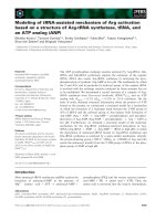
Fig. 1. Comparison of
1
H-
13
C constant-time HSQC NMR spectra of 0.6 mM of UL At3g16450.1 and 0.2 mM of SAIL At3g16450.1. (A) Full
spectrum of UL At3g16450.1. (B) Full spectrum of SAIL At3g16450.1. (C) Methylene region of UL At3g16450.1. (D) Methylene region of
SAIL At3g16450.1. (E) Methyl region of UL At3g16450.1. (F) Methyl region of SAIL At3g16450.1. Spectra were recorded at 27.5°Cat
1
H fre-
quency of 800 MHz. In the case of the SAIL protein,
2
H decoupling was applied during the
13
C chemical shift evolution.
SAIL-NMR structure of a myrosinase-binding protein M. Takeda et al.
5874 FEBS Journal 275 (2008) 5873–5884 ª 2008 The Authors Journal compilation ª 2008 FEBS
M. Takeda et al. SAIL-NMR structure of a myrosinase-binding protein
FEBS Journal 275 (2008) 5873–5884 ª 2008 The Authors Journal compilation ª 2008 FEBS 5875
Comparison of NMR spectra of SAIL and UL
At3g16450.1
Although the concentration of the SAIL protein was
lower than that of the UL protein by a factor of three
(SAIL, 0.2 mm; UL, 0.6 mm), the NMR spectra of
SAIL At3g16450.1 exhibited higher signal-to-noise
ratios than those of UL At3g16450.1. The
1
H-
13
C
constant-time HSQC spectrum of SAIL At3g16450.1
was less crowded and better resolved than that of UL
At3g16450.1 (Fig. 1A,B). The extensive stereo- and
regio-specific deuteration of the SAIL protein led to
diminished overlaps and sharpened peaks, particularly
in the methylene region, without compromising essential
structural information (Fig. 1C,D). In the methyl
region, the regio-specifically labeled methyl resonances
from the SAIL sample were much less crowded
(Fig. 1E,F). As a result of these striking spectral
improvements, it became possible to use established
methods [14] to assign 95.5% of the resonances of SAIL
At3g16450.1. The chemical shifts for SAIL At3g16450.1
have been deposited in the Biological Magnetic Reso-
nance Data Bank (BMRB) [15] with accession number
15607. In addition, 93% of the backbone carbonyl
13
C
shifts had been assigned previously using uniformly
13
C ⁄
15
N-labeled protein [13]. These assigned chemical
shifts were used as input for the talos program [16] to
obtain dihedral angle constraints.
Solution structure of SAIL At3g16450.1
Assignment of the NOE peaks of At3g16450.1 and the
structure determination were accomplished by use of
the cyana program [17,18]. The structural statistics are
summarized in Table 1. Although the 20 conformers
representing the structures of At3g16450.1 did not
superimpose well when the full sequence was considered
(residues 1-299), each individual domain (residues 1-144
or residues 153-299) superimposed well when considered
separately (Fig. 2A,B). Residues 16–21 and 45–47 exhib-
ited severe line broadening, probably arising from inter-
nal dynamics of these residues on the intermediate time
scale for chemical shifts. As a result, these are the least
well-defined regions of the N-terminal domain. The
C-terminal domain yielded reasonably well-converged
structures, including the side-chain conformations of
residues in its core (Fig. 2C,D).
Residues 145–152 in the linker region between the
two domains are highly disordered. In addition, a care-
ful search failed to reveal any inter-domain NOE peaks.
Thus the relative orientations of the two domains
appear not to be fixed, and the overall structure of
At3g16450.1 is best described as two tandem structural
domains connected by a flexible linker (Fig. 3A). The
secondary structural elements of At3g16450.1, extracted
from the coordinates of the three-dimensional structure
using the dssp algorithm [19], showed that each domain
has a similar structure consisting of three b-sheets
related by pseudo three-fold symmetry (Fig. 3B).
The coordinates of the 20 energy-refined conformers
that represent the solution structure of At3g16450.1
have been deposited in the Protein Data Bank with
accession code 2JZ4. A structural homology search
using the program dali at the European Molecular
Biology Laboratory (EMBL) [20,21] yielded the aggluti-
nin from Maclura promifera (Protein Data Bank code
Table 1. NMR constraints and structure calculation statistics for
At3g16450.1
a
.
Completeness of the chemical shift assignments (%)
All 95.5
Backbone 97.8
Side chain 93.3
NOE distance constraints
Total 1982
Short-range, |i – j| £ 1 1192
Medium-range, 1 < |i – j| < 5 111
Long-range, |i – j| ‡ 5, intra-molecular 679
Maximal violation (A
˚
) 0.18
Torsion angle constraints
/ 138
w 136
Maximal violation (°) 2.6
Restrained hydrogen bonds 124
CYANA target function value (A
˚
2
) 1.77 ± 0.56
AMBER energies (kcalÆmol
)1
)
Total )7508 ± 21
van der Waals )2239 ± 30
Ramachandran plot statistics (%) [35]
Residues in most favored regions 89.0
Residues in additional allowed regions 9.5
Residues in generously allowed regions 1.0
Residues in disallowed regions 0.5
Root mean square deviation from
the averaged coordinates (A
˚
)
Backbone atoms of residues
2–144 (N-domain)
1.12 ± 0.19
Heavy atoms of residues
2–144 (N-domain)
1.65 ± 0.16
Backbone atoms of residues
153–297 (C-domain)
0.69 ± 0.10
Heavy atoms of residues
153–297 (C-domain)
1.08 ± 0.09
a
The completeness of the
1
H,
13
C and
15
N chemical shift assign-
ments was evaluated for the aliphatic, aromatic, backbone amide
and Asn ⁄ Gln ⁄ Trp side-chain amide nuclei, excluding the carbon and
nitrogen atoms not bound to
1
H. Where applicable, the value given
corresponds to the average over the 20 energy-refined conformers
that represent the solution structure.
CYANA target function values
were calculated before energy refinement.
SAIL-NMR structure of a myrosinase-binding protein M. Takeda et al.
5876 FEBS Journal 275 (2008) 5873–5884 ª 2008 The Authors Journal compilation ª 2008 FEBS
1JOT), a plant lectin, as the closest structure. The root
mean square deviation values for the N- and C-terminal
domains versus the agglutinin are 2.2 and 2.0A
˚
, respec-
tively. Thus each of the two domains of At3g16450.1
adopts a lectin fold. The orientation of the N-terminal
domain relative to the C-terminal domain could not be
defined owing to the absence of inter-domain NOEs. To
confirm the molecular organization of the tandem
arrangement, expression vectors were constructed that
separately encoded the N-terminal half (residues 1–153)
and the C-terminal half (residues 151–299) of
At3g16450.1, and these were used to prepare
15
N-
labeled samples of each domain. The
1
H-
15
N HSQC
spectrum of each domain was well dispersed, and, when
overlaid, closely approximated the spectrum of full-
length At3g16450.1 (Fig. 4A,B). This result confirms
the structural arrangement of At3g16450.1 as two
independent tandem structural domains.
Interaction analysis of At3g16450.1 with sugars
Because each structural domain of At3g16450.1 was
found to adopt a lectin fold, we assayed At3g16450.1
for possible sugar-binding activity. We utilized 13 fluo-
rescence-labeled oligosaccharides (PA sugars) as candi-
dates. Four PA sugars eluted more slowly than the
tetra-sialyl PA-glycan as a control PA sugars from a
column of immobilized At3g16450.1 (Fig. 5A,B and
Table 2). On the basis of the elution profiles, the K
d
values for the four PA sugars to At3g16450.1 were
estimated to be low, at most 10
)4
m. To further examine
the observed interaction, we acquired
1
H-
15
N HSQC
spectra of
15
N-labeled At3g16450.1 in the presence and
absence of maltohexaose, (Glca1-4Glc)
3
. However,
addition of (Glca1-4Glc)
3
did not cause any perturba-
tion of NMR resonances, even when the concentration
of the sugar was ten times higher than that of the pro-
tein (data not shown). By contrast, NMR titration of
At3g16450.1 with (Glca1-4Glc)
3
-PA led to distinct
chemical shift changes for certain NMR resonances
(Fig. 5C), but addition of PA as the ligand resulted only
in limited subtle changes. These results suggest that
both PA and the (Glca1-4Glc)
3
elements contribute to
the observed interactions. Residues in both the N- and
C-terminal domains of At3g16450.1 were affected by
the presence of PA sugars (Fig. 5C, blue and red boxes).
Taken together, these binding analyses suggest that
At3g16450.1 has the potential to bind PA sugars with
specificity for the sugar structure, although none of the
various sugars tested exhibited a strong affinity.
Discussion
In this study, we determined the solution structure of
the 32 kDa At3g16450.1 protein from A. thaliana by
the SAIL-NMR method. This is the first application of
SAIL-NMR in a structural genomics study. It pro-
vided the first structure for a member of the hitherto
structurally unexplored MyroBP family.
At3g16450.1 consists of two tandem domains, each
composed of three b-sheets. The fold of each domain
is nearly identical to that of an agglutinin (Protein
Data Bank code 1JOT), which shares sequence identi-
ties of 26 and 33% with the N- and C-terminal
domains of At3g16450.1, respectively. Sequence simi-
larity searches performed by psi-blast [22] identified
other MyroBPs and MyroBP-like proteins from
A. thaliana and B. napus, with sequence identities to
Fig. 2. Three-dimensional NMR structure of At3g16450.1. (A)
Superposition of the 20 energy-minimized conformers that repre-
sent the 3D solution structure of the N-terminal domain. (B) Super-
position of conformers representing the C-terminal domain. (C)
Aromatic side chains and one backbone trace of the NMR struc-
tures for the N-terminal domain. (D) Aromatic side chains and one
backbone trace of the NMR structure of the C-terminal domain.
M. Takeda et al. SAIL-NMR structure of a myrosinase-binding protein
FEBS Journal 275 (2008) 5873–5884 ª 2008 The Authors Journal compilation ª 2008 FEBS 5877
the At3g16450.1 domains ranging from 30% to 70%.
The most highly conserved regions correspond to the
b-strands (Fig. 6). The N- and C-terminal domains of
At3g16450.1, with 51% sequence identity to each
other, are superimposed with root mean square devia-
tions of 1.3 A
˚
for the backbone of the b-strands and
1.7 A
˚
if the loop regions are included, indicating that
all of these family members adopt a similar fold.
It has been reported that seed MyroBP from
B. napus possesses lectin activity, binding to p-amino-
phenyl-a-d-mannopyranoside and to some extent to
N-acetylglucosamine [10]. Because myrosinase contains
potential N-linked sugar-binding sites [23], the sugar-
binding activity of MyroBP is implicated in binding to
myrosinase. In the case of At3g16450.1, the protein
did not show a significant affinity for sugar structures
specific to N-linked glycan, but rather showed weak
affinity for starch or glycolipid, raising the possibility
that the lectin activity of the MyroBP family is also
involved in interaction between a myrosinase complex
and other molecules. It is also noteworthy that a Uni-
Gene database search [24] suggested that At3g16450.1
is expressed in leaf and root. Because myrosinases have
also been shown to be expressed in A. thaliana leaf
[6,8], it may be suspected that At3g16450.1 forms a
complex with myrosinase, thereby guiding the myrosin-
ase to a damaged site in the leaf via weak interactions
with starch in the leaf or glycolipid from foreign
pathogens. However, it is obvious that further study
will be required to determine the biological importance
of MyroBP–sugar interactions.
Many MyroBP and MyroBP-related proteins contain
tandem lectin domains as shown in Fig. 6. The tandem
domains present in MyroBP family members may par-
ticipate in multivalent sugar binding as observed with
other carbohydrate binding proteins with multiple
domains. Results of the NMR chemical-shift pertur-
bation experiments (Fig. 5C) suggest that both domains
of At3g16450.1 can participate in a bivalent sugar bind-
ing. It is also probable that each homologous domain
of the MyroBP family possesses different ligand-bind-
ing properties, thereby providing a broad binding speci-
ficity. In some proteins containing tandem homologous
domains, inter-domain interactions fix the relative ori-
entation of the domains in a specific multi-domain
structure that is essential for biological function. Other
proteins with tandem domains contain a flexible linker,
and a specific structure may be adopted only when a
target is bound. The present study suggests that
At3g16450.1 belongs to the latter category.
The major problems with structural genomics studies
using NMR are low solubility and molecular-weight
limitations [2]. As shown by this study, the SAIL-
NMR method provides a promising approach to over-
coming both of these problems. One important aspect
of the SAIL technology is that the signal intensities for
the SAIL protein are several times stronger than for
the corresponding UL sample [3], thus making it possi-
ble to perform structure determination for proteins
even at low concentration. In this study, the structure
was determined using a 0.2 mm sample of SAIL
Fig. 3. Secondary structure of At3g16450.1. (A) Ribbon representa-
tion of the NMR structure of At3g16450.1. These figures were pre-
pared using
MOLMOL [25]. Due to the lack of NOEs, the relative
orientation between the N- and C-terminal domains could not be
defined. (B) Primary sequence of At3g16450.1. The sequences that
correspond to the N-terminal (residues 1-144) and C-terminal (resi-
dues 153-299) structural domains are highlighted in blue and pink,
respectively, and b-strands are indicated by arrows above the
sequence.
SAIL-NMR structure of a myrosinase-binding protein M. Takeda et al.
5878 FEBS Journal 275 (2008) 5873–5884 ª 2008 The Authors Journal compilation ª 2008 FEBS
Fig. 4. Comparison of the NMR spectra of
full-length At3g16450.1 and its isolated
N- and C-terminal halves. (A)
1
H-
15
N HSQC
spectrum of full-length (residues 1–299)
SAIL At3g16450.1. (B) Overlay of
1
H-
15
N
HSQC spectra of the N-terminal (residues
1–153, blue) and C-terminal (residues
151–299, red) halves of [U-
15
N]-labeled
At3g16450.1. These spectra were acquired
at 27.5°C, pH 6.8, using a Bruker DRX600
NMR spectrometer. The pattern of the over-
laid spectra is almost identical to that of the
full-length construct, showing that the two
domains of At3g16450.1 are largely inde-
pendent.
Fig. 5. Investigation of sugar-binding proper-
ties of At3g16450.1. (A) Elution profile from
the FAC binding assay for (Glca1-4Glc)
3
-PA
(red) and control sugar (black). (B) FAC bind-
ing assay for Gala1-4Galb1-4Glc-PA (red)
and control PA sugar (black). (C) Overlay of
the
1
H-
15
N HSQC spectra of uniformly
15
N-labeled At3g16450.1 in the absence
(black) and presence (red) of (Glca1-4Glc)
3
-
PA. Assignments and boxes (blue for the
N-terminal domain; red for the C-terminal
domain) indicate some of the perturbed
resonances.
M. Takeda et al. SAIL-NMR structure of a myrosinase-binding protein
FEBS Journal 275 (2008) 5873–5884 ª 2008 The Authors Journal compilation ª 2008 FEBS 5879
At3g16450.1. The SAIL-NMR method offers the
opportunity to determine structures of proteins with
low solubility or poor yield. The SAIL method can
also accelerate the process of structural analysis. The
spectral simplification achieved by SAIL with this lar-
ger protein makes it possible to use semi- or fully auto-
mated methods developed for use with smaller proteins
to analyze the NMR data. We are developing a soft-
ware package that exploits the benefits of the SAIL
method [25–27]. Finally, the SAIL method is expected
to enable functional investigations of larger proteins.
Experimental procedures
Plasmid construction
The construction of pET15b (Novagen, Madison, WI, USA)
harboring At3g16450.1 was performed as described previ-
ously [13]. The vector used for cell-free production of
At3g16450.1 was constructed according to a strategy
described previously [28]. DNA coding for the N-terminal
histidine tag followed by the At3g16450.1 was subcloned into
pIVEX2.3d (Roche, Pleasanton, CA, USA) between the
NcoI ⁄ NdeI and NdeI ⁄ BamHI sites, respectively. Silent muta-
tions were introduced into the N-terminal sequence to
enhance the expression rate [28]. Expression vectors coding
for the N-terminal (residues 1–153) and C-terminal (residues
151–299) domains of At3g16450.1 were constructed by clon-
ing the corresponding target sequence into the NdeI and
BamHI sites of pET15b.
Preparation of labeled proteins
[U-
15
N]- and [U-
13
C, U-
15
N]-labeled proteins were produced
by culturing Escherichia coli BL21 (DE3) strain harboring
the corresponding expression vector in M9 medium contain-
ing
15
NH
4
Cl and ⁄ or [U-
13
C]-labeled glucose as the sole nitro-
gen and carbon sources. Cells were cultured at 30 °C with
shaking. Expression was induced by the addition of isopropyl
thio-b-d-galactoside (IPTG) at a final concentration of
1mm, and cells were harvested 6.5 h after induction.
SAIL At3g16450.1 was produced by E. coli cell-free
expression. A total of 110 mg of SAIL amino acid mixture
was used, with the amount of each individual SAIL amino
acid proportional to the amino acid composition of
At3g16450.1. A home-made E. coli S30 extract was used,
and the reaction was performed as previously described
[25,28]. The volumes of the inner and outer solutions were
10 and 40 mL, respectively. The reaction was carried out at
30 °C for 15 h with shaking. To prevent degradation of the
produced protein, a protease inhibitor cocktail (Roche) was
added to the reaction. The At3g16450.1 protein was puri-
fied as described previously [13].
NMR spectroscopy
The NMR sample used for the structure determination
contained 0.2 mm SAIL At3g16450.1 protein in 20 mm bis-
Tris(2-carboxymethyl)phosphine: HCl(D19, 98%) (Cam-
bridge Isotope Laboratories Andover, MA, USA), 100 mm
KCl, 10% D
2
O, pH 6.8. NMR spectra were recorded on a
Bruker (Tsukuba, Japan) Avance 600 MHz spectrometer
equipped with a 5 mm
1
H-observe triple-resonance cryogenic
probe (Bruker TXI cryoProbe), and on a Bruker Avance
800 MHz spectrometer at 27.5 °C. The spectra were pro-
cessed using the programs xwinnmr version 3.5 (Bruker) or
nmrpipe [29], and analyzed using the program sparky
(T. D. Goddard and D. G. Kneller, Department of Phar-
maceutical Chemistry, University of California, San Fran-
cisco, CA, USA). Backbone and b-CH resonances were
assigned using 2D HSQC, and 3D HN(CO)CACB and
HBHA(CO)NH spectra. Side-chain resonances were
assigned using 3D H(CCCO)NH, (H)CC(CO)NH, HCCH-
TOCSY, constant time-HCCH-COSY,
13
C-edited NOESY
Table 2. Summary of results of the FAC binding assay for
At3g16450.1 with various PA sugars.
Major natural
location
PA sugars that showed affinity for At3g16450.1
(Glca1-4Glc)
3
maltohexaose Starch of higher
plants
(Glca1-6Glc)
3
isomaltohexaose Starch of higher
plants
Gala1-4Galb1-4Glc Glycolipid
GalNAca1-3(Fuca1-2)
Galb1-3(Fuca1-4)GlcNAcb1-3Galb1-4Glc
Glycolipid
PA sugars that did not show affinity
for At3g16450.1
Galb1-3(Fuca1-4)GlcNAcb1-3Galb1-4Glc Glycolipid
Galb1-4(Fuca1-3)GlcNAcb1-3Galb1-4Glc Glycolipid
(GlcNAcb1-4GlcNAc)
3
Chitohexaose Insects and
crustaceans
(Glcb1-4Glc)
3
Cellohexaose Cell walls of
higher plants
(Glcb1-3Glc)
3
Laminarihexaose Pachyman of
Poria cocos
Man9GN2 (high-mannose type)
(code no. M9.1)
N-glycan
GlcNAcb1-2Mana1-6
(GlcNAcb1-2Mana1-3)
Manb1-4GlcNAcb1-4(Fuca1-6)
GlcNAc (code no. 210.1)
N-glycan
Galb1-4GlcNAcb1-2Mana1-6
(Galb1-4GlcNAcb1-2
Mana1-3)Manb1-4GlcNAcb1-4(Fuca1-6)
GlcNAc (code no. 210.4)
N-glycan
GlcNAcb1-2Mana1-6(GlcNAcb1-2Mana1-3)
Manb1-4(Xylb1-2)GlcNAcb1-4
(Fuca1-3)GlcNAc (code no. 210.1FX)
N-glycan
SAIL-NMR structure of a myrosinase-binding protein M. Takeda et al.
5880 FEBS Journal 275 (2008) 5873–5884 ª 2008 The Authors Journal compilation ª 2008 FEBS
and
15
N-edited NOESY spectra.
15
N- and
13
C-edited NO-
ESY spectra were recorded with a mixing time of 75 ms,
and the inter-proton distance constraints were obtained
from the NOESY peaks, which were selected and manually
filtered using sparky.
Collection of conformational constraints,
structure calculation and refinement
Automated NOE cross-peak assignments [30] and structure
calculations with torsion-angle dynamics were performed
A
t3g16450.1N AQKVEAGGGAGGASWDDG-VHDGVRKVHVGQGQDGVSSINVVYAKDSQDVEGGEHGKKTL
A
t3g16450.1C AKKLSAIGGDEGTAWDDG-AYDGVKKVYVGQGQDGISAVKFEYNKGAENIVGGEHGKPTL
||* | * || | | | * |*
MBPfromB.napus1-125 MSWDDG-KHTKVKKIQLT-FDDVIRSIEVEYEGTN LKSQRRGTVGT
MBPfromB.napus194-336 KVGPLGGEKGNVFEDV-GFEGVKKITVGADQYSVTYIKIEYIKDGQ-VVVREHGTVR
G
MBPfromB.napus356-498 KKGPLGGEKGEEFNDV-GFEGVKKITVGADQYSVTYIKIEYVKDGK-VEIREHGTSR
G
A
t1g52030.2-154 SEKVGAMGGNKGGAFDDG-VFDGVKKVIVGKDFNNVTYIKVEYEKDGK-FEIREHGTNR
G
A
t1g52030.161-289 PQGGNGGSAWDDG-AFDGVRKVLVGRNGKFVSYVRFEYAKGER-MVPHAHGKRQE
A
t3g16400.2-142 AQKLEAKGGEMGDVWDDG-VYENVRKVYVGQAQYGIAFVKFEYVNGSQVVVGDEHGKKTE
A
t3g16440.2-144 AQKVEAQGGIGGDVWDDG-AHDGVRKVHVGQGLDGVSFINVVYENGSQEVVGGEHGKKSL
A
t3g16440.154-300 AKKLPAVGGDEGTAWDDG-AFDGVKKVYIGQAQDGISAVKFVYDKGAEDIVGDEHGNDTL
A
t3g16470.2-145 AKKLEAQGGRGGEEWDDGGAYENVKKVYVGQGDSGVVYVKFDYEKDGK-IVSHEHGKQTL
A
t3g16470.158-297 KLEAQGGRGGDVWDDGGAYDNVKKVYVGQGDSGVVYVKFDYEKDGK-IVSLEHGKQTL
A
t3g16470.308-450 TIPAQGGDGGVAWDDG-VHDSVKKIYVGQGDSCVTYFKADYEKASKPVLGSDHGKKTL
A
t3g21380.7-130 SWDDG-KHMKVKRVQIT-YEDVINSIEAEYDGDT HNPHHHGTPG
K
A
t3g16450.1N LG FETFEVD-ADDYIVAVQVTYDNVFG QDSDIITSITFNTFKGKTSPPYG
A
t3g16450.1C LG FEEFEIDYPSEYITAVEGTYDKIFG SDGLIITMLRFKTNK-QTSAPFG
| | | | | | | | ||*
MBPfromB.napus1-125 K SDGFTLS-TDEYITSVSGYYKTTFS G-DHITALTFKTNK-KTYGPYG
MBPfromB.napus194-336 E LKEFSVDYPNDNITAVGGTYKHVYT YDTTLITSLYFTTSKGFTSPLFG IDS
MBPfromB.napus356-498 E LQEFSVDYPNDSITEVGGTYKHNYT YDTTLITSLYFTTSKGFTSPLFG INS
A
t1g52030.2-154 Q LKEFSVDYPNEYITAVGGSYDTVFG YGSALIKSLLFKTSYGRTSPILGHTTLL
G
A
t1g52030.161-289 A PQEFVVDYPNEHITSVEGTIDG YLSSLKFTTSKGRTSPVFG
A
t1g52030.491-634 LG TETFELDYPSEYITSVEGYYDKIFG VEAEVVTSLTFKTNK-RTSQPFG
A
t3g16400.2-142 LG VEEFEID-ADDYIVYVEGYREKVND MTSEMITFLSIKTFKGKTSHPIE
A
t3g16440.2-144 IG IETFEVD-ADDYIVAVQVTYDKIFG YDSDIITSITFSTFKGKTSPPYG
A
t3g16440.154-300 LG FEEFQLDYPSEYITAVEGTYDKIFG FETEVINMLRFKTNK-KTSPPFG
A
t3g16470.2-145 LG TEEFVVD-PEDYITSVKIYYEKLFG SPIEIVTALIFKTFKGKTSQPFG
A
t3g16470.158-297 LG TEEFEID-PEDYITYVKVYYEKLFG SPIEIVTALIFKTFKGKTSQPFG
A
t3g16470.308-450 LG AEEFVLG-PDEYVTAVSGYYDKIFS VDAPAIVSLKFKTNK-RTSIPYG
A
t3g21380.7-130 K SDGVSLS-PDEYITDVTGYYKTTGA E-DAIAALAFKTNK-TEYGPYG
A
t3g16450.1N LETQKKFVLKDKNGGKLVGFHGRAG-EALYALGAYFA
A
t3g16450.1C LEAGTAFELKE-EGHKIVGFHGKAS-ELLHQFGVHVMPLTN
| || *| * |
MBPfromB.napus1-125 NKTQNYFSADAPKDSQIAGFLGTSG-ALL FA
MBPfromB.napus194-336 EKKGTEFEFKGENGGKLLGFHGRGG-NAIDAIGAYF
MBPfromB.napus356-498 EKKGTEFEFKDENGGKLIGLHGRGG-NAIDAIGAYF
A
t1g52030.2-154 NPAGKEFMLESKYGGKLLGFHGRSG-EALDAIGPHFFAVNS
A
t1g52030.161-289 NVVGSKFVFE-ETSFKLVGFCGRSG-EAIDALGAHF
A
t1g52030.336-476 METEKKLELKDGKGGKLVGFHGKAS-DVLYALGAYFA
A
t3g16400.2-142 KRPGVKFVL HGGKIVGFHGRST-DVLHSLGAYVS
A
t3g16440.2-144 LDTENKFVLKEKNGGKLVGFHGRAG-EILYALGAYF
A
t3g16440.154-300 IEAGTAFELKE-EGCKIVGFHGKVS-AVLHQFGVHILPVTN
A
t3g16470.2-145 LTSGEEAELG GGKIVGFHGSSS-DLIHSVGVYIIPST-
A
t3g16470.158-297 LTSGEEAELG GGKIVGFHGTSS-DLIHSLGAYIIP
A
t3g16470.308-450 LEGGTEFVLEK-KDHKIVGFYGQAG-EYLYKLGVNVAPIA-
A
t3g21380.7-130 NKTRNQFSIHAPKDNQIAGFQGISS-NVLNSIDVHFA
Fig. 6. Alignment of MyroBP-related sequences. Sequences of the N- and C-terminal domains of At3g16450.1 are aligned with those of
MyroBP from B. napus and MyroBP-like proteins from A. thaliana (At1g52030, At3g16400, At3g16440, At3g16470 and At3g21380). Asterisks
and vertical bars indicate identical and similar residues, respectively. The b-strands of At3g16450.1 are indicated by arrows above the sequence.
M. Takeda et al. SAIL-NMR structure of a myrosinase-binding protein
FEBS Journal 275 (2008) 5873–5884 ª 2008 The Authors Journal compilation ª 2008 FEBS 5881
using the program cyana, version 2.2 [31]. Backbone tor-
sion-angle constraints obtained from database searches
using the program talos [16] were incorporated into the
structure calculation. Simulated annealing with 20 000
torsion-angle dynamics time steps per conformer was
performed during the cyana structure calculations. In the
final cycle of the cyana protocol, 100 conformers were
generated and further refined using the amber 9 software
package [32] with a full-atom force field [33]. The refine-
ment comprised three stages: initial minimization, molecu-
lar dynamics, and final minimization. Minimization and
molecular dynamics consisted of 1500 steps and 20 ps dura-
tion, respectively. A generalized Born implicit solvent
model was used to account for the solvent effects [34]. The
force constants for distance and torsion-angle constraints
were 50 kcalÆmol
)1
ÆA
˚
)2
and 200 kcalÆmol
)1
Ærad
)2
respec-
tively. From the resulting structures of this first amber
refinement, we extracted backbone hydrogen-bond
constraints in the regular secondary elements that were
present in more than 75% of the 100 conformers. With
these as additional constraints, we repeated the refinement.
From the conformers that did not significantly violate
experimental constraints, we selected the 20 lowest-energy
structures for analysis. The structural quality was evaluated
using procheck-nmr [35]. The program molmol [36] was
used to visualize the structures. The coordinates of the 20
energy-refined cyana conformers of At3g16450.1 have been
deposited in the Protein Data Bank (accession code 2JZ4).
The chemical shifts of At3g16450.1 have been deposited in
the BioMagResBank (accession code 15607).
Frontal affinity chromatography
M9.1, 210.1, 210.4 and 210.1FX were purchased from
Seikagaku Kogyo Co (Tokyo, Japan). The code numbers
and structures of pyridylaminated oligosaccharides refer to
the GALAXY website at o/
ENG/index.html [37]. Two kinds of PA-oligosaccharides,
GalNAca1-3(Fuca1-2)Galb1-3(Fuca1-4)GlcNAcb1-3Galb1-
4Glc-PA and Neu5Aca2-6Galb1-4GlcNAcb1-2Mana1-
6(Neu5Aca2-3Galb1-3(Neu5Aca2-6)GlcNAcb1-4(Neu5Aca2-
6Galb1-4GlcNAcb1-2)Mana1-3)Manb1-4GlcNAcb1-4Glc-
NAc-PA were obtained from Takara Bio. Inc. (Otsu, Shiga,
Japan). Other PA glycans were prepared by amination of the
commercial oligosaccharides using 2-aminopyridine [38].
Lewis A- and Lewis X-type glycans, Galb1-3(Fuca1-4)Glc-
NAcb1-3Galb1-4Glc and Galb1-4(Fuca1-3)GlcNAcb1-
3Galb1-4Glc were purchased from Calbiochem (San Diego,
CA, USA). Cellohesaose, chitohesaose, isomaltohexaose,
laminarihesaose and maltohexaose were purchased from
Seikagaku Kogyo Co.
The protein At3g16450.1 containing the N-terminal histi-
dine tag was dissolved in 10 mm HEPES buffer, pH 7.6,
containing 150 mm NaCl, 1 mm CaCl
2
, and bound to Ni-
NTA agarose. After immobilization, the agarose beads were
packed into a stainless steel column (4.0 · 10 mm, GL
Sciences, Tokyo, Japan).
Frontal affinity chromatography analysis was performed
as described previously [39]. PA oligosaccharides were dis-
solved at a concentration of 10 nm in 10 mm HEPES,
pH 7.6, containing 150 mm NaCl, 1 mm CaCl
2
, and applied
onto the At3g16450.1 column at a flow rate of 0.25 mLÆmin
)1
at 20 °C. The elution profile was monitored by the fluores-
cence intensity at 400 nm (excitation at 320 nm). Tetrasialyl
PA glycan Neu5Aca2-6Galb1-4GlcNAcb1-2Mana1-6(Neu5-
Aca2-3Galb1-3(Neu5Aca2-6)GlcNAcb1-4(Neu5Aca2-6Galb1-
4GlcNAcb1-2)Ma na1-3)Manb1-4GlcNAcb1-4GlcNA-PA
was used as a control sugar to determine the elution volume
of the unbound oligosaccharide.
NMR chemical-shift perturbation mapping
NMR samples were prepared using free [U-
15
N]-labeled
At3g16450.1 (0.1 mm protein, 10 mm HEPES, pH 7.6,
150 mm KCl, 1 mm CaCl
2
) and its complex with PA sugar
[same solvent composition plus 0.5 mm PA-(Glca1-4Glc)
3
].
1
H-
15
N HSQC spectra of the isolated and titrated samples
were acquired at 27.5 °C using a Bruker Avance 600 MHz
NMR spectrometer.
Acknowledgements
This work was supported by the Technology Develop-
ment for Protein Analyses and Targeted Protein
Research Program of the Ministry of Education,
Culture, Sports, Science and Technology of Japan
(MEXT), by Core Research for Evolutional Science
and Technology (CREST) of the Japan Science and
Technology Agency (JST), by a Grant-in-Aid for
Scientific Research from the Japan Society for the
Promotion of Science (JSPS), by the National Insti-
tutes of Health Protein Structure Initiative (grants P50
GM64598 and U54 GM074901), and by the Volk-
swagen Foundation.
References
1 The Arabidopsis Initiative (2000) Analysis of the gen-
ome sequence of the flowering plant Arabidopsis thali-
ana. Nature 408, 796–815.
2 Vinarov DA, Loushin Newman CL & Markley JL
(2006) Wheat germ cell-free platform for eukaryotic
protein production. FEBS J 273, 4160–4169.
3 Kainosho M, Torizawa T, Iwashita Y, Terauchi T, Ono
AM & Gu
¨
ntert P (2006) Optimal isotope labelling for
NMR protein structure determinations. Nature 440,
52–57.
4 Rask L, Andre
´
asson E, Ekbom B, Eriksson S, Pontopp-
idan B & Meijer J (2000) Myrosinase: gene family
SAIL-NMR structure of a myrosinase-binding protein M. Takeda et al.
5882 FEBS Journal 275 (2008) 5873–5884 ª 2008 The Authors Journal compilation ª 2008 FEBS
evolution and herbivore defense in Brassicaceae. Plant
Mol Biol 42, 93–113.
5Lo
¨
nnerdal B & Janson JC (1973) Studies on myrosinas-
es. II. Purification and characterization of a myrosinase
from rapeseed (Brassica napus L.). Biochim Biophys
Acta 315, 421–429.
6 Xue J, Jørgensen M, Pihlgren U & Rask L (1995) The
myrosinase gene family in Arabidopsis thaliana: gene
organization, expression and evolution. Plant Mol Biol
27, 911–922.
7 Takechi K, Sakamoto W, Utsugi S, Murata M & Mo-
toyoshi F (1999) Characterization of a flower-specific
gene encoding a putative myrosinase binding protein in
Arabidopsis thaliana. Plant Cell Physiol 40, 1287–1296.
8 Capella AN, Menossi M, Arruda P & Benedetti CE
(2001) COI1 affects myrosinase activity and controls the
expression of two flower-specific myrosinase-binding pro-
tein homologues in Arabidopsis. Planta 213, 691–699.
9 Eriksson S, Andre
´
asson E, Ekbom B, Grane
´
r G, Pon-
toppidan B, Taipalensuu J, Zhang J, Rask L & Meijer J
(2002) Complex formation of myrosinase isoenzymes in
oilseed rape seeds are dependent on the presence of
myrosinase-binding proteins. Plant Physiol 129, 1592–
1599.
10 Taipalensuu J, Eriksson S & Rask L (1997) The myro-
sinase-binding protein from Brassica napus seeds pos-
sesses lectin activity and has a highly similar
vegetatively expressed wound-inducible counterpart. Eur
J Biochem 250, 680–688.
11 Falk A, Taipalensuu J, Ek B, Lenman M & Rask L
(1995) Characterization of rapeseed myrosinase-binding
protein. Planta 195, 387–395.
12 Kasai K, Oda Y, Nishikawa M & Ishii S (1986) Frontal
affinity chromatography: theory for its application to
studies on specific interactions of biomolecules. J Chro-
matogr 376, 33–47.
13 Sugimori N, Torizawa T, Aceti DJ, Thao S, Markley
JL & Kainosho M (2004)
1
H,
13
C and
15
N backbone
assignment of a 32 kDa hypothetical protein from
Arabidopsis thaliana, At3g16450.1. J Biomol NMR 30,
357–358.
14 Cavanagh J, Fairbrother WJ, Palmer AG III, Skelton
NJ & Rance M (2006) Protein NMR Spectroscopy.
Principles and Practice, 2nd edn. Academic Press, San
Diego, CA.
15 Seavey BR, Farr EA, Westler WM & Markley JL
(1991) A relational database for sequence-specific pro-
tein NMR data. J Biomol NMR 1, 217–236.
16 Cornilescu G, Delaglio F & Bax A (1999) Protein back-
bone angle restraints from searching a database for
chemical shift and sequence homology. J Biomol NMR
13, 289–302.
17 Gu
¨
ntert P, Mumenthaler C & Wu
¨
thrich K (1997) Torsion
angle dynamics for NMR structure calculation with the
new program DYANA. J Mol Biol 273, 283–298.
18 Gu
¨
ntert P (2003) Automated NMR protein structure cal-
culation. Prog Nucl Magn Reson Spectrosc 43, 105–125.
19 Kabsch W & Sander C (1983) Dictionary of protein
secondary structure – pattern-recognition of hydrogen-
bonded and geometrical features. Biopolymers 22, 2577–
2637.
20 Holm L, Ouzounis C, Sander C, Tuparev G & Vriend
G (1992) A database of protein structure families with
common folding motifs. Protein Sci 1, 1691–1698.
21 Holm L & Sander C (1993) Protein structure compari-
son by alignment of distance matrices. J Mol Biol 233,
123–138.
22 Altschul SF, Madden TL, Scha
¨
ffer AA, Zhang J,
Zhang Z, Miller W & Lipman DJ (1997) Gapped
BLAST and PSI-BLAST: a new generation of protein
database search programs. Nucleic Acids Res 25,
3389–3402.
23 Falk A, Ek B & Rask L (1995) Characterization of a
new myrosinase in Brassica napus. Plant Mol Biol 27,
863–874.
24 Schuler GD (1997) Pieces of the puzzle: expressed
sequence tags and the catalog of human genes. J Mol
Med 75, 694–698.
25 Takeda M, Ikeya T, Gu
¨
ntert P & Kainosho M (2007)
Automated structure determination of proteins with the
SAIL-FLYA NMR method. Nat Protoc 2, 2896–2902.
26 Lo
´
pez-Me
´
ndez B & Gu
¨
ntert P (2006) Automated pro-
tein structure determination from NMR spectra. JAm
Chem Soc 128, 13112–13122.
27 Scott A, Lo
´
pez-Me
´
ndez B & Gu
¨
ntert P (2006) Fully
automated structure determinations of the Fes SH2
domain using different sets of NMR spectra. Magn
Reson Chem 44, S83–S88.
28 Torizawa T, Shimizu M, Taoka M, Miyano H &
Kainosho M (2004) Efficient production of isotopically
labeled proteins by cell-free synthesis: a practical proto-
col. J Biomol NMR 30, 311–325.
29 Delaglio F, Grzesiek S, Vuister GW, Zhu G, Pfeifer J
& Bax A (1995) NMRPipe – a multidimensional spec-
tral processing system based on Unix pipes. J Biomol
NMR 6, 277–293.
30 Herrmann T, Gu
¨
ntert P & Wu
¨
thrich K (2002) Protein
NMR structure determination with automated NOE
assignment using the new software CANDID and the
torsion angle dynamics algorithm DYANA. J Mol Biol
319, 209–227.
31 Gu
¨
ntert P (2004) Automated NMR structure calcula-
tion with CYANA. Methods Mol Biol 278, 353–378.
32 Case DA, Cheatham TE, Darden T, Gohlke H, Luo R,
Merz KM, Onufriev A, Simmerling C, Wang B &
Woods RJ (2005) The Amber biomolecular simulation
programs. J Comput Chem 26, 1668–1688.
33 Cornell WD, Cieplak P, Bayly CI, Gould IR, Merz
KM, Ferguson DM, Spellmeyer DC, Fox T, Caldwell
JW & Kollman PA (1995) A second generation force
M. Takeda et al. SAIL-NMR structure of a myrosinase-binding protein
FEBS Journal 275 (2008) 5873–5884 ª 2008 The Authors Journal compilation ª 2008 FEBS 5883
field for the simulation of proteins, nucleic acids, and
organic molecules. J Am Chem Soc 117, 5179–5197.
34 Tsui V & Case DA (2000) Theory and applications of
the generalized Born solvation model in macromolecu-
lar simulations. Biopolymers 56, 275–291.
35 Laskowski RA, Rullmann JAC, MacArthur MW, Kap-
tein R & Thornton JM (1996) AQUA and PRO-
CHECK-NMR: programs for checking the quality of
protein structures solved by NMR. J Biomol NMR 8,
477–486.
36 Koradi R, Billeter M & Wu
¨
thrich K (1996) MOLMOL:
a program for display and analysis of macromolecular
structures. J Mol Graphics 14 , 51–55.
37 Takahashi N & Kato K (2003) GALAXY (glycoanaly-
sis by the three axes of MS and chromatography): a
web application that assists structural analyses of N-gly-
cans. Trends Glycosci Glycotechnol 15, 235–251.
38 Yamamoto S, Hase S, Fukuda S, Sano O & Ikenaka T
(1989) Structures of the sugar chains of interferon-c
produced by human myelomonocyte cell line HBL-38.
J Biochem (Tokyo) 105, 547–555.
39 Arata Y, Hirabayashi J & Kasai K (2001) Sugar bind-
ing properties of the two lectin domains of the tandem
repeat-type galectin LEC-1 (N32) of Caenorhabditis ele-
gans. Detailed analysis by an improved frontal affinity
chromatography method. J Biol Chem 276, 3068–3077.
SAIL-NMR structure of a myrosinase-binding protein M. Takeda et al.
5884 FEBS Journal 275 (2008) 5873–5884 ª 2008 The Authors Journal compilation ª 2008 FEBS