Tài liệu Báo cáo khoa học: Hypoxic resistance to articular chondrocyte apoptosis – a possible mechanism of maintaining homeostasis of normal articular cartilage pdf
Bạn đang xem bản rút gọn của tài liệu. Xem và tải ngay bản đầy đủ của tài liệu tại đây (571.03 KB, 11 trang )
Hypoxic resistance to articular chondrocyte
apoptosis – a possible mechanism of maintaining
homeostasis of normal articular cartilage
J W. Seol
1
, H B. Lee
1
, Y J. Lee
1
, Y H. Lee
2
, H s. Kang
1
, I s. Kim
1
, N S. Kim
1
and S Y. Park
1
1 Center for Healthcare Technology Development, Bio-Safety Research Institute, College of Veterinary Medicine, Chonbuk National
University, Jeonju, Jeonbuk, South Korea
2 Institute of Oral Bioscience, School of Dentistry, Chonbuk National University, Jeonju, Jeonbuk, South Korea
Keywords
chondrocytes; hypoxia; proteasome; reactive
oxygen species; tumour necrosis
factor-related apoptosis-inducing ligand
(TRAIL)
Correspondence
S Y. Park, College of Veterinary Medicine,
Chonbuk National University, Jeonju,
Jeonbuk 561-756, South Korea
Fax: +82 63 270 3780
Tel: +82 63 270 3886
E-mail:
(Received 21 August 2009, revised 10
October 2009, accepted 20 October 2009)
doi:10.1111/j.1742-4658.2009.07451.x
Hypoxia and hypoxia-related genes are important factors in articular chon-
drocytes during cartilage homeostasis and osteoarthritis. We have investi-
gated the various apoptotic factors that show significance in synovial fluid
obtained from normal and experimental osteoarthritic animal models and
have evaluated the effect of hypoxia on articular chondrocyte apoptosis
induced by these apoptotic factors. Mature beagle dogs underwent surgical
transections of ligaments and medial meniscectomies to explore the under-
lying mechanisms of osteoarthritis. Cartilage and synovial fluid obtained
from normal animals and those with osteoarthritis were evaluated via pro-
teasome inhibition, tumour necrosis factor-related apoptosis-inducing
ligand (TRAIL) protein expression, mitochondrial transmembrane poten-
tial and levels of reactive oxygen species. Canine chondrocytes were
exposed to the proteasome inhibitor N-acetyl-Leu-Leu-Norleu-al and trea-
ted with recombinant TRAIL protein under normoxic and hypoxic condi-
tions, measuring chondrocyte cell viability, proteasome activity and levels
of apoptotic factors. TRAIL protein expression and ubiquitinated proteins
were increased significantly, but the proteasome activity in the synovial
fluid of osteoarthritic joints relative to that in normal joints was not. Pri-
mary cultured articular chondrocytes cotreated with the proteasome inhibi-
tor and TRAIL progressed to severe apoptosis under normoxic conditions,
but the sensitization caused by the combined treatment was suppressed by
exposure to hypoxia. Caspase-8 activation, c-Jun N-terminal kinase phos-
phorylation, the mitochondrial transmembrane potential and the genera-
tion of reactive oxygen species involved in cell death regulation were
significantly inhibited under hypoxic conditions. These findings suggest that
proteasome inhibition and TRAIL may be possible mechanisms in cartilage
degradation and joint-related diseases. Furthermore, the maintenance
of hypoxic conditions or therapy with hypoxia-related genes in the joint
may be successful for the treatment of joint-related diseases, including
osteoarthritis.
Abbreviations
ALLN, N-acetyl-Leu-Leu-Norleu-al; DCFH
2
-DA, 2¢,7¢-dichlorodihydrofluorescein diacetate; DR-5, death receptor-5; JC-1, 5,5¢,6,6¢-tetrachloro-
1,1¢3,3¢-tetraethylbenzimidazol-carbocyanine iodide; JNK, c-Jun N-terminal kinase; JNK-SAPK, c-Jun N-terminal kinase-stress-activated protein
kinase; MTP, mitochondrial transmembrane potential; OA, osteoarthritis; ROS, reactive oxygen species; Suc-LLVY-AMC, Suc-Leu-Leu-Val-
Tyr-7-amino-4-methylcoumarin; TRAIL, tumour necrosis factor-related apoptosis-inducing ligand.
FEBS Journal 276 (2009) 7375–7385 ª 2009 The Authors Journal compilation ª 2009 FEBS 7375
Introduction
A hallmark of osteoarthritis (OA) is a decrease in the
number of chondrocytes, as they are the only resident
cells in articular cartilage. Chondrocytes regulate the
enzymatic breakdown of the extracellular matrix,
thereby maintaining the equilibrium between synthetic
and degradative processes in the cartilage [1]. There-
fore, the metabolic and structural changes of chondro-
cytes in the articular cartilage play a significant role in
the initiation and progression of the disease. Several
studies have examined cell death in human articular
cartilage affected by OA or in experimental models of
OA [2,3].
Tumour necrosis factor-related apoptosis-inducing
ligands (TRAILs) are type II transmembrane mole-
cules that trigger the apoptotic signal cascade by bind-
ing to cognate receptors expressed on the cell surface
[4,5]. TRAIL is highly expressed on the surfaces of
natural killer (NK) cells, as well as on CD4+ and
CD8+ T cells. It promotes apoptosis, which may aid
in the resolution of infection and the attenuation of
the development of streptoxotocin-induced diabetes
and collagen-induced arthritis [6–9]. Some studies have
reported a role for TRAIL protein in articular joint
disease [10,11]. TRAIL alone can induce apoptosis in
primary cultured chondrocytes from different animal
species, such as humans and rats [11,12], but the exact
role of TRAIL in chondrocytes has not been clearly
defined to date.
Ubiquitin-proteasome-mediated protein degradation
pathways have been shown to play an important role
in regulating both cell proliferation and cell death [13].
Most recent studies have suggested that ubiquitin-pro-
teasome-dependent proteolysis is also involved in
apoptosis, although its exact role remains controversial
[14]. Proteasome inhibitors block the process of pro-
grammed cell death in thymocytes and neurons, but
induce apoptosis in various human cancer cell lines.
Proteasome inhibition suppresses growth plate prolifer-
ation and induces chondrocyte apoptosis [15]. Human
chondrocytes are also sensitive to proteasome-induced
apoptosis [16]. Although the treatment of cells with
this compound causes marked increases in a large
number of cellular proteins, including cyclin-dependent
protein kinase inhibitors, it is not clear how this agent
actually induces apoptosis [17,18]. More research is
required to fully characterize the types of cell death in
aging and arthritic cartilage, together with their respec-
tive frequencies.
Articular cartilage is an avascular tissue that func-
tions at an oxygen tension lower than that of most
other tissues, and derives both its nutrition and oxygen
supply by diffusion from the synovial fluid and
subchondral bone [19,20]. It has been estimated that
articular chondrocytes in the deepest layers may have
access to no more than 1–6% O
2
[21,22]. Oxygen can
be processed to generate reactive oxygen species
(ROS), which play an important role in intracellular
signalling and thus in cell physiology and cellular
destruction. ROS are known to induce a wide range of
responses, depending on cell type and levels of ROS
within the cell [23,24].
The aims of this study were to investigate the major
signalling pathways and effects of hypoxic conditions
in experimental osteoarthritic cartilage degeneration
and cell death of primary cultured chondrocytes. In
particular, we focused on the role of proteasome
inhibition and TRAIL in osteoarthritic disease and
chondrocyte apoptosis, and the hypoxic inhibition of
cartilage and chondrocyte degeneration.
Results
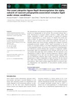
Macroscopic and radiographic examination of the
articular cartilage after experimentally induced
OA
Articular cartilage of the femoral condyles from the
experimental joints was examined to assess any macro-
scopic damage caused by experimentally induced OA.
Cartilage damage was visualized on the tibial plateau
of the experimental joints when compared with normal
control joints. The medial tibia plateau cartilage in the
experimental joints was fibrillated with erosive lesions
(Fig. 1A), and radiographic findings revealed joint
distension. There was no evidence of sclerosis, erosions
or osteophyte and enthesophyte formation in the
experimental joints (Fig. 1B).
TRAIL and ubiquitinated protein expression were
significantly increased, but proteasome activity
was not, in the synovial fluid of osteoarthritic
joints
Proteasome activity was assayed in the synovial fluid
from experimental osteoarthritic joints via Suc-Leu-
Leu-Val-Tyr-7-amino-4-methylcoumarin (Suc-LLVY-
AMC) hydrolysis, and was significantly lower than the
activity in control joints. The ubiquitinated protein lev-
els in synovial fluid from osteoarthritic joints were
higher than in the control joint group (Fig. 2A). We
also examined TRAIL protein expression in experi-
mental osteoarthritic synovial fluid. The protein
Hypoxic condition inhibits articular chondrocyte death J W. Seol et al.
7376 FEBS Journal 276 (2009) 7375–7385 ª 2009 The Authors Journal compilation ª 2009 FEBS
expression of TRAIL was increased in osteoarthritic
joints compared with the control group (Fig. 2B). In
this experiment, the elevated TRAIL protein of osteo-
arthritic synovial fluid may have originated from vari-
ous inflammatory cells, such as lymphocytes, and other
studies support this [6,8].
Combined treatment with proteasome inhibitor
and TRAIL markedly enhanced apoptosis in
cultured canine chondrocytes
To investigate the proteasome inhibition effect on
TRAIL-induced apoptosis in canine chondrocytes,
N-acetyl-Leu-Leu-Norleu-al (ALLN) was used as a
proteasome inhibitor. Canine chondrocytes were
exposed to ALLN (10 lm) for 12 h and then treated
with recombinant TRAIL protein for an additional
12 h. TRAIL and ALLN alone did not induce apopto-
sis, but combined treatment markedly induced apopto-
sis to 60% in canine chondrocytes (Fig. 3A). The
examination of cell morphology also supported the
enhancing effect of combined ALLN and TRAIL
treatment in canine chondrocytes (Fig. 3B).
To determine whether treatment with the protea-
some inhibitor ALLN affected proteasome-mediated
degradation in canine chondrocytes, proteasome activ-
ity was assayed in cell lysates as a measure of the
hydrolysis of the fluorogenic substrate Suc-LLVY-
AMC. Proteasome activity was significantly inhibited
by treatment with ALLN only and cotreatment with
ALLN and TRAIL (Fig. 3C). Western blot analysis
was also used to investigate whether ALLN induced
proteasome inhibition in canine chondrocytes. In the
absence of ALLN, smears of ubiquitinated proteins
were not observed in control and TRAIL-treated
canine chondrocytes. In the presence of ALLN,
marked accumulation of polyubiquitinated proteins
was observed in canine chondrocytes (Fig. 3D).
Proteasome inhibition increased significantly
TRAIL-mediated caspase-8 activation and JNK
phosphorylation
To determine the mechanism by which proteasome
inhibition enhanced TRAIL-induced apoptosis in
canine chondrocytes, we examined caspase-8 activation
and death receptor-5 (DR-5) and TRAIL protein
A
B
Fig. 1. Evaluation of articular cartilage after experimentally induced
osteoarthritis. (A) Photomicrographs of articular cartilage. (B) The
evaluation of osteoarthritis in the right and left joints of dogs was
graded 12 weeks after surgery by the evaluation of radiographs
using established parameters. OA, osteoarthritis sample.
A
B
Fig. 2. TRAIL protein and ubiquitinated protein levels were signifi-
cantly higher and proteasome activity was lower in osteoarthritic
joints. (A) Proteasome activity was measured using the synthetic
fluorogenic substrate Suc-LLVY-AMC. Fluorescence was measured
at 380 nm excitation and 440 nm emission. The fluorescence value
for control cells was set at 100%, and the fluorescence values rela-
tive to the control are presented. The experiments were performed
in triplicate at least twice independently. (A, B) Proteins were sepa-
rated on an 8–15% SDS gel, and apoptotic proteins were detected
by western blot analysis. b-Actin was used to normalize equal pro-
tein loading. Blot images represent one of three independent exper-
iments. *P < 0.05 versus normal sample was calculated using
Student’s t-test. OA, osteoarthritis sample; Ubi, ubiquitin.
J W. Seol et al. Hypoxic condition inhibits articular chondrocyte death
FEBS Journal 276 (2009) 7375–7385 ª 2009 The Authors Journal compilation ª 2009 FEBS 7377
expression in cells treated with ALLN and ⁄ or TRAIL
protein. Western blot analysis showed that caspase-8
was slightly activated in control, ALLN-treated and
TRAIL-treated chondrocytes. However, under protea-
some inhibition induced by pretreatment with ALLN,
TRAIL treatment unexpectedly increased the activa-
tion of caspase-8. In addition, the phosphorylation of
JNK protein was markedly increased by combined
treatment with ALLN and TRAIL when compared
with other groups. The expression of DR-5 and
TRAIL protein was also investigated, but these were
not altered in cells with proteasome inhibition (Fig. 4).
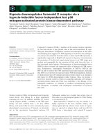
Proteasome inhibition and the expression of
TRAIL protein induced the dissipation of the
mitochondrial transmembrane potential (MTP)
and ROS generation
MTP was investigated in order to address the possible
mechanism by which proteasome inhibition enhances
TRAIL-induced apoptosis in canine chondrocytes.
MTP evaluation is based on the ability of a fluorescent
probe to enter the mitochondria selectively and
reversibly change its colour from green to red as the
mitochondrial potential increases. 5,5¢,6,6¢-Tetrachloro-
1,1¢3,3¢-tetraethylbenzimidazol-carbocyanine iodide
(JC-1; Molecular Probes, Eugene, OR, USA) exists as
a monomer at low MTP values and shows green fluo-
rescence, whereas it forms an aggregate at high MTP
and shows red fluorescence. The fluoroscopic results
presented in Fig. 5A show a red and slightly green flu-
orescence in cells treated with ALLN and TRAIL
alone, but a highly green fluorescence after combined
treatment. Photomicrographs indicated that ALLN
and TRAIL alone induced a small change in MTP,
whereas combined treatment with ALLN and TRAIL
caused a significant dissipation of MTP relative to neg-
ative controls. When ROS generation was examined,
the results showed that pretreatment of the cells with
the proteasome inhibitor increased ROS levels, and
that significant ROS generation was induced with
TRAIL cotreatment in canine chondrocytes (Fig. 5B).
A
C
B D
Fig. 3. Proteasome inhibition markedly enhanced TRAIL-induced apoptosis and significantly inhibited proteasome activity in primary cultured
canine chondrocytes. (A) Cell viability was determined by the crystal violet staining method. The viability of control cells was set at 100%,
and the viability relative to the control is presented. The experiments were performed in triplicate at least twice independently. The bars
describe the standard deviation. (B) Cell morphology was photographed (·200) under the various conditions. (C) Proteasome activity was
measured using the synthetic fluorogenic substrate Suc-LLVY-AMC. Fluorescence was measured at 380 nm excitation and 440 nm emis-
sion. The fluorescence value for control cells was set at 100%, and the fluorescence values relative to the control are presented. The experi-
ments were performed in triplicate at least twice independently. The bars describe the standard deviation. (D) Whole-cell lysates were
prepared and total protein (40 lgÆmL
)1
) was electrophoretically resolved on SDS gel. Ubiquitin protein levels were detected by western blot-
ting analysis. b-Actin was used to normalize equal protein loading. Blot images represent one of three independent experiments.
**P < 0.01, *P < 0.05 versus control were calculated using Student’s t-test.
Hypoxic condition inhibits articular chondrocyte death J W. Seol et al.
7378 FEBS Journal 276 (2009) 7375–7385 ª 2009 The Authors Journal compilation ª 2009 FEBS
To investigate the effects of MTP in cartilage by the
induction of OA, chondrocytes were isolated from
experimentally induced osteoarthritic cartilage. The
photomicrographs and fluorescence values indicated
that these chondrocytes showed a decrease in MTP
compared with normal cartilage (Fig. 5C). In addition,
the chondrocytes isolated from osteoarthritic joints
demonstrated significantly greater ROS generation
than did the controls (Fig. 5D).
Hypoxia inhibited the apoptosis of primary
cultured canine chondrocytes induced by
proteasome inhibition and TRAIL treatment
In order to examine the functional role of ALLN and
TRAIL in apoptotic cell death under hypoxic condi-
tions, canine chondrocytes were exposed to hypoxia
and ALLN (10 lm) for 12 h, and were then treated
with recombinant TRAIL protein for an additional
12 h under normoxic and hypoxic conditions. Hypoxic
conditions inhibited significantly the apoptosis of
chondrocytes induced by cotreatment of the cells with
the proteasome inhibitor and TRAIL (Fig. 6A). Chon-
drocyte survival under hypoxic conditions was
enhanced by 25% compared with the survival of cells
Fig. 4. Proteasome inhibition and TRAIL treatment significantly
increased caspase-8 activation and JNK phosphorylation. Whole-cell
lysates were prepared and total protein (40 lgÆmL
)1
) was electro-
phoretically resolved on SDS gel. Apoptotic proteins were detected
by western blotting analysis. b-Actin was used to normalize equal
protein loading. Blot images represent one of three independent
experiments.
AC
B
D
Fig. 5. The decrease in MTP and ROS generation induced by proteasome inhibition and TRAIL. (A, C) MTP was determined using a JC-1
probe. The cells were photographed using a fluoroscope. The green fluorescence intensity was measured under the conditions described in
Materials and methods. The experiments were performed in triplicate at least twice independently. (B, D) The ROS level was measured
using DCFH-DA. DCFH fluorescence was determined with a fluorescence plate reader with 490 and 525 nm as excitation and emission
wavelengths, respectively. The fluorescence value for control cells was set at 100%; fluorescence values relative to the control are pre-
sented. The experiments were performed in triplicate at least twice independently. **P < 0.01, *P < 0.05 versus control were calculated
using Student’s t-test. MFI, mean fluorescence intensity; OA, osteoarthritis sample.
J W. Seol et al. Hypoxic condition inhibits articular chondrocyte death
FEBS Journal 276 (2009) 7375–7385 ª 2009 The Authors Journal compilation ª 2009 FEBS 7379
that were cotreated with ALLN and TRAIL under
normoxic conditions. Moreover, photomicrographs
revealed that cells showed a decreased death rate under
hypoxic conditions when they were cotreated with
ALLN and TRAIL (Fig. 6A).
Hypoxia inhibited chondrocyte apoptosis through
the inhibition of caspase activation, JNK
phosphorylation, restoration of MTP loss and
ROS generation
To examine why hypoxia inhibited the combined
effects of ALLN and TRAIL in canine chondrocytes,
western blot analysis was performed. It was shown
that cotreatment of cells with ALLN and TRAIL
increased caspase-8 activation and JNK phosphoryla-
tion. However, both caspase-8 activation and JNK
phosphorylation were inhibited under hypoxic condi-
tions (Fig. 6B). MTP and ROS were investigated in
order to address the inhibitory mechanism exerted by
hypoxic conditions. Canine chondrocytes were
exposed to ALLN (10 lm) for 12 h and were then
treated with recombinant TRAIL protein for an addi-
tional 12 h under normoxic and hypoxic conditions.
The fluoroscopic results presented in Fig. 6C show
that the cells fluoresce green after cotreatment with
ALLN and TRAIL, indicating lower MTP under
normoxic conditions. However, the green fluorescence
indicating lower MTP declined under hypoxic condi-
tions (Fig. 6C). Cotreatment of cells with ALLN and
TRAIL induced ROS generation significantly under
normoxic conditions, but hypoxia prevented ROS
generation after cotreatment with ALLN and TRAIL
(Fig. 6D).
A
C
B D
Fig. 6. Hypoxia inhibited chondrocyte death and apoptosis-related signals induced by proteasome inhibition and TRAIL. (A) Cell viability was
determined using the crystal violet staining method. The control cell viability was set at 100%; viability relative to the control is presented.
The experiments were performed in triplicate at least twice independently. The cell morphology was photographed (·200). (B) Whole-cell
lysates were prepared and total protein (40 lgÆmL
)1
) was electrophoretically resolved on SDS gel and then tested for apoptotic proteins by
western blotting analysis. b-Actin was used to normalize equal protein loading; Blot images represent one of three independent experiments.
(C) MTP was determined using a JC-1 probe. The cells were photographed using a fluoroscope. The green fluorescence intensity was mea-
sured under the conditions described in Materials and methods. (D) The ROS level was measured using DCFH-DA. The fluorescence value
for control cells was set at 100%; fluorescence values relative to the control are presented. The experiments were performed in triplicate at
least twice independently. **P < 0.01, *P < 0.05 versus control were calculated using Student’s n-test. Nor, normoxia; Hypo, hypoxia; A,
ALLN; T, TRAIL; MFI, mean fluorescence intensity.
Hypoxic condition inhibits articular chondrocyte death J W. Seol et al.
7380 FEBS Journal 276 (2009) 7375–7385 ª 2009 The Authors Journal compilation ª 2009 FEBS
Discussion
TRAIL is a good candidate for cancer therapy as it
selectively induces apoptosis in tumour cells, with little
or no effect on normal cells [25]. It has recently been
reported that rheumatoid arthritis synovial tissue and
fibroblasts both express high levels of DR5 (TRAIL-
R2), are highly susceptible to DR5-mediated apoptosis,
and DR5 may be a selective marker for rheumatoid
arthritis [10]. In addition, TRAIL protein is produced
in rat arthritic cartilage and plays an important role in
the pathogenesis of OA [11]. Our study showed that
TRAIL and ubiquitin protein expression in the syno-
vial fluid from osteoarthritic joints was increased com-
pared with that in control joints. Changes in TRAIL
and ubiquitin levels may be linked to the progression
of inflammation and may be detected in the synovial
fluid. These changes could be associated with a natural
history of OA and may be beneficial in the detection
of patients at risk of rapidly progressing disease.
Articular cartilage is an avascular tissue that func-
tions at an oxygen tension lower than that of most
other tissues. Articular cartilage derives both its nutri-
tion and oxygen supply through diffusion from the
synovial fluid and the subchondral bone [19,20,26]. It
has been reported that the partial pressure of oxygen
in the synovial fluid of joints affected by OA is
between 40 and 85 mmHg, corresponding to an oxy-
gen concentration of approximately 6–11% [27]. It has
been estimated that articular chondrocytes in the deep-
est layers have access to no more than 1–6% O
2
[20,22]. Moreover, mitochondria are sparse in the
articular chondrocytes, occupying only 1–2% of the
intracellular volume [28], compared with 15–20% in
other typical animal cells (for example, the liver).
Marcus [29] and Otte [30] observed that chondrocytes
produced ATP mostly through substrate-level phos-
phorylation during glycolysis. However, oxygen
tensions below 1% inhibit both glucose uptake and
lactate production, as well as cellular RNA synthesis
[29,30]. This indicates that chondrocytes need at least
some oxygen for their basal metabolic activity. There-
fore, hypoxia is considered to be a key factor in the
growth and survival of chondrocytes.
Hypoxia is known to regulate the expression of
many genes, but little is known about its role in either
apoptosis or anti-apoptosis, especially in canine chon-
drocytes. In this article, we investigated the possible
effects of proteasome inhibition on TRAIL-induced
apoptosis under normoxic and hypoxic conditions.
We found that TRAIL and ALLN alone did not
induce apoptosis, but combined treatment of the cells
with ALLN and TRAIL increased apoptosis markedly
to 60% in canine chondrocytes. However, ALLN ⁄
TRAIL cotreatment-induced apoptosis of canine
chondrocytes was inhibited significantly under hypoxic
conditions. This suggests that hypoxia can inhibit
apoptotic activity in canine chondrocytes, and may
therefore suppress the development and progression of
OA.
Cell death in osteoarthritic cartilage possesses cer-
tain features of apoptosis or programmed cell death
[31]. Apoptosis is mediated by a cascade of aspartate-
specific cysteine proteases or caspases, and increased
caspase expression has been correlated with reduced
cell density in human osteoarthritic cartilage [32]. The
present study demonstrated that ALLN pretreatment
with TRAIL increased the activation of caspase-8
under normoxic conditions, but that caspase-8
activation was inhibited under hypoxic conditions. In
addition, the enhancing effect of proteasome inhibition
on TRAIL-induced apoptosis was completely inhibited
by the pan-caspase inhibitor, z-VAD-fmk. Taken
together, our data indicated that proteasome inhibition
enhanced TRAIL-induced cell death via the caspase
pathway, the key regulator of the TRAIL-induced cell
death pathway in canine chondrocytes. Furthermore,
cotreatment of cells with both ALLN and TRAIL
increased JNK phosphorylation under normoxic condi-
tions, but this increase was inhibited under hypoxic
conditions. This suggests that the protective role of
hypoxia involves the inhibition of caspase activation
and JNK phosphorylation.
Mitochondria are central regulators of apoptosis
[33,34] and may also be involved in chondrocyte death
during bone development. The activities of respiratory
chain complexes II and III and the mitochondrial
membrane potential are significantly reduced in cul-
tured human chondrocytes from osteoarthritic donors
when compared with normal donors [35]. In this study,
we demonstrated that hypoxic conditions prevented
ROS generation and restored the loss of MTP seen
after cotreatment with ALLN and TRAIL. These find-
ings suggest that the mitochondrial respiratory chain
complexes are probable sites of ROS production, and
that the inhibition of depolarization of MTP during
hypoxia probably induces a decrease in ROS levels in
canine chondrocytes.
In conclusion, the present study has demonstrated
that proteasome activity, ubiquitinated protein,
TRAIL and ROS are altered significantly in synovial
fluid acquired from experimentally induced osteoar-
thritic joints. At the cellular level, proteasome inhibi-
tion markedly enhances TRAIL-induced apoptosis
through the activation of caspase-8, the phosphoryla-
tion of JNK protein, a decrease in MTP and the gener-
J W. Seol et al. Hypoxic condition inhibits articular chondrocyte death
FEBS Journal 276 (2009) 7375–7385 ª 2009 The Authors Journal compilation ª 2009 FEBS 7381
ation of ROS in primary cultured canine chondrocytes.
However, the enhanced apoptosis of chondrocytes
induced by this combined treatment is inhibited under
hypoxic conditions. All these findings suggest that pro-
teasome inhibition and TRAIL play a pivotal role in
canine chondrocyte death and cartilage degradation.
These findings indicate that the maintenance of
hypoxic conditions in cartilage inhibits articular chon-
drocyte apoptosis and may suppress the progression
of arthritis.
Materials and methods
Induction of OA
Beagle dogs (n = 20) with a mean ± SD age of
1.4 ± 0.4 years and a mean ± SD weight of 10.2 ± 1.4 kg
were used. A right stifle joint medial arthrotomy was per-
formed. The cranial cruciate and the medial collateral liga-
ments were transected and a medial meniscectomy was
performed. The experimental animals were given intrave-
nous crystalloid fluids (10 mLÆkg
)1
Æh
)1
). The surgical area
was shaved and prophylactic antibiotic, cephalexin (Methi-
lexin InjÒ; Union Korea Pharm. Co. Ltd., Seoul, South
Korea), 25 mgÆkg
)1
intravenously, was administered 1 h
before surgery. The experimental animals were premedicat-
ed with atropine sulfate (Atropin Sulfate InjÒ; Dai Han
Pharm. Co. Ltd., Seoul, South Korea), 0.05 mgÆkg
)1
, sub-
cutaneously. Anaesthesia was induced with propofol (Ane-
pol InjÒ; Hana Pharm. Co. Ltd., Seoul, South Korea),
6mgÆkg
)1
intravenously, and maintained with enflurane
and oxygen. During surgery, the jaw reflex, ocular reflex,
heart rate (using electrocardiogram) and respiratory rate
(using capnography) were monitored. Based on these data,
we changed the vaporizer settings if the experimental ani-
mals were in deep or light anaesthesia. After surgery, post-
operative treatment was given with butophanol (Butopan
InjÒ; Hana Pharm. Co. Ltd.), 10 mgÆkg
)1
intramuscularly,
every 12 h for 7 days for pain relief. After 7 days, no anal-
gesic drug was given as the progress of OA was graded
using a clinical scoring system, such as lameness, joint
mobility and weight bearing. All procedures employed in
the animal experiments were approved by the Standard
Operation Procedure of the Institutional Animal Care and
Use Committee, Jeonju, South Korea.
Evaluation of OA
Experimental animals were sacrificed at 12 weeks to evalu-
ate the severity of OA after surgery. Levels of macroscopic
synovial inflammation and cartilage damage were evaluated
with digital high-resolution photographs. The severity of
synovial inflammation was graded on the basis of colour,
angiogenesis and fibrillation: grade 0, no inflammation;
grade 1, slight inflammation; grade 2, strong inflammation.
The cartilage damage severity of the femoral condyles
and tibial plateau was graded from 0 to 4: grade 0,
smooth surface; grade 1, slight fibrillation; grade 2, fibrilla-
tion with shallow grooves; grade 3, deep and sharp
grooves; grade 4, deep and sharp grooves with surrounding
damage.
Radiographic examinations were also performed. The
severity of osteophyte formation in the femoropatellar, lat-
eral femorotibial, medial femorotibial and central femoroti-
bial joints was graded from 0 to 3: grade 0, absent; grade
1, mild; grade 2, moderate; grade 3, severe. The degree of
synovial effusion was graded from 0 to 3: grade 0, absent;
grade 1, mild; grade 2, moderate; grade 3, severe. Two
independent observers assigned individual scores, and all
values were averaged and used in the statistical analyses.
Synovial fluid preparation
Synovial fluid was collected 12 weeks after the induction of
OA. Briefly, experimental animals were sedated with ace-
promazine (Sedazect Inj; Samwoo Pharm. Co. Ltd., Seoul,
South Korea), 0.2 mgÆkg
)1
intravenously, and placed in
ventrodorsal recumbency with the right stifle joints flexed.
Digital pressure was applied to the medial side of the
straight patellar ligament. A 21-gauge spinal needle was
inserted through the fat pad into the intercondylar space
lateral to the straight patellar ligament.
Chondrocyte isolation
Normal canine knee cartilage was obtained from the knee
joints of beagles (2-year-old females). The cartilage surfaces
were first rinsed with sterile NaCl ⁄ P
i
. The cartilage slices
were chopped and incubated with 0.25% trypsin for
30 min, followed by 0.1% collagenase (Sigma-Aldrich, St.
Louis, MO, USA; #C6885) treatment for 6 h in Dulbecco’s
modified Eagle’s medium (Invitrogen-Gibco, Grand Island,
NY, USA) supplemented with 10% (v ⁄ v) fetal bovine
serum (Invitrogen-Gibco) and antibiotics (100 lgÆmL
)1
gentamycin and 100 lgÆmL
)1
penicillin–streptomycin). Cells
were filtered through a 70 lm cell strainer (Falcon, Frank-
lin Lakes, NJ, USA), washed twice with NaCl ⁄ P
i
and then
seeded into tissue culture flasks. The total cell number was
calculated using a haemocytometer.
Cell viability test
Canine chondrocytes were adjusted to 1.0 · 10
6
cells per
well in 12-well plates, pretreated with ALLN (Sigma,
St Louis, MO, USA) for 12 h, and then further incubated
with recombinant TRAIL protein for 12 h under normoxic
(21% O
2
) and hypoxic (1% O
2
) conditions at the indicated
doses. Cellular morphology was photographed under light
Hypoxic condition inhibits articular chondrocyte death J W. Seol et al.
7382 FEBS Journal 276 (2009) 7375–7385 ª 2009 The Authors Journal compilation ª 2009 FEBS
microscopy (Nikon, Tokyo, Japan), and cell viability was
determined using the crystal violet staining method, as
described previously [36]. Briefly, the cells were stained for
10 min at room temperature with staining solution (0.5%
crystal violet in 30% ethanol and 3% formaldehyde),
washed four times with water and then dried. The cells
were then lysed with 1% SDS solution and the absorbance
was measured at 550 nm. The cell viability was calculated
based on the relative dye intensity compared with
controls.
Western blot assay
To prepare whole-cell lysates, cells were harvested and
resuspended in lysis buffer (25 mm Hepes, pH 7.4, 100 mm
NaCl, 1 mm EDTA, 5 mm MgCl
2
, 0.1 mm dithiothreitol
and protease inhibitor mixture). Synovial fluid was diluted
10 times with NaCl ⁄ P
i
. Proteins were electrophoretically
resolved on an 8–15% SDS gel, and western blots were per-
formed as described previously [37]. Equal amounts of the
lysate protein were also resolved on an 8–15% SDS-PAGE
gel and then electrophoretically transferred to a nitrocellu-
lose membrane. The immunoreactivity was detected
through sequential incubation with horseradish peroxidase-
conjugated secondary antibodies and ECL reagents (Amer-
sham corp., Burlington, MA, USA). The antibodies used
for western blotting analyses were caspase-8 (AAP-118)
(Stressgen, Victoria, Canada), Ubiquitin (Cell Signaling
Technology, Danvers, MA, USA), TRAIL (Santa Cruz
Biotechnology, Santa Cruz, CA, USA; sc-8440), c-Jun
N-terminal kinase–stress-activated protein kinase (JNK)
and the phosphorylated form (p-JNK) (Upstate Biotechnol-
ogy, Lake Placid, NY, USA).
Proteasome activity test
Proteasome activity was measured as described previously
[38]. The cells were collected by centrifugation and the
synovial fluid was diluted 10 times with NaCl ⁄ P
i
. Protein
concentrations of synovial fluid and cytoplasm were deter-
mined using the Bradford protein assay kit (Bio-Rad, Her-
cules, CA, USA). Two hundred micrograms of synovial
fluid protein and cytoplasm protein were added to the assay
buffer (20 mm Tris ⁄ HCl, pH 8.0, 1 mm ATP, 2 mm MgCl
2
)
in the presence of the synthetic fluorogenic substrate Suc-
LLVY-AMC to a final concentration of 60 lm (Sigma-
Aldrich) suspended in a final volume of 1 mL. The tubes
were incubated at 30 °C for 30 min, after which the reac-
tion was terminated through the addition of 1 mL of cold
ethanol. The lysate was spun at 12 000 g for 10 min at
4 °C. Fluorescence was measured at 380 nm excitation and
440 nm emission using a fluorescence plate reader (Spectra-
Max fluorometer with the softmax program; Molecular
Probes, Eugene, OR, USA).
Evaluation of MTP
The level of MTP was determined using a lipophilic cation,
JC-1 (Molecular Probes). Briefly, chondrocytes were iso-
lated from cartilage obtained from osteoarthritic joints. The
cells were collected by centrifugation, washed twice with
NaCl ⁄ P
i
and resuspended in 500 lL of NaCl ⁄ P
i
containing
JC-1 at a concentration of 10 lm. After 30 min of incuba-
tion at 37 °C, the cells were photographed using a micro-
scope (ECLIPSE 80 i, Nikon), and red fluorescence was
monitored with a fluorescence plate reader (SpectraMax
fluorometer with the softmax program; Molecular Probes),
with 490 and 590 nm as excitation and emission wave-
lengths, respectively.
Determination of ROS
ROS levels, particularly the levels of intracellular hydroper-
oxides, were assessed using the oxidant-sensitive dye 2¢,7¢-
dichlorodihydrofluorescein diacetate (DCFH
2
-DA). The
cells treated with ALLN and TRAIL for 12–24 h were
washed twice with NaCl⁄ P
i
and incubated with 10 lm
DCFH
2
-DA in sodium pyruvate containing Dulbecco’s
modified Eagle’s medium for 1 h at 37 °C. After DCFH
2
-
DA incubation, the cells were washed and further incubated
in sodium-containing medium for 10 min to allow de-esteri-
fication to occur. The cells were then collected, and the flu-
orescence signals corresponding to intracellular ROS were
monitored at 490 nm excitation and 525 nm emission using
a fluorescence plate reader (SpectraMax fluorometer with
the softmax program, Molecular Probes).
Hypoxic conditions
A sealed chamber was used to culture the chondrocytes at
low oxygen tension (1%). A gas mixture of 1% O
2
,5%
CO
2
and 94% N
2
was added to the sealed chamber, and
ambient air was evacuated through an outlet tube. The
oxygen flow was allowed to stream through the chamber
for 2–3 min to maintain the desired oxygen tension inside
the chamber. Culture plates were incubated in sealed cham-
bers containing 1% O
2
at 37 °C. For the normoxic condi-
tion (21% O
2
tension), the chondrocytes were incubated at
37 °C in a 95% humidified atmosphere with 5% CO
2
.
There were two controls (normoxia and hypoxia) in this
experiment. The hypoxia control was handled in the same
type of sealed unit as used for 1% O
2
.
Statistical evaluation
All data are expressed as the mean ± SD, and were com-
pared using Student’s t-test and the ANOVA Duncan test
with the sas statistical package. The results were considered
to be significant at P < 0.05 and P < 0.01.
J W. Seol et al. Hypoxic condition inhibits articular chondrocyte death
FEBS Journal 276 (2009) 7375–7385 ª 2009 The Authors Journal compilation ª 2009 FEBS 7383
Acknowledgement
This work was supported by a Korea Research Foun-
dation Grant from the Regional Research Universities
Program ⁄ Center for Healthcare Technology Develop-
ment.
References
1 Hashimoto S, Ochs RL, Komiya S & Lotz M (1998)
Linkage of chondrocyte apoptosis and cartilage degra-
dation in human osteoarthritis. Arthritis Rheum 41,
1632–1638.
2 Aigner T, Hemmel M, Neureiter D, Gebhard PM,
Zeiler G, Kirchner T & McKenna L (2001) Apoptotic
cell death is not a widespread phenomenon in normal
aging and osteoarthritis human articular knee cartilage:
a study of proliferation, programmed cell death (apop-
tosis), and viability of chondrocytes in normal and
osteoarthritic human knee cartilage. Arthritis Rheum 44,
1304–1312.
3 Hashimoto S, Takahashi K, Amiel D, Coutts RD &
Lotz M (1998) Chondrocyte apoptosis and nitric oxide
production during experimentally induced osteoarthritis.
Arthritis Rheum 41, 1266–1274.
4 Hymowitz SG, Christinger HW, Fuh G, Ultsch M,
O’Connell M, Kelley RF, Ashkenazi A & de Vos AM
(1999) Triggering cell death: the crystal structure of
Apo2L ⁄ TRAIL in a complex with death receptor 5.
Mol Cell 4, 563–571.
5 Cha SS, Kim MS, Choi YH, Sung BJ, Shin NK, Shin
HC, Sung YC & Oh BH (1999) 2.8 A
˚
resolution crystal
structure of human TRAIL, a cytokine with selective
antitumor activity. Immunity 11, 253–261.
6 Collison A, Foster PS & Mattes J (2009) The emerging
role of TRAIL as key regulator of inflammatory
responses. Clin Exp Pharmacol Physiol, doi:10.1111/j.
1440-1681.2009.05258.x.
7 Brincks EL, Katewa A, Kucaba TA, Griffith TS &
Legge KL (2008) CD8 T cells utilize TRAIL to
control influenza virus infection. J Immunol 181,
4918–4925.
8 Lamhamedi-Cherradi SE, Zheng SJ, Maguschak KA,
Peschon J & Chen YH (2003) Defective thymocyte
apoptosis and accelerated autoimmune diseases in
TRAIL– ⁄ – mice. Nat Immunol 4, 255–260.
9 Robertson NM, Zangrilli JG, Steplewski A, Hastie A,
Lindemeyer RG, Planeta MA, Smith MK, Innocent N,
Musani A, Pascual R et al. (2002) Differential expres-
sion of TRAIL and TRAIL receptors in allergic asth-
matics following segmental antigen challenge: evidence
for a role of TRAIL in eosinophil survival. J Immunol
169, 5986–5996.
10 Ichikawa K, Liu W, Fleck M, Zhang H, Zhao L,
Ohtsuka T, Wang Z, Liu D, Mountz JD, Ohtsuki M
et al. (2003) TRAIL-R2 (DR5) mediates apoptosis of
synovial fibroblasts in rheumatoid arthritis. J Immunol
171, 1061–1069.
11 Lee SW, Lee HJ, Chung WT, Choi SM, Rhyu SH, Kim
DK, Kim KT, Kim JY, Kim JM & Yoo YH (2004)
TRAIL induces apoptosis of chondrocytes and influ-
ences the pathogenesis of experimentally induced rat
osteoarthritis. Arthritis Rheum 50, 534–542.
12 Pettersen I, Figenschau Y, Olsen E, Bakkelund W,
Smedsrod B & Sveinbjornsson B (2002) Tumor necrosis
factor-related apoptosis-inducing ligand induces apopto-
sis in human articular chondrocytes in vitro. Biochem
Biophys Res Commun 296, 671–676.
13 Ishizawa J, Yoshida S, Oya M, Mizuno R, Shinojima
T, Marumo K & Murai M (2004) Inhibition of the
ubiquitin-proteasome pathway activates stress kinases
and induces apoptosis in renal cancer cells. Int J Oncol
25, 697–702.
14 Santer FR, Bacher N, Moser B, Morandell D, Ressler
S, Firth SM, Spoden GA, Sergi C, Baxter RC, Jansen-
Durr P et al. (2006) Nuclear insulin-like growth factor
binding protein-3 induces apoptosis and is targeted to
ubiquitin ⁄ proteasome-dependent proteolysis. Cancer
Res 66, 3024–3033.
15 Wu S & De Luca F (2006) Inhibition of the proteaso-
mal function in chondrocytes down-regulates growth
plate chondrogenesis and longitudinal bone growth.
Endocrinology 147, 3761–3768.
16 Kuhn K, Shikhman AR & Lotz M (2003) Role of nitric
oxide, reactive oxygen species, and p38 MAP kinase in
the regulation of human chondrocyte apoptosis. J Cell
Physiol 197, 379–387.
17 An WG, Hwang SG, Trepel JB & Blagosklonny MV
(2000) Protease inhibitor-induced apoptosis: accumula-
tion of wt p53, p21WAF1 ⁄ CIP1, and induction of
apoptosis are independent markers of proteasome inhi-
bition. Leukemia 14, 1276–1283.
18 Kim OH, Lim JH, Woo KJ, Kim YH, Jin IN, Han ST,
Park JW & Kwon TK (2004) Influence of p53 and
p21Waf1 expression on G2 ⁄ M phase arrest of colorectal
carcinoma HCT116 cells to proteasome inhibitors. Int J
Oncol 24, 935–941.
19 Falchuk KH, Goetzl EJ & Kulka JP (1970) Respiratory
gases of synovial fluids. An approach to synovial tissue
circulatory–metabolic imbalance in rheumatoid arthritis.
Am J Med 49, 223–231.
20 Lund-Olesen K (1970) Oxygen tension in synovial flu-
ids. Arthritis Rheum 13, 769–776.
21 Silver IA (1975) Measurement of pH and ionic compo-
sition of pericellular sites. Philos Trans R Soc London
B: Biol Sci 271, 261–272.
22 Treuhaft PS & McCarty DJ (1971) Synovial fluid
pH, lactate, oxygen and carbon dioxide partial
pressure in various joint diseases. Arthritis Rheum 14,
475–484.
Hypoxic condition inhibits articular chondrocyte death J W. Seol et al.
7384 FEBS Journal 276 (2009) 7375–7385 ª 2009 The Authors Journal compilation ª 2009 FEBS
23 Kannan K, Holcombe RF, Jain SK, Alvarez-Hernandez
X, Chervenak R, Wolf RE & Glass J (2000) Evidence
for the induction of apoptosis by endosulfan in a
human T-cell leukemic line. Mol Cell Biochem 205,
53–66.
24 Kannan K & Jain SK (2000) Oxidative stress and apop-
tosis. Pathophysiology 7, 153–163.
25 Walczak H, Miller RE, Ariail K, Gliniak B, Griffith
TS, Kubin M, Chin W, Jones J, Woodward A, Le T
et al. (1999) Tumoricidal activity of tumor necrosis fac-
tor-related apoptosis-inducing ligand in vivo. Nat Med
5, 157–163.
26 Kiaer T, Gronlund J & Sorensen KH (1988) Subchon-
dral pO2, pCO2, pressure, pH and lactate in human
osteoarthritis of the hip. Clin Orthop Relat Res 229,
149–155.
27 Grimshaw MJ & Mason RM (2000) Bovine articular
chondrocyte function in vitro depends upon oxygen
tension. Osteoarthritis Cartilage 8, 386–392.
28 Brighton CT, Kitajima T & Hunt RM (1984) Zonal
analysis of cytoplasmic components of articular carti-
lage chondrocytes. Arthritis Rheum 27, 1290–1299.
29 Marcus RE (1973) The effect of low oxygen concentra-
tion on growth, glycolysis, and sulfate incorporation by
articular chondrocytes in monolayer culture. Arthritis
Rheum 16, 646–656.
30 Otte P (1991) Basic cell metabolism of articular carti-
lage. Manometric studies. Z Rheumatol 50, 304–312.
31 Kuhn K, D’Lima DD, Hashimoto S & Lotz M (2004)
Cell death in cartilage. Osteoarthritis Cartilage 12, 1–16.
32 Sharif M, Whitehouse A, Sharman P, Perry M &
Adams M (2004) Increased apoptosis in human osteoar-
thritic cartilage corresponds to reduced cell density and
expression of caspase-3. Arthritis Rheum 50, 507–515.
33 Ghafourifar P, Bringold U, Klein SD & Richter C
(2001) Mitochondrial nitric oxide synthase, oxidative
stress and apoptosis. Biol Signals Recept 10, 57–65.
34 Salvioli S, Bonafe M, Capri M, Monti D & Franceschi
C (2001) Mitochondria, aging and longevity – a new
perspective. FEBS Lett 492, 9–13.
35 Maneiro E, Martin MA, de Andres MC, Lopez-
Armada MJ, Fernandez-Sueiro JL, del Hoyo P, Galdo
F, Arenas J & Blanco FJ (2003) Mitochondrial respira-
tory activity is altered in osteoarthritic human articular
chondrocytes. Arthritis Rheum 48, 700–708.
36 Chaudhari AA, Seol JW, Kim SJ, Lee YJ, Kang HS,
Kim IS, Kim NS & Park SY (2007) Reactive oxygen
species regulate Bax translocation and mitochondrial
transmembrane potential, a possible mechanism for
enhanced TRAIL-induced apoptosis by CCCP. Oncol
Rep 18, 71–76.
37 Park SY, Billiar TR & Seol DW (2002) Hypoxia inhibi-
tion of apoptosis induced by tumor necrosis factor-
related apoptosis-inducing ligand (TRAIL). Biochem
Biophys Res Commun 291, 150–153.
38 Jamaluddin M, Casola A, Garofalo RP, Han Y, Elliott
T, Ogra PL & Brasier AR (1998) The major component
of IkappaBalpha proteolysis occurs independently of the
proteasome pathway in respiratory syncytial virus-
infected pulmonary epithelial cells. J Virol 72, 4849–4857.
J W. Seol et al. Hypoxic condition inhibits articular chondrocyte death
FEBS Journal 276 (2009) 7375–7385 ª 2009 The Authors Journal compilation ª 2009 FEBS 7385