Tài liệu Báo cáo khoa học: Oxidation inhibits amyloid fibril formation of transthyretin ppt
Bạn đang xem bản rút gọn của tài liệu. Xem và tải ngay bản đầy đủ của tài liệu tại đây (592.66 KB, 7 trang )
Oxidation inhibits amyloid fibril formation of transthyretin
Simin D. Maleknia
1
, Nata
`
lia Reixach
2
and Joel N. Buxbaum
2
1 School of Biological, Earth and Environmental Sciences, University of New South Wales, Sydney, NSW, Australia
2 Division of Rheumatology Research, Department of Molecular and Experimental Medicine, Scripps Research Institute, La Jolla, CA, USA
Protein oxidation has been implicated in a wide range
of diseases, and ageing [1–4]. Reactive oxygen species
(ROS) contribute to processes that induce irreversible
structural damage and alter protein activity. Oxygen-
containing radicals, in particular the hydroxy radical,
react with proteins through hydrogen abstraction,
addition and elimination reactions at both the amino
acid side chains and backbone amide bonds to produce
oxidized, degraded, and cross-linked proteins [2,5,6].
The oxidized cross-linked products and protein aggre-
gates have been identified as insoluble proteins in
many diseased tissues including amyloid fibrils [7,8].
We are investigating the role of amino acid side chain
oxidation in amyloid assemblies by comparing the
kinetics of fibril formation of native and oxidized
proteins.
Interactions between amino acid side chains help to
stabilize protein structures and control folding and the
assembly of complexes [9,10]. The nature of amino
acid side chain bonds and their thermodynamic stabil-
ity direct the formation of secondary structure in pro-
teins [11,12], and these types of information are useful
in predicting misfolding or aggregation events in rela-
tion to disease [13,14]. Oxidation of amino acids may
alter their tertiary structure contacts, and oxidation
can be used as a facile method of investigating the
Keywords
amyloid fibril; footprinting; radical probe
mass spectometry; reactive oxygen species;
transthyretin
Correspondence
S. D. Maleknia, School of Biological, Earth
and Environmental Sciences, University of
New South Wales, Sydney, NSW 2052,
Australia
E-mail:
(Received 11 July 2006, revised 28 Septem-
ber 2006, accepted 9 October 2006)
doi:10.1111/j.1742-4658.2006.05532.x
The role of amino acid side chain oxidation in the formation of amyloid
assemblies has been investigated. Chemical oxidation of amino acid side
chains has been used as a facile method of introducing mutations on pro-
tein structures. Oxidation promotes changes within tertiary contacts that
enable identification of residues and interactions critical in stabilizing pro-
tein structures. Transthyretin (TTR) is a soluble human plasma protein.
The wild-type (WT) and several of its variants are prone to fibril forma-
tion, which leads to amyloidosis associated with many clinical syndromes.
The effects of amino acid side chain oxidations were investigated by com-
paring the kinetics of fibril formation of oxidized and unoxidized proteins.
The WT and V30M TTR mutant (valine 30 substituted with methionine)
were allowed to react over a time range of 10 min to 12 h with hydroxy
radical and other reactive oxygen species. In these timescales, up to five
oxygen atoms were incorporated into WT and V30M TTR proteins.
Oxidized proteins retained their tetrameric structures, as determined by
cross-linking experiments. Side chain modification of methionine residues
at position 13 and 30 (the latter for V30M TTR only) were dominant oxi-
dative products. Mono-oxidized and dioxidized methionine residues were
identified by radical probe mass spectometry employing a footprinting type
approach. Oxidation inhibited the initial rates and extent of fibril forma-
tion for both the WT and V30M TTR proteins. In the case of WT TTR,
oxidation inhibited fibril growth by 76%, and for the V30M TTR by
nearly 90%. These inhibiting effects of oxidation on fibril growth suggest
that domains neighboring the methionine residues are critical in stabilizing
the tetrameric and folded monomer structures.
Abbreviations
ROS, reactive oxygen species; TTR, transthyretin.
5400 FEBS Journal 273 (2006) 5400–5406 ª 2006 The Authors Journal compilation ª 2006 FEBS
residues and interactions that are critical in stabilizing
protein structures and folding.
The amyloidoses are a group of protein-misfolding
diseases that result from deposition of proteins nor-
mally soluble under physiological conditions [15–18].
These include Alzheimer’s disease, Creutzfeldt–Jakob
disease, familial amyloidotic polyneuropathy, familial
amyloidotic cardiomyopathy and senile systemic amy-
loidosis. Transthyretin (TTR) is a homotetrameric
plasma protein associated with the transport of thyrox-
ine and vitamin A [19]. Deposition of the wild-type
(WT) protein has been associated with senile systemic
amyloidosis [20], and more than 80 TTR variants have
been linked to familial amyloidotic polyneuropathy
and familial amyloidotic cardiomyopathy when depos-
ition occurs in peripheral nerve and heart, respectively
[21]. The kinetics of fibril formation of TTR and its
variants have been the subject of many studies [22–24],
and TTR makes an ideal model system for investi-
gating the effects of protein oxidation.
Although the onset of amyloidogenesis is not well
understood, in vitro studies suggest that the molecular
mechanism of amyloid fibril formation is based on dis-
sociation of the tetrameric protein into its monomeric
subunits, which, upon misfolding, self-assemble to
form insoluble fibrils [25,26]. Further studies have
shown that mutant proteins with modified disulfide
bonds are more susceptible to fibril formation, suggest-
ing that tetramer dissociation may not be the rate-
limiting step in fibril kinetics [27]. Moreover, mutations
of single amino acids alter the kinetics of fibril forma-
tion. For example, familial mutations in which valine
at position 30 has been substituted with methionine
(V30M) or leucine at position 55 has been replaced
with proline (L55P) increase fibril formation kinetics
[28,29]. Accordingly in this study, we investigated the
effects of protein oxidation by comparing the kinetics
of fibril formation of WT and V30M TTR mutant
with their oxidized counterparts.
Results and Discussion
Reactions of proteins with ROS induce predominantly
covalent modification of amino acid side chains [2,5,6].
The amino acids methionine, cysteine, phenylalanine,
tyrosine, tryptophan, proline, histidine, leucine and
lysine are most susceptible to reactions with ROS
[5,6,30,31]. When reactions are restricted to millisecond
timescales, limited oxidation of amino acid side chains
occurs without structural damage. This limited oxida-
tion method, termed radical probe mass spectometry
[6,30], has been utilized for probing protein structure
[32], folding [33] and interactions [34,35]. As the reac-
tion timescale increases, backbone cleavage and aggre-
gation reactions occur [6], resulting in the possibility of
structural damage [36]. The dose-dependent oxidation
method has been applied to the study of protein stabil-
ity and the onset of oxidative damage [36]. The present
study expands the utility of radical probe mass specto-
metry in investigating side chain interactions that are
critical in stabilizing protein assemblies.
Oxidized proteins for this study were prepared by
reaction with hydrogen peroxide [37] in a timescale
range of 10 min to 12 h. Oxidation of WT and
V30M TTR proteins in these timescales increased
their molecular masses by 80 Da, indicating that
up to five oxygen atoms were incorporated into the
protein structure. Electrospray mass spectometry
(ESI-MS) analysis also revealed that, after reaction
with hydrogen peroxide, these proteins were nearly all
oxidized (i.e. oxidized samples did not contain unre-
acted proteins). To verify that this level of oxidation
did not disturb the tetrameric structure of TTR, glu-
taraldehyde cross-linking reactions were performed
for WT and V30M TTR and their oxidized forms.
Products of cross-linking reactions were analyzed by
gel electrophoresis (data not shown). The unoxidized
and oxidized proteins contained similar cross-linking
products, and a dominant band of 55 kDa signified
that tetrameric structures of WT and V30M TTR
were preserved after oxidation. These results suggest
that oxidation in these timescales did not alter the
structure of TTR significantly, and the oxidized pro-
teins maintained tetrameric structures.
In vitro fibril formation of TTR was performed to
compare the effects of amino acid side chain oxidation.
Structural transitions of proteins to amyloid fibrils can
be followed under laboratory conditions by exposing
the folded protein to mildly denaturing conditions such
as low pH or elevated temperatures [28]. TTR can be
converted into amyloid fibrils through a pH-mediated
tetramer-dissociation step. The in vitro mechanism of
fibril formation is believed to involve tertiary structural
changes at low pH resulting in the formation of mono-
meric amyloidogenic intermediates that can self-assem-
ble into fibrils [21,26]. Oxidation of amino acid side
chains is used in this study to facilitate generation of
new TTR variants, and the kinetics of fibril formation
of these oxidized proteins reveal the amino acid inter-
actions that are critical in the onset of amyloido-
genesis.
The rates of amyloid fibril formation for WT and
V30M TTR and their oxidized forms were monitored
by turbidity measurement at 330 nm and 400 nm.
These absorbance measurements detect both fibrils and
aggregates [24]. The results of measurements at 330
S. D. Maleknia et al. Oxidation inhibits amyloid fibril formation of TTR
FEBS Journal 273 (2006) 5400–5406 ª 2006 The Authors Journal compilation ª 2006 FEBS 5401
and 400 nm in this study were similar, and therefore
only the 330-nm data are discussed here. The kinetics
of fibril formation for the unoxidized proteins and
oxidized proteins resulting from the 12-h reaction with
hydrogen peroxide are shown in Fig. 1. Fibril growth
was followed as a function of time for up to 14 days.
These results show that both the unoxidized and oxid-
ized proteins could form fibrils. The absorbance meas-
urements (Fig. 1) show the normal pattern of an initial
exponential fibril growth over the 5-day period fol-
lowed by a slower growth period as a function of time.
As the concentration and buffers for all samples were
similar and the oxidized samples did not contain signi-
ficant amounts of unreacted protein, differences in tur-
bidity measurements reflect the effects of amino acid
side chain oxidation on fibril growth kinetics. Oxida-
tion had a dramatic affect on initial rates (slopes of
tangent lines to experimental curves up to t ¼ 24 h) of
fibril growth for both WT and V30M TTR. Larger
effects on the kinetics of fibril formation were seen for
oxidized V30M TTR compared to the unoxidized
V30M TTR than for oxidized WT TTR compared to
unoxidized WT TTR, consistent with the fact that in
V30M TTR there is one more methionine available for
oxidation than in WT TTR.
While fibril growth progressed over the 14 days, oxi-
dation inhibited the extent of fibril formation overall
for both the WT and V30M TTR proteins. The extent
of fibril formation can be calculated as the percentage
of the turbidity (absorbance at 330 nm) of the oxidized
proteins divided by the turbidity of the unoxidized
proteins. Oxidation reduced fibril growth of the WT
protein by 76% after 1 day to 60% after 14 days.
In the case of V30M TTR protein, oxidation reduced
fibril growth by 90% after 1 day and 74% after
14 days. After 1 day of incubation, 60% of the unoxi-
dized V30M TTR was in the supernatant, whereas
80% of the oxidized protein was in the supernatant.
After 3 days of incubation, the values were 27% for
the unoxidized V30M TTR and 44% for the oxidized
protein. These data show that the decrease in turbidity
is not due to different properties of the fibril formed
by oxidized relative to unoxidized protein, rather the
differences observed reflect true inhibition of fibril
formation.
A similar effect was observed for both the WT and
V30M TTR when they were reacted with ROS on
shorter timescales. The percentages of fibril formation
over time for V30M TTR are compared in Fig. 2 for
unoxidized and oxidized proteins from reactions with
hydrogen peroxide for 10 min and 1 h. These results
show that shorter reaction times of 10 min are suffi-
cient to inhibit the growth of fibrils, although the
extent is somewhat smaller; for example, after 1 day,
inhibition of fibril formation decreased from 90% for
the 1 h oxidation treatment to 84% for the 10 min oxi-
dation preparation.
Oxidation of amino acid side chains follows their
order of solvent accessibility when oxidative reactions
are performed in millisecond timescales [6,30–36]. The
reaction time influences the level of oxidation at each
reactive residue. The site of oxidation of amino acid
side chains was investigated after proteolysis by mass
spectometry sequencing. Methionine residues are
highly reactive and oxidize readily in the presence of
ROS [5,6,37]. The WT contains methionine at posi-
tions )1 (methionine resulting from the recombinant
preparation) and 13. V30M TTR contains an
additional methionine at position 30 [38]. These
methionine residues were highly oxidized to their
mono-oxidized and di-oxidized forms. The oxidation
of Met13 can be explained by an accessible surface
area of 22.8 A
˚
2
[solvent accessible surface area calcu-
lated for V30M TTR monomer (Protein Data Bank
entry1TTC) and based on the percentage of the
maximum possible exposure of the C-terminal Glu127
350 300
250
200 150 100
50 0
0.0
0.1
0.2
0.3
0.4
0.5
incubation time (h)
A 330 nm
V30M TTR
V30M TTR Oxidize
d
WT TTR
WT TTR Oxidized
Fig. 1. Kinetics of fibril formation monitored at 330 nm for WT TTR,
V30M TTR and their oxidation products after reaction with hydro-
gen peroxide for 1 h.
0
25
50
75
100
336120
72246
incubation time (h)
% fibril formation
60 min oxidation10 min oxidationunoxidized
Fig. 2. Percentage of TTR fibril formation over time for V30M TTR
and its oxidized forms from reaction with hydrogen peroxide for
10 min and 1 h. Absorbance measurements (A
330
) for each dataset
normalized to absorbance of unoxidized V30M TTR on day 14.
%Fibrils ¼ [A
330nm
(oxidized) ⁄ A
330nm
(unoxidized)] x 100.
Oxidation inhibits amyloid fibril formation of TTR S. D. Maleknia et al.
5402 FEBS Journal 273 (2006) 5400–5406 ª 2006 The Authors Journal compilation ª 2006 FEBS
residue]. However, Met30 is not solvent accessible
and was completely oxidized [39].
Oxidation of the methionine residues to their mono-
oxidized and di-oxidized forms was confirmed by mass
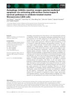
spectometry sequencing. Figure 3 shows post-source
decay sequencing mass spectra for the di-oxidized
(after reaction with ROS) and unoxidized tryptic pep-
tides covering residues 23–35 for V30M TTR. The
protonated di-oxidized tryptic peptide is observed at
m/z 1430.5. Oxidation of the methionine residue is
verified, as C-terminus fragment ions from y
5
(MHVFR) to y
8
(NVAMHVFR) are shifted by 32u,
indicating the addition of two oxygen atoms on this
methionine residue. The y
1
to y
4
remain unchanged,
signifying that the C-terminal HVFR portion of this
peptide was not oxidized. The N-terminal fragment
ions b
3
to b
8
remain unchanged, indicating that the
GSPAINVA portion is not oxidized, and (b
10
+32)
and (b
11
+32) ions signify that oxidation is exclusive
to the methionine residue. These results confirm that
the methionine residues of WT TTR and V30M TTR
are highly reactive toward oxidative modification.
The inhibition effects of fibril formation for these
oxidized proteins are intriguing and show that side
chain oxidation can be used as a method of inducing
mutations in protein sequences to investigate amino
acids that are critical in preserving a protein’s structure
and stability [36]. Interestingly, in vitro studies of a
17-residue peptide showed that replacement of methi-
onine residues with their oxidized forms eliminated
fibril formation [40]. In the case of TTR, dissociation
of the tetramers into monomers is believed to be a pre-
liminary and limiting step of the fibril formation pro-
cess [26]. This inhibition of fibril formation seen in the
oxidized proteins suggests that they are more stable
than the unoxidized forms. Whereas changing the
valine residue at position 30 to methionine increases
the amyloidogenesis of TTR [28,29], oxidation of the
methionine is shown here to partially inhibit fibril
growth. The amino acid side chain oxidation may have
Fig. 3. Post-source decay sequencing mass
spectra for (top) di-oxidized and (bottom)
unoxidized tryptic peptides showing the
oxidation of methionine after reaction of
V30M TTR with ROS.
S. D. Maleknia et al. Oxidation inhibits amyloid fibril formation of TTR
FEBS Journal 273 (2006) 5400–5406 ª 2006 The Authors Journal compilation ª 2006 FEBS 5403
altered tertiary contacts in a manner that stabilized the
oxidized tetramers. We speculate that oxidation may
have introduced new tertiary contacts that stabilized
the folded monomeric structure of the oxidized pro-
teins and inhibited the formation of the unfolded
monomer, which has been proposed [25,26] to be a
prerequisite for fibril growth. Together these effects
caused a delay in the onset of amyloid fibril formation.
Alternatively, the inhibition of fibril formation may
purely be the result of an increase in solubility of oxid-
ized proteins [6,41]. Limited oxidation increases the
hydrophilicity of proteins as determined by their elu-
tion times from hydrophobic columns [6,31,32]. On the
basis of liquid chromatography ⁄ ESI-MS analysis under
similar conditions, oxidized TTR proteins were eluted
40 s faster than their unoxidized forms, indicating
an increase in their hydrophilicity.
These results show that amino acid side chain oxida-
tion can be used as a method of investigating regions of
proteins that are critical in the onset of amyloid forma-
tion. This study reveals that domains neighboring
methionine residues are critical in the formation of fibril
assemblies. These oxidation reactions are being followed
in shorter timescales to possibly distinguish between the
oxidation of Met13 and Met30 in order to more accu-
rately define the key residues of amyloid fibril inhibition.
The timescales of reactions with hydrogen peroxide are
limiting, yet ROS can be generated by an electrospray
discharge source [30] that has been shown to generate a
high flux of ROS on millisecond timescales for studies of
protein structures [6,30–36]. Alternatively, other mutant
proteins could be designed to further investigate the
effect of fibril formation by substituting amino acids
neighboring methionine residues.
Studies revealing the onset and growth of amyloid
fibrils are necessary to understand the pathological con-
ditions that lead to many diseases. Valuable information
can be gained on why certain mutants have a greater
propensity to form fibrils or to inhibit fibrils in compar-
ison with their respective native proteins. Identifying
protein sequences or domains that are critical in preser-
ving protein stability and function should provide
opportunities for prevention and treatment of diseases.
Experimental procedures
Two variants of WT TTR and V30M TTR were selected
for this study. These proteins were expressed in an
Escherichia coli system as described elsewhere [29]. The
proteins were purified by gel-filtration chromatography on
a Superdex 75 column (Amersham Biosciences, Uppsala,
Sweden) in 10 mm sodium phosphate buffer (pH 7.6) ⁄
100 mm KCl ⁄ 1mm EDTA. Oxidized proteins were pre-
pared by allowing the proteins (35 lm) to react with
hydrogen peroxide (reagent-grade; 30 mgÆmL
)1
; Sigma
Chemicals, St Louis, MO, USA) at a concentration of
2.7% peroxide. The oxidation reactions were performed at
pH 7.6 in a timescale range of 10 min to 12 h. The oxid-
ized proteins were then purified from the hydrogen perox-
ide reagent through extensive buffer exchange [10 mm
phosphate buffer (pH 7.6) ⁄ 100 mm KCl ⁄ 1mm EDTA]
with centriprep devices with 10-kDa filters (Millipore, Bill-
erica, MA, USA). The concentrations of all protein solu-
tions were adjusted to 10 lm with the sodium phosphate
buffer at pH 7.6 based on A
280
. The proteins were ana-
lyzed by liquid chromatography ⁄ ESI-MS to verify their
molecular masses and extent of oxidation. Proteins were
also digested with trypsin, and post-source decay sequen-
cing experiments identified the site of amino acid side
chain modification.
Kinetics of amyloid fibril formation
Chemical cross-linking was performed to check that the
tetrameric structure of proteins was preserved after the oxi-
dation reactions. Glutaraldehyde (25%) was added to pro-
tein solutions (10% v ⁄ v), and incubated for 4 min. The
reaction was quenched by the addition of NaBH
4
(7%
in 0.1 m NaOH). The samples were analyzed by
1D SDS ⁄ PAGE, and protein bands were visualized with
Coomassie blue stain.
The in vitro amyloid fibril formation procedure is well
established [42] and was initiated by diluting the protein
solutions with an equal volume of 200 mm acetate buffer
(pH 4.2) ⁄ 100 mm KCl ⁄ 1mm EDTA. The protein solutions
were then distributed into a series of cluster tubes and incu-
bated at 37 °C. The rates of fibril formation were monit-
ored over the course of 14 days by measuring absorbance
at 330 and 400 nm in UV 96-well plates; triplicate experi-
ments were used for each time point. The results are
expressed as mean ± SD from triplicate determinations.
Acknowledgements
The MALDI-TOF MS instrument (Axima-CFR;
Shimadzu Biotech, Manchester, UK) utilized for post-
source decay experiments was purchased through a
Griffith University Infrastructure grant provided to
Simin D. Maleknia.
References
1 Stadtman ER (1992) Protein oxidation and aging.
Science 257, 1220–1224.
2 Berlett BS & Stadtman ER (1997) Protein oxidation in
aging, disease, and oxidative stress. J Biol Chem 272,
20313–20316.
Oxidation inhibits amyloid fibril formation of TTR S. D. Maleknia et al.
5404 FEBS Journal 273 (2006) 5400–5406 ª 2006 The Authors Journal compilation ª 2006 FEBS
3 Stadtman ER, Van Remmen H, Richardson A, Whr
NB & Levine RL (2005) Methionine oxidation and
aging. Biochim Biophys Acta 1703, 135.
4 Dean RT, Fu S, Stocker R & Davies MJ (1997) Bio-
chemistry and pathology of radical-mediated protein
oxidation. Biochem J 324, 1–18.
5 Maleknia SD, Brenowitz M & Chance MR (1999) Milli-
second radiolytic modification of peptides by synchro-
tron X-rays identified by mass spectrometry. Anal Chem
71, 3965–3973.
6 Maleknia SD & Downard KM (2001) Radical approaches
to probe protein structure, folding, and interactions by
mass spectrometry. Mass Spectrom Rev 20, 388–401.
7 Stadtman ER (1995) The status of oxidatively modified
proteins as a marker of aging. In Molecular Aspect of
Aging (Esser, K & Martin, GM, eds), pp. 129–143. John
Wiley & Sons Ltd., New York, NY.
8 Oliver CN, Starke-Reed Stadtman ER, Liu GJ, Carney
JM & Floyd RA (1990) Oxidative damage to brain
proteins, loss of glutamine synthetase activity, and
production of free radicals during ischemia ⁄ reperfusion-
induced injury to gerbil brain. Proc Natl Acad Sci USA
87, 5144–5147.
9 Hilser VJ, Dowdy D, Oas TG & Freire E (1998) The
structural distribution of cooperative interactions in
proteins: analysis of the native state ensemble. Proc Natl
Acad Sci USA 95 , 9903–9908.
10 Fleming PJ & Richards FM (2000) Protein packing:
dependence on protein size, secondary structure and
amino acid composition. J Mol Biol 299, 487–498.
11 Lee KL, Xie D, Freire E & Amzel LM (1994) Estima-
tion of changes in side chain configurational entropy in
binding and folding: general methods and application to
helix formation. Proteins 20, 68–84.
12 Kay MS & Baldwin RL (1996) Packing interactions in
the apomyglobin folding intermediate. Nat Struct Biol
3, 439–445.
13 Thomas PJ, Qu B & Pedersen PL (1995) Defective pro-
tein folding as a basis of human disease. Trends Bio-
chem Sci 20, 456–459.
14 Taubes G (1996) Misfolding the way to disease. Science
271, 1493–1495.
15 Kelly JW (1998) The alternative conformations of amy-
loidogenic proteins and their multi-step assembly path-
ways. Curr Opin Struct Biol 8, 101–106.
16 Dobson CM (2003) Protein folding and misfolding.
Nature 426, 884–890.
17 Selkoe DJ (2003) Folding proteins in fatal ways. Nature
426, 900–904.
18 Buxbaum JN & Tagoe CE (2000) The genetics of the
amyloidoses. Annu Rev Med 51, 543–569.
19 Hornberg A, Eneqvist T, Olofsson A, Lundgren E &
Sauer-Eriksson AE (2000) A comparative analysis of 23
structures of the amyloidogenic protein transthyretin.
J Mol Biol 302 , 649–669.
20 Westermark P, Sletten K, Johansson B & Cornwell GG
(1990) Fibril in senile systemic amyloidosis is derived
from normal transthyretin. Proc Natl Acad Sci USA 87,
2843–2845.
21 Saraiva MJ (2001) Transthyretin mutations in hyperthy-
roxinemia and amyloid diseases. Hum Mutat 17 , 493–
503.
22 Johnson SM, Wiseman RL, Sekijima Y, Green NS,
Adamski-Werner SL & Kelly JW (2005) Native state
kinetic stabilization as a strategy to ameliorate protein
misfolding diseases: a focus on the transthyretin amyloi-
doses. Acc Chem Res 38, 911–921.
23 Olofsson A, Ippel HJ, Baranov V, Horstedt P, Wijm-
enga S & Lundgren E (2001) Capture of a dimeric inter-
mediate during transthyretin amyloid formation. J Biol
Chem 276, 39592–39599.
24 Reixach N, Deechongki S, Jiang X, Kelly JW & Bux-
baum JN (2004) Tissue damage in the amyloidoses:
transthyretin monomers and nonnative oligomers are
the major cytotoxic species in tissue culture. Proc Natl
Acad Sci USA 101 , 2817–2822.
25 Liu K, Cho HS, Hoy DW, Nguyen TN, Olds P, Kelly JW
& Wemmer DE (2000) Deuterium-proton exchange on
the native wild-type transthyretin tetramer identifies the
stable core of the individual subunits and indicates mobi-
lity at the subunit interface. J Mol Biol 303, 555–565.
26 Hammarstrom P, Wiseman RL, Powers ET & Kelly JW
(2003) Prevention of transthyretin amyloid disease by
changing protein misfolding energetics. Science 299,
713–716.
27 Zhang Q & Kelly JW (2003) Cys10 mixed disulfides
make transthyretin more amyloidogenic under mildly
acidic conditions. Biochemistry 42, 8756–8761.
28 Lashuel HA, Lai Z & Kelly JW (1998) Characterization
of the transthyretin acid denaturation pathways by ana-
lytical ultracentrifugation: implications for wild-type,
V30M, and L55P amyloid fibril formation. Biochemistry
37, 17851–17864.
29 McCutchen SL, Colon W & Kelly JW (1993) Transthyr-
etin mutation Leu-55-Pro significantly alters tetramer
stability and increases amyloidogenicity. Biochemistry
32, 12119–12127.
30 Maleknia SD, Downard KM & Chance MR (1999)
Electrospray-assisted modification of proteins: a radical
probe of protein structure. Rapid Commun Mass Spec-
trom 13, 2352–2358.
31 Maleknia SD, Wong JW & Downard KM (2004)
Photochemical and electrophysical production of radi-
cals on millisecond timescales to probe the structure,
dynamics and interactions of proteins. Photochem
Photobiol Sci 3, 741–748.
32 Maleknia SD, Kiselar JG & Downard KM (2002)
Hydroxyl radical probe of the surface of lysozyme by
synchrotron radiolysis and mass spectrometry. Rapid
Commun Mass Spectrom 16, 53–61.
S. D. Maleknia et al. Oxidation inhibits amyloid fibril formation of TTR
FEBS Journal 273 (2006) 5400–5406 ª 2006 The Authors Journal compilation ª 2006 FEBS 5405
33 Maleknia SD & Downard KM (2001) Unfolding of
apomyoglobin helices by synchrotron radiolysis and
mass spectrometry. Eur J Biochem 268, 5578–5588.
34 Wong JH, Maleknia SD & Downard KM (2003) Study
of the ribonuclease-S–protein-peptide complex using a
radical probe and electrospray ionization mass spectro-
metry. Anal Chem 75, 1557–1563.
35 Wong JH, Maleknia SD & Downard KM (2005)
Hydroxyl radical probe of the calmodulin–melittin
complex interface by electrospray ionization mass
spectrometry. J Am Soc Mass Spectrom 16, 225–233.
36 Shum WK, Maleknia SD & Downard KM (2005) Onset
of oxidative damage in alpha-crystallin by radical probe
mass spectrometry. Anal Biochem 344, 247–256.
37 Teh LC, Murphy LJ, Huq NL, Surus AS, Friesen HG,
Lazarus L & Chapman GE (1987) Methionine oxida-
tion in human growth hormone and human chorionic
somatomammotropin. Effects on receptor binding and
biological activities. J Biol Chem 262, 6472–6477.
38 Hamilton JA, Steinrauf LK, Braden BC, Liepnieks J,
Benson MD, Holmgren G, Sandgren O & Steen L
(1993) The x-ray crystal structure refinements of normal
human transthyretin and the amyloidogenic Val-30 fi
Met variant to 1.7-A
˚
resolution. J Biol Chem 268, 2416.
39 Willard L, Ranjan A, Zhang H, Monzavi1 H, Boyko
RF, Sykes BD & Wishart DS (2003) VADAR: a web
server for quantitative evaluation of protein structure
quality. Nucleic Acids Res 31, 3316–3319 (http://redpoll.
pharmacy.ualberta.ca/vadar/).
40 Kammerer RA, Kostrewa D, Surdo J, Detken A,
Garcia-Echeverria C, Green JD, Muller SA, Meier BH,
Winkler FK, Dobson CM, et al. (2004) Exploring amy-
loid formation by a de novo design. Proc Natl Acad Sci
USA 101, 4435–4440.
41 Cervera J & Levine RL (1998) Modulation of the
hydrophobicity of glutamine synthetase by mixed-func-
tion oxidation. FASEB J 2, 2591–2595.
42 Hammarstrom P, Jiang X, Hurshman AR, Powers
ET & Kelly JW (2002) Sequence-dependent denatura-
tion energetics: a major determinant in amyloid
disease diversity. Proc Natl Acad Sci USA 99,
16427–16432.
Oxidation inhibits amyloid fibril formation of TTR S. D. Maleknia et al.
5406 FEBS Journal 273 (2006) 5400–5406 ª 2006 The Authors Journal compilation ª 2006 FEBS