Tài liệu Báo cáo khóa học: Further insights into the assembly of the yeast cytochrome bc1 complex based on analysis of single and double deletion mutants lacking supernumerary subunits and cytochrome b pdf
Bạn đang xem bản rút gọn của tài liệu. Xem và tải ngay bản đầy đủ của tài liệu tại đây (299.67 KB, 10 trang )
Further insights into the assembly of the yeast cytochrome
bc
1
complex based on analysis of single and double deletion mutants
lacking supernumerary subunits and cytochrome
b
Vincenzo Zara
1
, Ilaria Palmisano
1
, Laura Conte
1
and Bernard L. Trumpower
2
1
Dipartimento di Scienze e Tecnologie Biologiche ed Ambientali, Universita
`
di Lecce, Italy;
2
Department of Biochemistry,
Dartmouth Medical School, Hanover, NH, USA
The cytochrome bc
1
complex of the yeast Saccharomyces
cerevisiae is composed of 10 different subunits that are
assembled as a symmetrical dimer in the inner mitochondrial
membrane. Three of the subunits contain redox centers and
participate in catalysis, whereas little is known about the
function of the seven supernumerary subunits. To gain fur-
ther insight into the function of the supernumerary subunits
in the assembly process, we have examined the subunit
composition of mitochondrial membranes isolated from
yeast mutants in which the genes for supernumerary sub-
units and cytochrome b were deleted and from yeast
mutants containing double deletions of supernumerary
subunits. Deletion of any one of the genes encoding cyto-
chrome b, subunit 7 or subunit 8 caused the loss of the other
two subunits. This is consistent with the crystal structure
of the cytochrome bc
1
complex that shows that these three
subunits comprise its core, around which the remaining
subunits are assembled. Absence of the cytochrome b/sub-
unit 7/subunit 8 core led to the loss of subunit 6, whereas
cytochrome c
1
, iron–sulfur protein, core protein 1, core
protein 2 and subunit 9 were still assembled in the mem-
brane, although in reduced amounts. Parallel changes in the
amounts of core protein 1 and core protein 2 in the mito-
chondrial membranes of all of the deletion mutants suggest
that these can be assembled as a subcomplex in the mito-
chondrial membrane, independent of the presence of any
other subunits. Likewise, evidence of interactions between
subunit 6, subunit 9 and cytochrome c
1
suggests that a
subcomplex between these two supernumerary subunits and
the cytochrome might exist.
Keywords: cytochrome bc
1
; assembly; supernumerary sub-
units; Saccharomyces cerevisiae.
The cytochrome bc
1
complex is a multisubunit complex
embedded in the inner membrane of mitochondria [1,2].
This respiratory enzyme catalyzes the transfer of electrons
from ubiquinol to cytochrome c and couples the electron
transfer to vectorial proton translocation across the inner
mitochondrial membrane. The bc
1
complex has been
crystallized and analyzed from bovine, chicken and yeast
mitochondria [3–7].
In mitochondria of the yeast Saccharomyces cerevisiae,
the cytochrome bc
1
complex is composed of 10 different
subunits organized in the lipid bilayer as a homo-dimer as
shown in Fig. 1A [8,9]. There are three catalytic subunits
that contain redox prosthetic groups, cytochrome b,cyto-
chrome c
1
and the Rieske iron–sulfur protein (ISP). In
addition, there are seven supernumerary subunits that lack
any cofactors. The supernumerarysubunits arecore protein 1
and core protein 2 [10,11], with apparent molecular masses of
44 and 40 kDa on SDS/PAGE, respectively, and five
smaller proteins. The latter are Qcr6p [12], Qcr7p [13], Qcr8p
[14], Qcr9p [15] and Qcr10p [8] with apparent molecular
masses of about 17, 14, 11, 7.3 and 8.5 kDa, respectively.
Although the supernumerary subunits of the mitochond-
rial bc
1
complexes were discovered one to two decades ago
[16], little is known about their function. It is also not known
how these peripheral subunits are assembled around the
catalytic core of the enzyme to arrive at the three dimen-
sional organization revealed by the crystal structures
(Fig. 1A). The supernumerary subunits and the catalytic
subunits of the yeast cytochrome bc
1
complex show
sequence similarities to those of the bc
1
complexes of higher
eucaryotes [1,2,9]. In addition, the crystallographic analysis
of the Saccharomyces cerevisiae cytochrome bc
1
complex
has revealed an essentially identical overall structure of this
complex and that of chicken and beef [6]. In yeast and
higher eukaryotes, cytochrome b is encoded by mito-
chondrial DNA, while the remaining subunits of the bc
1
complex are encoded in the nucleus, synthesized by cytosolic
polysomes, and then imported into mitochondria, thereby
reaching their final location in the inner membrane [17]. The
similarities of the yeast bc
1
complex to the bc
1
complexes
of higher eukaryotes suggest that the yeast enzyme may
serve as a paradigm to understand how this oligomeric
protein complex is assembled into the inner mitochondrial
membrane.
Correspondence to V. Zara, Dipartimento di Scienze e Tecnologie
Biologiche ed Ambientali, Universita
`
di Lecce, Via Prov.le Lecce-
Monteroni, I-73100 Lecce, Italy. Fax: + 39 0832 298626,
Tel.: + 39 0832 298705, E-mail:
Abbreviations: DFP, diisopropyl fluorophosphate; ISP, Rieske
iron–sulfur protein.
(Received 8 January 2004, revised 23 January 2004,
accepted 6 February 2004)
Eur. J. Biochem. 271, 1209–1218 (2004) Ó FEBS 2004 doi:10.1111/j.1432-1033.2004.04024.x
In this study, we have investigated the role of the
supernumerary subunits in the assembly of the bc
1
complex
in S. cerevisiae mitochondria. To this end we have prepared
single and double deletion yeast mutants in which one or
two nuclear genes encoding the supernumerary subunits
Qcr6p, Qcr7p, Qcr8p, Qcr9p and Qcr10p have been deleted
and analyzed the bc
1
subunits present in mitochondrial
membranes using antibodies directed against the various
subunits. Yeast mutant strains containing single deletions of
genes for supernumerary subunits were described previously
[8,15,18–20], even though an exhaustive analysis of cyto-
chrome bc
1
subunit composition in these yeast strains has
not been reported. We have also created two yeast strains
in which the mitochondrial gene encoding cytochrome b
has been deleted or truncated and examined the subunit
composition of membranes in which the catalytic and
structural core of the enzyme is absent.
Experimental procedures
Materials
Yeast extract and bacto-peptone were purchased from
Difco. Yeast nitrogen base without amino acids, Coomas-
sie Brilliant Blue, phenylmethylsulfonyl fluoride, glass
beads, acrylamide, bis-acrylamide, N,N,N¢N¢-tetramethyl-
ethylenediamine, ammonium persulfate, diisopropyl fluoro-
phosphate (DFP), glucose and glycerol were from Sigma.
Anti-mouse and anti-rabbit IgG, coupled to peroxidase,
were from Bio-Rad. The ECL detection system for
Western blotting was from Amersham. Nitrocellulose
was from Pall Life Sciences, New York, NY, USA
1
.
Polyclonal and monoclonal antibodies against the various
subunits of the yeast cytochrome bc
1
complex were
prepared in the Trumpower laboratory. The anti-Tom40
Igs were a gift of N. Pfanner
2
(Institute for Biochemistry
and Molecular Biology, Freiburg, Germany). All other
reagents were of analytical grade.
Yeast strains, media and genetic methods
The S. cerevisiae strains used in this study are listed in
Table 1. The construction of the QCR7 deletion strain
(VZ1) was performed following the procedure of homolog-
ous recombination as described previously [21]. A DNA
fragment prepared by PCR and carrying the coding region
for the selectable TRP1 marker, plus the flanking sequences
of the QCR7 open-reading frame at the 5¢-and3¢-regions,
was used to transform yeast cells by treatment with lithium
acetate [22]. The transformants were then selected for
tryptophan prototrophy.
The double deletion strains were constructed as follows.
The haploid strains VZ1 (D7) and MES8 (D6), VZ2 (D7) and
LLD9 (D8), JDP1 (D9) and LLD9 (D8), JDP2 (D9) and
UBL2 (D10), were mated and the resulting diploids were
sporulated to obtain the double deletion strains VZ4
(D6/D7), VZ6 (D7/D8), VZ14 (D8/D9) and VZ9 (D9/D10),
respectively. The selectable markers exhibited a 2 : 2 segre-
gation pattern, and some spores were prototrophic for both
markers. Haploid spores of VZ4, VZ6, VZ14 and VZ9 were
then selected for Trp
+
and Leu
+
,Trp
+
and His
+
,His
+
and
Ura
+
,orHis
+
and Leu
+
prototrophy, respectively. Other
yeast genetic methods used were as described in [23].
The expected absence of the corresponding protein pro-
ducts in mitochondrial membranes from the deletion strains
was assessed by Western blot analysis (Results).
The respiratory capacity of the yeast strains was checked
on nonfermentable solid medium containing 1% (w/v) yeast
extract, 2% (w/v) bacto-peptone, 2% (w/v) agar, 3% (v/v)
glycerol and 2% (v/v) ethanol (YPEG). Viability of the
strains on fermentable medium was confirmed on 1% (w/v)
yeast extract, 2% (w/v) bacto-peptone, 2% (w/v) agar and
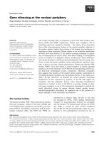
Fig. 1. The yeast cytochrome bc
1
complex. (A) The structure of the dimeric yeast bc
1
complex with the redox subunits, cytochrome b,cyto-
chrome c
1
, and the Rieske ISP colored blue, red and yellow, respectively. The supernumerary subunits are colored gray. The structure is oriented as
it would appear in the inner mitochondrial membrane, with the mitochondrial matrix at the bottom. (B) The structure of cytochrome b and
supernumerary subunits 7 and 8 in one monomer (the Ôcytochrome b, subunit 7, subunit 8 coreÕ). Cytochrome b is colored blue, subunit 7 is colored
pink, and subunit 8 is colored green. The arrow labeled (a) points to the N-terminus of cytochrome b where it is enveloped by subunit 7. The arrows
labeled (b) and (c) point to the areas of interaction between the transmembrane helix of subunit 8 and helices G and H1 of cytochrome b and
between the N-terminus of subunit 8 and helix a of cytochrome b. The figure was constructed from the crystal structure of the yeast bc
1
complex [6].
1210 V. Zara et al. (Eur. J. Biochem. 271) Ó FEBS 2004
2% (w/v) glucose (YPD). For the isolation of mitochondrial
membranes, the yeast strains were grown in liquid YPD
medium containing 1% (w/v) yeast extract, 2% (w/v) bacto-
peptone and 2% (w/v) glucose, pH 5.0.
Isolation of mitochondrial membranes
Mitochondrial membranes were isolated from the various
yeast strains by a modification of a previously described
method [24]. Yeast cells were grown overnight at 30 °C,
unless otherwise specified, in 800 mL of YPD until expo-
nential growth phase was reached (D
600
3
of 1–2). Cells were
recovered by centrifugation at 3200 g for 15 min and then
washed once with distilled water. The pellet was resuspended
in 25 mL of MTE buffer (400 m
M
mannitol, 50 m
M
Tris/
HCl, 2 m
M
EDTA, pH 7.4). Acid-washed glass beads were
added up to a final volume of 30 mL to the mixture kept
at 4 °Cand1m
M
DFP was then added. Afterwards, the
cells were mixed with a vortex mixer at maximum speed for
10 min at 4 °C. After the further addition of MTE buffer to a
final volume of 50 mL, the mixture was centrifuged at 1000 g
for 10 min at 4 °C. The supernatant was then centrifuged
at 18 500 g for 30 min at 4 °C in order to pellet the
mitochondrial membranes. The pellet was washed with
20–30 mL of MTE and re-isolated by centrifugation as
described above. The mitochondrial membranes were then
resuspended in 1 mL of MTE buffer, divided in aliquots of
50 lL each, and stored at )80 °C for subsequent analysis
by SDS/PAGE and Western blotting.
SDS/PAGE and Western blotting
Mitochondrial membranes were analyzed by standard
SDS/PAGE with 15% (w/v) acrylamide and an acryl-
amide/bis-acrylamide ratio of 30 : 0.8 (w/w) [25]. The
proteins were then stained with Coomassie Blue or
transferred to nitrocellulose membranes. Immunodetection
of the yeast mitochondrial proteins was carried out with
monoclonal and polyclonal antibodies by chemilumines-
cence. The stained polyacrylamide gels and the fluoro-
graphs containing the immunodetected proteins were
scanned and quantified using an Imaging Densitometer
GS-700 from Bio-Rad.
Other methods
Protein concentrations were determined by the Bradford
method [26] or the modified Lowry method [27]. Electro-
phoretic analysis of DNA on agarose gels, restriction
endonuclease analysis, ligation of DNA fragments,
Table 1. Yeast strains used in this study.
Strain Genotype Reference
W303–1 A (WT) MATa, ade2–1, his3–11,15, trp1–1, leu2–3,112,
ura3–1, can1–100
Gift from A. Tzagoloff, Columbia University,
New York
W303–1B (WT) MATa, ade2–1, his3–11,15, trp1–1, leu2–3,112,
ura3–1, can1–100
Gift from A. Tzagoloff, Columbia University,
New York
MES8 (D6) MATa, ade2–1, his3–11,15, trp1–1, leu2–3,112,
ura3–1, can1–100, qcr6D::LEU2
[37]
VZ1 (D7) MATa, ade2–1, his3–11,15, trp1–1, leu2–3,112,
ura3–1, can1–100, qcr7D::TRP1
This study
VZ2 (D7) MATa, ade2–1, his3–11,15, trp1–1, leu2–3,112,
ura3–1, can1–100, qcr7D::TRP1
This study
LLD9 (D8) MATa, ade2–1, his3–11,15, trp1–1, leu2–3,112,
ura3–1, can1–100, qcr8D::HIS3
Daniels and Trumpower, unpublished data
JDP1 (D9) MATa, ade2–1, his3–11,15, trp1–1, leu2–3,112,
ura 3–1, can1–100, qcr9D1::URA3
[15]
JDP2 (D9) MATa, leu2–3,112, his3, can 1–11, qcr9D2::HIS3 [15]
UBL2 (D10) MATa, ade2–1, his3–11,15, leu2–3,112, ura3–1,
can1–100, qcr10D2::LEU2
[8]
VZ4 (D6/D7) MATa, ade2–1, his3–11,15, trp1–1, leu2–3,112,
ura3–1, can1–100, qcr6D::LEU2, qcr7D::TRP1
This study
VZ6 (D7/D8) MATa, ade2–1, his3–11,15, trp1–1, leu2–3,112,
ura3–1, can1–100, qcr7D::TRP1, qcr8D::HIS3
This study
VZ14 (D8/D9) MATa, ade2–1, his3–11,15, trp1–1, leu2–3,112,
ura3–1, can1–100, qcr8D::HIS3, qcr9D1::URA3
This study
VZ9 (D9/D10) MATa, ade2–1, leu2–3,112, qcr9D2::HIS3,
qcr10D2::LEU2
This study
SUY 106-a MATa, his3-D200, leu2-D, qcr6D1::LEU2,
qcr10D1::HIS3
[8]
W303–1B q° MATa, ade2–1, his3-, trp1–1, leu2–3,112, ura3–1
(no mtDNA)
Gift from B. Meunier, UCL
CKWT MATa, leu1, kar1–1 (WT mtDNA, intronless) Gift from B. Meunier, UCL
CKL57 MATa, leu1, kar1–1 (intronless mtDNA,
point mutation in cytochrome b gene)
Gift from B. Meunier, UCL
Ó FEBS 2004 Assembly of the yeast cytochrome bc
1
complex (Eur. J. Biochem. 271) 1211
transformation of Escherichia coli and isolation of plasmid
DNA from bacterial cells were carried out by standard
procedures [28].
Results
Growth phenotype of single and double deletion
mutants
The growth phenotype of the yeast strains with deletions of
genes encoding various subunits of the bc
1
complex was
determined by plating the cells on solid media containing
fermentable or nonfermentable carbon sources and then
incubating at 30 °C. The results are summarized in Table 2.
Among the single deletion mutants, only the subunit 6
(MES8) and subunit 10 (UBL2) deletion strains were able to
grow on nonfermentable carbon source at a rate compar-
able to the wild-type strain (W303). Under the same
conditions, the strain JDP1, in which the nuclear gene
encoding subunit 9 had been deleted, exhibited a reduced
growth rate with respect to the wild-type strain as reported
previously [15,29]. The yeast mutants with deletions for the
genes encoding subunit 7 (VZ1) or subunit 8 (LLD9) failed
to grow on the nonfermentable YPEG medium.
Among the double deletion mutants, the strain with the
genes encoding subunits 9 and 10 deleted (VZ9) and that
with the genes encoding subunits 6 and 10 deleted (SUY
106-a) grew on nonfermentable medium, although at a
reduced rate compared to the wild-type strain. In the case
of the VZ9 strain, this was to be expected, based on the
reduced growth rate of the single deletion strain lacking
subunit 9. The remaining double deletion mutants, VZ4
(D6/D7), VZ6 (D7/D8) and VZ14 (D8/D9), were unable to
grow on the same medium.
Cytochrome
bc
1
subunit analysis of single deletion
mutants
We sought to determine how the absence of individual
supernumerary subunits affected the composition of bc
1
subunits in the mitochondrial membranes. For this purpose,
mitochondrial membranes were isolated from the single
deletion strains grown at 30 °C in YPD, then transferred
to nitrocellulose and probed with an antiserum against
Tom40p, an outer membrane protein belonging to the
import machinery of yeast mitochondria (data not shown).
In this way we adjusted the amount of mitochondrial
membranes in order to use comparable amounts of protein
for the subsequent immunoblot experiments.
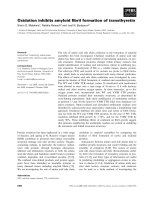
The blot in Fig. 2 shows the cytochrome bc
1
subunits in
the mitochondrial membranes from the mutants in which
genes for subunit 6 (MES8), 7 (VZ1), 8 (LLD9), 9 (JDP1) or
10 (UBL2) were deleted. Relative amounts of the subunits
determined by densitometry scanning of the stained gels are
tabulated in Table 3. The relative amounts of cytochrome b
and the mature forms of both cytochrome c
1
and Rieske
ISP decreased to 52, 64 and 68%, respectively, in the
subunit 6 deletion strain compared to the wild-type strain.
Table 2. Growth phenotype of single and double deletion mutants. All
the strains were first grown in liquid YPD medium to the same original
density and subsequently plated on solid media containing ferment-
able (YPD) or nonfermentable carbon sources (YPEG). Normal
growth, +; reduced growth rate (+); no growth, ).
Strain
Lacking
subunit(s)
Growth
YPD YPEG
W303 – + +
MES8 Qcr6p + +
VZ1 Qcr7p + –
LLD9 Qcr8p + –
JDP1 Qcr9p (+) (+)
UBL2 Qcr10p + +
VZ4 Qcr6p/Qcr7p + –
VZ6 Qcr7p/Qcr8p + –
VZ14 Qcr8p/Qcr9p + –
VZ9 Qcr9p/Qcr10p + (+)
SUY 106-a Qcr6p/Qcr10p (+) (+)
Fig. 2. Subunit composition of mitochondrial membranes from yeast
mutants with single deletions of genes for each of the nuclear encoded
supernumerary subunits. Yeast strains were grown on YPD medium
and mitochondrial membranes were analyzed by SDS/PAGE and
Western blotting with antibodies to the subunits of the yeast bc
1
complex indicated on the left side of the blots.
1212 V. Zara et al. (Eur. J. Biochem. 271) Ó FEBS 2004
Interestingly, the absence of subunit 6 also resulted in an
increase in the ratio of intermediate to mature cyto-
chrome c
1
and a disappearance of the intermediate form
of the Rieske protein. At the same time, the levels of
subunits 7, 8 and 9 significantly decreased in this mutant
strain. However, the amounts of core protein 1 and core
protein 2 were relatively unaffected. Therefore, the absence
of subunit 6 appeared to alter the rates of processing of two
of the redox subunits and caused minor changes in amounts
of the small supernumerary subunits, but it did not cause
dramatic changes in the cytochrome bc
1
composition.
Accordingly, this yeast strain was respiratory-competent.
Deletion of the gene encoding either subunit 7 or subunit
8 resulted in a more severe phenotype, and the changes in
bc
1
subunit composition of the membranes were compar-
able in these two deletion strains, as can be seen from the
blot in Fig. 2. In addition, the absence of subunit 7 caused
a strong decrease in subunit 8 and vice versa, suggesting
a correlation between these two subunits. In both strains,
cytochrome b and the Rieske protein were almost unde-
tectable, while the amounts of cytochrome c
1
were similar to
that found in the wild-type strain. Subunit 9 decreased to
36% of the wild-type level in both mutant strains, and the
twocoreproteinsdecreasedinparallelinbothmutants,with
lower amounts found in the subunit 8 deletion strain. The
only difference between the two strains was that subunit 6
was present in small amounts in the subunit 7 deletion strain
but completely absent in the subunit 8 deletion strain.
In mitochondrial membranes from the strain JDP1, in
which the gene encoding subunit 9 had been deleted, there
was a significant decrease in cytochrome c
1
(45% of the
wild-type content), a barely detectable amount of Rieske
protein and low levels of cytochrome b (12% of the wild-
type content). Core protein 1 and core protein 2 decreased
significantly to about 40% of the wild-type levels. Subunit 8
decreased to the same extent as the core proteins, whereas a
greater decrease was seen in the case of both subunits 6 and
7. Interestingly, a higher amount of cytochrome b,almost
equivalent to that of wild-type cells, was detected in the
JDP1 (D9) mitochondrial membranes when this mutant
strain was grown at 25 °C instead of 30 °C (results not
shown). This effect of temperature on cytochrome b content
was not observed in the case of the other single deletion
mutants.
Among the single deletion strains tested, UBL2, in which
the gene for subunit 10 was deleted, was the only one
showing wild-type levels of all of the bc
1
subunits (Fig. 2
and Table 3). It is also worth noting that the mitochondrial
membranes from these mutant cells also showed the same
ratio of intermediate to mature form of ISP when compared
to the wild-type membranes (Fig. 2). Accordingly, deletion
of QCR10 did not affect mitochondrial respiration, even
though bc
1
activity was significantly reduced [8]. This is due
tothefactthatactivityofthebc
1
complex in wild-type yeast
is significantly greater than what is required to support
normal rates of respiration.
Cytochrome
bc
1
subunit analysis of double deletion
mutants
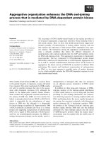
Mitochondrial membranes were isolated from the double
deletion strains and processed by SDS/PAGE and Western
blotting using Tom40p to normalize the protein load in the
same manner as for the single deletion strains. The immuno-
detection of bc
1
subunits in the mitochondrial membranes
isolated from the double deletion mutants is shown in
Fig. 3, and the corresponding quantifications are reported
in Table 4. A comparison of the immunoblots in Figs 3 and
2 reveals that the double deletions of genes encoding bc
1
subunits had more marked effects on the composition of
bc
1
subunits in the mitochondrial membranes than was
observed with the single deletion mutants.
The membranes from the D6/D7 double deletion strain
(VZ4) exhibited the strongest defect in the assembly of the
catalytic subunits of cytochrome bc
1
complex. This strain
showed only 18% and 6% of the wild-type levels of iron–
sulfur protein and cytochrome b, respectively, while mature
cytochrome c
1
disappeared completely and only a small
amount of the intermediate form was visible. Subunits 8 and
9 were reduced to about one third of the original levels.
However, the core proteins were only slightly diminished.
The most notable difference between this double deletion
strain and the others (see below) was the complete absence
of mature cytochrome c
1
.
The mitochondrial membranes from the D7/D8 double
deletion strain (VZ6) showed no cytochrome b and only a
negligible amount of ISP, as expected on the basis of the
results obtained with the single deletion strains. The relative
amount of cytochrome c
1
decreased by 50%, as did both
core proteins. There was also a strong reduction in the
amounts of both subunit 6 and subunit 9 in this strain.
The highest amount of cytochrome c
1
, approximately
80% of the normal amount, was found in the mitochondrial
membranes from the D8/D9 double deletion strain (VZ14).
However, there was a strong defect in both cytochrome b
and ISP in this strain, similar to what was observed in the
other double deletion strains. Core proteins 1 and 2 were
reduced to approximately half of the wild-type levels, while
subunits 6 and 7 were present only in small amounts (18%
and 8%, respectively).
The mitochondrial membranes from the D9/D10 double
deletion strain (VZ9), which is one of the two respiratory
Table 3. Cytochrome bc
1
subunit analysis of single deletion mutants.
The values represent the percentages of the amounts of the individual
subunits present in the yeast mutant strains with respect to the
amounts present in the wild-type strain W303, which were set to 100%.
The numbers are the averages of at least three independent experi-
ments.
Subunits
Yeast mutant strains
MES8
(D6)
VZ1
(D7)
LLD9
(D8)
JDP1
(D9)
ULB2
(D10)
Cytochrome b 52 5 2 12 109
Cytochrome c
1
64 90 90 45 106
ISP 68 10 9 3 113
Core 1 103 57 30 38 101
Core 2 104 58 28 45 116
Qcr6p – 18 – 10 120
Qcr7p 64 – 5 28 111
Qcr8p 43 12 – 40 113
Qcr9p 36 36 36 – 100
Ó FEBS 2004 Assembly of the yeast cytochrome bc
1
complex (Eur. J. Biochem. 271) 1213
competent double deletion strains characterized here,
showed decreased levels of all three catalytic subunits,
cytochrome b, ISP and cytochrome c
1
. In addition, core
proteins 1 and 2 and subunit 6 were reduced to about half of
their original levels, while subunits 7 and 8 were reduced to
about one quarter of their original levels. The deletion of
both genes encoding subunit 6 and 10 in the strain SUY
106-a caused significant changes in the amount of catalytic
subunits not observed previously with the single deletion
strains lacking either subunit 6 or subunit 10. In fact,
cytochrome b and ISP were reduced to about 12% and
27% of the original levels. Cytochrome c
1
and core proteins
1 and 2 decreased by about 50%, whereas a greater decrease
was found in the case of subunits 7, 8 and 9.
Cytochrome
bc
1
subunit analysis of cytochrome
b
deletion mutants
Crystal structures of the bc
1
complexes indicate that
cytochrome b is the organizing component of the bc
1
complex, providing eight transmembrane helices that form
the central core of the complex [6]. This central core is
surrounded by four additional transmembranes helices
contributed by cytochrome c
1
, the Rieske protein, and
subunits 8 and 9. It is therefore clear that cytochrome b
plays a fundamental role in organizing and stabilizing the
structure of the entire complex in the inner mitochondrial
membrane. For this reason, we investigated the composition
of cytochrome bc
1
complex subunits in mitochondrial
membranes from yeast strains in which the gene encoding
cytochrome b had been deleted or truncated.
To this end, we used the yeast strain W303–1B q°,devoid
of mitochondrial DNA, and therefore without the gene
encoding cytochrome b. We performed similar experiments
with the strain CKL57 that contains a point mutation
(L263-STOP) in the cytochrome b gene that results in a
nonfunctional, truncated protein (Table 1). Both of these
yeast strains were respiratory-deficient.
Figure 4 shows the subunit composition of the mito-
chondrial membranes from the W303–1B q° and CKL57
strains and from the corresponding wild-type cells grown in
YPD at 30 °C. In general, the pattern of subunits present in
the mitochondrial membranes was identical for these two
mutant strains, although the decrease in amounts of the
subunits was more severe in the q° strain. As expected,
cytochrome b was absent from the W303–1B q° strain.
Likewise, no cytochrome b protein was detectable in the
CKL57 strain. We do not know whether the lack of
immunoreactivity in the latter strain was due to the inability
of the truncated protein to insert into and be stable in the
inner mitochondrial membrane or lack of detection of the
truncated protein by the antibodies.
In the W303–1B q° strain the amounts of the other two
catalytic subunits, cytochrome c
1
and the ISP, were reduced
by about 70–80% (Fig. 4A,C). In the case of the strain
Table 4. Cytochrome bc
1
subunit analysis of double deletion mutants.
The values represent the percentages of the amounts of individual
subunits present in the yeast mutant strains with respect to the
amounts present in the wild-type strain W303, which were set to 100%.
The numbers are the averages of at least three independent experi-
ments.
Subunits
Yeast mutant strains
VZ4
(D6/D7)
VZ6
(D7/D8)
VZ14
(D8/D9)
VZ9
(D9/D10)
SUY 106-a
(D6/D10)
Cytochrome b 6 – 7 26 12
Cytochrome c
1
–507940 50
ISP 18 15 15 33 27
Core 1 74 42 42 59 45
Core 2 83 54 49 53 45
Qcr6p – 23 18 51 –
Qcr7p – – 8 26 23
Qcr8p 28 – – 27 33
Qcr9p 30 31 – – 40
Fig. 3. Subunit composition of mitochondrial membranes from yeast
mutants with double deletions of genes for nuclear encoded super-
numerary subunits. Yeast strains were grown on YPD medium and
mitochondrial membranes were analyzed by SDS/PAGE and Western
blotting with antibodies to the subunits of the yeast bc
1
complex
indicated on the left side of the blots.
1214 V. Zara et al. (Eur. J. Biochem. 271) Ó FEBS 2004
CKL57 the amount of ISP was 40%, while the cyto-
chrome c
1
content was almost unaffected (Fig. 4B,D). Core
1 and core 2 proteins were significantly reduced in both
mutant strains, being 33 and 29%, respectively, in the
W303–1B q° strain (Fig. 4A,C) and 57 and 53%, respect-
ively, in the CKL57 strain (Fig. 4B,D). Interestingly, the
small subunits 6, 7 and 8 were totally absent in both mutant
strains. Only a small amount (22%) of subunit 9 was present
in the W303–1B q° strain, Fig. 4A,C, while essentially
normal amounts of this subunit were present in the CKL57
strain.
When the cytochrome b mutant strains were grown in
YPD at 25 °C instead of 30 °C, the defects in subunit
composition appeared less evident, especially in the case of
the W303–1B q° strain (results not shown). In the mito-
chondrial membranes from this strain, the content of
cytochrome c
1
increased from 29 to 73%, and the amounts
of core proteins 1 and 2 increased from 33 to 74% and 29 to
81%, respectively. Likewise, the relative amount of sub-
unit 9 increased from 22 to 48%. The amount of ISP
changed only slightly at the lower growth temperature, from
23 to 32%. In the CKL57 mutant strain, the amounts of all
subunits increased by about 10–20%. Subunit 9, as already
seen at 30 °C, was present in wild-type amounts. Subunit 6
was present in only small amounts in the W303–1B q° strain
(22%) and in considerably greater amounts (80%) in the
CKL57 strain. Interestingly, subunits 7 and 8 remained
undetectable, even at the lower growth temperature, in both
cytochrome b mutants.
Discussion
We have analyzed the composition of cytochrome bc
1
subunits in mitochondrial membranes of yeast mutants in
which genes for individual and pairs of bc
1
subunits have
been deleted. As far as we know, this is the first time that
such a large collection of single and double deletion mutants
of the yeast cytochrome bc
1
complex has been characterized
simultaneously. Our results add to and extend previous
work on the assembly of the yeast bc
1
complex from the
laboratories of Berden [30] and Tzagoloff [31]. It has been
demonstrated previously that gene expression, import of
proteins into mitochondria and sorting to the inner
membrane are not influenced by the absence of subunits
of the bc
1
complex [19,20,30]. Thus, this experimental
strategy allows the determination of which subunits are
present in the inner mitochondrial membrane independent
of previous steps in bc
1
complex assembly. Defects in the
mitochondrial membrane composition of bc
1
subunits in the
deletion strains can be ascribed to an altered process of
assembly of the multisubunit complex in the inner mito-
chondrial membrane. The bc
1
subunits that are imported
but not assembled into the multisubunit complex or
subcomplexes thereof are probably more susceptible to
proteolysis, as previously proposed [19,20,30,32]. This is
reflected in decreased amounts or absence of the non-
assembled subunits in the mitochondrial membranes.
With all of the single and double deletion mutants there
appeared to be a strict correlation in the amounts of
Fig. 4. Subunit composition of mitochondrial membranes from a yeast mutant lacking mitochondrial DNA and a yeast mutant with a truncated
cytochrome b gene. The wild-type (WT), q°, and CKL57 yeast strains were grown on YPD medium and mitochondrial membranes were analyzed
by SDS/PAGE and Western blotting with antibodies to the subunits of the yeast bc
1
complex indicated on the left side of the blots. The Western
blots are shown in panels A and B and the relative amounts of each of the subunits determined by densitometry scanning of the stained Western
blots of the q° and CKL57 membranes are shown in panels C and D, respectively.
Ó FEBS 2004 Assembly of the yeast cytochrome bc
1
complex (Eur. J. Biochem. 271) 1215
cytochrome b, subunit 7 and subunit 8. Deletion of the gene
for any one of these proteins caused a strong decrease or the
disappearance of the other two components. Accordingly,
the double deletion mutant VZ6, in which both QCR7 and
QCR8 had been deleted, showed no cytochrome b.This
agrees with the crystal structures that show that these two
supernumerary subunits both interact with cytochrome b.
As shown in Fig. 1B, subunit 7 envelopes the N-terminus of
cytochrome b within the membrane near the inner mem-
brane surface. Subunit 8 exhibits a single transmembrane
helix that spans the membrane parallel to cytochrome b,
interacting extensively with transmembrane helices G and
H1 and also interacting with helix a of cytochrome b
parallel to the inner membrane surface. This structural
relationship and the coincidental behavior of these three
subunits in the deletion strains lend support to previous
suggestions [13,30,31] that cytochrome b, subunit 7 and
subunit 8 may form a nucleating subcomplex in the lipid
bilayer of the inner mitochondrial membrane, around which
the other subunits are assembled (Fig. 5).
Subunit 8 interacts with several other subunits of the
complex in addition to cytochrome b [6]. Our results with
the single deletion mutant lacking subunit 8 extend the
previous findings of Maarse coworkers [19]. In addition
to the strong decrease or disappearance of subunit 7,
cytochrome b and ISP as reported previously [19], we
observed the disappearance of subunit 6 and a strong
decrease of subunit 9 and both core proteins. Accordingly,
the QCR8 gene deletion resulted in the most severe
phenotype among the single deletion strains tested.
In the deletion mutant lacking subunit 7 we found an
almost complete lack of cytochrome b, subunit 8 and ISP,
in agreement with previous studies [20]. However, unlike
previous results [20], we also found a significant decrease of
both core proteins, and low levels of subunits 6 and 9. In
fact, concomitant and significant decreases of almost all
remaining subunits, except cytochrome c
1
,wereobserved.
These results were confirmed by those obtained with the
double deletion strain VZ6 (D7/D8). The results with
the deletion mutant lacking subunit 7 further corroborate
the interdependence among subunits 7, 8 and cytochrome b
and the role of this core subcomplex in organizing the
cytochrome bc
1
complex. It was proposed previously that
the N-terminus of subunit 7 plays an important role during
the assembly of the cytochrome bc
1
complex [33,34]. In
support of this proposal, it is the N-terminal 30 amino acids
of subunit 7 that envelopes the N-terminus of cytochrome b
near the matrix side of the inner membrane (Fig. 1B).
Cytochrome c
1
appears to be the cytochrome bc
1
com-
ponent least influenced by the absence of other subunits of
the complex. In fact, only marginal variations in cyto-
chrome c
1
were observed in the single deletion mutants
tested, except for the increase of the intermediate form of
c
1
in the strain lacking subunit 6. Subunit 6 is an acidic
protein that interacts with cytochrome c
1
on the cytosolic
surface of the membrane. The retardation in c
1
maturation
in the absence of subunit 6 suggests that the apo-
cytochrome must associate with this subunit before the
c-type heme can be inserted. The formation of a subcom-
plex between cytochrome c
1
and subunit 6 has previously
been proposed on the basis of biochemical [35] and genetic
evidence [18].
Interestingly, the D6/D7 double deletion strain is the only
one showing a complete lack of mature cytochrome c
1
and
also showed only a small amount of the cytochrome c
1
intermediate form. This is probably due to the combination
of two phenomena, the maturation delay caused by the
absence of subunit 6, and the pleiotropic effects due to the
deletion of QCR7, including almost complete disappearance
of cytochrome b and subunit 8. Similar effects, including the
presence of the intermediate form of cytochrome c
1
along
with a complete lack of the mature form, were previously
seen in the QCR6 deletion strain grown at nonpermissive
temperatures [36]. In that study, also a complete block of
cytochrome c
1
maturation was found together with a
simultaneous lack of both subunits 6 and 8 and low levels
of cytochrome b. Together, these results suggest that the
absence of subunit 6 delays cytochrome c
1
maturation while
the absence of the cytochrome b subcomplex (formed by
Fig. 5. Schematic model summarizing the putative cytochrome bc
1
subcomplexes involved in bc
1
complex assembly. The double arrows
indicate that the sequence of events by which the three subcomplexes
associate to form a subcomplex containing both cytochromes b and c
1
prior to insertion of ISP and subunit 10 (Qcr10p) in the inner mito-
chondrial membrane is currently not known.
1216 V. Zara et al. (Eur. J. Biochem. 271) Ó FEBS 2004
cytochrome b, subunit 7 and subunit 8) hinders the insertion
of mature cytochrome c
1
into the complex. However, when
the cytochrome b subcomplex is missing, but the gene
encoding subunit 6 is not deleted, as in several of the single
and double deletion strains, mature cytochrome c
1
is
present in the mitochondrial membranes in considerable
amount.
As reported previously [18,37], the strain lacking the gene
for subunit 6 showed only moderate defects in the levels of
most of the other subunits of the bc
1
complex when grown
at permissive temperatures. However, we found that subunit
9 was present in this deletion strain at about only one-third
of the normal level, which suggests that subunit 6 stabilizes
subunit 9, although the crystal structure shows that these
two subunits do not interact directly [6]. Deletion of the gene
encoding subunit 9 resulted in a respiratory deficient yeast
strain with very low bc
1
complex activity, particularly at
high temperatures [15,29]. In this strain we found a
significant decrease of both cytochrome c
1
and subunit 6.
Interestingly, previous studies suggested an interaction
between subunit 9 and cytochrome c
1
[24,38,39]. Taken
together, these results suggest that a subcomplex between
cytochrome c
1
and the two supernumerary subunits 6 and 9
is possible (Fig. 5). This would be consistent with the crystal
structure, which shows that these two supernumerary
subunits interact with cytochrome c
1
[6].
The level of ISP was significantly influenced in almost all
of the deletion strains. This catalytic subunit was present in
very low amounts in the D7, D8andD9 single deletion
mutants, and in all of the double deletion mutants prepared
in this study. The extensive loss of ISP in the yeast strain
lacking the gene for subunit 9 is in agreement with previous
results indicating that this catalytic subunit is protease-
sensitive in the absence of subunit 9 [29]. In addition, recent
findings show a synergistic interaction between cyto-
chrome b and subunit 9 in yeast mitochondria [40]. These
authors proposed a stabilizing role of subunit 9 on the
interactions among the catalytic subunits of the cyto-
chrome bc
1
complex, especially at high temperatures. In this
regard, it is noteworthy that the level of cytochrome b
increased in the strain lacking the gene for subunit 9 when
the cells were grown at 25 °C instead of 30 °C. In addition,
less dramatic changes in subunit composition were found in
cytochrome b mutant strains grown at 25 °C instead of
30 °C (Results). A critical effect of the temperature on the
level of various subunits of cytochrome bc
1
complex is
therefore evident in the yeast strains in which the genes for
subunit 6, subunit 9 and cytochrome b had been deleted.
Core protein 1 and core protein 2 interact with each other
and with the membrane-embedded subunits of the bc
1
complex and protrude, almost completely, into the mito-
chondrial matrix [6]. In contrast to previous results with
several yeast bc
1
complex mutants [31], the amounts of
core 1 and core 2 proteins were significantly influenced by
the absence of other subunits of the bc
1
complex. Deletion
of the genes for subunit 7, subunit 8 or subunit 9 caused a
strong reduction of the two core proteins in the mito-
chondrial membranes (Fig. 2 and Table 3). These results
were confirmed by those obtained with the double deletion
strains (Fig. 3 and Table 4). Furthermore, deletion of the
gene for cytochrome b caused a decrease of both core
proteins (Fig. 4). The low levels of both core proteins found
in this study may be due to the fact that we examined
mitochondrial membranes instead of mitochondria. Using
mitochondria there is still the possibility to detect proteins in
transit and not yet inserted into the inner mitochondrial
membrane. The fact that both core proteins decreased by
the same extent in the various deletion strains suggests that
they probably form a subcomplex as hypothesized previ-
ously [30,31] (Fig. 5).
Our results allow some insight into the sequence of events
in assembly of the bc
1
complex. Two of the supernumerary
subunits, 7 and 8, along with cytochrome b,appeartoplay
an important role in the structural organization of the
bc
1
complex. This suggests that these subunits associate at
an early step in the assembly pathway. In contrast, the
supernumerary subunit 10 seems to play only a minor role
in the overall structure of the bc
1
complex. Deletion of the
QCR10 gene has no effect on the composition of bc
1
subunits in the mitochondrial membrane. This subunit is
readily lost during purification and is not present in the
crystal structure of the bc
1
complex [6]. This suggests that
subunit 10 is in a peripheral location on the bc
1
complex
and that it is added late in the assembly pathway. In
general, our results agree with and extend the model for the
assembly of the yeast bc
1
complex proposed by Berden and
coworkers [30]. In fact, these authors proposed the
existence of three distinct subcomplexes, essentially con-
firmed by the present data (Fig. 5). In addition, our results
revealed a strict interdependence between the cytochrome b
subcomplex and the supernumerary subunit Qcr6p. It is
also evident that Qcr9p plays an important role in the
temperature-sensitive stabilization of the yeast bc
1
complex.
At present it is not possible to deduce the sequence in which
subunits of the bc
1
complex are assembled into subcom-
plexes or the sequence in which the putative subcomplexes
are assembled to form the bc
1
complex. The sequence in
which the subunits and subcomplexes are assembled is
under investigation.
Acknowledgements
This study was supported by the Ministero dell’Istruzione, dell’Uni-
versita
`
e della Ricerca (MIUR), PRIN 2002, and by NIH Grant GM
20379 to B. L. T.
References
1. Trumpower, B.L. (1990) Cytochrome bc
1
complexes of micro-
organisms. Microbiol. Rev. 54, 101–129.
2. Berry,E.A.,Guergova-Kuras,M.,Huang,L.S.&Crofts,A.R.
(2000) Structure and function of cytochrome bc complexes. Annu.
Rev. Biochem. 69, 1005–1075.
3. Xia, D., Yu, C A., Kim, H., Xia, J Z., Kachurin, A.M., Zhang,
L., Yu, L. & Deinsenhofer, J. (1997) Crystal structure of the
cytochrome bc
1
complex of bovine heart mitochondria. Science
277, 60–66.
4. Zhang, Z., Huang, L., Shulmeister, V.M., Chi, Y., Kim, K.K.,
Hung, L., Crofts, A.R., Berry, E.A. & Kim, S. (1998) Electron
transfer by domain movement in cytochrome bc
1
. Nature 392,
677–684.
5. Iwata, S., Lee, J.W., Okada, K., Lee, J.K., Iwata, M., Rasmussen,
B., Link, T.A., Ramaswamy, S. & Jap, B.K. (1998) Complete
structure of the 11-subunit bovine mitochondrial bc
1
complex.
Science 281, 64–71.
Ó FEBS 2004 Assembly of the yeast cytochrome bc
1
complex (Eur. J. Biochem. 271) 1217
6. Hunte, C., Koepke, J., Lange, C., Rossmanith, T. & Michel, H.
(2000) Structure at 2.3 A
˚
resolution of the cytochrome bc
1
complex
from the yeast Saccharomyces cerevisiae co-crystallized with an
antibody fragment. Structure 8, 669–684.
7. Lange, C. & Hunte, C. (2002) Crystal structure of the yeast
cytochrome bc
1
complex with its bound substrate cytochrome c.
Proc.NatlAcad.Sci.USA99, 2800–2805.
8. Brandt, U., Uribe, S., Scha
¨
gger, H. & Trumpower, B.L. (1994)
Isolation and characterization of QCR10, the nuclear gene
encoding the 8.5-kDa subunit 10 of the Saccharomyces cerevisiae
cytochrome bc
1
complex. J. Biol. Chem. 269, 12947–12953.
9. Tzagoloff, A. (1995) Ubiquinol-cytochrome-c oxidoreductase
from Saccharomyces cerevisiae. Methods Enzymol. 260, 51–63.
10. Tzagoloff, A., Wu, M. & Crivellone, M. (1986) Assembly of the
mitochondrial membrane system. Characterization of COR1, the
structural gene for the 44-kilodalton core protein of yeast coen-
zyme QH2-cytochrome c reductase. J. Biol. Chem. 261, 17163–
17169.
11. Oudshoorn, P., Van Steeg, H., Swinkels, B.W., Schoppink, P. &
Grivell, L.A. (1987) Subunit II of yeast QH2: cytochrome-c
oxidoreductase. Nucleotide sequence of the gene and features of
the protein. Eur. J. Biochem. 163, 97–103.
12. Van Loon, A.P., De Groot, R.J., Haan, M., Dekker, A. & Grivell,
L.A. (1984) The DNA sequence of the nuclear gene coding for the
17-kd subunit VI of the yeast ubiquinol-cytochrome c reductase: a
protein with an extremely high content of acidic amino acids.
EMBO J. 3, 1039–1043.
13. De Haan, M., Van Loon, A.P.G.M., Kreike, J., Vaessen,
R.T.M.J. & Grivell, L.A. (1984) The biosynthesis of the ubiquinol-
cytochrome c reductase complex in yeast. DNA sequence analysis
of the nuclear gene coding for the 14 kDa subunit. Eur. J. Bio-
chem. 138, 169–177.
14. Maarse, A.C. & Grivell, L.A. (1987) Nucleotide sequence of the
gene encoding the 11-kDa subunit of the ubiquinol-cytochrome c
oxidoreductase in S. cerevisiae. Eur. J. Biochem. 165, 419–425.
15. Phillips, J.D., Schmitt, M.E., Brown, T.A., Beckmann, J.D. &
Trumpower, B.L. (1990) Isolation and characterization of QCR9,
a nuclear gene encoding the 7.3-kDa subunit 9 of the Saccharo-
myces cerevisiae ubiquinol-cytochrome c oxidoreductase complex.
J. Biol. Chem. 265, 20813–20821.
16. Yang, X. & Trumpower, B.L. (1988) Protonmotive Q cycle
pathway of electron transfer and energy transduction in the three-
subunit ubiquinol-cytochrome c oxidoreductase complex of
Paracoccus denitrificans. J. Biol. Chem. 263, 11962–11970.
17. Pfanner, N. & Geissler, A. (2001) Versatility of the mitochondrial
protein import machinery. Nat. Rev. Mol. Cell Biol. 2, 339–349.
18. Schoppink, P.J., Hemrika, W., Reynen, J.M., Grivell, L.A. &
Berden, J.A. (1988) Yeast ubiquinol: cytochrome c oxidoreductase
is still active after inactivation of the gene encoding the 17-kDa
subunit VI. Eur. J. Biochem. 173, 115–122.
19. Maarse, A.C., De Haan, M., Schoppink, P.J., Berden, J.A. &
Grivell, L.A. (1988) Inactivation of the gene encoding the 11-kDa
subunit VIII of the ubiquinol-cytochrome-c oxidoreductase in
Saccharomyces cerevisiae. Eur. J. Biochem. 172, 179–184.
20. Schoppink, P.J., Berden, J.A. & Grivell, L.A. (1989) Inactivation
of the gene encoding the 14-kDa subunit VII of yeast ubiquinol
cytochrome c oxidoreductase and analysis of the resulting mutant.
Eur. J. Biochem. 181, 475–483.
21. Baudin, A., Ozier-Kalogeropoulos, O., Denouel, A., Lacroute, F.
& Cullin, C. (1993) A simple and efficient method for direct gene
deletion in Saccharomyces cerevisiae. Nucleic Acids Res. 21,
3329–3330.
22. Ito, H., Fukuda, Y., Murata, K. & Kimura, A. (1983) Transfor-
mation of intact yeast cells treated with alkali cations. J. Bacteriol.
153, 163–168.
23. Guthrie, C. & Fink, G.R. (1991) Guide to Yeast Genetics and
Molecular Biology.AcademicPress,SanDiego,CA.
24. Schmitt, M.E. & Trumpower, B.L. (1991) The petite phenotype
resulting from a truncated copy of subunit 6 results from loss of
assembly of the cytochrome bc
1
complex and can be suppressed
by overexpression of subunit 9. J. Biol. Chem. 266, 14958–14963.
25. Laemmli, U.K. (1970) Cleavage of structural proteins during
the assembly of the head of bacteriophage T4. Nature 227,
680–685.
26. Bradford, M.M. (1976) A rapid and sensitive method for the
quantitation of microgram quantities of protein utilizing the
principle of protein-dye binding. Anal. Biochem. 72, 248–254.
27. Dulley, J.R. & Grieve, P.A. (1975) A simple technique for elimi-
nating interference by detergents in the Lowry method of protein
determination. Anal. Biochem. 64, 136–141.
28. Sambrook, J., Fritsch, E.F. & Maniatis, T. (1989) Molecular
Cloning: a Laboratory Manual, 2nd edn. Cold Spring Harbor
Laboratory, Cold Spring Harbor, NY.
29. Phillips, J.D., Graham, L.A. & Trumpower, B.L. (1993) Subunit 9
of the Saccharomyces cerevisiae cytochrome bc
1
complex is
required for insertion of EPR-detectable iron-sulfur cluster into
the Rieske iron-sulfur protein. J. Biol. Chem. 268, 11727–11736.
30. Berden, J.A., Schoppink, P.J. & Grivell, L.A. (1988) A model for
the assembly of ubiquinol: cytochrome c oxidoreductase in yeast.
In Molecular Basis of Biomembrane Transport (Palmieri, F. &
Quagliariello, E., eds), pp. 195–208. Elsevier, Amsterdam.
31. Crivellone,M.D.,Wu,M.&Tzagoloff,A.(1988)Assemblyofthe
mitochondrial membrane system. J. Biol. Chem. 263, 14323–
14333.
32. Rep, M. & Grivell, L.A. (1996) The role of protein degradation
in mitochondrial function and biogenesis. Curr. Genet. 30, 367–
380.
33. Malaney, S., Trumpower, B.L., Debers, C.M. & Robinson, B.H.
(1997) The N terminus of the Qcr7 protein of the cytochrome bc
1
complex is not essential for import into mitochondria in
Saccharomyces cerevisiae but is essential for assembly of the
complex. J. Biol. Chem. 272, 17495–17501.
34. Lee, S.Y., Hunte, C., Malaney, S. & Robinson, B.H. (2001) The
N-terminus of the Qcr7 protein of the cytochrome bc
1
complex in
S. cerevisiae may be involved in facilitating stability of the sub-
complex with the Qcr8 protein and cytochrome b. Arch. Biochem.
Biophys. 393, 215–221.
35. Kim, C.H., Balny, C. & King, T.E. (1987) Role of the hinge
protein in the electron transfer between cardiac cytochrome c
1
and
c. Equilibrium constants and kinetic probes. J. Biol. Chem. 262,
8103–8108.
36. Yang, M. & Trumpower, B.L. (1994) Deletion of QCR6, the gene
encoding subunit six of the mitochondrial cytochrome bc
1
com-
plex, blocks maturation of cytochrome c
1
, and causes tempera-
ture-sensitive petite growth in Saccharomyces cerevisiae. J. Biol.
Chem. 269, 1270–1275.
37. Schmitt, M.E. & Trumpower, B.L. (1990) Subunit 6 regulates
half-of-the-sites reactivity of the dimeric cytochrome bc
1
complex in Saccharomyces cerevisiae. J. Biol. Chem. 265, 17005–
17011.
38. Gonzales-Halphen, D., Lindorfer, M.A. & Capaldi, R.A. (1988)
Subunit arrangement in beef heart complex III. Biochemistry 27,
7021–7031.
39. Scha
¨
gger, H., Link, T.A., Engel, W.D. & von Jagow, G. (1986)
Isolation of the eleven protein subunits of the bc
1
complex from
beef heart. Methods Enzymol. 126, 224–237.
40. Saint-Georges, Y., Bonnefoy, N., di Rago, J.P., Chiron, S. &
Dujardin, G. (2002) A pathogenic cytochrome b mutation reveals
new interactions between subunits of the mitochondrial bc
1
com-
plex. J. Biol. Chem. 277, 49397–49402.
1218 V. Zara et al. (Eur. J. Biochem. 271) Ó FEBS 2004