Tài liệu Báo cáo khoa học: N-Methyl-L-amino acid dehydrogenase from Pseudomonas putida A novel member of an unusual NAD(P)-dependent oxidoreductase superfamily ppt
Bạn đang xem bản rút gọn của tài liệu. Xem và tải ngay bản đầy đủ của tài liệu tại đây (154.29 KB, 7 trang )
N-Methyl-L-amino acid dehydrogenase from Pseudomonas
putida
A novel member of an unusual NAD(P)-dependent oxidoreductase
superfamily
Hisaaki Mihara
1,
*, Hisashi Muramatsu
1,
*, Ryo Kakutani
1
, Mari Yasuda
2
, Makoto Ueda
2
,
Tatsuo Kurihara
1
and Nobuyoshi Esaki
1
1 Institute for Chemical Research, Kyoto University, Uji, Kyoto 611-0011, Japan
2 Yokohama Research Center, Mitsubishi Chemical Corp., Yokohama, Japan
n-Methyl amino acids occur naturally as components
of several depsipeptides, including cyclosporin A [1],
vancomycin, destruxins [2], didemnins [3], dolastatin
[4], and enniatin [5], as well as a calpain inhibitor from
Streptomyces griseus [6] and a pilin from Pseudomonas
aeruginosa [7]. Enniatins consist of alternating residues
of d-2-hydroxyisovalerate and N-methyl-l-valine (or
N-methyl-l-isoleucine) [5]. N-Methylphenylalanine is
the N-terminal amino acid of pilins of most bacterial
pathogens expressing type IV pili [7]. The synthesis of
these N-methylated peptides is catalyzed by specific
N-methyltransferases such as enniatin synthetase [8]
and PilD [9] using S-adenosyl-l-methionine as the
methyl group donor.
Free forms of N-methyl amino acids also occur in
bivalves [10,11], several plant species [12], and some
bacteria [13,14]. In Aminobacter aminovorans (formerly
Pseudomonas MA) [15], N-methylglutamate and
Keywords
methylamine; NADPH; N-methyl-
L-amino
acid dehydrogenase; N-methyl-
L-amino acid;
Pseudomonas putida
Correspondence
N Esaki, Institute for Chemical Research,
Kyoto University, Uji, Kyoto 611-0011, Japan
Fax: 81 774 38 3248
Tel: 81 774 38 3240
E-mail:
*Note
Equivalent first authors.
Database
Nucleotide sequence data reported are avail-
able in the DDBJ ⁄ EMBL ⁄ GenBank databas-
es under the accession number AB190215.
(Received 8 October 2004, revised 12
December 2004, accepted 22 December
2004)
doi:10.1111/j.1742-4658.2004.04541.x
We found N-methyl-l-amino acid dehydrogenase activity in various bacter-
ial strains, such as Pseudomonas putida and Bacillus alvei, and cloned the
gene from P. putida ATCC12633 into Escherichia coli. The enzyme purified
to homogeneity from recombinant E. coli catalyzed the NADPH-dependent
formation of N-alkyl-l-amino acids from the corresponding a-oxo acids
(e.g. pyruvate, phenylpyruvate, and hydroxypyruvate) and alkylamines (e.g.
methylamine, ethylamine, and propylamine). Ammonia was inert as a sub-
strate, and the enzyme was clearly distinct from conventional NAD(P)-
dependent amino acid dehydrogenases, such as alanine dehydrogenase (EC
1.4.1.1). NADPH was more than 300 times more efficient than NADH as
a hydrogen donor in the enzymatic reductive amination. Primary structure
analysis revealed that the enzyme belongs to a new NAD(P)-dependent
oxidoreductase superfamily, the members of which show no sequence
homology to conventional NAD(P)-dependent amino acid dehydrogenases
and opine dehydrogenases.
Abbreviation
NMAADH, N-methyl-
L-amino acid dehydrogenase.
FEBS Journal 272 (2005) 1117–1123 ª 2005 FEBS 1117
ammonia are formed from methylamine and glutamate
by N-methylglutamate synthase (EC 2.1.1.21) [16].
Another A. aminovorans strain, formerly called Pseudo-
monas MS, produces N-methylalanine from methyl-
amine and pyruvate in the presence of NADPH with
N-methylalanine dehydrogenase (EC 1.4.1.17) [17].
Therefore, in contrast with the N-methyl groups of
depsipeptides and pilins, those of free N-methyl amino
acids are derived from methylamine.
The reaction catalyzed by N-methylalanine dehydro-
genase resembles that of alanine dehydrogenase (EC
1.4.1.1) [18], which acts on ammonia instead of meth-
ylamine. However, their mechanisms for discriminating
between methylamine and ammonia are poorly under-
stood. N-Methyl amino acids are structurally similar
to opines such as nopaline, octopine, lysopine, and
histopine, which are N-substituted amino acids found
in crown gall tumor tissues induced by infection with
Agrobacterium tumefaciens [19,20]. d-Octopine dehy-
drogenase (EC 1.5.1.11) [21], alanopine dehydrogenase
(EC 1.5.1.17) [22] and d-nopaline dehydrogenase (EC
1.5.1.19) [23], involved in the metabolism of opines,
have been characterized and shown to be similar to
NAD(P)-dependent amino acid dehydrogenases [24].
Dairi & Asano [25] have shown that the primary struc-
ture of the opine dehydrogenase from Arthrobacter sp.
strain 1C is similar to d-lysopine dehydrogenase (EC
1.5.1.16), d-nopaline dehydrogenase, and phenylalan-
ine dehydrogenase (EC 1.4.1.20). They share the gly-
cine-rich nucleotide-binding motif, which is strongly
conserved among NAD(P)-dependent dehydrogenases
[24,26].
We have screened bacterial strains that produce an
enzyme catalyzing the production of N-methyl-l-
phenylalanine from phenylpyruvate and methylamine
in order to develop a new method for the industrial
production of N-methyl-l-phenylalanine. We found
that Pseudomonas putida ATCC12633 shows high
N-methyl-l-phenylalanine dehydrogenase activity [27].
We here describe the gene cloning and characterization
of the enzyme, which belongs to a protein superfamily
completely different from that of conventional amino
acid dehydrogenases and opine dehydrogenases.
Results and Discussion
Identification of a gene encoding N-methyl-L-
amino acid dehydrogenase (NMAADH)
We tested about 100 bacterial strains for the ability to
form N-methylphenylalanine from phenylpyruvate and
methylamine. We found such activity in several strains,
including P. putida, Bacillus alvei, Brevibacterium sac-
chrolyticum, Brevibacterium linens, Agrobacterium
viscosum, Aerobacter aerogenes, P. aeruginosa, and
P. fluorescens. The crude extract from P. putida
ATCC12633 exhibited the highest activity, and this
strain was chosen for further studies.
We cultivated the P. putida strain in 200 L Luria–
Bertani medium and obtained about 2.5 kg wet cells.
NMAADH was purified 150-fold with a yield of
0.066% by purification steps with such chromatogra-
phy columns as SuperQ-Toyopearl, Butyl-Toyopearl,
DEAE-Toyopearl, Green-Sepharose, RESOURCE
PHE, and Blue-Sepharose. SDS ⁄ PAGE analysis and a
TLC-based assay of fractions from the Blue-Sepharose
column revealed that a 36-kDa protein was the enzyme
exhibiting the NMAADH activity. The N-terminal
amino-acid sequence of this protein was determined to
be XAPSTSTVVRVPFTEL. We carried out a blast
search using the unfinished microbial genome database
at TIGR ( with this sequence
and found that it was very similar to that of a putative
protein PP3591 (NCBI database number, AAN69191)
encoded by the genome of P. putida KT2440. PP3591
contained 341 amino-acid residues with a calculated
molecular mass of 35 972.9 Da and showed moderate
sequence similarity to malate dehydrogenase homo-
logs.
Cloning and sequencing of the gene encoding
NMAADH
A DNA fragment containing a gene for NMAADH,
named dpkA, from P. putida ATCC12633 was
obtained by PCR with primers designed on the basis
of sequences of PP3591 from P. putida KT2440. The
nucleotide sequence of the cloned gene (1026 bp; 341
amino acids; molecular mass, 35 972.8 Da) matched
completely that directly sequenced with the chromo-
somal DNA of P. putida ATCC12633: no nucleotide
substitution occurred on PCR (DDBJ ⁄ EMBL ⁄
GenBank accession number: AB190215). The nucleo-
tide and deduced amino-acid sequences of the gene
showed high identities (97.9% and 99.1%, respectively)
with those of the PP3591 gene of P. putida KT2440.
Purification and subunit structure of NMAADH
The NMAADH gene was inserted into pET21a(+) to
yield the plasmid pDPKA. NMAADH was purified to
homogeneity by chromatography using a Green-Seph-
arose and a DEAE-Toyopearl column from the recom-
binant Escherichia coli BL21(DE3) cells carrying
pDPKA with a yield of 23%. The N-terminal amino-
acid sequence of the purified enzyme was identical with
N-Methyl-L-amino acid dehydrogenase from P. putida H. Mihara et al.
1118 FEBS Journal 272 (2005) 1117–1123 ª 2005 FEBS
the deduced amino-acid sequence of the enzyme, except
that the initial Met was removed. The initial Met was
also missing in the enzyme purified from P. putida
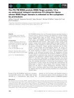
ATCC12633. The purified enzyme gave a single band
with a molecular mass of 37 kDa on SDS ⁄ PAGE
(Fig. 1). The molecular mass of the native enzyme was
found to be 74 kDa by gel filtration. Therefore, the
enzyme probably consists of two identical subunits.
Substrate specificity and effects of various
compounds on the enzymatic activity
The substrate specificity of the enzyme in NADPH-
dependent reductive amination was examined with
various a-oxo acids and amines. Pyruvate was the best
substrate of the various a-oxo acids tested (Table 1).
The enzyme also acted on a-oxohexanoate, phenylpyru-
vate, a-oxobutyrate, fluoropyruvate, a-oxovalerate,
a-oxoisocaproate, a-oxo-octanoate, and hydroxypyru-
vate. However, branched-chain a-oxo acids, such as
a-oxoisovalerate and a-oxo-b-methylvalerate, were
inert. The best substrate was methylamine, but the
enzyme also showed activity towards ethylamine (4.4%,
relative to methylamine), 2-chloroethylamine (0.74%),
2-bromoethylamine (0.27%), n-propylamine (0.16%)
and dimethylamine (0.12%). Weak activities (0.06–
0.03%, relative to methylamine) were found with
ethylenediamine, hydroxylamine, isopropylamine, n-
butylamine, n-amylamine, n-hexylamine, 1,6-diamino-
hexane, and spermidine. Interestingly, the enzyme was
unable to use ammonia as a substrate and was distinct
from alanine dehydrogenase [18] and N-methylalanine
dehydrogenase found by Lin & Wagner [17]. Lysine
and ornithine were inert as an amine substrate; the
enzyme shows no opine dehydrogenase activity with
these amino acids. The enzyme was sensitive to bivalent
metal ions, such as CuCl
2
, HgCl
2
, and CoCl
2
. Concen-
trations of the enzyme causing 50% inhibition were
0.35 lm for CuCl
2
, 0.96 lm for HgCl
2
, and 160 lm for
CoCl
2
. Carbonyl reagents, including hydroxylamine,
phenylhydrazine, semicarbazide, and aminoguanidine,
did not inhibit the enzyme. Chelating agents, such as
o-phenanthroline and EDTA, hardly inhibited the
enzyme. Thiol reagents, such as N-ethylmaleimide and
iodoacetate, and a serine-modifying reagent, phenyl-
methanesulfonyl fluoride, did not affect the enzyme.
Non-substrate a-oxo acids, a-oxoisovalerate and a-oxo-
b-methylvalerate, did not inhibit the enzyme.
Other enzymological properties
The enzyme showed maximum activity at pH 10.0 for
both reductive amination of phenylpyruvate and oxi-
dative deamination of N-methyl-l-alanine. It was sta-
ble between pH 6.0 and 10.0 and showed maximum
activity at 35 °C. However, it was unstable at this tem-
perature and lost 30% of its original activity after
a 30-min incubation in 20 mm Tris ⁄ HCl buffer at
pH 7.0. The enzyme was stable at temperatures below
30 °C for at least 30 min and used both NAD
+
and
NADH as coenzymes. However, the specific activity of
the enzyme with NADPH (42 UÆmg
)1
) was more than
300 times higher than with NADH (0.13 UÆmg
)1
)in
reductive amination of pyruvate. Oxidative deamina-
tion of N-methyl-l-alanine with NADP
+
(0.36 UÆ
mg
)1
) was markedly lower than reductive amination of
pyruvate with NADPH.
Fig. 1. Overexpression and purification of recombinant NMAADH.
Protein samples from various stages of the purification were sub-
jected to SDS ⁄ PAGE and stained with Coomassie Brilliant Blue.
Lane 1, marker proteins (sizes in kDa are shown); lane 2, crude
extract; lane 3, after the Green-Sepharose CL-4B chromatography;
lane 4, after the DEAE-Toyopearl chromatography.
Table 1. Substrate specificity of NMAADH in the forward reaction.
NMAADH activity was determined in reaction mixture containing
10 m
M a-oxo acid, 60 mM methylamine (pH 10.0), and 0.2 m M
NADPH at 30 °C.
Substrate Relative activity (%)
Pyruvate 100
a-Oxohexanoate 52
Phenylpyruvate 30
a-Oxobutyrate 30
Fluoropyruvate 27
a-Oxovalerate 16
a-Oxoisocaproate 9.1
a-Oxo-octanoate 7.6
Hydroxypyruvate 5.8
H. Mihara et al. N-Methyl-
L-amino acid dehydrogenase from P. putida
FEBS Journal 272 (2005) 1117–1123 ª 2005 FEBS 1119
Kinetic studies
The kinetic mechanism of NMAADH was studied
by steady-state kinetic analyses. NADPH-dependent
methylamination of pyruvate showed hyperbolic sat-
uration curves and gave linear double-reciprocal plots
in the range 1–10 mm pyruvate, 5–60 mm methyl-
amine, and 0.05–0.3 mm NADPH. The families of
lines obtained from the double-reciprocal plots of reac-
tion velocities against pyruvate concentrations at var-
ious fixed concentrations of methylamine intersected to
the left of the 1 ⁄ v axis. Similar results were obtained
with various concentrations of NADPH and fixed con-
centrations of pyruvate. These indicate a sequential
mechanism for the NMAADH reactions. In fact, the
data obtained gave a good global fit to the equation of
an ordered Ter Bi mechanism.
To test the kinetic mechanism further, we performed
inhibition studies with both products of the reaction,
NADP
+
and N-methyl-l-alanine. At a fixed subsaturat-
ing concentration of N-methyl-l-alanine (100 mm), the
inhibition by NADPH was competitive linearly with
respect to NADP
+
. With N-methyl-l-alanine as the
variable substrate, at a fixed subsaturating concentra-
tion of NADP
+
(0.2 mm), NADPH was a linear mixed-
type inhibitor. Pyruvate behaved as a noncompetitive
inhibitor with regard to NADP
+
and N-methyl-l-alan-
ine. Methylamine behaved as a mixed-type inhibitor
with respect to NADP
+
and N -methyl-l-alanine. These
results indicate that NADPH-dependent N -methyl-l-
alanine formation proceeds in an ordered sequential Ter
Bi mechanism, in which NADPH, pyruvate, and meth-
ylamine are bound to the enzyme in this order, and
N-methyl-l-alanine and then NADP
+
are released from
the enzyme. Therefore, the kinetic mechanism of the
enzyme is the same as those of conventional NAD(P)-
dependent amino acid dehydrogenases [25–27].
Comparison with other NAD(P)-dependent
dehydrogenases
The reaction catalyzed by NMAADH is similar to
those by conventional NAD(P)-dependent amino acid
dehydrogenases, such as glutamate dehydrogenase [28],
leucine dehydrogenase [29], and phenylalanine dehy-
drogenase [30], as well as opine dehydrogenase [25,31],
which have a common structure called the Rossmann
fold for their NAD(P)-binding sites [26]. However,
NMAADH shows little (< 15%) sequence similarity to
the NAD(P)-dependent amino acid dehydrogenases and
opine dehydrogenases and has no sequence motif char-
acteristic of the Rossmann fold, indicating that the
structure of the nicotinamide nucleotide-binding site of
NMAADH is completely different from those with the
Rossmann fold. A homology search revealed that
NMAADH belongs to a new protein superfamily
including NAD(P)-dependent malate ⁄ l-lactate dehy-
drogenases with no sequence homology to conventional
malate dehydrogenases or lactate dehydrogenases [32].
The superfamily contains, in addition to the malate ⁄
l-lactate dehydrogenases, various NAD(P)-dependent
dehydrogenases, such as l-sulfolactate dehydrogenase
[33], ureidoglycolate dehydrogenase [34], and 2,3-
diketo-l-gulonate reductase [35]. The action on a-oxo
acids as substrate is common to all these enzymes, and
NMAADH is a new addition to this superfamily.
Conclusion
We have identified an enzyme catalyzing the NADPH-
dependent formation of N -methyl-l-phenylalanine from
phenylpyruvate and methylamine. The enzyme is
unique in that it does not act on ammonia at all and
shows broad specificity for various a-oxo acids. Accord-
ingly, we named the enzyme N-methyl-l-amino acid
dehydrogenase to distinguish it from the previously
reported N-methylalanine dehydrogenase [17]. More-
over, this study shows that the enzyme belongs to a new
NAD(P)-dependent oxidoreductase family and is struc-
turally distinct from conventional NAD(P)-dependent
amino acid dehydrogenases and opine dehydrogenases.
Experimental procedures
Materials
N-Methyl-l-phenylalanine and d-amino acid oxidase from
porcine kidney were purchased from Sigma (St Louis, MO,
USA). RESOURCE PHE, Superose 12, Sepharose CL-4B,
and molecular-mass marker proteins were obtained from
Amersham Pharmacia Biotech (Uppsala, Sweden). SuperQ-
Toyopearl, DEAE-Toyopearl, and Butyl-Toyopearl were
from Tosoh (Tokyo, Japan). Green-Sepharose was prepared
as described previously [36]. NADH, NADPH, and mole-
cular-mass marker proteins for gel filtration were from
Oriental Yeast (Tokyo, Japan). Restriction enzymes and
kits for genetic manipulation were from Takara Shuzo
(Kyoto, Japan), Toyobo (Osaka, Japan), and New England
Biolabs (Beverly, MA, USA). All other reagents were of
analytical grade from Nacalai Tesque (Kyoto, Japan) and
Wako Pure Chemical Industries (Osaka, Japan).
Culture and screening of bacteria
Bacterial strains were cultivated for 15–20 h at 30 °Cin
0.5 mL screening medium containing citric acid (0.5 gÆL
)1
),
N-Methyl-L-amino acid dehydrogenase from P. putida H. Mihara et al.
1120 FEBS Journal 272 (2005) 1117–1123 ª 2005 FEBS
N-methyl-l-phenylalanine (1 gÆL
)1
), glucose (2 gÆL
)1
),
K
2
HPO
4
(7 gÆL
)1
), KH
2
PO
4
(3 gÆL
)1
), and MgSO
4
Æ7H
2
O
(0.1 gÆL
)1
) (pH 6.9). The cells were harvested by centrifu-
gation, resuspended in 0.1 mL reaction mixture containing
5mm calcium phenylpyruvate and 1 m methylam-
ine ⁄ H
2
SO
4
(pH 8.9), and incubated for 3 h at 30 °C. The
formation of N-methylphenylalanine in the reaction mix-
ture was analyzed by TLC on a silica gel 60 plate (Merck,
Darmstadt, Germany) or by HPLC with an Ultron
ES-PhCD column (Shinwa Kako, Kyoto, Japan). The sol-
vent system for TLC was ethyl acetate ⁄ ethanol ⁄ acetic
acid ⁄ water (5 : 2 : 1 : 1, by vol.), and N-methylphenyl-
alanine was visualized with a coloring reagent containing
0.2% ninhydrin, 0.5% acetic acid, and 95% butan-1-ol.
HPLC analysis was performed with a solvent containing
16 mm KH
2
PO
4
, 20% acetonitrile, and 0.04% phosphoric
acid at a flow rate of 0.85 mLÆmin
)1
at 40 °C, and eluates
were monitored at 210 nm.
Enzyme assays
For the purification of the enzyme from P. putida,
NMAADH activity was determined in a reaction mixture
containing 12 mm phenylpyruvate, 240 mm methyl-
amine ⁄ H
2
SO
4
(pH 10.0), and 8.0 mm NADPH in a final
volume of 50 lLat37°C. The N-methylphenylalanine
formed was analyzed by HPLC as described above. Other
NMAADH assays were carried out by measuring the
decrease in the amount of NADPH at 340 nm with an
MPS-2000 spectrophotometer (Shimadzu, Kyoto, Japan) at
30 °C. A standard reaction mixture contained 10 mm
phenylpyruvate, 60 mm methylamine ⁄ H
2
SO
4
(pH 10.0),
0.2 mm NADPH, and the enzyme in a final volume of
1 mL.
Cloning and expression of NMAADH in E. coli
PCR primers were designed based on the nucleotide
sequence of ORF PP3591 located on the 4 080 935–
4 081 933-bp region of the P. putida KT2440 genome [37].
A 1-kb DNA fragment containing the NMAADH gene was
amplified from the genomic DNA of another P. putida
strain ATCC12633 by PCR with a Perkin–Elmer Thermal
Cycler 480 (Wellesley, MA, USA) in a 50-lL reaction mix-
ture containing 1· LA Taq buffer (Takara Shuzo), 2.5 mm
MgCl
2
, 0.4 mm dNTP, 0.2 mm each primer (5¢-GGAAT
TCCATATGTCCGCACCTTCCACCAGCACCG-3¢ and
5¢-GGGAAGCTTTCAGCCAAGCAGCTCTTTCAGG-3¢),
2.5 U LA Taq DNA polymerase, and 115 ng genomic
DNA from P. putida ATCC12633: preincubation at 94 °C
for 1 min and then 30 cycles between 98 °C for 20 s and
68 °C for 3 min and finally at 72 °C for 10 min. The PCR
product was digested with NdeI and HindIII and ligated
into pET21a(+) previously digested with the same restric-
tion enzymes. The resultant plasmid, pDPKA, was intro-
duced into E. coli BL21(DE3) to provide us with
recombinant DpkA.
Purification of recombinant NMAADH
E. coli BL21(DE3) carrying pDPKA was cultivated in
Luria–Bertani medium containing 100 lgÆL
)1
ampicillin at
37 °C for 14 h. The culture was supplemented with 1 mm
isopropyl b-d-thiogalactopyranoside and grown for 3 h.
The wet cells (3.3 g) obtained by centrifugation were sus-
pended in 28 mL of our standard buffer: a 20 mm Tris ⁄ HCl
buffer (pH 7.0) containing 1 mm phenylmethanesulfonyl
fluoride. The crude extract obtained by sonication was loa-
ded on to a Green-Sepharose CL-4B column (100 mL)
equilibrated with the standard buffer. The enzyme was elut-
ed with a linear gradient of 0–1 m NaCl in the buffer. The
enzyme fractions were pooled, concentrated with Centriprep
YM-10 (Millipore, Bedford, MA, USA), and dialyzed
against the buffer. The enzyme solution was applied to a
DEAE-Toyopearl column (60 mL) equilibrated with the
same buffer and eluted with a linear gradient of 0–0.35 m
NaCl in the buffer. The enzyme fractions were collected,
concentrated, and dialyzed against the standard buffer. The
final preparation of the enzyme was stored at )80 °C until
use without significant inactivation.
Effect of various compounds on the activity
The enzyme was incubated with various reagents in 20 mm
Tris ⁄ HCl (pH 7.0) at 25 °C for 15 min. The remaining
activity was determined as described above.
Kinetic analysis
Initial rates of the NMAADH reactions were measured
with various concentrations of one substrate and fixed
(and excess) concentrations of the other substrates. Data
were fitted to the hyperbolic Michaelis–Menten equation,
and kinetic parameters were calculated using nonlinear
least-squares regression with kaleida graph software
(Adelbeck Software, Reading, PA, USA) or IGOR Pro
software (WaveMetrics, Inc., Lake Oswego, OR, USA).
Eqn (1) describes a sequential mechanism: K
a
, K
b
, and K
c
represent the Michaelis constants for the NADPH (A),
pyruvate (B), and methylamine (C), respectively; K
ia
and
K
ib
are the dissociation constants for the enzyme–NADPH
complex and the enzyme–pyruvate complex, respectively.
Product inhibition studies were performed with various
concentrations of either NADP
+
or N-methyl-l-alanine as
one substrate and a fixed saturating concentration of the
other. The data were fitted to Eqns (2), (3) and (4), which
describe the competitive, uncompetitive, and noncompeti-
tive inhibition patterns, respectively. P is the concentration
of the product, K
is
is the inhibition constant from the slope
H. Mihara et al. N-Methyl-L-amino acid dehydrogenase from P. putida
FEBS Journal 272 (2005) 1117–1123 ª 2005 FEBS 1121
term, and K
ii
is the inhibition constant from the intercept
term.
v ¼ VABC=ðK
ia
K
ib
K
c
þ K
ib
K
c
A þ K
ia
K
b
C
þ K
c
AB þ K
b
AC þ K
a
BC þ ABCÞð1Þ
v ¼ VA=½K
a
ð1 þ P=K
is
ÞþAð2Þ
v ¼ VA=½K
a
þ Að1 þ P=K
ii
Þ ð3Þ
v ¼ VA=½K
a
ð1 þ P=K
is
ÞþAð1 þ P=K
ii
Þ ð4Þ
Analytical size-exclusion chromatography
The protein quaternary structure was analyzed by an
A
¨
KTAexplorer system (Amersham Biosciences, Amersham,
Buckinghamshire, UK) using a YMC-Pack Diol 200 col-
umn (YMC Co, Ltd, Kyoto, Japan). The column was
equilibrated and operated at a flow rate of 1.0 mLÆmin
)1
with a 0.1 m potassium phosphate buffer (pH 7.0) contain-
ing 0.2 m NaCl. The protein standards used were cyto-
chrome c, myokinase, enolase, lactate dehydrogenase, and
glutamate dehydrogenase from Oriental Yeast, Osaka,
Japan.
Other analytical methods
The N-terminal amino-acid sequence of the enzyme was
determined with an automated Shimadzu PPSQ10 protein
sequencer (Kyoto, Japan). The nucleotide sequence of
DNA was determined with an Applied Biosystems 370A
DNA sequencer (Foster City, CA, USA).
Acknowledgements
This work was supported in part by a Grant-in-Aid
for Scientific Research on Priority Areas (B) 13125203
(to NE) from the Ministry of Education, Culture,
Sports, Science, and Technology of Japan, by Grant-
in-Aid for Encouragement of Young Scientists
15780070 (to HM) from the Japan Society for the Pro-
motion of Science, by the National Project on Protein
Structural and Functional Analyses and by Grant-in-
Aid from the Ministry of Education, Culture, Sports,
Science and Technology, Japan (21st Century COE on
Kyoto University Alliance for Chemistry).
References
1 Walsh CT, Zydowsky LD & McKeon FD (1992)
Cyclosporin A, the cyclophilin class of peptidylprolyl
isomerases, and blockade of T cell signal transduction.
J Biol Chem 267 , 13115–13118.
2 Harris CM, Kopecka H & Harris TM (1983) Vancomy-
cin: structure and transformation to Cdp-I. J Am Chem
Soc 105, 6915–6922.
3 Vera MD & Joullie MM (2002) Natural products as
probes of cell biology: 20 years of didemnin research.
Med Res Rev 22, 102–145.
4 Poncet J (1999) The dolastatins, a family of promising
antineoplastic agents. Curr Pharm Des 5, 139–162.
5 Tomoda H, Nishida H, Huang XH, Masuma R, Kim
YK & Omura S (1992) New cyclodepsipeptides, ennia-
tins D, E and F produced by Fusarium sp. FO-1305.
J Antibiot (Tokyo) 45, 1207–1215.
6 Alvarez ME, Houck DR, White CB, Brownell JE, Bob-
ko MA, Rodger CA, Stawicki MB, Sun HH, Gillum
AM & Cooper R (1994) Isolation and structure elucida-
tion of two new calpain inhibitors from Streptomyces
griseus. J Antibiot (Tokyo) 47, 1195–1201.
7 Frost LS, Carpenter M & Paranchych W (1978)
N-Methylphenylalanine at the N-terminus of pilin iso-
lated from Pseudomonas aeruginosa K. Nature 271,
87–89.
8 Zocher R, Keller U & Kleinkauf H (1982) Enniatin
synthetase, a novel type of multifunctional enzyme cata-
lyzing depsipeptide synthesis in Fusarium oxysporum.
Biochemistry 21, 43–48.
9 Strom MS, Nunn DN & Lory S (1993) A single bifunc-
tional enzyme, PilD, catalyzes cleavage and N-methyla-
tion of proteins belonging to the type IV pilin family.
Proc Natl Acad Sci USA 90, 2404–2408.
10 Shibata K, Tarui A, Todoroki N, Kawamoto S,
Takahashi S, Kera Y & Yamada R (2001) Occurrence
of N-methyl-l-aspartate in bivalves and its distribution
compared with that of N-methyl-d-aspartate and d,l-
aspartate. Comp Biochem Physiol B Biochem Mol Biol
130, 493–500.
11 Tarui A, Shibata K, Takahashi S, Kera Y, Munegumi
T & Yamada RH (2003) N-methyl-d-glutamate and
N-methyl-l-glutamate in Scapharca broughtonii (Mol-
lusca) and other invertebrates. Comp Biochem Physiol B
Biochem Mol Biol 134, 79–87.
12 Cannon JR & Williams JR (1982) The alkaloids of
gastrolobium-callistachys. Aust J Chem 35, 1497–1500.
13 Shaw WV, Tsai L & Stadtman ER (1966) The enzy-
matic synthesis of N-methylglutamic acid. J Biol Chem
241, 935–945.
14 Kung HF & Wagner C (1970) The enzymatic synthesis
of N-methylalanine. Biochim Biophys Acta 201, 513–516.
15 Urakami T, Araki H, Oyanagi H, Suzuki KI &
Komagata K (1992) Transfer of Pseudomonas-Amino-
vorans (Dendooren Dejong 1926) to Aminobacter
General-Nov as Aminobacter-Aminovorans Comb-Nov
and description of Aminobacter-Aganoensis Sp-Nov
and Aminobacter-Niigataensis Sp-Nov. Int J Syst
Bacteriol 42, 84–92.
N-Methyl-L-amino acid dehydrogenase from P. putida H. Mihara et al.
1122 FEBS Journal 272 (2005) 1117–1123 ª 2005 FEBS
16 Pollock RJ & Hersh LB (1971) N-Methylglutamate
synthetase. Purification and properties of the enzyme.
J Biol Chem 246 , 4737–4743.
17 Lin MC & Wagner C (1975) Purification and characteri-
zation of N-methylalanine dehydrogenase. J Biol Chem
250, 3746–3751.
18 Ohashima T & Soda K (1979) Purification and proper-
ties of alanine dehydrogenase from Bacillus sphaericus.
Eur J Biochem 100, 29–30.
19 Nster EW, Gordon MP, Amasino RM & Yanofsky MF
(1984) Crown gall: a molecular and physiological analy-
sis. Annu Rev Plant Physiol 35, 387–413.
20 Hack E & Kemp JD (1977) Comparison of octopine,
histopine, lysopine, and octopinic acid synthesizing
activities in sunflower crown gall tissues. Biochem Bio-
phys Res Commun 78, 785–791.
21 Hack E & Kemp JD (1980) Purification and characteri-
zation of the crown gall-specific enzyme, octopine
synthase. Plant Physiol 65, 949–955.
22 Fields JH, Eng AK, Ramsden WD, Hochachka PW &
Weinstein B (1980) Alanopine and strombine are novel
imino acids produced by a dehydrogenase found in the
adductor muscle of the oyster Crassostrea gigas. Arch
Biochem Biophys 201, 110–114.
23 Kemp JD, Sutton DW & Hack E (1979) Purification
and characterization of the crown gall specific
enzyme nopaline synthase. Biochemistry 18, 3755–
3760.
24 Brunhuber NM & Blanchard JS (1994) The biochemis-
try and enzymology of amino acid dehydrogenases. Crit
Rev Biochem Mol Biol 29, 415–467.
25 Dairi T & Asano Y (1995) Cloning, nucleotide sequen-
cing, and expression of an opine dehydrogenase gene
from Arthrobacter sp. strain 1C. Appl Environ Microbiol
61, 3169–3171.
26 Rossmann MG, Moras D & Olsen KW (1974) Chemical
and biological evolution of nucleotide-binding protein.
Nature 250, 194–199.
27 Muramatsu H, Mihara H, Kakutani R, Yasuda M,
Ueda M, Kurihara T & Esaki N (2004) Enzymatic
synthesis of N-methyl-l-phenylalanine by a novel
enzyme, N-methyl-l-amino acid dehydrogenase, from
Pseudomonas putida. Tetrahedron: Asymmetr 15, 2841–
2843.
28 Smith EL, Austen BM, Blumenthal KM & Nyc JF
(1975) Glutamate dehydrogenase. In The Enzymes, 3rd
edn. (Boyer, P D, ed.), pp. 293–367. Academic Press,
New York.
29 Ohshima T, Misono H & Soda K (1978) Properties of
crystalline leucine dehydrogenase from Bacillus sphaeri-
cus. J Biol Chem 253, 5719–5725.
30 Hummel W, Weiss N & Kula MR (1984) Isolation and
characterization of a bacterium possessing l-phenylala-
nine dehydrogenase-activity. Arch Microbiol 137, 47–52.
31 Asano Y, Yamaguchi K & Kondo K (1989) A new
NAD
+
-dependent opine dehydrogenase from Arthro-
bacter sp. strain 1C. J Bacteriol 171, 4466–4471.
32 Jendrossek D, Kratzin HD & Steinbu
¨
chel A (1993) The
Alcaligenes eutrophus ldh structural gene encodes a novel
type of lactate dehydrogenase. FEMS Microbiol Lett
112, 229–235.
33 Honka E, Fabry S, Niermann T, Palm P & Hensel R
(1990) Properties and primary structure of the l-malate
dehydrogenase from the extremely thermophilic archae-
bacterium Methanothermus fervidus. Eur J Biochem 188,
623–632.
34 Cusa E, Obradors N, Baldoma L, Badia J & Aguilar J
(1999) Genetic analysis of a chromosomal region con-
taining genes required for assimilation of allantoin
nitrogen and linked glyoxylate metabolism in Escheri-
chia coli. J Bacteriol 181, 7479–7484.
35 Yew WS & Gerlt JA (2002) Utilization of l-ascorbate
by Escherichia coli K-12: assignments of functions to
products of the yjf-sga and yia-sgb operons. J Bacteriol
184, 302–306.
36 Ohshima T & Yamasaki T (1990) Dye-ligand affinity-
chromatography: simple preparation method of dye
ligand resins and their application. Seikagaku 62, 283–
287.
37 Nelson KE, Weinel C, Paulsen IT, Dodson RJ, Hilbert
H, Martins dos Santos VA, Fouts DE, Gill SR, Pop M,
Holmes M, et al. (2002) Complete genome sequence and
comparative analysis of the metabolically versatile Pseu-
domonas putida KT2440. Environ Microbiol 4, 799–808.
H. Mihara et al. N-Methyl-L-amino acid dehydrogenase from P. putida
FEBS Journal 272 (2005) 1117–1123 ª 2005 FEBS 1123