Tài liệu Age-related changes of the dental aesthetic zone at rest and during spontaneous smiling and speech pptx
Bạn đang xem bản rút gọn của tài liệu. Xem và tải ngay bản đầy đủ của tài liệu tại đây (509.91 KB, 8 trang )
doi:10.1093/ejo/cjn009
Advance Access publication 16 July 2008
European Journal of Orthodontics 30 (2008) 366–373
© The Author 2008. Published by Oxford University Press on behalf of the European Orthodontic Society.
All rights reserved. For permissions, please email:
Introduction
In social interaction, our attention appears mainly directed
towards the mouth and eyes of the face of the person speaking
( Thompson et al. , 2004 ). As the mouth is the centre of
communication in the face, the aesthetic appearance of the
oral region during smiling is a conspicuous part of facial
attractiveness. The aesthetic ( Garber and Salama, 1996 ) or
display ( Ackerman and Ackerman, 2002 ) zone is composed of
the size, shape, position and colour of the displayed teeth, the
gingival contour, the buccal corridor, and the framing of the
lips. The range of the aesthetic zone is defi ned by the movements
of the upper and lower lip during smiling and speech.
Lip position and the amount of tooth and gingival display
during smiling and speech are important diagnostic criteria in
orthodontics, dentofacial surgery, and aesthetic dentistry.
Smiles that entirely display the teeth including some gingiva
(2 – 4 mm) are perceived as the most aesthetic ( Kokich et al. ,
1999 ; Van der Geld et al. , 2007b ). Furthermore, a continuous
gingival contour should be parallel with the curve of the upper
lip ( Moskowitz and Nayyar, 1995 ; Peck and Peck, 1995 ). The
most ideal incisal line of the upper dentition is established in
relation to the curve of the lower lip ( Sarver, 2001 ; Ackerman
et al. , 2004 ). Therefore, adequate evaluation of lip lines is
required for the orthodontic diagnosis, especially in patients
with reduced tooth display, unaesthetic gingival contours,
exposed posterior gingiva, occlusal cants, asymmetry of the
upper lip during smiling, and ‘ gummy smiles ’ .
Age-related changes of the dental aesthetic zone at rest and
during spontaneous smiling and speech
Pieter Van der Geld , Paul Oosterveld and Anne Marie Kuijpers-Jagtman
Department of Orthodontics and Oral Biology, Radboud University Nijmegen Medical Centre, The Netherlands
SUMMARY The aims of this study were to analyse lip line heights and age effects in an adult male population
during spontaneous smiling, speech, and tooth display in the natural rest position and to determine
whether lip line height follows a consistent pattern during these different functions. The sample consisted
of 122 randomly selected male participants from three age cohorts (20 – 25 years, 35 – 40 years, and 50 – 55
years). Lip line heights were measured with a digital videographic method for smile analysis, which had
previously been tested and found reliable. Statistical analysis of the data was carried out using correlation
analysis, analysis of variance, and Tukey’s post hoc tests.
Maxillary lip line heights during spontaneous smiling were generally higher in the premolar area than
at the anterior teeth. The aesthetic zone in 75 per cent of the participants included all maxillary teeth up
to the fi rst molar. Coherence in lip line heights during spontaneous smiling, speech, and tooth display
in the natural rest position was confi rmed by signifi cant correlations. In older subjects, maxillary lip
line heights decreased signifi cantly in all situations. Lip line heights during spontaneous smiling were
reduced by approximately 2 mm. In older participants, the mandibular lip line heights also changed
signifi cantly and teeth were displayed less during spontaneous smiling. Mandibular tooth display in the
rest position increased signifi cantly. Upper lip length increased signifi cantly by almost 4 mm in older
subjects, whereas upper lip elevation did not change signifi cantly.
The signifi cant increasing lip coverage of the maxillary teeth indicates that the effects of age should be
included in orthodontic treatment planning.
In spite of the relevance of the aesthetic zone in orthodontic
treatment planning, relatively little research has been carried
out on lip line height and tooth and gingival exposure during
spontaneous smiling and speech. A drawback of most studies
is that only posed smiles have been measured. It is claimed
that such smiling on request has the advantage of
reproducibility ( Rigsbee et al. , 1988 ; Ackerman et al. , 1998 ),
yet it should be questioned whether the posed social smile
is the same as a spontaneous smile of joy. The smile in
fact is not a singular category of facial behaviour. In
psychophysiology, for example a difference is made between
emotion elicited spontaneous smiles of joy and voluntary
posed smiles ( Ekman, 1992 ). On the basis of structural
differences between spontaneous smiling and the posed
smile, spontaneous smiling is considered as a focus point for
lip line analysis in orthodontic treatment planning ( Tarantili
et al. , 2005 ). This is in line with the recommendations of oral
surgeons ( Allen and Bell, 1992 ) and aesthetic dentists
( Moskowitz and Nayyar, 1995 ). Ackerman et al. (2004)
proposed that the orthodontist should view the dynamics of
anterior tooth display as a continuum delineated by the time
points of rest, speech, posed social smile, and a (spontaneous)
Duchenne smile. Most of the methods for smile measurement,
however, are not designed to measure spontaneous smiles.
Consequently, limited data are available to serve as a
guideline for lip line heights in spontaneous smiling and
speech, particularly for the adult population.
367
AGE AND THE DENTAL AESTHETIC ZONE
Another important aspect, to consider when evaluating
the aesthetic zone, is the effect of age on lip line height.
Based on clinical experience, the prosthetic literature
demonstrates that with age the lips become less elastic and
less mobile. As a result of this, older people are reported to
show less of the maxillary and more of the mandibular teeth
during smiling ( Shillingburg et al. , 1997 ). Dong et al.
(1999) and Dickens et al. (2002) measured changes in the
smile as an effect of age. Both studies reported a decrease of
maxillary incisor display during smiling. Dong et al. (1999)
also found a slight increase of mandibular incisor display.
In the studies of Vig and Brundo (1978) and Al Wazzan
(2004) , the maxillary incisor display at rest was found to
gradually reduce with an increase in age, while mandibular
incisor display increased. It should be noted, however, that
most of these results were not statistically tested.
From the starting point that the lip line height is an
essential diagnostic criterion in (adult) orthodontics,
dentofacial surgery, and aesthetic restorative dentistry, a
digital videographic method to measure both spontaneous
smiling and speech was developed ( Van der Geld et al. ,
2007a ). The specifi c aims of the present study were fi rstly to
analyse lip line heights and age effects in an adult male
population during spontaneous smiling, speech, and in
natural rest position with a digital videographic measurement
method and secondly to determine if lip line heights followed
a consistent pattern during these different functions.
Subjects and methods
The research proposal was approved by the ethical
committee of the Academic Centre of Dentistry, Amsterdam.
Informed consent was obtained from the subjects according
to the guidelines of that institution.
Participants
Of 1069 military males on an air force base, 122 were
randomly selected from three age cohorts (20 – 25 years,
35 – 40 years, and 50 – 55 years). Selection criteria were full
maxillary and mandibular dental arches up to and including
the fi rst molar, Caucasian, no excessive facial disharmonies,
and no visible periodontal disease or caries.
Recording and measurement during spontaneous smiling,
speech, and at rest
A digital videographic measurement method was used to
capture records of a spontaneous smile of joy and during
speech. In addition, a record of a spontaneous natural rest
position (with the lips slightly parted) and a full dentition
record with the aid of cheek retractors were made. The
reliability and clinical application of this digital videographic
measurement method has been tested previously. The
method appeared to be reliable with intraclass coeffi cients
ranging from 0.99 to 0 .80 ( Van der Geld et al. , 2007a ).
On the full dentition record, the lengths of the teeth were
measured to obtain the actual length of the tooth crowns. On
the spontaneous smiling and speech records, the display of
teeth and gingiva was measured. In the maxilla and
mandible, a central and lateral incisor, a canine, a fi rst and
second premolar, and a fi rst molar were measured from the
left and right side alternately to exclude infl uences of facial
asymmetry. Digital horizontal lines were used to mark the
most incisal point of each tooth (line 1) and the lip edge
(line 2, Figure 1 ). These marking lines were parallel to the
inter pupil line. The vertical distance between these lines
was measured (see lip position measurement, Figure 1 ).
Following the concept of Peck and Peck (1995) , lip line
height was expressed relative to the gingival margin (line 3)
and thus is a measurement for both tooth and gingival
visibility ( Figure 1 ). Lip line height was calculated as the
difference between lip position and tooth length. When the
gingival margin was displayed, positive values were assigned
both for the maxilla and the mandible. When the teeth
remained partly covered, negative values were given. If the
upper and lower lip covered both gingival margin and incisal
point, lip line height was denoted as not measurable. If a tooth
was not visible, lip line height was recorded as missing.
On the record in the natural rest position, the amount of
tooth display was measured from the incisal point of each
tooth to the edge of the lip. If a tooth was not visible, the
tooth display was denoted as zero.
The vertical length of the upper lip was measured between
the lower edge of the upper lip and subnasion on the
spontaneous smiling record and the record in the natural
rest position. The amount of lip elevation during spontaneous
smiling was calculated as the percentage difference between
upper lip length in the rest position and upper lip length
during spontaneous smiling.
Data analysis
Correlation analysis was used to determine if the lip line
heights of a subject were coherent during the situations of
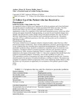
Figure 1 Measurement of lip line height; Line 1: the most incisal point of
the central incisor; Line 2: the lip edge on the central incisor; Line 3: cervical
margin of the central incisor. Lip line height is lip position minus tooth length.
When the gingival margin is displayed, lip line height has positive values.
When the teeth are partly covered, lip line height has negative values.
P. VAN DER GELD ET AL.
368
spontaneous smiling, speech, and in the rest position. Following
the conventions set by Cohen (1988) , correlations of 0.10,
0.30, and 0.50 were considered weak, moderate, and strong,
respectively. The signifi cance level P < 0.05 was chosen.
Analysis of variance (ANOVA) was used to compare lip
line heights for each tooth between the three age cohorts in
the situations, spontaneous smiling and speech. ANOVA
was performed on each tooth separately as the number of
teeth displayed varied between the situations. When the lip
line heights were found to differ signifi cantly between the
age cohorts, Tukey’s post hoc tests were performed to
identify the cohorts that differed signifi cantly. The same
procedure was performed for tooth display in the natural
rest position, lip elevation, and upper lip length.
Results
Lip line heights and frequencies of displayed teeth
Lip line heights during spontaneous smiling and speech are
shown for the three age groups, for the maxilla and mandible,
in Figures 2 and 3 , respectively. In Figure 2 , the minimum
and maximum graphs of lip line heights show a considerable
individual variation in some subjects compared with the
majority of the sample. In contrast to spontaneous smiling,
the maxillary lip line heights during speech were generally
lower. The cervical gingival margins were mostly covered
by the upper lip.
During spontaneous smiling and speech, the mandibular lip
line heights were mostly positioned on the tooth ( Figure 3 ).
The cervical gingival margins were thus covered by
the lower lip. Contrary to the maxilla, during speech,
the mandibular teeth were displayed more than during
spontaneous smiling.
The collected data showed that in 75 per cent of the sample,
the maxillary fi rst molar was substantially displayed during
spontaneous smiling and was part of the aesthetic zone. The
mandibular anterior teeth formed part of the aesthetic zone
especially during speech in 93 per cent of the participants.
Relationships between lip line heights in different
situations
Table 1 shows the correlation analysis used to determine if
the lip line heights followed a coherent pattern during
spontaneous smiling, speech, and tooth display in the
natural rest position. The lip line heights of all maxillary
teeth demonstrated a signifi cant and strong to moderate
relationship between spontaneous smiling and speech. In the
mandible, this applied to the anterior teeth and the fi rst
premolar.
Maxillary anterior lip line heights during spontaneous
smiling and tooth display in the natural rest position were
highly signifi cant and strongly correlated. Maxillary anterior
lip line heights during speech and tooth display in the natural
rest position also showed a signifi cant and strong to
moderate relationship. No correlations between these
situations were found for the mandibular teeth.
Age effects on the aesthetic zone
The results of ANOVA, comparing the lip line heights of the
three age cohorts during spontaneous smiling, are given in
Table 2 . The suggestion ( Figure 2 ) that lip line heights
gradually decrease with age was confi rmed by the signifi cant
results for all maxillary teeth. Post hoc analysis showed that
the signifi cant effects occurred mainly between the 20 – 25
and 50 – 55 year cohorts. The mandibular lip line heights
also decreased with age; the lateral incisor, the canine, and
the fi rst premolar were signifi cantly covered by the lower
lip in the older age cohorts.
During speech the effect of decreasing lip line heights
with age was signifi cantly manifested in the maxillary
anterior region ( Table 3 ). Beside signifi cant effects for all
anterior teeth between the 20 – 25 and 50 – 55 year cohorts,
both incisors also showed signifi cant effects between the
20 – 25 and 35 – 40 year cohorts.
In the mandible, no signifi cant age effects on lip line heights
during speech were found apart from the central incisor. This
single signifi cant effect was possibly caused by a differing
mean in the second cohort. As this is not in line with the other
results, the fi ndings should be interpreted with caution.
The same as lip line heights during spontaneous smiling
and speech, maxillary anterior tooth display in the natural rest
position showed a signifi cant decrease with age ( Table 4 ).
Signifi cant differences between all age cohorts were found
for the maxillary incisors. Opposite to the maxillary decrease
of tooth display, mandibular anterior tooth display increased
highly signifi cantly in the older subjects.
The upper lip length during spontaneous smiling and in
the natural rest position both showed very high signifi cant
lengthening with age ( Table 5 ). For both situations, the
signifi cant effects occurred between the 20 – 25 and 35 – 40
year cohorts and 20 – 25 and 50 – 55 year cohorts. For the
upper lip elevation during spontaneous smiling, no
signifi cant changes were found.
Discussion
Spontaneous smiling and speech have a dynamic nature,
which requires a dynamic registration method. However,
ear rods are often used for standardization of the head
position. This is not a favourable position to elicit
a spontaneous smile of joy in patients. Therefore, a
less intrusive dynamic registration method based on
videographic measurement of spontaneous smiling and
speech was developed ( Van der Geld et al. , 2007a ). Since
this approach is relatively new in smile analysis, no data
were available of adult lip line heights during spontaneous
smiling, speech, and tooth display in the natural rest
position. This makes a comparison with other studies
diffi cult.
369
AGE AND THE DENTAL AESTHETIC ZONE
In the present investigation, the sample used was restricted
to males. Selection of the sample according to the criteria
was accurate because adequate dental documentation was
present. Furthermore, a homogeneous sample was needed
to exclude factors such as race or gender. This means that
the results of this study are valid for Caucasian males only.
As shown in Figure 2 , the maxillary lip line heights
during spontaneous smiling tended to be generally higher
in the premolar area and, for a considerable number of
patients, the posterior maxillary region was also part of the
aesthetic zone. This fi nding is in line with a study of posed
smiling, in which Kapagiannidis et al. (2005) reported that
maxillary gingival display was greater for premolars
compared with the central incisor and canine. This is
important with respect to orthodontic diagnosis and
treatment planning. Obviously, during orthodontic treatment
more attention is given to incisor lip line heights but at a
risk of overexposure of the posterior gingiva. This gingival
overexposure is undesirable in the smile and diffi cult to
correct ( Mackley, 1993 ).
Compared with spontaneous smiling, during speech the
maxillary teeth were covered more by the upper lip and less
displayed. Especially, the maxillary anterior teeth and the
fi rst premolar were visible. In the mandible, by contrast, the
lower lip moved more towards the gingival margin during
speech than during spontaneous smiling ( Figure 3 ). During
Figure 2 Median, quartiles, and ranges of maxillary lip line heights in millimetres relative to the gingival margin for the upper incisors, canine, premolars,
and fi rst molar. The grey shaded areas represent the gingiva. Percentages of (measurable) displayed teeth in the total sample are show in pie charts.
P. VAN DER GELD ET AL.
370
speech a larger number of mandibular teeth (the anterior
teeth and the fi rst premolar) were in view and were also
more exposed than during smiling.
Ackerman et al. (2004) found clinically and statistically
signifi cant changes in anterior lip – tooth relationships
between posed smiling and speech. In addition, in the present
study, the coherence of lip line heights during spontaneous
smiling, speech, and tooth display in the natural rest position
was determined. This means, e.g., that patients showing
higher lip line heights during spontaneous smiling, also
showed higher lip line heights during speech as well as a
greater amount of tooth display in the natural rest position.
The patients’ coherence of lip line heights during these
situations provides an unambiguous orthodontic strategy as
the one functional situation does not require a totally
different treatment approach from another.
Limited studies are available that provide data
concerning the effect of age on the aesthetic zone. These
data are relevant, among others, for predictable long-
term aesthetic results of orthodontic therapy. The general
assumption, mostly based on clinical experience, that lip
line height decreases with age was statistically confi rmed
for the maxilla in this study. Moreover, the age effect on
the perioral tissues is not equal for the maxilla and
mandible or for each situation. With age, a decrease of
maxillary lip line height and tooth display was found
Figure 3 Median, quartiles, and ranges of mandibular lip line heights in millimetres relative to the gingival margin for the lower incisors, canine, premolars,
and fi rst molar. The grey shaded areas represent the gingiva. Percentages of (measurable) displayed teeth in the total sample are show in pie charts.
371
AGE AND THE DENTAL AESTHETIC ZONE
in combination with an increase of upper lip length.
For the upper central incisor, lip line heights during
spontaneous smiling decreased by 2 mm. Both tooth
display and upper lip length in the natural rest position
decreased by almost 4 mm.
The age-related increase of upper lip length appeared
approximately equal to the reduction of maxillary incisor
display in the natural rest position. An interesting fi nding
was that the age-related decrease in lip line height during
spontaneous smiling was considerably less than in the
natural rest position. It was also interesting to note that in
the natural rest position, the age-related effects occurred
between all age cohorts. These intercohort effects were
less obvious during speech whereas during spontaneous
smiling, the age-related effects only occurred between the
youngest and oldest age cohorts. At fi rst, the age-related
effects appear to diminish in situations where more
musculature activity is required. It is presumed that in
situations with more perioral musculature activity, as in
spontaneous smiling, the initial effects of age on the soft
tissues are compensated ( Gosain et al. , 1996 ). This is
supported by the fact that lip elevation was the same for all
ages ( Table 5 ).
In this investigation, a combination of perioral muscle
activity and lower lip soft tissue atrophy was considered to
play a key role in the opposite mandibular age effects. In the
Table 2 Analysis of variance and Tukey’s post hoc test of lip line heights during spontaneous smiling between the three age cohorts.
Spontaneous smiling
Maxilla Mandible
I1 I2 C P1 P2 M1 I1 I2 C P1 P2 M1
Cohort 20 – 25 years
Mean (mm) 0.4 1.8 1.9 3.1 3.6 3.3 − 3.6 − 4.0 − 5.5 − 4.1 − 3.5 − 2.9
Standard deviation (mm) 2.2 2.5 2.8 2.7 2.6 2.6 2.9 2.8 2.5 1.6 1.9 1.6
Cohort 35 – 40 years
Mean (mm) − 0.3 1.1 0.6 2.4 2.5 2.5 − 4.0 − 5.1 − 6.5 − 6.0 − 5.0 − 4.1
Standard deviation (mm) 2.0 2.3 2.6 2.4 2.6 2.5 2.3 1.8 2.2 1.5 1.5 0.4
Cohort 50 – 55 years
Mean (mm) − 1.3 0.1 − 0.6 1.4 1.6 0.8 − 4.7 − 6.0 − 7.4 − 5.1 − 5.4 − 3.6
Standard deviation (mm) 2.3 2.6 2.7 2.8 2.7 2.8 2.7 3.0 2.3 2.2 2.1 2.3
N
122 122 117 118 116 91 78 82 77 49 28 14
P value
0.003** 0.014* 0.000*** 0.026* 0.006** 0.002** 0.288 0.020* 0.014* 0.015* 0.092 0.702
Post hoc Tukey’s HSD
Cohort 1 – 2 0.303 0.410 0.091 0.572 0.173 0.500 0.255 0.307 0.011
*
Cohort 2 – 3 0.102 0.209 0.114 0.224 0.294 0.035
*
0.427 0.301 0.305
Cohort 1 – 3 0.002** 0.010* 0.000*** 0.020* 0.004** 0.001** 0.016* 0.010* 0.283
* P < 0.05, **P < 0.01, *** P < 0.001.
Table 1 Correlation analysis of coherence in lip line heights of subjects during functional situations. The situations of spontaneous
smiling, speech, and tooth display are mutually compared.
Maxilla Mandible
I1 I2 C P1 P2 M1 I1 I2 C P1 P2 M1
Spontaneous smiling
speech
Correlation ( r )
0.64 0.64 0.68 0.54 0.48 0.57 0.68 0.56 0.62 0.42 0.55 0.07
P value
0.000*** 0.000*** 0.000*** 0.000*** 0.002** 0.027* 0.000*** 0.000*** 0.000*** 0.005** 0.053 0.955
Spontaneous smiling
at rest
Correlation ( r )
0.54 0.56 0.50 — — — 0.15 − 0.09 0.18 — — —
P value
0.000*** 0.000*** 0.001** — — — 0.404 0.632 0.351 — — —
Speech- at rest
Correlation ( r )
0.54 0.46 0.35 — — — 0.26 0.23 0.30 — — —
P value
0.000*** 0.000*** 0.024* — — — 0.096 0.142 0.072 — — —
No data or N < 10% of the sample.
* P < 0.05, ** P < 0.01, *** P < 0.001
P. VAN DER GELD ET AL.
372
natural rest position with the least perioral musculature activity,
mandibular tooth display increased because of ‘ sagging ’ of
the lower lip with age. During speech no signifi cant age
effects were found. During spontaneous smiling, however,
line heights decreased, which means that the lower lip was
elevated somewhat higher in the older age group.
The above results show that the effects of age on lip line
heights and tooth display for the long-term aesthetic outcome
of orthodontic treatment are less relevant for the mandible
than for the maxilla. Especially, when intrusion of the upper
anterior teeth is indicated in younger patients, caution should
be exercised. In patients with short clinical crowns in
combination with gingival excess, periodontal surgery is the
fi rst choice to improve the harmony between tooth length and
displayed cervical gingiva. Furthermore, it should be borne in
mind that smiles displaying the teeth including some gingiva
Table 3 Analysis of variance and Tukey’s post hoc test of lip line heights during speech between the three age cohorts.
Speech
Maxilla Mandible
I1 I2 C P1 P2 M1 I1 I2 C P1 P2 M1
Cohort 20 – 25 years
Mean (mm) − 2.3 − 2.5 − 3.7 − 2.9 − 2.8 − 4.0 − 3.0 − 3.6 − 5.0 − 4.7 − 3.8 − 2.6
Standard deviation
(mm)
2.4 2.5 2.4 2.1 1.6 1.5 2.2 1.8 2.2 1.6 1.7 0.9
Cohort 35 – 40 years
Mean (mm) − 3.1 − 2.4 − 4.3 − 3.0 − 2.0 − 2.3 − 2.1 − 3.2 − 4.7 − 4.7 − 4.2 − 3.1
Standard deviation
(mm)
2.0 2.4 2.5 2.2 2.1 3.0 2.3 2.3 2.1 1.7 1.5 1.2
Cohort 50 – 55 years
Mean (mm) − 4.7 − 3.9 − 5.8 − 4.2 − 4.0 − 2.9 − 3.5 − 3.9 − 5.6 − 4.7 − 4.6 − 3.8
Standard deviation
(mm)
2.4 2.4 2.7 2.6 2.5 2.3 2.6 2.9 2.2 2.0 2.1 1.5
N
121 119 102 76 43 15 118 118 112 92 51 15
P value
0.000*** 0.009** 0.004** 0.094 0.055 0.639 0.036* 0.381 0.199 0.995 0.430 0.511
Post hoc Tukey’s HSD
Cohort 1 – 2 0.318 0.993 0.545 0.176
Cohort 2 – 3 0.004** 0.016* 0.056 0.032*
Cohort 1 – 3 0.000*** 0.023* 0.003** 0.723
* P < 0.05, ** P < 0.01, *** P < 0.001.
Table 4 Analysis of variance and Tukey’s post hoc test of tooth display in the rest position between the three age cohorts.
Rest position
Maxilla Mandible
I1 I2 C I1 I2 C
Cohort 20 – 25 years
Mean (mm) 5.5 4.0 2.1 0.5 0.3 0.3
Standard deviation (mm) 2.2 2.1 1.2 1.0 0.9 1.0
Cohort 35 – 40 years
Mean (mm) 3.8 2.7 0.7 0.7 0.7 0.8
Standard deviation (mm) 1.8 1.9 1.4 1.2 1.2 1.3
Cohort 50 – 55 years
Mean (mm) 2.0 1.1 0.7 1.5 1.7 1.4
Standard deviation (mm) 1.6 1.5 1.3 1.5 1.6 1.6
N
122 122 122 122 122 122
P value
0.000*** 0.000*** 0.000*** 0.004** 0.000*** 0.001***
Post hoc Tukey’s HSD
Cohort 1 – 2 0.000*** 0.006** 0.001** 0.774 0.304 0.223
Cohort 2 – 3 0.000*** 0.000*** 0.999 0.032* 0.002** 0.088
Cohort 1 – 3 0.000*** 0.000*** 0.001** 0.005** 0.000*** 0.001**
* P < 0.05, ** P < 0.01, *** P < 0.001.
373
AGE AND THE DENTAL AESTHETIC ZONE
(2 – 4 mm) are perceived as the most aesthetic ( Kokich et al. ,
1999 ; Van der Geld et al. , 2007b ). Even in the 50 – 55 year
group, lip line heights were reduced by approximately 2 mm
during spontaneous smiling and almost 4 mm in the natural
rest position. In patients with less than 4 mm of gingival
display in adolescence or young adulthood, intrusion of
maxillary teeth, rather than focussing on a harmonious gingival
contour and smile arc, is therefore questionable. Intrusion will
inevitably lead to a reduced tooth display at a later age. This is
often unacceptable as it is associated with ageing.
Conclusions
1. The upper premolars and fi rst molar are part of the
aesthetic zone in most patients.
2. Lip – tooth relationships during spontaneous smiling,
speech, and at rest follow a consistent pattern.
3. The signifi cant reduction in maxillary lip line heights
with age should be taken into consideration in orthodontic
treatment planning.
Address for correspondence
Professor Anne Marie Kuijpers-Jagtman
Department of Orthodontics and Oral Biology
Radboud University Nijmegen Medical Centre
309 Tandheelkunde
P.O. Box 9101
6500 HB Nijmegen
The Netherlands
E-mail:
References
Ackerman M B , Ackerman J L 2002 Smile analysis and design in the
digital era . Journal of Clinical Orthodontics 36 : 221 – 236
Ackerman J L , Ackerman M B , Brensinger C M , Landis J R 1998 A
morphometric analysis of the posed smile . Clinical Orthodontics and
Research 1 : 2 – 11
Ackerman M B , Brensinger C , Landis J R 2004 An evaluation of dynamic
lip-tooth characteristics during speech and smile in adolescents . Angle
Orthodontist 74 : 43 – 50
Al Wazzan K A 2004 The visible portion of anterior teeth in rest position .
Journal of Contemporary Dental Practice 5 : 1 – 7
Allen E , Bell W 1992 . Enhancing facial esthetics through gingival surgery .
Bell W H (ed). Modern practice in orthognathic and reconstructive
surgery . Saunders , Philadelphia , pp. 235 – 251
Cohen J 1988 Statistical power analysis for the behavioural sciences . 2nd
edn. Lawrence Erlbaum , Hillsdale, New Jersey
Dickens S T , Sarver D M , Profi tt W R 2002 Changes in frontal soft tissue
dimensions of the lower face by age and gender . World Journal of
Orthodontics 3 : 313 – 320
Dong J K , Jin T H , Cho H W , Oh S C 1999 The esthetics of the smile: a review
of some recent studies . International Journal of Prosthodontics 12 : 9 – 19
Ekman P 1992 Facial expressions of emotion: an old controversy and new
fi ndings . Philosophical transactions of the Royal Society of London.
Series B, Biological Sciences 335 : 63 – 69
Garber D A , Salama M A 1996 The aesthetic smile: diagnosis and treatment .
Periodontology 2000 : ( 11 ), 18 – 28
Gosain A K , Amarante M T , Hyde J S , Yousif N J 1996 A dynamic analysis
of changes in the nasolabial fold using magnetic resonance imaging:
implications for facial rejuvenation and facial animation surgery . Plastic
and Reconstructive Surgery 98 : 622 – 636
Kapagiannidis D , Kontonasaki E , Bikos P , Koidis P 2005 Teeth and
gingival display in the premolar area during smiling in relation to gender
and age . Journal of Oral Rehabilitation 32 : 830 – 837
Kokich V , Kiyak H , Shapiro P 1999 Comparing the perception of dentists
and lay people to altered dental esthetics . Journal of Esthetic Dentistry
11 : 311 – 324
Mackley R J 1993 ‘ Animated ’ orthodontic treatment planning . Journal of
Clinical Orthodontics 27 : 361 – 365
Moskowitz M E , Nayyar A 1995 Determinants of dental esthetics: a
rationale for smile analysis and treatment . The Compendium of
Continuing Education in Dentistry 16 : 1164 – 1166
Peck S , Peck L 1995 Selected aspects of the art and science of facial
esthetics . Seminars in Orthodontics 1 : 105 – 126
Rigsbee III O H , Sperry T P , BeGole E A 1988 The infl uence of facial
animation on smile characteristics . The International Journal of Adult
Orthodontics and Orthognathic Surgery 3 : 233 – 239
Sarver D M 2001 The importance of incisor positioning in the esthetic
smile: the smile arc . American Journal of Orthodontics and Dentofacial
Orthopedics 120 : 98 – 111
Shillingburg Jr H T , Hobo S , Whitset L D 1997 Fundamentals of fi xed
prosthodontics . 3rd edn. Quintessence , Chicago , pp. 419 – 420
Tarantili V V , Halazonetis D J , Spyropoulos M N 2005 The spontaneous
smile in dynamic motion . American Journal of Orthodontics and
Dentofacial Orthopedics 128 : 8 – 15
Thompson L , Malmberg J , Goodell N , Boring R 2004 The distribution of
attention across a talker’s face . Discourse Processes 38 : 145 – 168
Van der Geld P , Oosterveld P , Van Waas M , Kuijpers-Jagtman A M 2007a
Digital videographic measurement of tooth display and lip position in
smiling and speech: reliability and clinical application . American Journal
of Orthodontics and Dentofacial Orthopedics 131 : 301.e1 – 301.e8
Van der Geld P , Oosterveld P , Van Heck G , Kuijpers-Jagtman A M 2007b
Smile attractiveness: self perception and infl uence on personality . Angle
Orthodontist 77 : 759 – 765
Vig R , Brundo G 1978 The kinetics of anterior tooth display . Journal of
Prosthetic Dentistry 39 : 502 – 504
Table 5 Analysis of variance and Tukey’s post hoc test of upper
lip lengths and lip elevation during spontaneous smiling, and upper
lip lengths in the rest position between the three age cohorts.
Rest position Spontaneous smiling
Lip length
in mm
Lip length
in mm
Lip elevation
in %
Cohort 20 – 25 years
Mean 20.3 16.0 21.3
Standard deviation 2.7 2.7 7.1
Cohort 35 – 40 years
Mean 23.3 18.0 22.2
Standard deviation 2.3 1.9 7.0
Cohort 50 – 55 years
Mean 24.0 18.3 23.5
Standard deviation 2.6 2.6 6.9
N
122 122 122
P value
0.000*** 0.000*** 0.364
Post hoc Tukey’s HSD
Cohort 1 – 2 0.000*** 0.001**
Cohort 2 – 3 0.412 0.837
Cohort 1 – 3 0.000*** 0.000***
** P < 0.01, *** P < 0.001.