Tài liệu Báo cáo khoa học: Calcium signalling by nicotinic acid adenine dinucleotide phosphate (NAADP) doc
Bạn đang xem bản rút gọn của tài liệu. Xem và tải ngay bản đầy đủ của tài liệu tại đây (202.14 KB, 9 trang )
MINIREVIEW
Calcium signalling by nicotinic acid adenine dinucleotide
phosphate (NAADP)
Michiko Yamasaki, Grant C. Churchill and Antony Galione
Department of Pharmacology, University of Oxford, UK
Intracellular Ca
2+
signals are coordinated to elicit spa-
tiotemporal patterns. These include repetitive Ca
2+
transients, which may be localized or propagated as
regenerative waves that may also pass into neighbour-
ing cells [1–3]. d-myo-Inositol 1,4,5-trisphosphate
(InsP
3
) is a well-established intracellular Ca
2+
mobil-
izing messenger in many cell types [3], and is a para-
digm for additional molecules that release Ca
2+
from
intracellular Ca
2+
stores. Cyclic ADP-ribose (cADPR)
and nicotinic acid adenine dinucleotide phosphate
(NAADP) were first discovered in the sea urchin egg
as novel Ca
2+
mobilizing agents [4–6]. In this cell,
cADPR was shown to target ryanodine receptors
(RyRs) to release Ca
2+
from the endoplasmic reticu-
lum (ER), and now has been established as an intracel-
lular messenger in several cell types [7,8]. In contrast,
NAADP was found to activate a Ca
2+
release mech-
anism distinct from those activated by InsP
3
and
cADPR, based on pharmacology and self-induced
inactivation of the different Ca
2+
release mechanisms.
It has thus been of great interest to investigate the
physiology, enzymology and pharmacology of the
NAADP signalling pathway. Recent reports have shown
increases in NAADP levels in response to cellular stim-
uli fulfilling a major criterion for the classification of
NAADP as a second messenger not only in sea urchin
eggs but also in mammalian cells [9–12]. Here we focus
on the Ca
2+
mobilizing properties of NAADP and
compare them with the actions of InsP
3
and cADPR.
Distinct properties of NAADP
Since the discovery of NAADP as a Ca
2+
mobilizing
molecule in sea urchin egg homogenates, the sea urchin
egg has remained an important system in which to
study the actions of NAADP. NAADP has an ability
to release Ca
2+
from intracellular Ca
2+
stores and is
the most potent Ca
2+
mobilizing agent described so
Keywords
acidic stores; cADPR; endoplasmic
reticulum; InsP
3
; NAADP
Correspondence
A. Galione, Department of Pharmacology,
University of Oxford, Mansfield Road,
Oxford OX1 3QT, UK
Fax: +44 1865 271853
Tel: +44 1865 271633
E-mail:
(Received 28 April 2005, accepted 30 June
2005)
doi:10.1111/j.1742-4658.2005.04860.x
Nicotinic acid adenine dinucleotide phosphate (NAADP) is a recently
described Ca
2+
mobilizing messenger, and probably the most potent. We
briefly review its unique properties as a Ca
2+
mobilizing agent. We present
arguments for its action in targeting acidic calcium stores rather than the
endoplasmic reticulum. Finally, we discuss possible biosynthetic pathways
for NAADP in cells and candidates for its target Ca
2+
release channel,
which has eluded identification so far.
Abbreviations
cADPR, cyclic ADP-ribose; CICR, Ca
2+
-induced Ca
2+
release; ER, endoplasmic reticulum; InsP
3
, D-myo-inositol 1,4,5-trisphosphate; NAADP,
nicotinic acid adenine dinucleotide phosphate; RyR, ryanodine receptor.
4598 FEBS Journal 272 (2005) 4598–4606 ª 2005 FEBS
far. Perhaps the most intriguing property of NAADP
is its profound self-desensitization mechanism that is
unparalleled by any other intracellular messenger. Sub-
threshold concentrations of NAADP inactivate the
NAADP evoked Ca
2+
release that normally shows a
robust Ca
2+
release response [13–15]. Although similar
effects have been seen in plant cell preparations [16], in
intact mammalian cells only high concentrations of
NAADP cause such self-desensitization [10,11,17–20],
which is also interesting as this occurs in the apparent
absence of any Ca
2+
release (Fig. 1).
In sea urchin eggs and egg homogenates, heparin
(an InsP
3
receptor antagonist), ruthenium red, pro-
caine and 8-NH
2
-cADPR (ryanodine or cADPR recep-
tor antagonists), inhibit InsP
3
- and cADPR-induced
Ca
2+
signals, whilst the NAADP-evoked Ca
2+
release
persists. In these preparations, thapsigargin, an ER
Ca
2+
-ATPase inhibitor, depletes InsP
3
and ryanodine
sensitive Ca
2+
stores, resulting in the inhibition of
InsP
3
and cADPR responses. However, NAADP-
induced Ca
2+
release remains [21]. Similar results were
seen in intact sea urchin eggs when photolysing caged
derivatives of these messengers. Both photoreleased
InsP
3
and cADPR failed to evoke Ca
2+
release in
thapsigargin-treated cells, whilst the response to photo-
released NAADP remained unaffected [22,23] (Fig. 2).
Pharmacological analyses extended to mammalian
preparations have also confirmed the distinct nature of
the NAADP-sensitive Ca
2+
release mechanism from
those regulated by InsP
3
or cADPR, particularly in
brain [24], and cardiac microsomes [25] as well as
in arterial smooth muscle cells [26]. Furthermore, in
sea urchin eggs, NAADP-sensitive Ca
2+
stores can
be separated physically from thapsigargin-sensitive
stores sensitive to InsP
3
and cADPR by cell fraction-
ation of egg homogenates or intact egg stratification
[6,27,28].
The pharmacology of NAADP-induced Ca
2+
release
in sea urchin egg homogenates has been found to be
different from known Ca
2+
release channels. For
example, it is sensitive to l-type Ca
2+
channel inhibi-
tors, such as dihydropyridines, D600 and diltiazem,
and to certain K
+
channel blockers, without affecting
Ca
2+
release via either InsP
3
or ryanodine receptors
[14,21,24]. Furthermore, NAADP-mediated Ca
2+
release is neither potentiated by Ca
2+
or Sr
2+
, nor
inhibited by Mg
2+
[14,21,29]. Therefore in contrast to
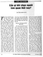
Fig. 1. Inactivation properties of NAADP-induced release. (A) Sea urchin eggs: The top panel illustrates the unusual phenomenon whereby in
sea urchin egg homogenates, a low concentration of NAADP (1 n
M) that induces no apparent Ca
2+
release (right hand trace), fully desensiti-
zes the NAADP receptor mechanism so that a subsequent application of a maximal concentration of NAADP (500 n
M, see left hand trace) is
now without effect [13,14]. (B) Mammalian cells: NAADP-induced Ca
2+
release in MIN6 cells. Percentage of the increase in normalized fluor-
escence (F ⁄ F
0
%) demonstrated for each caged NAADP concentration. The inset bar symbolizes the least significant difference (LSD) that
was calculated from observed errors. The numbers in brackets over the bars present number of replicates. Data are means ± SEM. The
graph shows a bell-shaped concentration-response curve which is typical in mammalian systems. Higher concentrations of NAADP inactivate
release by this messenger under conditions where little if any release occurs. Modified from [10].
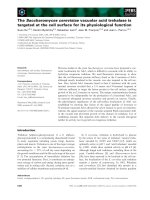
Fig. 2. NAADP-induced Ca
2+
release from a thapsigargin-insensitive
Ca
2+
store. Effect of thapsigargin on the initial response to photo-
release of an InsP
3
-cADPR mixture and NAADP. Eggs were treated
with thapsigargin (2 l
M) for > 30 min and then exposed to UV. The
final intracellular concentrations were (l
M): Oregon green 488
BAPTA Dextran, 10; caged NAADP, 0.5; and both caged cADPR
and caged InsP
3
, 5. Modified from [22].
M. Yamasaki et al. Calcium signalling by NAADP
FEBS Journal 272 (2005) 4598–4606 ª 2005 FEBS 4599
Ca
2+
release channels modulated by either InsP
3
or
cADPR that participate in Ca
2+
-induced Ca
2+
release
mechanism (CICR), the NAADP-sensitive Ca
2+
release
mechanism is unlikely to do so directly. The apparent
inability of NAADP to induce regenerative Ca
2+
signals itself implies a role in initiating localized Ca
2+
signals, which may then be propagated by recruiting
CICR mechanisms. Additional interactions of NAADP
signalling pathways with Ca
2+
signals may arise since
the metabolism of NAADP to inactive NAAD is regu-
lated by a Ca
2+
-dependent 2¢-phosphatase [30].
Radioligand binding studies employing [
32
P]NAADP
support the idea that NAADP acts on a fundamentally
different Ca
2+
releasing channel from those gated by
InsP
3
or cADPR. Binding of radiolabelled NAADP to
sea urchin egg homogenate membranes is highly speci-
fic [13,31,32] and is unaffected by InsP
3
or cADPR
[13,31]. Binding studies have revealed another peculiar
property of the NAADP receptor where NAADP
binds to its receptor in an essentially irreversible man-
ner in the sea urchin egg homogenates [13,31,32]. In
mammalian systems, however, [
32
P]NAADP binding to
membrane preparations from rat brain [31], rat heart
[25] and MIN-6 cells [10] is reversible. The apparent
irreversibility of NAADP binding in sea urchin egg
preparations is dependent on high K
+
concentrations
in the binding medium routinely used [15].
Ca
2+
mobilizing messengers and
multiple stores
Studies of the Ca
2+
mobilizing effects of NAADP in
intact cells have revealed that this Ca
2+
release mech-
anism rarely operates in isolation. Rather the resultant
Ca
2+
signals evoked by this molecule are often boos-
ted by Ca
2+
release by RyRs, InsP
3
Rs or both. Inter-
actions between different Ca
2+
release mechanisms are
critical for shaping Ca
2+
signals in response to agon-
ists in many different cell types [33]. The effects of
NAADP on Ca
2+
release are often abolished or
attenuated by both heparin and 8-NH
2
-cADPR, anta-
gonists for InsP
3
and cADPR receptors, indicating that
the different Ca
2+
release channels are tightly coupled
functionally. In sea urchin eggs, ascidian oocytes, and
arterial smooth muscle, antagonists of InsP
3
Rs or
RyRs reduce responses to NAADP [26,34,35], whereas
in T-lymphocytes, starfish oocytes and pancreatic aci-
nar cells, little effect of NAADP is seen in the presence
of these inhibitors [18,36–39]. To explain these phe-
nomena two models have currently been proposed.
The first describes a single pool, the ER, expressing
InsP
3
Rs and RyRs. Here NAADP interacts either
directly with RyRs or via a separate protein that may
indirectly activate RyRs [40,41]. This model accounts
for the apparent complete abolition of NAADP
evoked release by either RyR blockers or thapsigargin.
A direct action of NAADP on RyRs is also supported
by the findings that NAADP was shown to activate
isolated RyRs reconstituted in lipid bilayers from
rabbit skeletal muscle (RyR1) [42] and cardiac micro-
somes (RyR2) [43]. A second model, the two pool or
trigger hypothesis, is based on the idea that there is a
distinct NAADP-sensitive storage organelle, possibly
an thapsigargin-insensitive acidic store [28], that is
responsible for a localized signal which is amplified
by InsP
3
Rs and RyRs the on the ER by CICR
[22,34,36,38]. This model accounts for the finding in
some cells that localized NAADP-induced signals per-
sist in the presence of InsP
3
Rs and RyR antagonists or
thapsigargin, but are abolished by agents that dissipate
storage of by acidic organelles, such as the vacuolar
H
+
pump inhibitor, bafilomycin A1. This has been
most clearly demonstrated in the sea urchin egg [28],
but also extended to several mammalian cell types
[11,44–46]. Two types of pharmacological manipula-
tion of acidic stores have been investigated with regard
to NAADP-evoked release. Glycyl-phenylalanyl-naph-
thylamide (GPN) is an agent that penetrates cellu-
lar membranes but is a substrate for the luminal
lysosomal enzyme cathepsin C trapping membrane
impermeant products within lysosomes resulting in
disruption of lysosomal-related organelles by osmotic
lysis [47]. The other approach is aimed at collapsing
proton gradients thought to power Ca
2+
uptake into
acidic stores by Ca
2+
⁄ H
+
exchange, such as bafilo-
mycin A1, FCCP and NH
3
[48]. These agents selec-
tively inhibit NAADP-induced Ca
2+
release, whilst
having little effect on the effects of either InsP
3
or
cADPR [11,28,45,46].
Changes in endogenous levels of
NAADP
Only recently have NAADP levels been measured
directly by using a radioreceptor assay with the
NAADP binding protein from sea urchin eggs [9–
12,49] and shown to change in response to extracellu-
lar stimuli [9–12]. This provided the final piece of
evidence required to classify NAADP as a second mes-
senger. NAADP levels have been shown to change in
sea urchin sperm during activation before fertilization
[9], in pancreatic beta cells in response to glucose [10],
in smooth muscle cells in response to endothelin [11],
and in pancreatic acinar cells in response to
gut-peptide cholecystokinin [12], which has been the
most detailed study so far. As outlined above, mouse
Calcium signalling by NAADP M. Yamasaki et al.
4600 FEBS Journal 272 (2005) 4598–4606 ª 2005 FEBS
pancreatic acinar cells have been an important system
in which investigate mechanisms for the generation of
intracellular Ca
2+
signals. It has been suggested that
interactions between a subset and all three messengers
are used to generate specific Ca
2+
signatures in
response to extracellular agonists such as cholecysto-
kinin and neurotransmitter acetylcholine [20,38,39,50–
52]. In this cell type, it has been proposed that an
initial increase in NAADP in response to cholecysto-
kinin triggers a primary Ca
2+
release, followed by
recruitment of InsP
3
Rs and RyRs by CICR. Although
there is much circumstantial evidence from physiologi-
cal and pharmacological studies that cholecystokinin
increases NAADP and cADPR levels, changes in the
levels of NAADP or cADPR had not been character-
ized in response to this agonist until recently. We have
recently provided the strong evidence to establish
NAADP as a second messenger in pancreatic acinar
cells [12]. Significant elevations of both NAADP and
cADPR levels in response to a specific agonist, chole-
cystokinin, in a concentration-dependent manner were
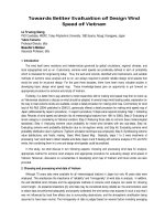
reported (Fig. 3). Cholecystokinin A receptors, expres-
sed on mouse pancreatic acinar cells, possess two bind-
ing sites for cholecystokinin, high and low-affinity
binding sites [53–55]. Concentration-response data sug-
gest that production of NAADP and cADPR can be
Fig. 3. Effects of cholecystokinin on NAADP and cADPR production in pancreatic acinar cells. (A) Time course of cholecystokinin induced
NAADP (r). Data were normalized to the maximum obtained with each individual time-course experiment. The NAADP levels reach a maxi-
mum within 10 s and return to resting levels in about 60 s (n ¼ 12). (B) Concentration-response curve for cholecystokinin-induced NAADP
increases (d). The data were filled to the Hill equation with two-sites (EC
50
s of 11.0 ± 3.0 pM and 830 ± 6.6 pM). Lorglumide, a cholecysto-
kinin A receptor agonist, was present at 10 l
M (n ¼ 3–6) (open triangles). (C) The time course of cADPR production (d) was determined in
the presence of physiological concentration of cholecystokinin (10 p
M). The production of cAPDR showed prolonged elevations comparing
that of NAADP. Lorglumide inhibited cADPR production (m). (D) Cholecystokinin-induced cADPR elevations (j) occur in a concentration-
dependent manner. Data are mean ± SEM. Modified from [12].
M. Yamasaki et al. Calcium signalling by NAADP
FEBS Journal 272 (2005) 4598–4606 ª 2005 FEBS 4601
activated through both high and low-affinity sites on
cholecystokinin A receptor. This same study also dem-
onstrated receptor specificity for the production of
NAADP and cADPR, whereby increases in cADPR
levels via stimulation of acetylcholine muscarinic recep-
tors as well as cholecystokinin A receptors, whereas
NAADP increased only through the activation of chol-
ecystokinin A. Intriguingly, the striking difference seen
in time courses between NAADP and cADPR pro-
duction, where the increase in NAADP was rapid
and transient, whereas the increase in cADPR was
much prolonged, strongly supports the proposed
hypothesis that NAADP provides a localized Ca
2+
trigger signal at the apical region where InsP
3
Rs, RyRs
and NAADP-sensitive Ca
2+
stores coexist [45,56–62]
(Fig. 4), and subsequently this localized Ca
2+
signal is
amplified by a CICR mechanism via sensitization of
RyRs throughout of the cell [20,38,39,50–52,60–63].
Cell surface receptors are predominantly located in the
basolateral membrane, however, agonist-induced Ca
2+
signals initiate at the apical pole before propagating
into the basolateral domain by the CICR mechanism.
The abundance of RyRs in the basolateral region
together with the slow rise in the cADPR levels dem-
onstrated in our recent report [12] may also contribute
greatly to such spatiotemporal heterogeneities of Ca
2+
signals.
Although there are few reports of direct NAADP
measurements, an interesting correlation is emerging.
Inhibition of agonist-evoked signalling by inactivating
NAADP concentrations or bafilomycin A1 correlates
well with receptors whose stimulation leads to eleva-
tions in NAADP levels, whereas those that are not
sensitive to these pharmacological manipulations are
not [11,12,45].
Outstanding questions in NAADP
signalling pathways
There are still several important aspects of the NAADP
signalling pathway that are unclear. Foremost is
the nature of the NAADP receptor. Studies from the
sea urchin egg system have suggested that NAADP
probably acts on a distinct protein that is pharmacolo-
gically different from IsnP
3
Rs of RyRs [64], although
direct activation of RyRs has also been proposed [64].
The kinetics of Ca
2+
release evoked by NAADP are
consistent with the gating of a Ca
2+
release channel
rather than a transporter protein [65]. Preliminary
biochemical characterization of [
32
P]NAADP binding
proteins from sea urchin eggs have shown that such
proteins are likely to be integral membrane proteins,
and probably smaller than either InsP
3
Rs or RyRs [30].
However, it is possible that the NAADP binding pro-
teins may not form a pore themselves but rather inter-
act with and modulate other channels. Further, in line
with the multiplicity of InsP
3
Rs and RyR isoforms, it is
also possible that multiple isoforms of NAADP ‘recep-
tors’ exist which may go some way in reconciling con-
flicting pharmacological data from different systems
[64]. Perhaps the most detailed study of the functional
properties of NAADP receptors, in the absence of
their molecular isolation, has come from the study of
NAADP signalling in starfish oocytes [37,66]. Here
much emphasis has been placed on the ability of
NAADP to gate a cation influx in addition to release
from internal stores. In contrast to the situation in sea
urchin eggs where NAADP induces a brief Ca
2+
influx
(the ‘cortical flash’), followed by a more substantial
mobilization [9], the starfish oocytes exhibits a pro-
found Ca
2+
influx of in response to NAADP. It
has been proposed that NAADP receptors may be
expressed at the plasma membrane of these cells, and
thus electrophysiological analyses have been employed
to characterize such NAADP-induced currents [67]. An
interesting question is whether these currents arise from
direct activation of NAADP receptors on the plasma
membrane or activation of a plasma membrane channel
via calcium released from cortical NAADP-sensitive
stores. Taken together these observations imply the
widespread distribution of NAADP receptors and
multiple roles of NAADP. However, the ultimate
Fig. 4. Localization of NAADP-induced Ca
2+
signals in mouse pan-
creatic acinar cells. Pancreatic acinar cells were injected with Ore-
gon Green 488 BAPTA Dextran and caged NAADP (estimated final
concentration: 100 n
M). In response photoreleased NAADP, the
local Ca
2+
spikes were confined to the apical pole (blue trace), but
not to the basal pole (red trace) (n ¼ 8). These localized Ca
2+
spikes become progressively amplified. Modified from [45].
Calcium signalling by NAADP M. Yamasaki et al.
4602 FEBS Journal 272 (2005) 4598–4606 ª 2005 FEBS
resolution of many these questions will require isolation
of NAADP-binding proteins.
It may come as some surprise that the biosynthetic
pathway for NAADP synthesis is still unknown.
Endogenous levels have been reported in several cell
types and in some of these changes in levels in
response to agonists have been reported. A favoured
pathway for synthesis involves enzymes known as
ADP-ribosyl cyclases (Fig. 5). As the name suggests
these where first described as activities and then char-
acterized as membrane proteins that cyclized NAD to
form cADPR. In mammalian systems the best-charac-
terized cyclases so far are CD38 and CD157 (BST-1)
(Fig. 5), although other membrane-bound and soluble
forms have been reported [68]. However, in vitro, these
multifunctional enzymes can utilize NADP as an alter-
native substrate, and at acidic pH and in the presence
of nicotinic acid, they can catalyse NAADP synthesis
by a mechanism involving base exchange (Fig. 5).
Whether CD38 or CD157 are responsible for agonist-
promoted NAADP synthesis in mammalian cells
remains to be demonstrated. Two potential problems
may confound this possibility. The first is that both
CD38 and CD157 are largely ectoenzymes, although
several reports suggest intracellular localizations as
well. The second is that large, and perhaps nonphysio-
logical, concentrations of nicotinic acid are required
for any appreciable NAADP production by these
enzymes, although high localized concentrations may
be postulated. One possibility that has not been
explored is that synthesis of NAADP may occur inside
intracellular vesicles, or even extracellularly (as has
been proposed for cADPR [69]), with the products
then being transported back into the cytoplasm where
it can interact with its putative targets. If these vesicles
were also the acidic stores proposed as major sites for
NAADP-evoked release [28], then the pH of the lumi-
nal environment would also promote NAADP synthe-
sis, and could also be a site for the accumulation
of nicotinic acid. However, endogenous changes in
NAADP levels have been reported in several systems,
and we are now in a position to elucidate the mecha-
nisms of NAADP production. For example, an investi-
gation of agonist-induced NAADP changes in cells
and tissues from CD38 ⁄ CD157 knockout mice may be
informative. Other possibilities for NAADP synthetic
pathways also deserving investigation are phosphory-
lation of NAAD, better known as a biosynthetic pre-
cursor of NAD, or a direct deamination reaction of
NADP.
The identification of the enzymes involved in
NAADP synthesis with the recent identification of
various agonists that stimulate increases in cellular
NAADP levels may also lead to an understanding of
the coupling mechanisms between cell surface receptors
and NAADP production. Both activation of the chole-
cystokinin A receptor and the ET-1 receptor have been
shown to couple to NAADP synthesis. As these recep-
tors are G protein coupled it is possible that G protein
subunits may directly regulate enzymes catalysing
NAADP production. In addition, the finding that
cAMP stimulates the NAADP synthesis in the pres-
ence of sea urchin membranes [70] may indicate an
involvement of downstream regulators (Fig. 5).
Summary
NAADP has been reported to be an endogenous and
potent Ca
2+
mobilizing agent in several cell types of
many different organisms. NAADP evokes localized
signals, which may be amplified by recruiting InsP
3
Rs
and RyRs through CICR mechanisms. Changes in
NAADP levels are linked to the activation of several
cell surface receptors. All the criteria have now been
satisfied for its recognition as an intracellular messen-
ger, however, further studies required in the future are
to establish the cellular mechanisms for the regulation
of NAADP synthesis and metabolism as well as the
molecular mechanisms mediating NAADP-induced
Ca
2+
release.
Fig. 5. Putative synthesis pathway for
NAADP. In the presence of b-NADP, ADP-
ribosyl cyclase catalyses the synthesis of
NAADP by a base exchange reaction with
an optimum pH of 4 [71]. CD38 and CD157
have been shown to be capable of forming
NAADP under the same condition [71–73].
cAMP is a stimulator of NAADP synthesis
via ADP-ribosyl cyclase [70].
M. Yamasaki et al. Calcium signalling by NAADP
FEBS Journal 272 (2005) 4598–4606 ª 2005 FEBS 4603
Acknowledgements
AG is a Wellcome Trust Senior Fellow in Basic Bio-
medical Science; MY is a Wellcome Trust Prize Stu-
dent. Work in AG and GCC’s laboratories is funded
by the Wellcome Trust.
References
1 Berridge MJ (1993) Inositol trisphosphate and calcium
signalling. Nature 361, 315–325.
2 Thomas AP, Bird GST, Hajnoczky G, Robb-Gaspers
LD & Putney J (1996) Spatial and temporal aspects of
cellular calcium signaling. FASEB J 10, 1505–1517.
3 Berridge MJ, Lipp P & Bootman MD (2000) The versa-
tility and universality of calcium signalling. Nat Mol
Cell Biol Rev 1, 11–21.
4 Clapper DL, Walseth TF, Dargie PJ & Lee HC (1987)
Pyridine nucleotide metabolites stimulate calcium release
from sea urchin egg microsomes desensitized to inositol
trisphosphate. J Biol Chem 262 , 9561–9568.
5 Lee HC, Walseth TF, Bratt GT, Hayes RN & Clapper
DL (1989) Structural determination of a cyclic metabo-
lite of NAD with intracellular calcium-mobilizing activ-
ity. J Biol Chem 264, 1608–1615.
6 Lee HC, Aarhus R & Graeff RM (1995) Sensitization
of calcium-induced calcium release by cyclic ADP-ribose
and calmodulin. J Biol Chem 270, 9060–9066.
7 Galione A (2002) Regulation of synthesis of cADPR
and NAADP. In Cyclic ADP-Ribose and NAADP:
Structures, Metabolism and Functions. (Lee HC, ed),
pp. 45–64. Kluwer, Dordrecht, the Netherlands.
8 Galione A & Churchill GC (2000) Cyclic ADP ribose as
a calcium-mobilizing messenger. Sci STKE 2000, PE1.
9 Churchill GC, O’Neill JS, Masgrau R, Patel S,
Thomas JM, Genazzani AA & Galione A (2003)
Sperm deliver a new second messenger. NAADP. Curr
Biol 13, 125–128.
10 Masgrau R, Churchill GC, Morgan AJ, Ashcroft SJ &
Galione A (2003) NAADP. A new second messenger
for glucose-induced Ca
2+
responses in clonal pancreatic
beta cells. Curr Biol 13, 247–251.
11 Kinnear NP, Boittin FX, Thomas JM, Galione A &
Evans AM (2004) Lysosome-sarcoplasmic reticulum
junctions: a trigger zone for calcium signalling by
NAADP and endothelin-1. J Biol Chem 279, 54319–
54326.
12 Yamasaki M, Thomas JM, Churchill GC, Garnham C,
Lewis AM, Cancela J-M, Patel S & Galione A (2005)
Role of NAADP and cADPR in the induction and
maintenance of agonist-evoked Ca
2+
spiking in mouse
pancreatic acinar cells. Curr Biol 15, 874–878.
13 Aarhus R, Dickey DM, Graeff RM, Gee KR, Walseth
TF & Lee HC (1996) Activation and inactivation of
Ca
2+
release by NAADP
+
. J Biol Chem 271 , 8513–
8516.
14 Genazzani AA, Empson RM & Galione A (1996)
Unique inactivation properties of NAADP-sensitive
Ca
2+
release. J Biol Chem 271, 11599–11602.
15 Dickinson GD & Patel S (2003) Modulation of
NAADP receptors by K
+
ions: evidence for multiple
NAADP receptor conformations. Biochem J 375, 805–
812.
16 Navazio L, Bewell MA, Siddiqua A, Dickinson GD,
Galione A & Sanders D (2000) Calcium release from
the endoplasmic reticulum of higher plants elicited by
the NADP metabolite nicotinic acid adenine dinucleo-
tide phosphate. Proc Natl Acad Sci USA 97, 8693–8698.
17 Johnson JD & Misler S (2002) Nicotinic acid-adenine
dinucleotide phosphate-sensitive calcium stores initiate
insulin signaling in human beta cells. Proc Natl Acad
Sci USA 99, 14566–14571.
18 Berg I, Potter BV, Mayr GW & Guse AH (2000) Nico-
tinic acid adenine dinucleotide phosphate (NAADP
+
)is
an essential regulator of T-lymphocyte Ca
2+
-signaling.
J Cell Biol 150, 581–588.
19 Lee HC (2001) Physiological functions of cyclic ADP-
ribose and NAADP as calcium messengers. Annu Rev
Pharmacol Toxicol 41, 317–345.
20 Patel S (2004) NAADP-induced Ca
2+
release – a new
signalling pathway. Biol Cell 9, 19–28.
21 Genazzani AA, Mezna M, Dickey DM, Michelangeli F,
Walseth TF & Galione A (1997) Pharmacological prop-
erties of the Ca
2+
-release mechanism sensitive to
NAADP in the sea urchin egg. Br J Pharmacol 121,
1489–1495.
22 Churchill GC & Galione A (2001) NAADP induces
Ca
2+
oscillations via a two-pool mechanism by priming
IP
3
- and cADPR-sensitive Ca
2+
stores. EMBO J 20,
2666–2671.
23 Churchill GC, Patel S, Thomas JM & Galione A (2002)
Spatial and temporal control of Ca
2+
signalling by
NAADP. In Cyclic ADP-Ribose and NAADP: Struc-
tures, Metabolism and Functions. (Lee HC, ed), pp. 199–
215. Kluwer, Dordrecht, the Netherlands.
24 Bak J, White P, Timar G, Missiaen L, Genazzani AA &
Galione A (1999) Nicotinic acid adenine dinucleotide
phosphate triggers Ca
2+
release from brain microsomes.
Curr Biol 9, 751–754.
25 Bak J, Billington RA, Timar G, Dutton AC & Genaz-
zani AA (2001) NAADP receptors are present and func-
tional in the heart. Curr Biol 11, 987–990.
26 Boittin FX, Galione A & Evans AM (2002) Nicotinic
acid adenine dinucleotide phosphate mediates Ca
2+
sig-
nals and contraction in arterial smooth muscle via a
two-pool mechanism. Circ Res 91, 1168–1175.
27 Lee HC & Aarhus R (2000) Functional visualization of
the separate but interacting calcium stores sensitive to
Calcium signalling by NAADP M. Yamasaki et al.
4604 FEBS Journal 272 (2005) 4598–4606 ª 2005 FEBS
NAADP and cyclic ADP-ribose. J Cell Sci 113, 4413–
4420.
28 Churchill GC, Okada Y, Thomas JM, Genazzani AA,
Patel S & Galione A (2002) NAADP mobilizes Ca
2+
from reserve granules, a lysosome-related organelle, in
sea urchin eggs. Cell 111, 703–708.
29 Chini EN & Dousa TP (1996) Nicotinate-adenine dinu-
cleotide phosphate-induced Ca
2+
-release does not
behave as a Ca
2+
-induced Ca
2+
-release system. Bio-
chem J 316, 709–711.
30 Berridge G, Dickinson G, Parrington J, Galione A &
Patel S (2002) Solubilization of receptors for the novel
Ca
2+
-mobilizing messenger, nicotinic acid adenine di-
nucleotide phosphate. J Biol Chem 277, 43717–43723.
31 Patel S, Churchill GC & Galione A (2000) Unique
kinetics of nicotinic acid–adenine dinucleotide phos-
phate (NAADP) binding enhance the sensitivity of
NAADP receptors for their ligand. Biochem J 352 Part
3, 725–729.
32 Billington RA & Genazzani AA (2000) Characterization
of NAADP
+
binding in sea urchin eggs. Biochem Bio-
phys Res Commun 276, 112–116.
33 Galione A & Churchill GC (2002) Interactions between
calcium release pathways: multiple messengers and mul-
tiple stores. Cell Calcium 32, 343–354.
34 Churchill GC & Galione A (2000) Spatial control of
Ca
2+
signaling by nicotinic acid adenine dinucleotide
phosphate diffusion and gradients. J Biol Chem 275,
38687–38692.
35 Albrieux M, Lee HC & Villaz M (1998) Calcium signal-
ing by cyclic ADP-ribose, NAADP, and inositol tri-
sphosphate are involved in distinct functions in ascidian
oocytes. J Biol Chem 273, 14566–14574.
36 Cancela JM, Churchill GC & Galione A (1999) Coordi-
nation of agonist-induced Ca
2+
-signalling patterns by
NAADP in pancreatic acinar cells. Nature 398, 74–76.
37 Santella L, Kyozuka K, Genazzani AA, De Riso L &
Carafoli E (2000) Nicotinic acid adenine dinucleotide
phosphate-induced Ca
2+
release. Interactions among
distinct Ca
2+
mobilizing mechanisms in starfish oocytes.
J Biol Chem 275, 8301–8306.
38 Cancela JM, Gerasimenko OV, Gerasimenko JV, Tepi-
kin AV & Petersen OH (2000) Two different but con-
verging messenger pathways to intracellular Ca
2+
release: the roles of nicotinic acid adenine dinucleotide
phosphate, cyclic ADP-ribose and inositol trisphos-
phate. EMBO J 19, 2549–2557.
39 Cancela JM, Van Coppenolle F, Galione A, Tepikin
AV & Petersen OH (2002) Transformation of local
Ca
2+
spikes to global Ca
2+
transients: the combinato-
rial roles of multiple Ca
2+
releasing messengers. EMBO
J 21, 909–919.
40 Gerasimenko JV, Maruyama Y, Yano K, Dolman NJ,
Tepikin AV, Petersen OH & Gerasimenko OV (2003)
NAADP mobilizes Ca
2+
from a thapsigargin-sensitive
store in the nuclear envelope by activating ryanodine
receptors. J Cell Biol 163, 271–282.
41 Dammermann W & Guse AH (2005) Functional ryano-
dine receptor expression is required for NAADP-
mediated local Ca
2+
signaling in T-lymphocytes. J Biol
Chem 280, 21394–21399.
42 Hohenegger M, Suko J, Gscheidlinger R, Drobny H &
Zidar A (2002) Nicotinic acid adenine dinucleotide
phosphate, NAADP, activates the skeletal muscle rya-
nodine receptor. Biochem J 367, 423–431.
43 Mojzisova A, Krizanova O, Zacikova L, Kominkova V
& Ondrias K (2001) Effect of nicotinic acid adenine
dinucleotide phosphate on ryanodine calcium release
channel in heart. Pflu
¨
g Arch 441, 674–677.
44 Mitchell KJ, Lai FA & Rutter GA (2003) Ryanodine
receptor type I and nicotinic acid adenine dinucleotide
phosphate (NAADP) receptors mediate Ca
2+
release
from insulin-containing vesicles in living pancreatic
beta-cells (MIN6). J Biol Chem 278, 11057–11064.
45 Yamasaki M, Masgrau R, Morgan AJ, Churchill GC,
Patel S, Ashcroft SJ & Galione A (2004) Organelle
selection determines agonist-specific Ca
2+
signals in
pancreatic acinar and beta cells. J Biol Chem 279, 7234–
7240.
46 Brailoiu E, Hoard JL, Filipeanu CM, Brailoiu GC, Dun
SL, Patel S & Dun NJ (2005) NAADP potentiates neur-
ite outgrowth. J Biol Chem 280, 5646–5650.
47 Jadot M, Andrianaivo F, Dubois F & Wattiaux R
(2001) Effects of methylcyclodextrin on lysosomes. Eur
J Biochem 268, 1392–1399.
48 Docampo R & Moreno SN (1999) Acidocalcisome: a
novel Ca
2+
storage compartment in trypanosomatids
and apicomplexan parasites. Parasitol Today 15, 443–
448.
49 Billington RA, Ho A & Genazzani AA (2002) Nicotinic
acid adenine dinucleotide phosphate (NAADP) is pre-
sent at micromolar concentrations in sea urchin sperma-
tozoa. J Physiol 544, 107–112.
50 Patel S, Churchill GC & Galione A (2001) Coordination
of Ca
2+
signalling by NAADP. Trends Biochem Sci 26,
482–489.
51 Cancela JM (2001) Specific Ca
2+
signaling evoked by
cholecystokinin and acetylcholine: the roles of NAADP,
cADPR, and IP
3
. Annu Rev Physiol 63, 99–117.
52 Cancela JM, Charpentier G & Petersen OH (2003) Co-
ordination of Ca
2+
signalling in mammalian cells by the
new Ca
2+
-releasing messenger NAADP. Pflu
¨
g Arch
446, 322–327.
53 Yule DI & Williams JA (1994) Stimulus-secretion cou-
pling in the pancreatic acinus. Physiology of the Gastro-
intestinal Tract (Johnson, L R, ed.), pp. 221–242. Raven
Press, New York.
54 Jensen RT (1994) Receptors on pancreatic acinar cells.
In Physiology of the Gastrointestinal Tract (Johnson LR,
ed), pp. 1377–1446. Raven Press, New York, USA.
M. Yamasaki et al. Calcium signalling by NAADP
FEBS Journal 272 (2005) 4598–4606 ª 2005 FEBS 4605
55 Matozaki T, Goke B, Tsunoda Y, Rodriguez M, Marti-
nez J & Williams JA (1990) Two functionally distinct
cholecystokinin receptors show different modes of
action on Ca
2+
mobilization and phospholipid hydro-
lysis in isolated rat pancreatic acini: studies using a new
cholecystokinin analog, JMV-180. J Biol Chem 15,
6247–6254.
56 Nathanson MH, Fallon MB, Padfield PJ & Maranto
AR (1994) Localization of the type 3 inositol 1,4,5-tris-
phosphate receptor in the Ca
2+
wave trigger zone of
pancreatic acinar cells. J Biol Chem 269, 4693–4696.
57 Gerasimenko OV, Gerasimenko JV, Belan PV &
Petersen OH (1996) Inositol trisphosphate and cyclic
ADP-ribose-mediated release of Ca2+ from single
isolated pancreatic zymogen granules. Cell 84, 473–
480.
58 Yule DI, Ernst SA, Ohnishi H & Wojcikiewicz RJ
(1997) Evidence that zymogen granules are not a phy-
siologically relevant calcium pool: defining the distribu-
tion of inositol 1,4,5-trisphosphate receptors in
pancreatic acinar cells. J Biol Chem 272, 9093–9098.
59 Lee MG, Xu X, Zeng W, Diaz J, Wojcikiewicz RJ, Kuo
TH, Wuytack F, Racymaekers L & Muallem S (1997)
Polarized expression of Ca
2+
channels in pancreatic and
salivary gland cells: correlation with initiation and pro-
pagation of [Ca
2+
]
i
waves. J Biol Chem 272, 15765–
15770.
60 Leite MF, Dranoff JA, Gao L & Nathanson MH
(1999) Expression and subcellular localization of the
ryanodine receptor in rat pancreatic acinar cells. Bio-
chem J 337, 305–309.
61 Leite MF, Burgstahler AD & Nathanson MH (2002)
Ca
2+
waves require sequential activation of inositol tris-
phosphate receptors and ryanodine receptors in pancre-
atic acini. Gastroenterology 122, 415–427.
62 Fitzsimmons TJ, Gukovsky I, McRoberts JA, Rodri-
guez E, Lai FA & Pandol SJ (2000) Multiple isoforms
of the ryanodine receptor are expressed in rat pancreatic
acinar cells. Biochem J 351, 265–271.
63 Straub SV, Giovannucci DR & Yule DI (2000) Calcium
wave propagation in pancreatic acinar cells: functional
interaction of inositol 1,4,5-trisphosphate receptors,
ryanodine receptors, and mitochondria. J Gen Physiol
116, 547–560.
64 Galione A & Petersen OH (2005) The NAADP recep-
tor: new receptors or new regulation? Mol Interv 5,
73–79.
65 Genazzani AA, Mezna M, Summerhill RJ, Galione A &
Michelangeli F (1997) Kinetic properties of nicotinic
acid adenine dinucleotide phosphate-induced Ca
2+
release. J Biol Chem 272, 7669–7675.
66 Lim D, Kyozuka K, Gragnaniello G, Carafoli E & San-
tella L (2001) NAADP
+
initiates the Ca
2+
response
during fertilization of starfish oocytes. FASEB J 15,
2257–2267.
67 Moccia F, Lim D, Kyozuka K & Santella L (2004)
NAADP triggers the fertilization potential in starfish
oocytes. Cell Calcium 36, 515–524.
68 Berger F, Ramirez-Hernandez MH & Ziegler M (2004)
The new life of a centenarian: signalling functions of
NAD(P). Trends Biochem Sci 29, 111–118.
69 De Flora A, Zocchi E, Guida L, Franco L & Bruzzone
S (2004) Autocrine and paracrine calcium signaling by
the CD38 ⁄ NAD+ ⁄ Cyclic ADP-ribose system. Ann NY
Acad Sci 1028, 176–191.
70 Wilson HL & Galione A (1998) Differential regulation
of nicotinic acid-adenine dinucleotide phosphate and
cADP-ribose production by cAMP and cGMP. Biochem
J 331, 837–843.
71 Aarhus R, Graeff RM, Dickey DM, Walseth TF & Lee
HC (1995) ADP-ribosyl cyclase and CD38 catalyze the
synthesis of a calcium- mobilizing metabolite from
NADP. J Biol Chem 270, 30327–30333.
72 Hirata Y, Kimura N, Sato K, Ohsugi Y, Takasawa S,
Okamoto H, Ishikawa J, Kaisho T, Ishihara K & Hir-
ano T (1996) ADP ribosyl cyclase activity of a novel
bone marrow stromal cell surface molecule, BST-1.
FEBS Lett 356, 244–248.
73 Itoh M, Ishihara K, Hiroi T, Lee BO, Maeda H, Iijima
H, Yanagita M, Kiyono H & Hirano T (1998) Deletion
of bone marrow stromal cell antigen-1 (CD157) gene
impaired systemic thymus independent-2 antigen-
induced IgG3 and mucosal TD antigen-elicited IgA
responses. J Immunol 161, 3974–3983.
Calcium signalling by NAADP M. Yamasaki et al.
4606 FEBS Journal 272 (2005) 4598–4606 ª 2005 FEBS