Tài liệu Báo cáo khoa học: Type I antifreeze proteins expressed in snailfish skin are identical to their plasma counterparts doc
Bạn đang xem bản rút gọn của tài liệu. Xem và tải ngay bản đầy đủ của tài liệu tại đây (363.26 KB, 10 trang )
Type I antifreeze proteins expressed in snailfish skin are
identical to their plasma counterparts
Robert P. Evans and Garth L. Fletcher
Ocean Sciences Centre, Memorial University of Newfoundland, St. John’s, Newfoundland, Canada
Teleost fish that inhabit icy seawater synthesize anti-
freeze proteins ⁄ polypeptides (AFPs) or antifreeze glyco-
proteins (AFGPs) for protection against freezing.
Diverse species from numerous taxonomic groups pro-
duce AFPs that are grouped into four distinct classes
(types I, II, III and IV) based on their primary and
secondary structural characteristics [1–3]. Regardless of
protein structure, all fish AFPs lower the solution
freezing point noncolligatively by binding to certain
surfaces of ice crystals, modifying their structure and
inhibiting further growth. The difference between the
lowered freezing point and unaltered melting point is
termed thermal hysteresis and is used as a measure of
antifreeze activity [1,3,4].
Of the four classes of AFPs described thus far, the
simplest is type I AFP found in right-eye flounders
(Pleuronectes) and a few sculpin species (e.g. Myoxo-
cephalus). These polypeptides have high alanine con-
tent (> 60 mol%), have an amphipathic a-helical
secondary structure, and are usually quite small (3.3–
4.5 kDa) [2,5]. Until the past decade, it was generally
accepted that the synthesis of AFPs was confined
solely to liver tissue (termed liver type) for secretion
into blood for extracellular freeze protection. How-
ever, more recently, a novel subclass of type I
AFPs was isolated and characterized from the skin of
winter flounder Pseudopleuronectes americanus (for-
merly Pleuronectes americanus) [6]. These AFPs, which
Keywords
antifreeze; cDNA; protein expression;
snailfish; type I
Correspondence
R. P. Evans, Department of Biochemistry,
University of Alberta, Edmonton, Alberta
T6G 2H7, Canada
Fax: +1 780 492 0886
Tel: +1 780 492 3481
E-mail:
(Received 13 June 2005, revised 8 August
2005, accepted 22 August 2005)
doi:10.1111/j.1742-4658.2005.04929.x
Type I antifreeze proteins (AFPs) are usually small, Ala-rich a-helical poly-
peptides found in right-eyed flounders and certain species of sculpin. These
proteins are divided into two distinct subclasses, liver type and skin type,
which are encoded by separate gene families. Blood plasma from Atlantic
(Liparis atlanticus) and dusky (Liparis gibbus) snailfish contain type I AFPs
that are significantly larger than all previously described type I AFPs. In
this study, full-length cDNA clones that encode snailfish type I AFPs
expressed in skin tissues were generated using a combination of library
screening and PCR-based methods. The skin clones, which lack both signal
and pro-sequences, produce proteins that are identical to circulating plasma
AFPs. Although all fish examined consistently express antifreeze mRNA in
skin tissue, there is extreme individual variation in liver expression – an
unusual phenomenon that has never been reported previously. Further-
more, genomic Southern blot analysis revealed that snailfish AFPs are
products of multigene families that consist of up to 10 gene copies per
genome. The 113-residue snailfish AFPs do not contain any obvious amino
acid repeats or continuous hydrophobic face which typify the structure of
most other type I AFPs. These structural differences might have implica-
tions for their ice-crystal binding properties. These results are the first to
demonstrate a dual liver ⁄ skin role of identical type I AFP expression which
may represent an evolutionary intermediate prior to divergence into distinct
gene families.
Abbreviations
AFGPs, antifreeze glycoproteins; AFPs, antifreeze proteins ⁄ polypeptides; IBM, ice-binding motif; ORF, open reading frame;
UTR, untranslated region.
FEBS Journal 272 (2005) 5327–5336 ª 2005 FEBS 5327
are encoded by a separate subset of genes, were desig-
nated as skin-type AFPs. They are synthesized as
mature polypeptides that lack both signal and pro-
sequences, which suggests that they remain intracellu-
lar [6]. Recent publications of skin-type AFP isolation
from shorthorn (Myoxocephalus scorpius) and long-
horn (M. octodecemspinosus) sculpins indicate that the
production of AFP in peripheral epithelial tissues may
be a common trait in many fish species [7,8]. The char-
acterization of known skin-type AFPs and the presence
of antifreeze activity in skin tissues of other species has
led to the hypothesis that skin-type AFPs are wide-
spread ancestors of liver-type (plasma) AFPs [6,9].
Atlantic snailfish (Liparis atlanticus) and dusky
snailfish (L. gibbus) belong to a large family (Cyclo-
pteridae) of benthic and pelagic marine fishes that
inhabit northern regions of the Atlantic Ocean. Snail-
fish are closely related to sculpins, which belong to a
different family of the same order Scorpaeniformes
[10]. Both species spawn during the winter months in
ice-laden inshore coastal regions around Newfound-
land, which makes them prime candidates for produc-
tion of AFPs. Type I AFPs were previously isolated
and characterized from the blood plasma of both
Atlantic and dusky snailfish which are the largest des-
cribed to date (> 9.3 kDa) [11]. We have also shown
recently that Atlantic snailfish skin tissues contain
type I AFPs that have identical molecular mass and
very similar amino acid content to their plasma counter-
parts [12].
Further analysis of the snailfish AFPs would be
helpful in determining the structure ⁄ function charac-
teristics of these unusually large type I AFPs and to
clarify the relationship between skin and plasma pro-
teins. Pursuant to this, an Atlantic snailfish skin
cDNA library was screened using a shorthorn sculpin
skin-type AFP clone as a probe. Full-length cDNA
sequences of both Atlantic and dusky snailfish skin
type I AFPs were generated using a combination of
library clones with RT-PCR and RACE techniques.
Results from this study show that skin AFPs from
both species are nearly identical to each other and
their skin transcripts produce proteins that are identi-
cal to their corresponding plasma proteins.
Results
cDNA library screening and cloning of snailfish
skin AFP
A cDNA library was constructed to investigate the
presence of type I AFP mRNA in skin tissues. Two
independent clones were identified from the library
screen using the open reading frame (ORF) of a
shorthorn sculpin skin cDNA as a probe. The
260 bp clones (clone-c1 and clone-c2) contained
identical sequences, apart from a small difference in
the length of poly(A) tail and a few nucleotides at their
5¢ ends. However, the clones appeared to be truncated
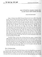
versions of complete type I AFP messages. As indica-
ted by the underline in Fig. 1, one reading frame gave
an Ala-rich 26 amino acid peptide, that lacks an
obligatory in-frame start codon. This sequence infor-
mation was then used in 5¢-RACE reactions to ascer-
tain the remainder of the skin AFP cDNA sequence.
RNA ligase-mediated RACE was used to clone the
remaining 5¢ portion of the snailfish AFP cDNA. The
full L. atlanticus skin cDNA is 568 bp and contains a
complete 342 bp ORF (Fig. 1). The ORF encodes an
Ala-rich protein of 113 residues and was designated as
Las-AFP (L. atlanticus skin AFP). The putative start
and stop codons are underlined as well as three pos-
sible polyadenylation signal sequences [13].
The Las-AFP sequence was utilized to design appro-
priate RT-PCR and 3¢)5¢-RACE primers for de novo
cloning of AFP sequence from dusky snailfish skin
RNA. The 3¢-RACE procedure (primers indicated in
Fig. 2) produced a single band that was 450 bp,
whereas 5¢-RACE gave a 370 bp product. The overlap-
ping sequences were combined into a 587 bp clone
which contained a 342 bp ORF that encodes a 113
residue, Ala-rich, protein designated as Lgs-AFP
(L. gibbus skin AFP). The putative start and stop
codons are underlined along with the standard poly-
adenylation signal sequence.
The AFP cDNAs cloned from the skin tissues of
Atlantic and dusky snailfish have strikingly similar nuc-
leotide sequences that encode nearly identical proteins
apart from a few minor differences. However, there is a
19 bp insertion in the Lgs-AFP 3¢-untranslated region
(UTR) just before the poly(A) tail region. Snailfish
AFP cDNA sequences are similar to skin-type AFPs
from winter flounder and sculpins in that they do not
appear to contain a signal sequence or pro-sequences
[6–8].
Surprisingly, the amino acid composition of proteins
purified from Atlantic snailfish skin tissue is quite sim-
ilar to the AFP predicted from the cDNA sequence
(Table 1). Any differences may be attributed to varia-
tions in analytical procedures and the fact that mix-
tures of AFPs were analyzed. Most importantly, the
predicted molecular mass and N-terminal sequence for
Las-AFP is identical to the isolated plasma proteins
[11]. Dusky snailfish also express the same type I AFP
in skin tissue that is circulating in their blood
(Table 1).
Expression of snailfish type I AFPs in skin tissue R. P. Evans and G. L. Fletcher
5328 FEBS Journal 272 (2005) 5327–5336 ª 2005 FEBS
Analysis of snailfish AFP genes
Total RNA from Atlantic snailfish tissues were probed
with a section from the 3¢-UTR of Las-AFP to evalu-
ate the distribution of snailfish AFP mRNA (Fig. 3A).
A specific band was visible after a short exposure in
skin tissue as well as a faint signal from gill is detect-
able with longer exposures. No other tissues gave
detectable signals on this northern blot. Similar results
were observed in another fish, except that there was a
Fig. 1. Nucleotide sequence and primary
translation product of L. atlanticus skin AFP
cDNA. The ORF is capitalized, whereas the
5¢- and 3¢-UTRs are in lower case letters.
The putative start and stop codons are
underlined in bold as are three possible
polyadenylation signal sequences. The
sequence obtained from the initial las-c1
and las-c2 cDNA clones are underlined.
RT-PCR or RACE primer sequences are
shown above (5¢fi3¢) or below (3¢fi5¢)
the nucleotide sequence. GenBank
Accession Number AY455862.
Fig. 2. Nucleotide sequence and primary
translation product of L. gibbus skin AFP
cDNA. The ORF is capitalized, whereas the
5¢- and 3¢-UTRs are in lower case letters.
The putative start and stop codons and the
polyadenylation signal are underlined.
RT-PCR or RACE primer sequences are
shown above (5¢fi3¢) or below (3¢fi5¢)
the nucleotide sequence. GenBank
Accession Number AY455863.
R. P. Evans and G. L. Fletcher Expression of snailfish type I AFPs in skin tissue
FEBS Journal 272 (2005) 5327–5336 ª 2005 FEBS 5329
definite detectable signal in liver tissue RNA (Fig. 3B).
RT-PCR performed using the same RNA (with ORF
primer set) gave positive bands for skin, gill, blood
cells, kidney, spleen and liver (Fig. 3C) and experi-
ments using 3¢-UTR primers gave the same expression
pattern (data not shown).
Additional northern blot experiments with 3¢-UTR
DNA probes gave very intense autoradiographic sig-
nals in skin tissues from four Atlantic snailfish and a
dusky snailfish (Fig. 4A), these were confirmed by
RT-PCR analysis (Fig. 4B). Results of a northern blot
using liver RNA from eight individual Atlantic snail-
fish and a dusky snailfish showed that three of the
Atlantic snailfish samples, but not the dusky snailfish,
gave positive signals of varying intensities (Fig. 4C).
RT-PCR analysis confirmed this result with the excep-
tion of one liver sample (Fig. 4D). A recently pub-
lished report of northern blots probed with shorthorn
sculpin skin ORF cDNA, indicated that snailfish liver
and skin tissues express type I AFP mRNA [11a].
To analyze snailfish genes, a Southern blot was
probed with the same DNA probe applied to the pre-
vious northern blots. At least nine individual frag-
ments can be distinguished when Atlantic snailfish
DNA is digested with HindIII (Fig. 5; lane 3), whereas
up to 10 are visible for dusky snailfish and the same
restriction enzyme (Fig. 5; lane 7). Results indicate
that snailfish AFPs are expressed via a multigene fam-
ily but the exact number gene copies cannot be deter-
mined precisely here.
Discussion
Using a combination of cDNA library screening and
5¢-RACE, a complete cDNA corresponding to type I
AFP was cloned from Atlantic snailfish skin tissue and
Table 1. Amino acid composition (mol%) and molecular mass of snailfish type I AFPs.
Amino
acid
LaP-AFP
(protein)
LaS-AFP
(protein)
Las-AFP
(cDNA)
LgP-AFP1
(protein)
LgP-AFP2
(protein)
Lgs-AFP
(cDNA)
Asx 3.6 5.5 3 5.4 5.5 3
Glx 3.0 4.9 2 2.6 2.6 2
Ser 2.8 4.7 5 2.0 2.0 5
Gly 4.6 3.7 2 3.9 3.9 2
Arg 1.6 2.4 1 1.8 0.9 3
Thr 10.3 10.8 15 8.9 9.0 15
Ala 58.8 45.9 69 51.2 51.7 66
Pro 2.5 2.9 2 4.2 4.2 3
Val 5.6 4.9 5 8.4 8.5 5
Ile 1.3 2.1 1 1.7 1.8 1
Leu 2.6 4.1 2 2.3 2.3 2
Lys 3.4 4.1 5 6.6 6.6 5
Mol mass (Da) 9344, 9344, 9415
a
9415
b
9646
a
9573, 9742
a
9742
b
9415
a
9457, 9387 9501 9514, 9814
a
Based on ESI-MS analysis of HPLC peaks [11,12].
b
Predicted from cDNA sequence excluding Met. LaP-AFP, L. atlanticus plasma AFP;
LaS-AFP, L. atlanticus skin AFP; LgP-AFP, L. gibbus plasma AFP; LgS-AFP, L. gibbus skin AFP.
A
B
C
Fig. 3. Tissue distribution of Atlantic snailfish skin AFP mRNA.
(A, B) Northern blots of samples from two individual fish with lanes
labeled with RNA tissue source. Each lane contains 5 lg total RNA
and blots were probed with a 175 bp fragment of the 3¢-UTR
sequence of snailfish type I AFP cDNA. (C) A typical result of
RT-PCR analysis. Numbers correspond with the tissue labels from
the northern blots above. c1 is a water only control; c2 is no RT
control. The lower panel shows RT-PCR products generated from
b-actin primers used as a loading control.
Expression of snailfish type I AFPs in skin tissue R. P. Evans and G. L. Fletcher
5330 FEBS Journal 272 (2005) 5327–5336 ª 2005 FEBS
subsequently in closely related dusky snailfish. The
nucleotide and protein sequences are almost identical,
clearly suggesting that these AFPs shared a common
ancestral gene prior to snailfish species divergence.
This differs from taxonomically related shorthorn and
longhorn sculpin skin AFPs which produce quite con-
trasting proteins, whereas the UTRs of mRNA are
nearly identical [8].
Based on the cDNA sequence, both snailfish species
express 113 residue type I AFPs that are the largest
described to date. The predicted proteins lack signal or
pro-sequences, which indicates that the mature poly-
peptides remain intracellular. This would imply that
their location and function is analogous to the pre-
sumptive intracellular skin AFPs of winter flounder [6]
and sculpins [7,8]. However, the molecular mass of
snailfish skin proteins predicted from cDNA and their
N-terminal sequence are identical to the results deter-
mined for their purified plasma AFPs [11,12]. Further-
more, northern blots indicate that snailfish AFP
mRNA has consistently significant expression only in
skin tissue. Taken together, the evidence indicates that
the circulating plasma AFPs and skin localized AFPs
are identical proteins that are normally expressed by
the same skin-specific gene.
These results represent the first definitive report of
fish that synthesize identical AFPs for protection in
two different physiological locations. The assumption
has been that skin-type AFPs are expressed via a
different subset of genes from the liver multigene fam-
ily [6–8]. The evidence from snailfish contradicts the
A
B
C
D
Fig. 4. Distribution of type I AFP mRNA in skin and liver tissues
from Atlantic and dusky snailfish. (A) Northern blot analysis of skin
tissue RNA from four individual Atlantic snailfish and one dusky
snailfish. Each lane contains 5 lg total RNA and blots were probed
with a 175 bp fragment of the 3¢-UTR sequence of snailfish type I
AFP cDNA. (B) The corresponding RT-PCR results from identical
tissue samples. Numbers correspond with the tissue labels from
the Northern blots. c1 is a water only control; c2 is no RT control.
The lower panel shows RT-PCR products generated from b-actin
primers used as a loading control. (C) Northern blot analysis of liver
tissue RNA from eight individual Atlantic snailfish and one dusky
snailfish. Blots were probed as indicated above. (D) The corres-
ponding RT-PCR results from the same tissue samples as des-
cribed above.
Fig. 5. Southern blot analysis of Atlantic and dusky snailfish AFP
genes. Ten micrograms of genomic DNA were digested with the
indicated restriction enzymes and run in each gel lane. The blot
was probed with the identical snailfish 3¢-UTR DNA fragment used
in the northern blots.
R. P. Evans and G. L. Fletcher Expression of snailfish type I AFPs in skin tissue
FEBS Journal 272 (2005) 5327–5336 ª 2005 FEBS 5331
original hypothesis that separate sets of genes code for
unique AFP isoforms to provide extracellular and
intracellular antifreeze protection. Although the exact
subcellular location has not yet been unequivocally
established for skin-type AFPs, evidence from winter
flounder indicates that skin AFPs are present in gill
cell cytoplasm as well as in contact with the plasma
membrane outside epithelial cells [14].
Clearly, snailfish AFPs produced in epithelial cells
are secreted into blood to provide extracellular protec-
tion but it is not clear whether some protein remains
inside these cells. It is uncertain exactly how snailfish
AFPs are secreted from the cells that expresses them if
they do not contain the requisite signal sequences.
There have been recent reports of mature type I AFPs
being exported from cells in winter flounder epidermis
despite the absence of a secretion signal or pro-
sequence [14,15]. Furthermore, alternative pathways
for protein export that circumvent the usual endo-
plasmic reticulum–Golgi complex have been described
previously [16,17].
The northern blot experiments exhibited unexpected
variation in AFP expression patterns among individual
fish. Whereas skin tissues consistently produced high
levels of AFP mRNA, expression in liver ranged from
undetectable to high levels. This extreme individual
variation in mRNA expression has not been reported
previously for any species producing antifreeze. How-
ever, studies have shown geographic-dependent popu-
lation differences in antifreeze gene copy number
[18,19]. In fact, individual fish from one population of
Newfoundland ocean pout had demonstrable differ-
ences in antifreeze gene copies that indicate the malle-
ability of antifreeze genes within a given fish genome
[18]. Furthermore, there is a report in the literature of
large variations in gene expression patterns in trans-
genic rainbow trout [20]. It would be informative to
determine if the diverse nature of the snailfish multi-
gene family or if regulatory control regions within
snailfish AFP gene(s) are responsible for the variation
in observed tissue-specific gene expression.
The physiological significance of the variegation in
snailfish mRNA expression is not clear because all fish
examined had significant levels of protein in blood and
skin during the winter. It is possible that different phy-
siological or environmental cues initiate expression in
each tissue separately. Previous studies have shown
that type I AFP expression in liver is seasonally adjus-
ted from low in summer to high in winter based on
environmental cues [1,3]. Moreover, skin AFP expres-
sion is uniformly high in winter flounder but has an
annual variation in shorthorn sculpin. It seems likely
that skin AFP expression provides the primary source
of AFP production in snailfish and the liver is an
ancillary site of expression for contributing supple-
mentary protection. Snailfish may rely more on skin
(and its AFP content) to provide the primary barrier
to ice crystal propagation.
The primary structure of snailfish AFPs is unlike
most other known type I AFPs. Although they are
extremely a-helical proteins – determined experiment-
ally by CD spectrometry – they possess only moderate
thermal hysteresis activity compared with other type I
AFPs [11]. Helical net and helical wheel representa-
tions (Fig. 6) indicate that Las-AFP contain none of
the ice-binding motifs (IBM) that were originally sug-
gested as important for ice binding [21–23]. Recently,
amino acid substitution experiments have determined
that it is the conserved Ala-rich hydrophobic surface
which is most important for ice-binding in type I AFPs
[5,24–26]. Las-AFP contain no full-length hydrophobic
surface is free from interfering polar residue side
chains. Furthermore, snailfish AFPs do not contain
Fig. 6. Schematic representations of Atlantic snailfish AFP secon-
dary structure. (A) Helical net, (B) helical wheel diagrams were
constructed by the
EMBOSS package located on the Canadian
Bioinformatics Resource web page. Hydrophilic residues DENQST
are marked with diamonds. Positively charged residues HKR are
marked with octagons. Aliphatic residues ILVM are marked with
squares.
Expression of snailfish type I AFPs in skin tissue R. P. Evans and G. L. Fletcher
5332 FEBS Journal 272 (2005) 5327–5336 ª 2005 FEBS
the requisite hydrogen bonding amino acids necessary
to create the elaborate terminal cap structures found in
most type I AFPs [21]. The lack of complete hydro-
phobic face and terminal caps might be responsible for
the low activity of these AFPs. It should be noted,
however, that the predicted structure of snailfish AFP
may not exactly correspond with structural data provi-
ded by experimental methods. It is possible that the
protein contains kinks or bends in the backbone
around the helix-breaking proline residues.
Based on protein primary structure, most type I
AFPs cluster into three distinct groups, depending
on the nature of their highly conserved N-terminal
sequences (Fig. 7). Two of the groups contain the clas-
sic 11 residue (ThrX
10
) repeat sequence, whereas the
third group contains no such repeat structure.
Although all polypeptides that fit in the three groups
are small ( 3.3 to 4.5 kDa), the unusually large snail-
fish and shorthorn sculpin skin AFPs are outliers that
do not conform to either of the categories. Similarly,
the novel hyperactive winter flounder type I AFP,
which is unusually large (15 kDa) and without obvious
amino acid repeats, would not fit into either major
group [27]. Interestingly, there seems to be no connec-
tion between the AFP structural groups and phylo-
genic classification or tissue source of the proteins.
With the discovery of snailfish skin proteins, it is
apparent that type I AFPs can be divided into distinct
structural subclasses based on size and the absence of
amino acid repeat structure. This subclass could have
unique evolutionary origins and a distinctive mechan-
ism for ice-binding separate from the three groups
mentioned above. Perhaps the fundamental property
of a type I AFP, as represented by snailfish AFPs, is
an Ala-rich protein with a-helical secondary structure
that is capable of ice binding.
Experimental procedures
Tissue sample collection
Twelve Atlantic snailfish (L. atlanticus) were collected by
divers near Logy Bay, Newfoundland, in winter 2000. Two
specimens of dusky snailfish (L. gibbus) were collected from
Placentia Bay, Newfoundland during winter 1999. Tissues
were removed from anesthetized fish, immediately frozen in
liquid nitrogen and stored at )70 °C.
Skin library construction and screening
Total RNA from Atlantic snailfish skin tissue was isolated
using TRIzolÒ reagent (Invitrogen Canada Inc, Burlington,
ON, Canada) and poly(A)
+
mRNA was isolated from total
RNA using an Oligotex mRNA Kit (Qiagen Inc, Mississ-
auga, ON, Canada). A skin cDNA library was constructed,
as described by the manufacturer, using Lambda ZAPÒ II
library and ZAP-cDNAÒ Synthesis Kit and Gigapack
Ò
Gold III packaging extracts (Stratagene, La Jolla, CA,
USA). The primary skin cDNA library contained around
5 · 10
5
clones. Normally, 50 000 plaques were grown on
15 cm NZYCM plates for primary screening; 9 cm plates
were used in secondary and tertiary screens.
Hybond-N nylon membranes (Amersham Biosciences,
Piscataway, NJ, USA) were prepared and screened
Fig. 7. Classification of known type I AFP
sequences based on primary structural char-
acteristics. Amino acid sequence alignments
of the groups created by
CLUSTALX analysis.
Columns of identical amino acids are shown
with black backgrounds, whereas those
with a majority of identical amino acids are
shaded gray. Abbreviations used: SS-3,
shorthorn sculpin plasma AFP 3; AS-3, Arc-
tic sculpin plasma AFP 3; GS-5, grubby scul-
pin plasma AFP 5; lss-AFP, longhorn sculpin
plasma AFP; wfs-AFP2, winter flounder skin
AFP 2; wfl-HPLC6, winter flounder liver
HPLC-6; AP-AFP, American plaice plasma
AFP; wfl-AFP9, winter flounder liver AFP9;
YT-AFP, yellowtail flounder AFP; sssAFP-2,
shorthorn sculpin skin AFP 2; Las-AFP,
L. atlanticus AFP.
R. P. Evans and G. L. Fletcher Expression of snailfish type I AFPs in skin tissue
FEBS Journal 272 (2005) 5327–5336 ª 2005 FEBS 5333
according to the manufacturer. Briefly, membranes were
hybridized at 42 °C overnight in the following buffer: 5·
NaCl ⁄ P
i
,5· Denhardt’s, 0.5% SDS, 50% formamide and
100 lgÆmL
)1
calf thymus DNA. Probe was labeled with
[
32
P]dCTP using an All-in-One Random-Primed Labeling
Mix (Sigma-Aldrich, Oakville, ON, Canada) and purified
prior to use with ProbeQuant G-50 Micro Columns (Amer-
sham Biosciences). The final wash was performed in 1.0·
NaCl ⁄ P
i
, and 0.1% SDS, at 52 °C for 20 min. A 300 bp
DNA fragment corresponding to the ORF of shorthorn
sculpin skin (s3–2) clone [7] was used as a probe to screen
2.0 · 10
5
clones of the primary cDNA library. Positive
plaques were first isolated and then pBluescriptÒ phage-
mids, to be used for sequencing inserts, were produced
using an in vitro excision protocol (Stratagene).
Northern blot analysis
Total RNA from various tissues of Atlantic and dusky
snailfish were isolated using TRIzolÒ reagent (Invitrogen
Canada Inc) as described by the manufacturer. Five-micro-
gram aliquots of total RNA were separated on 1% for-
maldehyde gels and analyzed by a nonradioactive northern
blotting procedure using positively charged nylon mem-
branes (Roche Diagnostics Canada, Laval, QC, Canada).
RNA was transferred to membranes using a VacuGene XL
Vacuum Blotting System (Amersham Biosciences) and
cross-linked with UV light. The membrane was hybridized
at 50 °C overnight in DIG Easy Hyb buffer (Roche Diag-
nostics). Probe was labeled with DIG-11–dUTP using a
DIG-High Prime DNA Labeling kit or in some cases with
a PCR DIG Probe Synthesis Kit (Roche Diagnostics) with
chemiluminescent signal detection using CDP-StarÒ. The
final wash was performed in 0.1· NaCl ⁄ P
i
, and 0.1% SDS,
at 50 °C for 2 · 15 min. A 175 bp DNA fragment corres-
ponding to the 3¢-UTR of the skin clone was used as a
probe.
Southern blot analysis
Genomic DNA was isolated from liver of Atlantic and
dusky snailfish using a WizardÒ Genomic DNA Purification
Kit (Promega, Madison, WI, USA). Aliquots of RNAse
A-treated genomic DNA were digested with various restric-
tion endonucleases (Invitrogen). Five-microgram aliquots of
the digestion products were separated in a 0.8% agarose gel,
transferred to positively charged nylon membranes using a
VacuGene XL Vacuum Blotting System (Amersham Bio-
sciences) and cross-linked with UV light. A chemilumines-
cent-based nonradioactive method was used to detect
sequences on the membrane. Briefly, the membrane was
hybridized at 42 °C overnight in DIG Easy Hyb buffer
(Roche Diagnostics). Probe was labeled with DIG-11–dUTP
using a PCR DIG Probe Synthesis Kit (Roche Diagnostics)
with chemiluminescent signal detection using CDP-Star
Ò
.
The final wash was performed in 0.5· NaCl ⁄ P
i
, and 0.1%
SDS, at 65 °C for 2 · 15 min. A 175 bp DNA fragment cor-
responding to the 3¢ UTR of the skin clone was used as a
probe.
RACE procedure
Both 5¢- and 3¢-RACE reactions were performed using the
RNA ligase-mediated GeneRacer
TM
Kit, as described by
the manufacturer (Invitrogen Canada Inc). One microgram
of DNase-treated total RNA combined with Thermo-
script
TM
reverse transcriptase (Invitrogen Canada Inc) was
used to generate adapter-linked first strand cDNA for 1 h
in a 50 °C reaction. The first-strand cDNA was combined
with the appropriate primers and touchdown PCR amplifi-
cation was performed using DyNAzyme EXT
TM
DNA
polymerase (Finnzymes, Oy, Finland) in an Eppendorf
MastercyclerÒ. The touchdown cycling conditions consisted
of an initial 95 °C denaturing step (2 min), followed by 10
cycles of 94 °C (15 s), 72 °C decreased to 60 °C (15 s),
72 °C (60 s) and 25 more cycles of 94 °C (15 s), 60 °C
(15 s), and 72 °C (60 s). In order to obtain a product in
most reactions, dimethylsulfoxide was added at 10% (v ⁄ v).
RACE reaction products were separated on 1% agarose
gels and then purified using spin columns provided in the
kit GeneRacer
TM
Kit or by CONCERT
TM
Gel Extraction
System (Invitrogen Canada Inc). A TOPO TA CloningÒ
kit was used to clone the purified RACE products for
sequencing into a pCRÒ4-TOPO cloning vector (Invitrogen
Canada Inc). At least three independent clones were isola-
ted and the purified plasmids sequenced.
RT-PCR analysis
One microgram of DNase-treated total RNA from each of
the specified tissues was combined with 70 pmol of an
anchored poly(T) primer. Thermoscript
TM
reverse transcrip-
tase (Invitrogen Canada Inc) was then used to generate
first-strand cDNA in a 1 h reaction at 50 °C, as described
by the manufacturer. Normally, 1 ⁄ 10th of the RT reaction
was combined with the appropriate primers and touchdown
PCR amplification was performed using DyNAzyme
EXT
TM
DNA polymerase in an Eppendorf MastercyclerÒ.
The touchdown cycling conditions consisted of an initial
95 °C denaturing step (2 min), followed by 10 cycles of
94 °C (15 s), 72 °C decreased to 60 °C (15 s), 72 °C (60 s)
and 25 more cycles of 94 °C (15 s), 60 °C (15 s), and 72 °C
(60 s). RT-PCR products were separated on 1% agarose
gels and visualized using ethidium bromide.
DNA sequencing
Sequencing was performed on the pBluescriptÒ phagemids
or pCRÒ4-TOPO plasmids using T3 and T7 primers at the
Expression of snailfish type I AFPs in skin tissue R. P. Evans and G. L. Fletcher
5334 FEBS Journal 272 (2005) 5327–5336 ª 2005 FEBS
DNA sequencing facility in The Centre for Applied Genom-
ics (Hospital for Sick Children, Toronto, ON, Canada).
Bioinformatics programs
Homologous nucleotide and protein sequences were
searched through blast searches on the NCBI web server.
The NCBI orf finder was utilized to identify putative
open reading frames in the nucleotide sequences. Helical
net and helical wheel diagrams were constructed using
emboss package located on the Canadian Bioinformatics
Resource web page (all located at .
gov/). Swiss PDB software used to generate a three-dimen-
sional model of Las-AFP. clustalx and treeview (1.6.1)
software were used to create an unrooted neighbor-joining
tree.
Acknowledgements
We thank M. King and Dr M. Shears at the OSC for
technical assistance and the OSC divers for sample col-
lection. We also thank Dr Ming Kao for help with
antifreeze activity measurements. This study was sup-
ported by a grant from NSERC.
References
1 Fletcher GL, Goddard SV, Davies PL, Gong Z, Ewart
KV & Hew CL (1998) New insights into fish antifreeze
proteins: physiological significance and molecular regu-
lation. In Cold Ocean Physiology (Po
¨
rtner HO & Playle
RC, eds), pp. 240–265. Cambridge University Press,
New York.
2 Ewart KV, Lin Q & Hew CL (1999) Structure, func-
tion and evolution of antifreeze proteins. Cell Mol Life
Sci 55, 271–283.
3 Fletcher GL, Hew CL & Davies PL (2001) Antifreeze
proteins of teleost fishes. Annu Rev Physiol 63, 359–390.
4 Hew CL & Yang DS (1992) Protein interaction with
ice. Eur J Biochem 203, 33–42.
5 Harding MM, Ward LG & Haymet AD (1999) Type I
‘antifreeze’ proteins. Structure–activity studies and
mechanisms of ice growth inhibition. Eur J Biochem
264, 653–665.
6 Gong Z, Ewart KV, Hu Z, Fletcher GL & Hew CL
(1996) Skin antifreeze protein genes of the winter
flounder, Pleuronectes americanus, encode distinct and
active polypeptides without the secretory signal and
pro-sequences. J Biol Chem 271, 4106–4112.
7 Low WK, Miao M, Ewart KV, Yang DS, Fletcher GL
& Hew CL (1998) Skin-type antifreeze protein from
the shorthorn sculpin, Myoxocephalus scorpius. Expres-
sion and characterization of a M
r
9700 recombinant
protein. J Biol Chem 273, 23098–23103.
8 Low WK, Lin Q, Stathakis C, Miao M, Fletcher GL
& Hew CL (2001) Isolation and characterization of
skin-type, type I antifreeze polypeptides from the long-
horn sculpin, Myoxocephalus octodecemspinosus. J Biol
Chem 276, 11582–11589.
9 Low WK, Lin Q, Ewart KV, Fletcher GL & Hew CL
(2002) The skin-type antifreeze polypeptides: a new
class of type I AFPs. In Fish Antifreeze Proteins (Ewart
KV & Hew C L, eds), pp. 161–186. World Scientific
Publishing, Singapore.
10 Scott WB & Scott MG (1988) Atlantic Fishes of
Canada. University of Toronto Press, Toronto, ON.
11 Evans RP & Fletcher GL (2001) Isolation and charac-
terization of type I antifreeze proteins from Atlantic
snailfish (Liparis atlanticus) and dusky snailfish (Liparis
gibbus). Biochim Biophys Acta 1547, 235–244.
11a Evans RP & Fletcher GL (2005) Type I antifreeze pro-
teins: Possible origins from chorion and keratin genes
in Atlantic snailfish. J Mol Evol. doi: org/10.1007/
s00239-004-0067-y.
12 Evans RP & Fletcher GL (2004) Isolation and purifica-
tion of antifreeze proteins from skin tissues of snailfish,
cunner and sea raven. Biochim Biophys Acta 1700,
209–217.
13 Graber JH, Cantor CR, Mohr SC & Smith TF (1999)
In silico detection of control signals: mRNA-3¢-end-
processing sequences in diverse species. Proc Natl Acad
Sci USA 96, 14055–14060.
14 Murray HM, Hew CL, Kao KR & Fletcher GL (2002)
Localization of cells from the winter flounder gill
expressing a skin type antifreeze protein gene. Can J
Zool 80, 110–119.
15 Murray HM, Hew CL & Fletcher GL (2003) Spatial
expression patterns of skin-type antifreeze protein
in winter flounder (Pseudopleuronectes americanus)
epidermis following metamorphosis. J Morph 257,
78–86.
16 Mignatti P, Morimoto T & Rifkin DB (1992) Basic
fibroblast growth factor, a protein devoid of secretory
signal sequence, is released by cells via a pathway inde-
pendent of the endoplasmic reticulum–Golgi complex.
J Cell Physiol 151, 81–93.
17 Menon RP & Hughes RC (1999) Determinants in the
N-terminal domains of galectin-3 for secretion by a
novel pathway circumventing the endoplasmic
reticulum–Golgi complex. Eur J Biochem 264, 569–576.
18 Hew CL, Wang NC, Joshi S, Fletcher GL, Scott GK,
Hayes PH, Buettner B & Davies PL (1988) Multiple
genes provide the basis for antifreeze protein diversity
and dosage in the ocean pout, Macrozoarces ameri-
canus. J Biol Chem 263, 12049–12055.
19 Hayes PH, Davies PL & Fletcher GL (1991) Popula-
tion differences in antifreeze gene copy number and
arrangement in winter flounder. Genome 34, 174–177.
R. P. Evans and G. L. Fletcher Expression of snailfish type I AFPs in skin tissue
FEBS Journal 272 (2005) 5327–5336 ª 2005 FEBS 5335
20 Uzbekova S, Amoros C, Cauty C, Mambrini M,
Perrot E, Hew CL, Chourrout D & Prunet P (2003)
Analysis of cell-specificity and variegation of transgene
expression driven by salmon prolactin promoter in
stable lines of transgenic rainbow trout. Transgenic Res
12, 213–227.
21 Sicheri F & Yang DS (1995) Ice-binding structure and
mechanism of an antifreeze protein from winter floun-
der. Nature 375, 427–431.
22 Lin Q, Ewart KV, Yang DS & Hew CL (1999) Stu-
dies of a putative ice-binding motif in winter floun-
der skin-type anti-freeze polypeptide. FEBS Lett 453,
331–334.
23 Zhang W & Laursen RA (1998) Structure–function
relationships in a type I antifreeze polypeptide. The
role of threonine methyl and hydroxyl groups in anti-
freeze activity. J Biol Chem 273, 34806–34812.
24 Baardsnes J, Kondejewski LH, Hodges RS, Chao H,
Kay C & Davies PL (1999) New ice-binding face for
type I antifreeze protein. FEBS Lett 463, 87–91.
25 Baardsnes J, Jelokhani-Niaraki M, Kondejewski LH,
Kuiper MJ, Kay CM, Hodges RS & Davies PL (2001)
Antifreeze protein from shorthorn sculpin: identification
of the ice-binding surface. Protein Sci 10, 2566–2576.
26 Fairley K, Westman BJ, Pham LH, Haymet ADJ,
Harding MM & Mackay JP (2002) Type I shorthorn
sculpin antifreeze protein. Recombinant synthesis,
solution conformation, and ice growth inhibition
studies. J Biol Chem 277, 24073–24080.
27 Marshall CB, Fletcher GL & Davies PL (2004) Hyper-
active antifreeze protein in a fish. Nature 429, 153.
28 Evans RP & Fletcher GL (2005) Type I antifreeze
proteins: possible origins from chorion and keratin
genes in atlantic snailfish. J Mol Evol in press.
Expression of snailfish type I AFPs in skin tissue R. P. Evans and G. L. Fletcher
5336 FEBS Journal 272 (2005) 5327–5336 ª 2005 FEBS