Tài liệu Báo cáo khoa học: Proteasome involvement in the degradation of the Gq family of Ga subunits pptx
Bạn đang xem bản rút gọn của tài liệu. Xem và tải ngay bản đầy đủ của tài liệu tại đây (493.48 KB, 13 trang )
Proteasome involvement in the degradation of the G
q
family of Ga subunits
Bente B. Johansson, Laura Minsaas and Anna M. Aragay
Department of Biomedicine, Faculty of Medicine, University of Bergen, Norway
One common feature of G protein-coupled receptor
(GPCR) signaling is the rapid loss of cellular sensitiv-
ity even in the presence of a stimulus. Insensitivity to
the extracellular stimuli reflects intracellular events
such as receptor⁄ G protein uncoupling, G protein
inactivation, and receptor sequestration and degrada-
tion that together regulate the duration and⁄ or the
magnitude of the signaling event. In particular, the
rapid degradation of signaling proteins by the protea-
some ⁄ ubiquitin system appears to play an important
role in the control of the duration of the signal. For
instance, ligand-stimulated ubiquitination of several
mammalian cell surface receptors has been reported
to induce internalization, followed by degradation in
lysosomes [1]. The ubiquitin-proteasome pathway
influences agonist-induced degradation of opioid
receptors [2], rhodopsin [3] and the yeast pheromone
receptors, ste2p and ste3p [4,5]. Recently, it has been
shown that agonist-stimulated ubiquitination of the b2
adrenergic receptor (b2AR) is required for receptor
degradation, whereas b-arrestin 2 ubiquitination is
essential for rapid receptor internalization [6]. In
addition, the turnover of G protein coupled receptor
kinase 2 (GRK2), the kinase that regulates the dur-
ation of receptor activation, is mediated by the protea-
some [7]. Also, it is becoming increasingly clear that
the degradation of members of the family of regulators
of G protein signaling (RGS) [8] and inositol (1,4,5)-
triphosphate [Ins(1,4,5)P3] receptors [9–12] is another
way to modulate cellular responses.
Keywords
degradation; G proteins; proteasome
Correspondence
A. M. Aragay, Department of Biomedicine,
Faculty of Medicine, University of Bergen,
N-5009 Bergen, Norway
Fax: +47 55586360
Tel: +47 55586379
E-mail:
(Received 28 June 2005, accepted
23 August 2005)
doi:10.1111/j.1742-4658.2005.04934.x
Metabolically unstable proteins are involved in a multitude of regulatory
networks, including those that control cell signaling, the cell cycle and in
many responses to physiological stress. In the present study, we have deter-
mined the stability and characterized the degradation process of some
members of the G
q
class of heterotrimeric G proteins. Pulse-chase experi-
ments in HEK293 cells indicated a rapid turnover of endogenously
expressed Ga
q
and overexpressed Ga
q
and Ga
16
subunits. Pretreatment
with proteasome inhibitors attenuated the degradation of both G alpha
subunits. In contrast, pretreatment of cells with inhibitors of lysosomal
proteases and nonproteasomal cysteine proteases had very little effect on
the stability of the proteins. Significantly, the turnover of these proteins is
not affected by transient activation of their associated receptors. Fraction-
ation studies showed that the rates of Ga
q
and Ga
16
degradation are accel-
erated in the cytosol. In fact, we show that a mutant Ga
q
which lacks its
palmitoyl modification site, and which is localized almost entirely in the
cytoplasm, has a marked increase in the rate of degradation. Taken
together, these results suggest that the G
q
class proteins are degraded
through the proteasome pathway and that cellular localization and ⁄ or
other protein interactions determine their stability.
Abbreviations
ALLN, N-acetyl-
L-leucyl-L-leucyl-L-norleucinal; GAP, GTPase activating protein; G protein, heterotrimeric guanine nucleotide-binding protein;
GPCR, G protein-coupled receptor; GRK, G protein-coupled receptor kinase; PLC, phosphoinositide phospholipase C; PLCb, phosphoinositide
phospholipase C; PMSF, phenylmethylsulphonylfluoride; RGS, regulator of G protein signaling.
FEBS Journal 272 (2005) 5365–5377 ª 2005 FEBS 5365
Activation of GPCRs by specific ligands, promotes
the exchange of GDP for GTP on Ga subunits, result-
ing in the dissociation from the Gbc dimer with the
result that the Ga subunit and Gbc become free to
affect downstream effectors. The activity of some G
alpha subunits is also controlled by RGS proteins [13–
15] or by downstream effectors which act as GTPase
activating proteins (GAPs) for Ga [16,17]. GRK2
regulates Ga
q
-mediated signaling by direct interaction
of its RGS domain with the transitional state of Ga
q
[18,19]. On the other hand, there is compelling evi-
dence that G proteins are regulated through co- and
post-translational modifications [20]. For instance, all
known Ga subunits undergo myristoylation and ⁄ or
palmitoylation and the lipid modifications are needed
for the full activity of G proteins. In addition, regula-
tion by serine ⁄ threonine or tyrosine phosphorylation
has been shown for different G protein alpha subunits.
Furthermore, chronic exposure to ligands leads to
receptor down regulation and can modulate the levels
of G protein alpha subunits [21]. Therefore, the regula-
tion of G protein turnover may be another mechanism
to regulate the signaling response.
In this study, we sought to characterize the degrada-
tion process of G
q
class of Ga subunits. This class
of Ga subunits stimulates phosphoinositide phospho-
lipase C (PLCb) enzymes to generate inositol 1,4,5-tris-
phosphate and release of Ca
2+
from intracellular
stores [22]. The G
q
class includes Ga
q
,Ga
11
,Ga
14
and
Ga
15 ⁄ 16
.Ga
q
,Ga
11
and Ga
14
are highly homologous
and have similar activities towards effector activation.
Ga
q
and Ga
11
are ubiquitously expressed. On the con-
trary, Ga
16
expression is confined to hematopoietic
cells derived mainly from early stages of differentiation
[23–26]. In addition, it appears that Ga
15
⁄ Ga
16
can be
activated by a greater variety of receptors than Ga
q
,
Ga
11
and Ga
14
[27], besides being phosphorylated by
protein kinase C [28]. Here, we show that two mem-
bers of the Gq class Ga subunits, namely Ga
q
and
Ga
16
have a fast rate of degradation. The results impli-
cate a prominent role for the proteasome pathway in
down regulation and basal turnover of the proteins.
The rate of degradation of the G
q
proteins does not
seem to be affected by receptor stimulation but instead
it is enhanced in the cytoplasm.
Results
In order to study the stability of the G
q
class of G
proteins, pulse and chase analysis of metabolically
labeled cells were performed. For this, HEK293 cells
were incubated for 30 min in presence of [
35
S]methio-
nine and then chased in the presence of unlabeled
medium for various time points. Subsequently, cells
were lysed and immunoprecipitated with the Ga
q
-spe-
cific antibodies for the recovery of proteins from the
membrane. The specificity of antibodies was verified
by analyzing HEK293 cells transiently transfected with
Ga
q
and Ga
16
cDNAs and immunoprecipitating with
the anti-Ga
q
(CT-12872 or sc-392) and anti-Ga
16
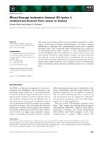
(CT56) antibodies prior to pulse-chase experiments. As
shown in Fig. 1(A,B), both anti-Ga
q
Igs detected a
band of 42 kDa that was more prominent in HEK293
cells transient transfected with the plasmid encoding
for Ga
q
. The CT56 antibody shows no apparent reac-
tion, at 43 kDa, in HEK293 cells that do not express
ABC
Fig. 1. Characterization of antibodies against
Ga
q
and Ga
16
proteins. The HEK293 cells
were transiently transfected with pCISLacZ
as control, pCISGa
q
or pCISGa
16
and labeled
with [
35
S]methionine as described in experi-
mental procedures. Cells were lysed and
protein extracts were immunoprecipitated
with Ga
q
or Ga
16
antiserum: (A) anti-G
q
CT-12178; (B) anti-G
q
sc-392; and (C)
anti-Ga
16
CT56, followed by SDS ⁄ PAGE (A
and C, 12.5% and B, 10% PAGE). The
figure shows representative autoradio-
graphies of whole SDS ⁄ PAGE gels loaded
with the
35
S immunoprecipitates and the
arrowheads indicate the position of Ga
q
and
Ga
16
. The molecular mass standards are
indicated. The arrow indicates the position
of some unspecific bands.
Proteasome degradation of G
q
proteins B. B. Johansson et al.
5366 FEBS Journal 272 (2005) 5365–5377 ª 2005 FEBS
the Ga
16
protein (Fig. 1C). Nevertheless, some unspe-
cific bands of higher molecular mass appear and are
labeled with arrows.
Figure 2A shows a representative experiment of the
pulse chase analysis of endogenous Ga
q
. As can be
seen, endogenous Ga
q
showed a rapid protein degra-
dation. The levels of endogenous Ga
q
decreased very
rapidly the first 3 h (60%) and then decreased progres-
sively at 6, 12 and 24 h to 44, 35 and 25% of zero time
controls, respectively. Based on curve fitting analysis,
the half life of Ga
q
was estimated 4 h if we assume a
monoexponential rate of decay. Control immuno-
precipitates of cells overexpressing Ga
q
are shown in
Fig. 1A (293 + Ga
q
). A similar degradation rate ( 3h)
was obtained for Ga
q
or Ga
16
proteins overexpressed
in cells (Fig. 2B,C). Control immunoprecipitates of
HEK293 cells expressing only endogenous Ga
q
or in
absence of Ga
16
are shown in Fig. 2B,C (293). Taken
together, these results suggest that the Ga
q
and Ga
16
proteins either endogenous or overexpressed display a
rapid turnover in HEK293 cells.
To investigate which proteases were responsible for
the degradation of Ga subunits, assays were performed
using cell-permeable protease inhibitors. Pulse and
chase experiments were performed after 3 h of preincu-
bation in the presence of the specific protease inhibitors
of different proteolytic pathways (Fig. 3). Leupeptin
(100 lgÆmL
)1
), an inhibitor of protein degradation in
lysosomes, had no effect on the stability of the Ga
q
and Ga
16
proteins. The presence of N-acetyl-l-leucyl-
l-leucyl-l-norleucinal (ALLN; 1 lm), which blocks non-
proteasomal proteases at 1 lm doses, did not influence
A
B
C
Fig. 2. Ga
q
and Ga
16
show a rapid turnover. Pulse-chase analysis of
endogenous Ga
q
(A), transfected Ga
q
(B), and transfected Ga
16
(C).
The HEK293 cells transfected with pCISLacZ as control (293), with
pCISGa
q
(293 + Ga
q
) or with pCISGa
16
(293 + Ga
16
) were meta-
bolically labeled and chased for the indicated hours. After pulse-
chase, protein extracts (900 lginA;100lg in B and C) were
immunoprecipitated with Ga
q
or Ga
16
antiserum (A, anti-G
q
sc-392;
B, anti-G
q
CT-12178 and C, anti-Ga
16
CT56) followed by SDS ⁄ PAGE
(A, 10%; B and C, 12.5% PAGE). Control immunoprecipitates of
cells expressing only endogenous Ga
q
(293) or in absence of Ga
16
(293) are shown in (B) and (C). The relative amounts of [
35
S]Ga
q
and [
35
S]Ga
16
were determined using a phophoimager and plotted
as a function of the chase time. Single experiments were per-
formed with triplicate samples and the mean of triplicates was nor-
malized by the mean at time zero. Data represent the mean of at
least four independent experiments where error bars are standard
deviations. Upper unspecific are shown by arrows. There are smalls
variations in the amount of these bands but no correspondence in
seen with the decrease G protein content during the chase taking
into account all experiments performed. Arrowheads indicate the
position of Ga
q
and Ga
16
.
B. B. Johansson et al. Proteasome degradation of G
q
proteins
FEBS Journal 272 (2005) 5365–5377 ª 2005 FEBS 5367
the turnover rate of the proteins, thus excluding their
involvement in protein breakdown. On the contrary,
treatment with the proteasome inhibitors MG132
(50 lm) and the highly specific lactacystin (30 lm)
clearly prevented the Ga
q
protein degradation after 3 h
of chase when compared with control conditions
A
BD
E
C
Fig. 3. The degradation of Ga
q
and Ga
16
is specifically decreased by proteasome inhibitors. The effect of protease inhibitors on the degrada-
tion of endogenous and transfected Ga
q
and Ga
16
was determined by incubating cells in presence or absence of 100 lgÆmL
)1
leupeptin
(lysosome inhibitor), 50 l
M MG132 (proteasome inhibitor), 1 lM ALLN (inhibitor of nonproteasomal cystein proteases) or 30 lM lactacystin
(proteasome inhibitor) prior to [
35
S]methionine labeling. Cells were metabolically labeled, chased for the indicated hours and protein extracts
(900 lg from endogenous G
q
expressing cells and 100 lg from transfected cells) were immunoprecipitated and analyzed by SDS ⁄ PAGE.
(A) Representative autoradiographies of endogenous Ga
q
(293), transfected Ga
q
(293 + Ga
q
) or transfected Ga
16
(293 + Ga
16
). The relative
amounts of the [
35
S]Ga subunits were determined using a phosphoimager and plotted as a function of treatment: (B) transfected Ga
q
;
(C) endogenous Ga
q
; and (D) transfected Ga
16
. Data represent mean of triplicates from a single experiment normalized by the mean at zero
hours where error bars are standard deviations. At least three independent experiments obtained similar results. (E) HEK293 cells transiently
transfected with either M2 muscarinic receptor or control vector in presence or absence of Ga
16
or with M1 muscarinic receptor and Ga
q
were labeled with myo-[2-
3
H]inositol (10 lCiÆmL
)1
) for 24 h before incubation in presence or absence of the proteasome inhibitor MG132
(50 l
M) 3 h prior to treatment with carbachol (10 lM). Values represent the means of duplicate determinants ± SD from a single experiment,
which is representative of two such experiments.
Proteasome degradation of G
q
proteins B. B. Johansson et al.
5368 FEBS Journal 272 (2005) 5365–5377 ª 2005 FEBS
(Fig. 3B). Consistently, similar effects were observed
with endogenous Ga
q
and cells expressing Ga
16
pro-
teins (Fig. 3C,D). We also studied the effect produced
by the treatment with the proteasome inhibitors on cell
viability and G protein activity by analyzing the release
of inositol phosphates by carbachol in cells expressing
the M1R (Fig. 3E). Carbachol-stimulation of HEK293
cells transiently transfected with the muscarinic recep-
tor 1 (M1R) and Ga
q
, showed the characteristic
increase in accumulation of inositol phosphates. Lig-
and-induced release of inositol phosphates was again
markedly increased after 3 h of preincubation with the
proteasome inhibitor MG132. Similar observations
were made in cells transiently transfected with the
muscarinic receptor 2 (M2R) and Ga
16
.
To explore whether receptor activation can modulate
Ga-turnover, receptors for carbachol (M1R and M2R)
were transfected in HEK293 cells that do not express
these receptors endogenously. These receptors were
chosen as it is well established the specificity of coup-
ling of G
q
with M1 receptor and G
16
with M2 recep-
tor. Cells were transfected with pCISM1R and analysis
of endogenous Ga
q
half-life after carbachol activation
was performed by pulse-chase (Fig. 4A). Under control
AB
CD
Fig. 4. Activation of Ga
q
and Ga
16
does not alter the half-life of the protein. HEK293 cells were transiently transfected with plasmids encoding
for M1R (A) and Ga
q
and Ga
q
R183C (B), Ga
16
and M2R (C), Ga
16
and Ga
16
Q212L (D), as indicated. Cells were metabolically labeled, stimula-
ted with carbachol (10 l
M) as indicated and chased for the indicated hours. Protein extracts [900 lg from endogenous Ga
q
expressing cells
(293 + M1R) and 100 lg from Ga
q
⁄ Ga
16
transfected cells (293 + Ga
q
⁄ 293 + Ga
16
)] were immunoprecipitated (anti-G
q
CT-12178 and anti-Ga
16
CT56) and analyzed by SDS ⁄ PAGE (12.5%). Relative amounts of [
35
S]Ga
q
and [
35
S]Ga
16
were determined using a phophoimager and plotted
as a function of the chase time. Data represent the mean of triplicates from a single representative experiment normalized by the mean at
zero hours where error bars are standard deviations. Representative autoradiographies are shown and arrow heads indicate the positions of
Ga
q
and Ga
16
. A minimum of three independent experiments obtained similar results in the all experiments shown.
B. B. Johansson et al. Proteasome degradation of G
q
proteins
FEBS Journal 272 (2005) 5365–5377 ª 2005 FEBS 5369
conditions, the levels of Ga
q
decay were essentially as
described in Fig. 2(A,B). Agonist stimulation did not
alter the degradation rate with 60 and 40% of the pro-
tein remaining at 3 and 6 h, respectively, both for the
ligand-activated and nonactivated Ga
q
. Adding new
media containing 10 lm carbachol every 30 min during
the chase period did not have any effect on the rate of
degradation either (data not shown). More surpris-
ingly, the activated mutant of Ga
q
,Ga
q
R183C,
showed no significant differences in stability regardless
of its constitutive activity, indicating that the activated
mutant is as stable as the wild-type form (Fig. 4B).
Accordingly, the levels of Ga
16
decay were the same in
the presence or absence of carbachol (Fig. 4C) in cells
expressing the M2R. Also the activated mutant of
Ga
16
,Ga
16
Q212L, showed no significant differences in
half-life compared with the wild-type Ga
16
(Fig. 4D).
Taken together, these results demonstrate that the rate
of G protein degradation is independent of ligand acti-
vation.
The observation that ligand activation did not pro-
duce any change in protein degradation could be due
to the inability of the transfected receptors to activate
the G proteins. To investigate this, the effect of ligand-
activation on inositol phosphate release was studied in
HEK293 cells coexpressing the M1R and endogenous
Ga
q
or M2R together with Ga
16
. As observed in
Fig. 5A, treatment of M1R-expressing cells with car-
bachol induces the typical increase responsiveness on
inositol phosphates. Equivalent results were observed
AB
CD
Fig. 5. Functional assay of the Ga subunits. HEK293 cells were transiently transfected with or without receptor in presence or absence of
Ga subunit and labeled with myo-[2-
3
H]inositol (10 lCiÆmL
)1
) for 24 h prior to treatment with ligand for 25 min. (A) Cells expressing the M1
muscarinic receptor in presence of endogenous Ga
q
and un-treated or treated with carbachol (10 lM). (B) Cells expressing Ga
q
,Ga
q
R183C
or control vector in absence of M1 receptor and treated as in (A). (C) Cells expressing Ga
16
and M2R and treated as in (A). (D) Cells expres-
sing Ga
16
,Ga
16
Q212L or control vector in absence of M2 receptor and treated as in (A). Expression of the G proteins in each of the assays
is shown in the lower panel. Values represent means of duplicate determinants from a single experiment, which is representative of
minimum two such experiments.
Proteasome degradation of G
q
proteins B. B. Johansson et al.
5370 FEBS Journal 272 (2005) 5365–5377 ª 2005 FEBS
for carbachol-induced M2 activation of Ga
16
(Fig. 5C)
and for the constitutive activate mutant forms of Ga
q
and Ga
16
(Fig. 5B,D). All these results confirm previ-
ous studies and demonstrate that in fact receptor sti-
mulation leads to activation of Ga
16
and Ga
q
subunits. Therefore the lack of change in the rate of
degradation cannot be due to lack of receptor-activa-
tion of the alpha subunits.
Given that the activation of G proteins by ligand-
stimulated receptors takes place in the cytoplasmic
leaflet of the cell membrane and that not all the Ga
subunits in the cell will be activated by receptor stimu-
lation, pulse-chase experiments were performed with
cell extracts enriched in particulated vs. cytoplasmic
fractions, in order to enrich in the GPCR-activated
pool. For this, cells were metabolically labeled, stimu-
lated and cell extracts were separated by centrifugation
(Fig. 6). Significantly, these experiments confirmed our
previous results that show no difference in the degra-
dation rate between the stimulated and nonstimulated
G proteins in both crude cytoplasmic and particulated
fractions and for both endogenous and overexpressed
Ga
q
and M1R or Ga
16
and M2R (Fig. 6A–C). How-
ever, a somehow surprising result was the fact that the
crude cytoplasmic fractions of both Ga
q
and Ga
16
were less stable than the membrane-enriched fraction.
After 6 h only 20% of the cytosolic proteins were
remaining vs. 50–60% of the membrane fraction.
These results are consistent with the idea that the
Ga subunits are more stable in the membrane than in
the cytoplasm. To study this further we designed a
mutant Ga
q
protein where the two palmitoylated cys-
teine residues CC9 ⁄ 10 were mutated to serine residues.
For Ga
q
and Ga
11
, subcellular distribution and the
role of N-terminal palmitoylation has been extensively
studied previously [29–33]. Consistent with these previ-
ous findings, fractionation and immunofluorescence
studies showed that the mutant Ga
q
CC9 ⁄ 10SS was
A
B
C
Fig. 6. Differences in Ga
q
and Ga
16
degradation rates in membrane
and cytosolic fractions. Cells were transiently transfected with plas-
mids encoding M1R and LacZ (A), M1R and Ga
q
(B) or M2R and
Ga
16
(C), metabolically labeled and chased for the indicated hours.
Cells were lysed (900 lg of total protein from endogenous Ga
q
expressing cells and 500 lg from transfected cells) and particulated
and cytosolic fractions were separated by centrifugation. Protein
extracts from each fraction were immunoprecipitated (anti-G
q
CT-12178 and anti-G a
16
CT56) and analyzed by SDS ⁄ PAGE (12%).
Relative amounts of [
35
S]Ga
q
and [
35
S]Ga
16
were determined using
a phophoimager and were plotted as a function of chase time. The
amount of Ga
q
and Ga
16
at time zero was set to 100%. The data
represent mean of triplicates from a single representative experi-
ment normalized by the mean at time zero where error bars are
standard deviations. A two-tailed Student’s t-test was run to com-
pare membrane fraction and cytosolic fraction. All tests show a sig-
nificance of *, **P < 0.001 where n varies from 5 to 9. No
significant difference was seen between ligand stimulated and
nonstimulated cells in the same experiments. Representative auto-
radiographies are shown and arrowheads indicate the positions of
Ga
q
and Ga
16
. Upper nonspecific bands are shown by arrows.
B. B. Johansson et al. Proteasome degradation of G
q
proteins
FEBS Journal 272 (2005) 5365–5377 ª 2005 FEBS 5371
present mainly in the cytoplasm-enriched fraction (data
not shown), in contrast both transfected and endo-
genous Ga
q
showed a similar distribution with the pro-
tein present mainly close to the cytoplasmic membrane
but also some present in the cytoplasmic fraction.
When pulse-chase analysis of Ga
q
CC9 ⁄ 10SS and Ga
q
were examined, a decrease in the total amount of the
Ga
q
CC9 ⁄ 10SS mutant was observed prior to chase
(Fig. 7). A decrease in the amount of
35
S associated
with Ga
q
CC9 ⁄ 10SS immediately after incubation with
[
35
S]methionine may indicate an alteration in the rate
of its degradation during the labeling period. In fact,
only 15% of
35
S-labeled mutant protein remains (vs.
100% of wild-type protein at 0 h). Nevertheless, it
could also be explained by changes in translation or
maturation of the protein. An increase was also
observed in the rate of degradation of the remaining
mutant protein (20% left of the protein) vs. the wild
type (40% left of the protein) at 3 h of chase
(Fig. 7A,B). We also analyzed the rate of degradation
of another mutant Ga
q
protein, Ga
q
IE25 ⁄ 26AA. This
mutant protein has two residues substituted to alanine
in the putative Gbc binding site [34,35]. An equivalent
region in Ga
i
was shown before to be in direct contact
to Gbc [36]. As shown in Fig. 7(A,B), no change in
the total amount of protein or in the rate of degrada-
tion was observed, which is an indication that the
binding of Ga to Gbc subunits may not the limiting
factor for its stability.
Discussion
In this report we present evidence that endogenous
Ga
q
, transfected Ga
q
and Ga
16
proteins degrade with
half-lives of around 3–4 h, if we assume a monoexpo-
nential rate of decay. Furthermore, we provide novel
data showing that members of the G
q
class proteins
are degraded through the proteasome pathway. The
degradation of the G
q
proteins is not dependent on
GPCR activation or on G protein activity. On the con-
trary, the association of Ga subunits to other proteins
close to or at the cytoplasmic membrane may play a
major role in protein stability.
Many signaling proteins with fast degradation rates
are degraded through the proteasome. Here, we have
provided evidence that the proteasomal pathway is
also responsible for the degradation of the members of
the G
q
alpha class. Specific degradation of the Ga sub-
unit by the proteasome-dependent pathway has been
shown for the yeast Gpa1 [37,38] and for Ga
o
[39].
Therefore it is increasingly evident that the protea-
some-dependent pathway plays an important role in
the regulation of G protein stability.
An open question in signal transduction studies has
been the effect that receptor activation has on the
turnover of downstream proteins. As is the case,
GRK2 degradation through proteasome is enhanced
by GPCR stimulation [7]. Chronic exposure to ligand
produces a decrease in G protein levels [21]. On the
other hand, our results have provided evidence for the
lack of ligand-induced degradation of Ga
q
proteins.
Neither receptor-activation of Ga
q
or Ga
16
in total
lysates nor in membrane or cytoplasm fractions had
any effect on the half-life of the proteins. Muta-
tional activation of Ga
q
through the inhibition of its
GTPase-activity, did not produce any enhancement in
the rate of degradation compared with the wild-type
protein. Our data is more consistent with an increased
destabilization of the protein in the cytoplasm, a pro-
cess that, in the case of the G
q
family of proteins, is
independent of receptor activation. Short-term receptor
A
B
Fig. 7. The mutant Ga
q
CC9 ⁄ 10SS has an increased rate of degrada-
tion compared to the Ga
q
wt. HEK293 cells were transiently
transfected with pcDNA3Ga
q
, pcDNA3Ga
q
CC9 ⁄ 10SS, pcDNA3Ga
q
IE25 ⁄ 26AA or empty pcDNA3 vector as a control (293) and
metabolically labeled with [
35
S]methionine and chased for 3 h.
(A) Shows the autoradiographies of the two mutant G proteins
compared with the wild-type protein at 0 and 3 h. (B) The relative
amounts of the [
35
S]Ga
q
subunits were determined using a phos-
phoimager and plotted as a function of chase time. Data represents
the mean of triplicates from a single representative experiment nor-
malized by the mean at zero hours where error bars are standard
deviations. Two experiments produce similar results in all the
experiments shown.
Proteasome degradation of G
q
proteins B. B. Johansson et al.
5372 FEBS Journal 272 (2005) 5365–5377 ª 2005 FEBS
activation of G
q
proteins does not induce translocation
of these subunits to the cytoplasm [31]. On the con-
trary, ligand activation of G
s
proteins induces trans-
location of these G proteins to the cytoplasm [40,41],
which could explain previous results showing that lig-
and activation of receptors coupled to G
s
promotes an
increase in the degradation of this subunit [41]. On the
other hand, chronic agonist treatment of receptors
coupled to Ga
q
could deplete the cytoplasmic mem-
brane from receptors and proteins associated to them,
and could explain the increased degradation of Ga
q
in
the experiments with persistent ligand stimulation [42–
44]. Interestingly, very recent work done with the yeast
Ga subunit Gpa1 have shown that poly ubiquitinated
Gpa1 exhibits a cytoplasmic localization [45]. On the
contrary a Gpa1 mutant that lacks the ubiquitinated
subdomain remains unmodified and is predominantly
localized at the plasma membrane.
Protein stability in the plasma membrane can be a
consequence of receptor association, but interactions of
Ga
q
subunits with other proteins could also be respon-
sible for the stabilization in the membrane. The fact
that the activated mutant forms of Ga
q
and Ga
16
have
the same behavior as the wild-type forms argues in
favor of the need of other proteins to stabilize the pro-
teins in the membrane. The interaction of Ga subunits
with Gbc could be a stabilizing factor. Mutations on
Ga
q
residues which locate in the Gbc binding region
and have been shown to diminish association with the
plasma membrane [34] did not have any consequence in
protein stability. It is still possible that this mutant
retains some binding to Gbc subunits, as it was shown
that localization at the plasma membrane could be res-
cued by expression of Gbc subunits [35]. Other proteins
that interact with the Ga
q
subunits and may have a
role in the stabilization of these subunits in the mem-
brane are the regulators of G-protein signaling (RGS)
[14] or the GRK2, which has been shown recently to
have a RGS-like domain that binds to Ga
q
[18,19].
Recent results have described an RGS–GAIP-inter-
acting protein, GIPN, that has E3-ubiquitin ligase
activity and promotes proteasome-dependent degrada-
tion of Ga
i3
[46]. The role of these proteins in the turn-
over of the Ga
q
protein should be further investigated.
Interestingly, G
q
, apparently without Gbc subunits,
stably associates with caveolin in caveolae structures
[47]. Caveolin has been suggested to act as a scaffold to
trap and stabilize Gq. On the other hand, the degrada-
tion of Ga
o
via the proteasome pathway is protected
by interaction of the Ga
o
subunit with Hsp90 [39]. Also
the Ga
12
subunit, which localizes in membrane frac-
tions [48], has been shown to associate to Hsp90 and
its association is important for Ga
12
signaling [49].
Work in progress indicates that the same interaction
could be taking place for Ga
q
subunits.
In summary, our results suggest that subcellular
localization and ⁄ or protein interactions at the mem-
brane are responsible for Ga protein stability. Further
work will help to elucidate the molecular bases of
those mechanisms that control the stability of G pro-
tein subunits close to the cytoplasmic membrane.
Experimental procedures
Materials
HEK293 cells (293-EBNA) and LipofectAMINE were
purchased from Invitrogen (Groningen, the Netherlands).
Carbachol and leupeptin were obtained from Sigma-Aldrich
(St Louis, MO, USA). C5a was a generous gift from
M. Oppermman (Georg-August Universita
¨
t, Gottingen,
Germany). ALLN and lactacystin were purchased from
Calbiochem (San Diego, CA, USA). MG132 was obtained
from BIOMOL Research Laboratories Inc (Plymouth
Meeting, PA, USA). C-Terminal peptide polyclonal anti-
bodies against Ga
q
and Ga
16
were generated in M. Simon’s
laboratory (California Institute of Technology, Pasadena,
CA, USA). In some experiments (indicated in the legends)
the C-terminal peptide polyclonal antibodies against Ga
q
were obtained from Santa Cruz Biotechnologies (Santa
Cruz, CA, USA). Secondary HRP-labeled antibody was
ordered from Zymed Laboratories. [
35
S]Methionine was
ordered from Amersham Pharmacia Biotech (Piscataway,
NJ, USA). Myo-[2-
3
H]inositol was purchased from Ameri-
can Radiolabeled Chemicals Inc (Saint Louis, MO, USA).
Enhanced chemiluminescence reagents were obtained from
Amersham Pharmacia Biotech. All other reagents were of
the highest grade commercially available.
DNA constructs
The cDNAs from Ga
q
and Ga
16
cloned into pCIS were
provided by M.I. Simon (California Institute of Technol-
ogy). The mutant deficient of Gbc-binding was generated
by using site-directed mutagenesis using pCISGa
q
as a tem-
plate and the following oligos: Ga
q
IE25 ⁄ 26AA: 5¢-ggat
caacgacgaggccgcgcggcagctgcgcaggg-3¢,Ga
q
IE25 ⁄ 26AA-cccc
tgcgcagctgccgcgcggcctcgtcgttgatcc. The palmitoylation-defi-
cient mutant Ga
q
CC9 ⁄ 10SS was generated by site-directed
mutagenesis and amplified using pCISGa
q
wt as a template.
PCR was carried out using a forward primer with a KpnI
site (underlined): Ga
q
-N-term-Kpn1: 5¢-cgcgggtaccatgatc
ctggagtccatcatggcgtgctgcctgagcgaggag-3¢,Ga
q
CC9 ⁄ 19SS-N-
term-Kpn1: 5¢-
cgcgggtaccatgactctggagtccatcatggcgtcctccctg
agcgaggag-3¢, and a reverse primer with a BamHI site
(underlined): Ga
q
-C-term- BamHI: 5¢-cgcggatccttagaccagat
tgtactcctt-3¢. The PCR products were digested and ligated
B. B. Johansson et al. Proteasome degradation of G
q
proteins
FEBS Journal 272 (2005) 5365–5377 ª 2005 FEBS 5373
into pcDNA3. Ga
16
was subcloned from pCIS vector to
pcDNA3 by PCR, with primers including restriction enzyme
sites. All the plasmids presented were sequenced prior to use.
Cell culture and transfection
HEK293 cells were cultured in Dulbecco’s modified Eagle’s
medium, supplemented with 10% (v ⁄ v) fetal bovine serum
at 37 °C in a humidified atmosphere containing 5% (v ⁄ v)
CO
2
. Transient transfections were performed on 70–80%
confluent monolayers by using LipofectAMINE reagent
according to the manufacturer’s instructions. Briefly,
HEK293 cells (1.5 · 10
6
cells) were seeded on a P60-plate a
day prior to transfection with 5 lg of total plasmid DNA:
pCISGa
q
or pCISGa
16
in presence or absence of pCISM1R
or pCISM2R ⁄ pCISC5aR (Ga ⁄ R ¼ 0.4 ⁄ 0.6), respectively.
The total amount of DNA was kept constant with the addi-
tion of pCISLacZ.
Metabolic labeling
Metabolic labeling was performed 48 h following transfec-
tion of cells kept with Dulbecco’s modified Eagle’s medium,
supplemented with 10% (v ⁄ v) fetal bovine serum. Cells
expressing Ga
q
or Ga
16
were incubated for 1 h in methion-
ine-free DMEM in absence of serum and then incubated
for 30 min in this medium supplemented with 50 lCiÆmL
)1
of [
35
S]methionine labeling mixture. The cell monolayers
were washed with phosphate-buffered saline (NaCl ⁄ P
i
) and
chased for indicated times in DMEM containing an excess
of cold methionine. To determine the effect of various pro-
tease inhibitors, cells were treated with inhibitors for 3 h
before the chase and were present throughout the chase at
the following concentrations: leupeptin 100 lgÆmL
)1
,
ALLN 1 lm, MG132 50 lm and lactacystin 30 lm. All
protease inhibitors, except leupeptin, were dissolved in
dimethylsulfoxide, and control cells were treated with equal
amounts of dimethylsulfoxide alone. In the experiments
with cells expressing Ga
q
or Ga
16
in presence or absence of
M2R ⁄ C5aR or M1R, respectively, the ligand was added at
the beginning of the chase (t ¼ 0 min) at concentrations of
10 lm carbachol or 100 nm C5a, respectively.
Immunoprecipitation
After the chase, cells were washed and lysed in RIPA buffer
[50 mm Tris pH 7.5, 300 mm NaCl, 1% (w ⁄ v) n-dodecyl-
b-d-maltoside, 0.1% (w ⁄ v) sodium dodecyl sulfate and 0.5%
(w ⁄ v) deoxycholate, with protease inhibitors] for 1 h at 4 °C
with continuous rocking. In early experiments, the total pro-
tein content in the samples was estimated before immuno-
precipitation by using Bradford analysis, later this step was
omitted due to good reproducibility of the samples. Protein
extracts (900 lg of total protein for endogenous Ga
q
samples and 100 lg for Ga
q
and Ga
16
transfected cells) sup-
plemented with 1 lgÆlL
)1
BSA were immunoprecipitated
overnight at 4 °C with the specific Ga
q
or Ga
16
antibodies,
followed by incubation with protein A-sepharose beads for
1.5 h. Immune complexes were then washed four times with
NaCl ⁄ P
i
, pH 7.2. Following SDS ⁄ PAGE resolution, the gel
was dried and later analyzed by phosphoimaging in a BAS
5000 system from Fuji (Fuji Foto Film, Tokyo, Japan). The
background level was subtracted from the values registered
for each band. As the background of some of the lanes was
variable some experiments were done with subtraction of
the background of the corresponding band, with no differ-
ences in the overall result. Every experiment was performed
with triplicate samples of each time point. The average value
of each time point was normalized with the average value at
0 h. A minimum of three independent experiments showed
similar results.
Determination of total inositol phosphate levels
Total inositol phosphate formation was measured essen-
tially as described previously [50]. Briefly, 1 · 10
5
cells were
seeded in 12-well plates and transfected after 24 h with
1 lg of total plasmid DNA. Cells were prelabeled with
10 lCiÆmL
)1
[
3
H]inositol for 24 h in inositol-free medium
containing 10% (v ⁄ v) dialyzed fetal bovine serum. Cells
were then washed and incubated in NaCl ⁄ P
i
containing
20 mm Li
+
with the agonist at 37 °C for 20 min. Cells were
then treated with 100 lL of 10% (v ⁄ v) perchloric acid and
10 lL of phytic acid (20 mgÆ mL
)1
) for 10 min. The mixture
was centrifuged and neutralized. After centrifugation, the
supernatants were subjected to anion exchange chromato-
graphy. The final eluant was dissolved in scintillation liquid
and counted in a scintillation counter.
Western blotting
For total G protein content analysis, cell extracts were pre-
pared by lysis in a hypotonic buffer (50 mm Hepes, 0.2 mm
EDTA, 1 mm dithiotreitol, pH 7.4) and cleared by centri-
fugation at 500 g for 5 min. Supernatants were boiled in Lae-
mmli sample buffer and resolved by SDS ⁄ PAGE. Proteins
were transferred to a nitrocellulose membrane and probed
with either Ga
16
or Ga
q
antibodies, respectively. Blots were
developed using a chemiluminiscence assay method.
Subcellular fractionation
Cells (1.5 · 10
6
) were seeded for transfection of cells with
Ga
q
and G a
16
, and 5 · 10
6
cells were used for studying
endogenously expressed Ga
q
. Cells were metabolically labe-
led as described. After the chase, the cells were harvested in
hypotonic buffer (50 mm Hepes, 0.2 mm EDTA, 1 mm,
pH 7.4) and lysed by several cycles of freezing and thawing.
Proteasome degradation of G
q
proteins B. B. Johansson et al.
5374 FEBS Journal 272 (2005) 5365–5377 ª 2005 FEBS
The cell lysates (approximately 500 lg of total protein for
transfected cells and 900 lg for endogenously expressed
proteins) were cleared by centrifugation at 400 g for
10 min. Supernatants were centrifuged at 100 000 g for
30 min at 4 °C. Crude membrane pellets were resuspended
in an equal volume with RIPA buffer (50 mm Tris pH 7.5,
300 mm NaCl, 1% (w ⁄ v) n-dodecyl-b-d-maltoside, 0.1%
(w ⁄ v) sodium dodecyl sulfate, 0.5% (w ⁄ v) deoxycholate
and protease inhibitors). Both fractions, crude membranes
and cytoplasm, were immunoprecipitated with the specific
antibodies against Ga
q
or Ga
16
as described before.
Acknowledgements
We thank I. Gavlen and T. Ellingsen for experimental
assistance and Dr Murga, Dr Ribas and Dr Penela for
insightful comments. This work was supported by
grants from The Norwegian Cancer Society.
References
1 Bonifacino JS & Weissman AM (1998) Ubiquitin and
the control of protein fate in the secretory and endo-
cytic pathways. Annu Rev Cell Dev Biol 14, 19–57.
2 Chaturvedi K, Bandari P, Chinen N & Howells RD
(2001) Proteasome involvement in agonist-induced
down-regulation of mu and delta opioid receptors.
J Biol Chem 276, 12345–12355.
3 Obin MS, Jahngen-Hodge J, Nowell T & Taylor A
(1996) Ubiquitinylation and ubiquitin-dependent proteo-
lysis in vertebrate photoreceptors (rod outer segments).
Evidence for ubiquitinylation of Gt and rhodopsin.
J Biol Chem 271, 14473–14484.
4 Roth AF & Davis NG (1996) Ubiquitination of the
yeast a-factor receptor. J Cell Biol 134, 661–674.
5 Hicke L & Riezman H (1996) Ubiquitination of a yeast
plasma membrane receptor signals its ligand-stimulated
endocytosis. Cell 84, 277–287.
6 Shenoy SK, McDonald PH, Kohout TA & Lefkowitz
RJ (2001) Regulation of receptor fate by ubiquitination
of activated beta 2-adrenergic receptor and beta-
arrestin. Science 294, 1307–1313.
7 Penela P, Ruiz-Gomez A, Castano JG & Mayor F Jr
(1998) Degradation of the G protein-coupled receptor
kinase 2 by the proteasome pathway. J Biol Chem 273,
35238–35244.
8 Davydov IV & Varshavsky A (2000) RGS4 is arginy-
lated and degraded by the N-end rule pathway in vitro.
J Biol Chem 275, 22931–22941.
9 Bokkala S & Joseph SK (1997) Angiotensin II-induced
down-regulation of inositol trisphosphate receptors in
WB rat liver epithelial cells: evidence for involvement of
the proteasome pathway. J Biol Chem 272, 12454–
12461.
10 Oberdorf J, Webster JM, Zhu CC, Luo SG & Wojcikie-
wicz RJ (1999) Down-regulation of types I, II and III
inositol 1,4,5-trisphosphate receptors is mediated by the
ubiquitin ⁄ proteasome pathway. Biochem J 339, 453–
461.
11 Wojcikiewicz RJ, Xu Q, Webster JM, Alzayady K &
Gao C (2003) Ubiquitination and proteasomal degrada-
tion of endogenous and exogenous inositol 1,4,5-tri-
sphosphate receptors in alpha T3–1 anterior pituitary
cells. J Biol Chem 278, 940–947.
12 Wojcikiewicz RJ (2004) Regulated ubiquitination of
proteins in GPCR-initiated signaling pathways. Trends
Pharmacol Sci 25, 35–41.
13 Berman DM & Gilman AG (1998) Mammalian RGS
proteins: barbarians at the gate. J Biol Chem 273, 1269–
1272.
14 Ross EM & Wilkie TM (2000) GTPase-activating pro-
teins for heterotrimeric G proteins: regulators of G pro-
tein signaling (RGS) and RGS-like proteins. Annu Rev
Biochem 69, 795–827.
15 Hollinger S & Hepler JR (2002) Cellular regulation of
RGS proteins: modulators and integrators of G protein
signaling. Pharmacol Rev 54, 527–559.
16 Berstein G, Blank JL, Jhon DY, Exton JH, Rhee SG &
Ross EM (1992) Phospholipase C-beta 1 is a GTPase-
activating protein for Gq ⁄ 11, its physiologic regulator.
Cell 70, 411–418.
17 Chidiac P & Ross EM (1999) Phospholipase C-beta1
directly accelerates GTP hydrolysis by Galphaq and
acceleration is inhibited by Gbeta gamma subunits.
J Biol Chem 274, 19639–19643.
18 Sallese M, Mariggio S, D’Urbano E, Iacovelli L & De
Blasi A (2000) Selective regulation of Gq signaling by
G protein-coupled receptor kinase 2: direct interaction
of kinase N terminus with activated galphaq. Mol
Pharmacol 57, 826–831.
19 Usui H, Nishiyama M, Moroi K, Shibasaki T, Zhou J,
Ishida J, Fukamizu A, Haga T, Sekiya S & Kimura S
(2000) RGS domain in the amino-terminus of G
protein-coupled receptor kinase 2 inhibits Gq-mediated
signaling. Int J Mol Med 5, 335–340.
20 Chen CA & Manning DR (2001) Regulation of G pro-
teins by covalent modification. Oncogene 20, 1643–1652.
21 Bohm SK, Grady EF & Bunnett NW (1997) Regulatory
mechanisms that modulate signalling by G-protein-
coupled receptors. Biochem J 322, 1–18.
22 Simon MI, Strathmann MP & Gautam N (1991) Diver-
sity of G proteins in signal transduction. Science 252,
802–808.
23 Grant KR, Harnett W, Milligan G & Harnett MM
(1997) Differential G-protein expression during B- and
T-cell development. Immunology 90, 564–571.
24 Amatruda TT, 3rd Steele DA, Slepak VZ & Simon MI
(1991) G alpha 16, a G protein alpha subunit specifically
B. B. Johansson et al. Proteasome degradation of G
q
proteins
FEBS Journal 272 (2005) 5365–5377 ª 2005 FEBS 5375
expressed in hematopoietic cells. Proc Natl Acad Sci
USA 88, 5587–5591.
25 Mapara MY, Bommert K, Bargou RC, Leng C, Beck
C, Ludwig WD, Gierschik P & Dorken B (1995) G
protein subunit G alpha 16 expression is restricted to
progenitor B cells during human B-cell differentiation.
Blood 85, 1836–1842.
26 Tenailleau S, Corre I & Hermouet S (1997) Specific
expression of heterotrimeric G proteins G12 and G16
during human myeloid differentiation. Exp Hematol 25,
927–934.
27 Offermanns S & Simon MI (1995) G alpha 15 and G
alpha 16 couple a wide variety of receptors to phospho-
lipase C. J Biol Chem 270, 15175–15180.
28 Aragay AM & Quick MW (1999) Functional regulation
of Galpha16 by protein kinase C. J Biol Chem 274,
4807–4815.
29 Hepler JR, Biddlecome GH, Kleuss C, Camp LA,
Hofmann SL, Ross EM & Gilman AG (1996) Func-
tional importance of the amino terminus of Gq alpha.
J Biol Chem 271, 496–504.
30 Edgerton MD, Chabert C, Chollet A & Arkinstall S
(1994) Palmitoylation but not the extreme amino-termi-
nus of Gq alpha is required for coupling to the NK2
receptor. FEBS Lett 354, 195–199.
31 Hughes TE, Zhang H, Logothetis DE & Berlot CH
(2001) Visualization of a functional Galpha q-green
fluorescent protein fusion in living cells: association with
the plasma membrane is disrupted by mutational activa-
tion and by elimination of palmitoylation sites, but not
be activation mediated by receptors or AlF4. J Biol
Chem 276, 4227–4235.
32 Wedegaertner PB, Chu DH, Wilson PT, Levis MJ &
Bourne HR (1993) Palmitoylation is required for
signaling functions and membrane attachment of
Gq alpha and Gs alpha. J Biol Chem 268, 25001–
25008.
33 McCallum JF, Wise A, Grassie MA, Magee AI, Guzzi
F, Parenti M & Milligan G (1995) The role of palmi-
toylation of the guanine nucleotide binding protein G11
alpha in defining interaction with the plasma membrane.
Biochem J 310, 1021–1027.
34 Evanko DS, Thiyagarajan MM & Wedegaertner PB
(2000) Interaction with Gbetagamma is required
for membrane targeting and palmitoylation of
Galpha(s) and Galpha(q). J Biol Chem 275, 1327–
1336.
35 Evanko DS, Thiyagarajan MM, Siderovski DP &
Wedegaertner PB (2001) Gbeta gamma isoforms selec-
tively rescue plasma membrane localization and palmi-
toylation of mutant Galphas and Galphaq. J Biol Chem
276, 23945–23953.
36 Lambright DG, Sondek J, Bohm A, Skiba NP, Hamm
HE & Sigler PB (1996) The 2.0 A
˚
crystal structure of a
heterotrimeric G protein. Nature 379, 311–319.
37 Madura K & Varshavsky A (1994) Degradation of G
alpha by the N-end rule pathway. Science 265, 1454–
1458.
38 Marotti LA Jr, Newitt R, Wang Y, Aebersold R &
Dohlman HG (2002) Direct identification of a G pro-
tein ubiquitination site by mass spectrometry. Biochem-
istry 41, 5067–5074.
39 Busconi L, Guan J & Denker BM (2000) Degradation
of heterotrimeric Galpha(o) subunits via the proteosome
pathway is induced by the hsp90-specific compound
geldanamycin. J Biol Chem 275, 1565–1569.
40 Wedegaertner PB, Bourne HR & von Zastrow M (1996)
Activation-induced subcellular redistribution of Gs
alpha. Mol Biol Cell 7, 1225–1233.
41 Levis MJ & Bourne HR (1992) Activation of the alpha
subunit of Gs in intact cells alters its abundance, rate of
degradation, and membrane avidity. J Cell Biol 119 ,
1297–1307.
42 Mullaney I, Caulfield MP, Svoboda P & Milligan G
(1996) Activation, cellular redistribution and enhanced
degradation of the G proteins Gq and G11 by endo-
genously expressed and transfected phospholipase
C-coupled muscarinic m1 acetylcholine receptors. Prog
Brain Res 109, 181–187.
43 Mitchell FM, Buckley NJ & Milligan G (1993)
Enhanced degradation of the phosphoinositidase
C-linked guanine-nucleotide-binding protein Gq
alpha ⁄ G11 alpha following activation of the human M1
muscarinic acetylcholine receptor expressed in CHO
cells. Biochem J 293, 495–499.
44 Shah BH, MacEwan DJ & Milligan G (1995) Gonado-
trophin-releasing hormone receptor agonist-mediated
down-regulation of Gq alpha ⁄ G11 alpha (pertussis
toxin-insensitive) G proteins in alpha T3–1 gonadotroph
cells reflects increased G protein turnover but not altera-
tions in mRNA levels. Proc Natl Acad Sci USA 92,
1886–1890.
45 Wang Y, Marotti LA Jr, Lee MJ & Dohlman HG
(2005) Differential regulation of G protein alpha
subunit trafficking by mono- and polyubiquitination.
J Biol Chem 280, 284–291.
46 Fischer T, De Vries L, Meerloo T & Farquhar MG
(2003) Promotion of G alpha i3 subunit down-regula-
tion by GIPN, a putative E3 ubiquitin ligase that inter-
acts with RGS-GAIP. Proc Natl Acad Sci USA 100,
8270–8275.
47 Oh P & Schnitzer JE (2001) Segregation of hetero-
trimeric G proteins in cell surface microdomains: G(q)
binds caveolin to concentrate in caveolae, whereas G(i)
and G(s) target lipid rafts by default. Mol Biol Cell 12,
685–698.
48 Yamazaki J, Katoh H, Yamaguchi Y & Negishi M
(2005) Two G(12) family G proteins, Galpha(12) and
Galpha(13), show different subcellular localization.
Biochem Biophys Res Commun 332, 782–786.
5376 FEBS Journal 272 (2005) 5365–5377 ª 2005 FEBS
Proteasome degradation of G
q
proteins B. B. Johansson et al.
49 Vaiskunaite R, Kozasa T & Voyno-Yasenetskaya TA
(2001) Interaction between the G alpha subunit of
heterotrimeric G(12) protein and Hsp90 is required for
G alpha(12) signaling. J Biol Chem 276, 46088–46093.
50 Wu DQ, Lee CH, Rhee SG & Simon MI (1992) Activa-
tion of phospholipase C by the alpha subunits of the
Gq and G11 proteins in transfected Cos-7 cells. J Biol
Chem 267, 1811–1817.
FEBS Journal 272 (2005) 5365–5377 ª 2005 FEBS 5377
B. B. Johansson et al. Proteasome degradation of G
q
proteins