Moses attrep, bernd kahn radioanalytical chemistry experiments springer (2007)
Bạn đang xem bản rút gọn của tài liệu. Xem và tải ngay bản đầy đủ của tài liệu tại đây (2.64 MB, 167 trang )
www.pdfgrip.com
Radioanalytical Chemistry Experiments
www.pdfgrip.com
Moses Attrep and Bernd Kahn
Radioanalytical
Chemistry
Experiments
www.pdfgrip.com
Moses Attrep
Los Alamos, NM, USA
344 Kimberly Lane
Los Alamos 87545
ISBN: 978-0-387-46914-0
Bernd Kahn
Health and Environmental Systems Lab
Georgia Institute of Technology
Atlanta, GA, USA
400 W. 10th St. NW
Atlanta 30332-0841
e-ISBN: 978-0-387-46925-6
Library of Congress Control Number: 2007938040
© 2008 Springer Science+Business Media, LLC
All rights reserved. This work may not be translated or copied in whole or in part without
the written permission of the publisher (Springer Science+Business Media, LLC, 233 Spring
Street, New York, NY 10013, USA), except for brief excerpts in connection with reviews
or scholarly analysis. Use in connection with any form of information storage and retrieval,
electronic adaptation, computer software, or by similar or dissimilar methodology now known
or hereafter developed is forbidden.
The use in this publication of trade names, trademarks, service marks and similar terms, even if
they are not identified as such, is not to be taken as an expression of opinion as to whether or
not they are subject to proprietary rights.
Printed on acid-free paper.
9 8 7 6 5 4 3 2 1
springer.com
www.pdfgrip.com
Table of Contents
Acknowledgments . . . . . . . . . . . . . . . . . . . . . . . . . . . . . . . . . . . . . . . . . . . . . . . . .
vii
Introduction . . . . . . . . . . . . . . . . . . . . . . . . . . . . . . . . . . . . . . . . . . . . . . . . . . . . . .
1
Experiment 1.
Practice in Pipetting and Weighing Samples
for Radioactivity Counting . . . . . . . . . . . . . . . . . . . . . . . . .
11
Experiment 2.
Radiation Detection Instrument Calibration and Quality
Assurance . . . . . . . . . . . . . . . . . . . . . . . . . . . . . . . . . . . . . . . . 15
Experiment 3.
Determination of Gamma Ray Self-absorption in a KCl
Sample . . . . . . . . . . . . . . . . . . . . . . . . . . . . . . . . . . . . . . . . . . . 31
Experiment 4.
Preparation of a Beta-particle Self-absorption Curve
for 40 K . . . . . . . . . . . . . . . . . . . . . . . . . . . . . . . . . . . . . . . . . . .
35
Experiment 5.
Preparation and Standardization of Carriers . . . . . . . . . .
41
Experiment 6.
Preparation and Counting of
Pu Tracer Solution . . .
45
Experiment 7.
Basic Radiochemical Techniques Applied to
Uranium-Thorium Separation: Precipitation, Solvent
Extraction, and Ion Exchange . . . . . . . . . . . . . . . . . . . . . .
51
Determination of Radium-226 and Radium-228 in
Drinking Water . . . . . . . . . . . . . . . . . . . . . . . . . . . . . . . . . . .
67
Radiochemical Determination of Tritium in Water . . .
79
Experiment 8.
Experiment 9.
Experiment 10. Determination of
131
242
I in Water . . . . . . . . . . . . . . . . . . . . .
85
Experiment 11. Modification of a Published Procedure for
the Determination of Picocurie Concentrations of
Iodine-131 in Milk . . . . . . . . . . . . . . . . . . . . . . . . . . . . . . . .
93
v
www.pdfgrip.com
vi
Table of Contents
Experiment 12. Vegetation Sample Preparation for Radiochemical
Analysis of Radio-strontium . . . . . . . . . . . . . . . . . . . . . . . .
97
Experiment 13. Determination of Radio-strontium Isotopes
in Environmental Samples . . . . . . . . . . . . . . . . . . . . . . . . . 103
Experiment 14. Determination of Radio-strontium in Water with a
Strontium-specific Solid-phase Extraction Column . . . 113
Experiment 15. Radiochemical Determination of Plutonium in Water
by Ion Exchange Chromatographic Separation . . . . . . . 119
Experiment 16. Radiochemical Determination of Plutonium in Water
by Solvent Extraction . . . . . . . . . . . . . . . . . . . . . . . . . . . . . . 131
Experiment 17. Selection of a Method for the Radiochemical
Determination of Plutonium in an Environmental
Sample . . . . . . . . . . . . . . . . . . . . . . . . . . . . . . . . . . . . . . . . . . . 137
Experiment 18. Gamma-ray Spectral Analysis of a Solution of Mixed
Fission Products . . . . . . . . . . . . . . . . . . . . . . . . . . . . . . . . . . 141
Experiment 19. Measurement of Uranium Isotopes by Mass
Spectrometer . . . . . . . . . . . . . . . . . . . . . . . . . . . . . . . . . . . . . . 151
Appendices . . . . . . . . . . . . . . . . . . . . . . . . . . . . . . . . . . . . . . . . . . . . . . . . . . . . . . . 157
www.pdfgrip.com
Acknowledgments
The authors thank the National Nuclear Security Administration, U.S.
Department of Energy, for providing financial support for writing and testing
these experiments under Grant DE-FG07-01ID14224, and in particular, Dan
Griggs and Stephen Chase, who provided encouragement as project officers.
We also thank the Georgia Institute of Technology, where this work was
performed.
The authors thank their associates in the Environmental Radiation Center,
EOSL, GTRI, especially Robert Rosson and Liz Thompson for their extensive
and valuable editorial support; and Jeff Lahr, Ramon Garcia, and David
Crowe for assistance in the laboratory. Our sympathy goes to the family of
Ramon Garcia, who died while work on the Radioanalytical Chemistry text
and experiments was in progress.
The authors thank the graduate students Ryan Cantor, Amir Saheb, and
Christina Hampton, in the School of Chemistry and Biochemistry at the
Georgia Institute of Technology, for performing most of these experiments.
Moreover, Ryan Cantor helped in developing some of the experiments and
selecting illustrations. We also thank Dr. Jiri Janata for reviewing early drafts
of the experiments.
The Editorial Advisory Board members who worked on the Radioanalytical
Chemistry Text also reviewed this manual and advised the authors. We thank:
Darleane Hoffman, Lawrence Berkeley Laboratory
Kenneth Inn, National Institute of Standards and Technology
John Keller, Oak Ridge National Laboratory
Harry Miley, Pacific Northwest National Laboratory
Stan Morton, General Engineering Laboratory
Glenn Murphy, University of Georgia
Richard Perkins, Pacific Northwest National Laboratory
Charles Porter, US Environmental Protection Agency, retired
John Wacker, Pacific Northwest National Laboratory
B.K. thanks his wife Gail for her support and encouragement. M.A. thanks his
wife Katherine for her support, comments, help, patience, and encouragement
during the preparation of these experiments.
vii
www.pdfgrip.com
Introduction to the Course
The experiments in this manual were selected to accompany the textbook Radioanalytical Chemistry. The manual is intended to acquaint the senior or graduate
student with the practices of radioanalytical chemistry and develop some familiarity with the various techniques and methods commonly used in the radioanalytical laboratory and the counting room. The authors believe that only hands-on
experience can translate the guidance provided by a textbook to an understanding
of the applications that form the basis of this aspect of radiochemistry.
These experiments are based on methods actually applied or developed
for instruction by the two authors during their careers in the radiochemistry
laboratory. Each experiment has been tested by students in an academic
laboratory setting. The experiments were practice-taught for one semester to a
group of graduate chemistry students. The students’ responses were then used
to modify the presentation of the experiments for inclusion in this manual.
The focus of the experiments is on the work in the radioanalytical chemistry
laboratory – initial sample processing, radioanalyte purification, and preparation of the sample for counting. Certain aspects of radiation detection,
such as counting efficiency and self-absorption for the various radiations, are
addressed in these experiments. We expect that the student learns about the
principles and applications of radiation detection instruments in a separate
radiation detection course, which can be presented before, after, or in parallel
with this course.
More experiments were included than can be assigned in the typical
15-week semester, to allow the instructor to select experiments that most
appropriately fit the selected instructional program or available resources.
Most experiments are intended for two 3-hour laboratories and one 1-hour
pre- or post-laboratory session per week. A few more elaborate experiments
are divided into several parts so that some parts may be dropped, or additional
laboratory time may be assigned to the entire experiment.
Two experiments (#11 and #17) require extensive reading of the radiochemical literature, design of a proposed experiment, discussions with the
instructor, and presentation to fellow students before the student begins to
perform laboratory work. If selected, these experiments should be assigned
near the beginning of the semester and evaluated near the end.
Selection of experiments undoubtedly will be guided by the availability (or
rather, unavailability) of specific radiation detection instruments, radionuclide
solutions, or chemicals required by certain experiments. Within the limits of
what is available, the instructor will wish to select those experiments that
provide the best learning experience for the students and are consistent with
the overall objectives of the teaching program. From personal experience, the
1
www.pdfgrip.com
2
Introduction to the Course
authors also expect the instructor to modify some experiments and substitute
others that have been found instructive and stimulating.
The specific practices and laboratory skills associated with individual
experiments are briefly identified below to provide the instructor with an
overview and assist in selecting experiments. A discussion of the principles
to be presented and emphasized is discussed in the following section of this
Introduction. The subsequent section emphasizes the safety precautions to be
maintained in the radioanalytical chemistry laboratory.
Practices addressed by individual experiments
•
•
•
•
•
•
•
•
•
•
•
•
•
•
Pipetting and weighing: #1
Precipitation separation: #7, 8, 10, 13, and 16
Ion exchange separation: #7, 13, 14, and 15
Solvent extraction separation: #7 and 16
Distillation separation: #9
Electrodeposition: #15
Mass spectrometry: #19
Carrier preparation: #5, 8, 10, 13, and 14
Tracer preparation and use: #6, 8, 15 and 16
Methods development: #11 and 17
Sample processing: #12
Reagent blank use: #8, 9, 14 and 19
Effect of radiation self-absorption: #2, 3 and 10
Choice of methods: #7, 11, 13/14, 15/16; 17
Use of specific radionuclides
•
•
•
•
•
•
•
•
•
•
•
•
•
Cesium-137: #2
Cobalt 60: #2
Iodine-131: #10 and 11
Potassium-40: #3 and 4
Plutonium-242 and -239: #6, 15, 16 and 17
Radium-226 and -228: #8
Strontium-90: #13 and 14
Strontium-89: #2
Thallium-204: #2
Thorium-234 (from uranium): #7
Tritium (H-3): #9
Uranium: #7 and 19
Mixed gamma-ray standard solution (europium-154 and -155,
antimony-125): # 2
• Mixed fission product solution (about 1-week old): #18
Use of specific radiation detectors
•
•
•
•
•
Alpha-particle spectrometer: #6, 7, 15 and 16
Gas-flow proportional counter (beta and alpha particles): #2, 4, 6, 13 and 14
Liquid scintillation counter (low-energy beta particles): #9
Gamma-ray spectrometer: #2, 3, 8 and 18
Radiation detector calibration: #2, 3, 4, 6, 8, 10, and 18
www.pdfgrip.com
Introduction to the Course
Before the students begin their laboratory work, the instructor is advised to
emphasize the importance of the following considerations:
• Laboratory safety, as discussed below. Consult the Material Data Safety
Sheets (MDSS) for information regarding hazards – carcinogens, flammability, corrosiveness, etc. – associated with chemicals used in the experiments.
• Knowledge of pertinent radioanalytical chemistry principles, including the
appropriate vocabulary.
• Good laboratory practices, in particular, preparation for performing the
experiment. This includes, but is not limited to, reading and comprehending
the assigned experiment prior to the laboratory period and having at hand
all required equipment and reagents.
Principles of Radioanalytical Chemistry
The primary function of the radioanalytical chemistry laboratory is to prepare
samples for radioactivity measurement. The radioactive species are identified
by detecting the radiation that they emit. The goal of the analysis is to produce
a sample for counting that has no interference from other radioactive species
and to quantify the recovery of the radioactive species in the analysis.
The method selected to prepare a radionuclide for counting depends on
the skills and preferences of the analyst, available detectors, and conditions
associated with the radioactive analyte, accompanying radionuclides, and the
sample matrix. All aspects have to be considered to obtain a measurement
that meets reliability and sensitivity specifications for radionuclide identification and detection. In some cases, the analyst has many options; in others,
choice is restricted by circumstances such as small samples, low radionuclide
concentration, half-life considerations, or unavailability of certain detector
types.
The more the analyst knows concerning the numerous nuclear and radiochemical properties and characteristics of the analyzed radionuclide, the easier
it is to select the most appropriate analysis and to resolve problems in ascertaining the quality and validity of the results. Especially for non-routine
sample analysis, the nuclear properties of the radionuclide of interest must
guide selection of the method of analysis and detection. The appropriate
passages of the accompanying text Radioanalytical Chemistry are referenced
when more detailed discussions are needed.
Overall, radiochemical analysis for the measurement of a radioactive
species requires three basic steps:
1) preparation of the radionuclide in the sample by chemical separation and
purification,
2) detection of the emitted radiation by an instrumental method, and
3) collection, analysis and treatment of data.
The execution of each step requires a range of activities, each of which
are covered thoroughly in the appropriate chapters of the Radioanalytical
Chemistry textbook. Sample separation and purification are of particular
concern, in the sense that no reasonable counting data can be obtained and
3
www.pdfgrip.com
4
Introduction to the Course
analyzed from improperly prepared samples. In brief, the following chemical
preparation steps are part of any radiochemical analysis and should be
performed with care and attention to detail to obtain good laboratory results.
Chemical Preparation of the Sample
Initial Sample Preparation. When received, the sample must be handled
according to the proper protocol to maintain its chemical and legal integrity.
Some pretreatment to preserve the sample usually is performed at collection
time and should be properly described in “chain of custody” documentation
that accompanies the sample, as described in the Radioanalytical Chemistry
text.
In the laboratory, a solid sample must be dissolved or leached to release
the analytes. The analysis will be conducted with the resultant solution if
chemical purification is needed.
Carrier or Tracer Addition. To quantify the purified final sample that will
be measured by a radiation detection instrument (as compared to a mass
spectrometer), a carrier or tracer is added to the sample. The carrier usually is
the same element as the radioanalyte (“isotopic carrier”) and is standardized,
typically at 5–20 mg/mL concentration. The carrier serves two purposes: to
provide macro quantities so that certain chemical steps (such as precipitation)
may be performed on the sample, and to determine the chemical yield, usually
by weight. A tracer serves only to determine the chemical yield of the process;
its nanogram quantities or less, comparable to the radioanalyte in the sample,
prevent use as carrier. The tracer is measured by its characteristic radiation
at the same time as the radioanalyte. An advantage in alpha-particle spectral
analysis is that the activity of the analyte can be calculated from the activity of
the tracer without knowledge of the detector counting efficiency, as discussed
below.
Isotopic Exchange/Equilibrium. Chemical steps are required at the outset
of the procedure to insure isotopic exchange between the radionuclide to be
analyzed (the radioanalyte) and the tracer or carrier that has been added. The
carrier or tracer and the radioanalyte must be in the same oxidation state and
chemical species in solution. This effort is not required for radionuclides that
exist in only a single form, such as Group 1A (Li, Na, K, Rb) elements that
are consistently in their +1 state in solution. Other elements (such as I or Ru)
that have multiple oxidation states, and also can form stable complexes, will
require steps to insure that the added carrier or tracer and the radioanalyte
exchange before the analysis is started.
To illustrate this concept, assume that a sample is to be analyzed for radiolanthanum. The lanthanum in the sample is complexed with some organic
ligand (L− ) that forms an anionic complex, LaL5 2− . Lanthanum carrier as
La+3 is added to the sample but no exchange steps are performed. If the
first step for the separation and purification of lanthanum is to adsorb it on a
cation-exchange resin, the carrier lanthanum cation may absorb on the column
while the radio-lanthanum as a complex anion will pass through the column.
When the analysis is completed, an adequate carrier yield but no lanthanum
radionuclide may be observed in the sample to give an erroneously low
radioanalytical result. This problem could have been avoided if the complex
had been destroyed in the first step or sufficient reagent had been added to
www.pdfgrip.com
Introduction to the Course
complex the carrier and allow the radio-lanthanum and carrier lanthanum to
be in the same state.
The case is different if, for instance, radio-sodium is being analyzed in a
sample. The sodium ion does not form complexes, nor does it have multiple
oxidation states. When mixed, the carrier ions and the radio-sodium ions are
immediately rendered indistinguishable in terms of the chemical separation
and purification steps. In this case, only mixing and no exchange step is
required.
Chemical Separation Steps. The radioanalytical or radiochemical procedure
is a series of chemical steps performed on the sample to insure that the
suitably exchanged radioanalyte plus carrier are separated from substances,
both radioactive and non-radioactive, that will interfere with the analysis.
The final sample must be free of radionuclides that could be mistaken as the
radioanalyte when counted. It also must be free of non-radioactive elements
that would falsely elevate the chemical yield (recovery), excessively attenuate
the emitted radiation, or otherwise interfere in the identification and quantification of the radioanalyte.
The first step after exchange often concentrates the radionuclide and carrier
to perform subsequent steps more easily in a smaller volume. Further steps by
precipitation, solvent extraction cycles, ion exchange, or distillation improve
sample purification. In these processes, the analyte is separated from various
known impurities. One type of separation step is adjusting the oxidation states
of analyte or impurity. A special step may be inserted to enhance separation
of an impurity that is difficult to remove.
As an example of concentration from a large volume, a silver radioanalyte can be precipitated as AgCl in an early step. This process combines
purification with concentration because only palladium follows silver under
specified conditions.
An example of removing multiple interfering elements is strontium
purification in the presence of fission products. Ferric ion is added as
a “holdback” carrier for the rare earths (and other radionuclides) and
then precipitated as Fe(OH)3 , the “scavenger” that carries these radioactive
impurities. This or any other step can be repeated for enhanced removal of
impurities.
Preparation of the Purified Sample for Counting. The last chemical step is
preparing a source for measuring the radiation emitted by the radioanalyte.
Traditionally, the sample is prepared as a stoichiometric compound that is
easily and reliably weighed to determine yield. The counting forms may
be metals such as silver or ruthenium, complex salt such as Cs2 IrCl6 for
measuring iridium, or – more commonly – a non-hydrated insoluble simple
salt or oxide. An unstable chemical form, such as one that is hygroscopic or
light sensitive, would not be chosen for yield determination and counting.
When the analyte and added tracer both emit alpha particles, the samples
typically are electrodeposited. Analyte and tracer are counted simultaneously,
and the analyte activity is determined from the tracer activity and the ratio of
the net count rate for the analyte relative to the tracer.
The sample in the final counting form must be handled with care and skill.
When radionuclides that emit alpha particles are poorly electrodeposited, the
quality of the counting data – the alpha-particle spectrum – may be degraded
due to extraneous material deposited over the active surface. The energy peak
5
www.pdfgrip.com
6
Introduction to the Course
and the count rate beneath the peak may not be as well-defined as they should
be, and one peak may interfere with another.
For samples that emit beta particles, the sample must be evenly distributed,
with defined and uniform thickness. Quantifying geometry and self-absorption
of beta particles is unreliable for an unevenly deposited source.
Characteristics of Successful Sample Preparation
The following characteristics should be examined to indicate whether a
radioanalytical separation is successful:
• Good Chemical Yield. If the yield is too low, the loss suggests a sample
processing problem, and measurement reliability is decreased. Many radioanalytical laboratories impose a cutoff at a minimum chemical yield value
such as 50% for an analysis to be acceptable.
• Reliable Sample for Radiation Detection. The sample must be prepared
for counting by an effective method. Many such methods have been
published, each appropriate for its element. For example, strontium may
be prepared for counting as strontium oxalate or carbonate, and iodine
as silver or palladium iodide. Samples submitted for mass spectrometric
analysis must be in a form that is suitably volatilized and processed by the
instrument.
• Low Count Rates in Blanks. Radioactive contamination in a “blank” should
be zero, or constant and extremely small. A procedure blank is a deionized
water sample that is processed through the complete analysis. Carrier is
added, every step is performed to the end of the analysis, and the final form
is counted. Blanks are processed as part of each sample batch to check
the quality of the analysis with regard to laboratory contamination for this
batch.
The blank shows whether the reagents, glassware, and work environment
contribute any radioactivity to samples. A blank will have a zero net count
rate – that is, the measured or gross count rate minus the detector background
count rate – if no radioactivity is observed. If the net count rate is above
zero, it should be low compared to the net count rate of the analyte. Efforts
should be made to find and reduce the source of contamination.
• Radiochemical Purity. A sample is radiochemically pure at the time of
counting if no other radionuclide is detected in it. As a general rule, the
radiochemical procedure is chosen to separate from the radioanalyte all
other radionuclides that are in the sample. Purification steps must be added
to the usual procedure if the level of contaminant radionuclides is very high
relative to the concentration of the radioanalyte.
Re-analysis with better purification should be considered if the count
rate is unexpectedly high or the observed half life and radiation energies
are not those of the radioanalyte. Contaminant radionuclides may be
tolerated if they do not interfere with counting the radioanalyte, or can
be subtracted from the count rate with only a minor increase in detection
uncertainty. In spectral analysis of alpha particles and gamma rays, for
example, contaminant radionuclides are tolerated in the sample if they
do not interfere with counting the characteristic spectral peaks of the
analyte.
www.pdfgrip.com
Introduction to the Course
• Chemical Yield versus Radiochemical Purity. The conditions for acceptance or rejection on the basis of chemical yield may have to be revised
when a sample contains major contaminants The needed additional purification steps invariably decrease the chemical yield, hence a chemical yield
below 50 percent may be acceptable. A skilled radiochemist is capable of
minimizing those losses while producing a high-purity sample. This ability
to balance purity and yield comes with practice, knowledge of radioanalytical techniques, and familiarity with the chemistry of the elements
involved.
Additional Considerations: Radionuclide Source and Category
Because more than 1,000 radionuclides have been observed, laboratories
generally specialize by categories such as source, half-life, type of emitted
radiation, sampling location, and amount. Overlap in categories is inevitable.
For example, the products of fission may be accompanied by activation
products; they may be short-lived at high levels in the process stream and
long-lived at low levels in the environment.
Analyte half-lives need to be considered to arrange for rapid collection,
transfer to the laboratory, and radioanalytical chemistry processing before
they decay to poorly-detectable low amounts. Types of emitted radiation
control the detector that must be purchased, calibrated, and operated. Radiochemists and radiation-detector operators who commonly handle a specific
category of radionuclides become skilled in purifying and counting these
radionuclides.
Among common radionuclide sources are the natural environment, fallout
from nuclear weapon tests, effluents from nuclear research laboratories, the
nuclear power fuel cycle, radiopharmaceutical development, manufacturing,
and various application, teaching and research uses. Decontamination and
decommissioning activities at former nuclear facilities and the potential of
terrorist radionuclide uses are current topics of interest for radioanalytical
chemistry laboratories. Simplified information on the numerous radionuclides is conveniently found in Charts of the Nuclides such as Nuclides
and Isotopes (revised by J. R. Parrington, H. D. Knox, S. L. Breneman,
E. M. Baum, and F. Feiner, 15th Edition, 1996, distributed by GE Nuclear
Energy).
The sources of the radionuclide samples sent to the laboratory include the
environment, nuclear facilities (for process control and bioassay monitoring),
and nuclear research laboratories. Each contributes its own set of radionuclides. In environmental samples, for example, one may find the naturallyoccurring radionuclides 226+228 Ra and isotopes of U and Th, and man-made
3
H (tritium), 90 Sr, 99 Tc, 131+129 I, 137 Cs, isotopes of Pu, and 241 Am. Appendix
1 has a list of commonly analyzed radionuclides in environmental samples,
and Appendices 2, 3, and 4 show the natural decay series for 238 U, 235 U, and
232
Th, respectively.
Laboratories specialize in analyzing radionuclides at either high or low
levels. Personnel-protection installations and practices are required for high
levels of radiation from high levels of radionuclides. Low-level radioanalytical
chemistry requires separation from high levels because results are easily
undermined by contamination.
7
www.pdfgrip.com
8
Introduction to the Course
Working with Radioactivity: Rules, Practices
and Safety Precautions
Any chemistry laboratory is a place that has many sources of hazards,
including explosive, toxic or flammable chemicals, noxious vapors, broken
glass, and hot liquids and solids. In addition to the rules given below, good
practice requires that the instructor and student review each experiment for
all potential hazards and discuss steps to avoid or mitigate such hazards. The
review should include considerations of dangerous chemicals and conditions
in each experiment.
Radioanalytical chemistry laboratories generally are divided into three
separate areas: (1) a sample-receiving facility, (2) a wet laboratory for initial
sample processing, radionuclide separations, and counting-source preparations, and (3) counting rooms where radiation detection and measurement
equipment is housed. The counting room is separated to maintain the detectors
and their electronic systems in a clean and stable environment. The student
is required to follow all procedures, rules, and protocols established for the
laboratory and counting room. Failure to do so may result in radioactive
contamination and the considerable expense and effort to restore the working
facilities to their original utility and cleanliness. The institution where this
course is being taught undoubtedly has training requirements that must be
met before the student embarks on any work with radioactive materials. The
instructor will inform the students of these requirements.
Safe handling of chemicals and radionuclides in these experiments requires
practice and the exercise of attention and care. This is true for all levels of
radioactivity, including levels in the becquerel range.
For the purposes of these experiments, the following practices are strongly
encouraged. The instructor may insist on additional practices and recommend
reading a designated safety manual. In addition, Chapter 14 of the textbook
Radioanalytical Chemistry covers the topic of laboratory safety. The student’s
responsibility is to follow all rules presented here, by the instructor, and in
the safety manual.
• Wear personal protective equipment when chemical and radioactive
materials are handled. This includes eye protection gear, a laboratory coat,
and gloves (rubber or plastic).
• Always treat laboratory equipment in a radioanalytical chemistry laboratory
as if it contained or had contained radioactive material. Glassware that
has been used with radioactive material should be segregated and properly
labeled.
• Always handle radioactive solids, solutions, and gases in a work area that
can be easily cleaned and checked for radioactivity.
• Never pipette by mouth.
• Do not perform any laboratory work in the counting room area.
• Do not eat, drink, smoke or chew in a radiological area.
• Perform experiments with volatile materials or radioactive gases in a wellventilated fume hood rated for that class of work. Good practice for safety
purposes is to conduct most work with radioactive materials in a fume hood.
• After a radioactive spill or other contamination of persons or environment,
immediately notify the laboratory instructor so that he/she can inform the
www.pdfgrip.com
Introduction to the Course
Radiological Safety staff and instruct you about the proper method and
protocol for cleaning up the spill, not spreading the contaminant, and
monitoring persons and areas.
• At the conclusion of the laboratory period or an experiment, and when
leaving the laboratory area for any reason, monitor hands, feet, head, body,
and clothing with a hand-held radiation counter before exiting, as specified
by your instructor. Your laboratory instructor will demonstrate the proper
technique and show you the proper instruments for the type of radiation
you that you will use.
• Dispose of all wastes in accord with instructions.
• Return all radioactive and chemical materials to their place of storage.
This may simply mean returning the radioactive materials to the laboratory
instructor and the chemicals to their storage cabinets.
• Keep all laboratory equipment and materials in the radiological work areas.
They should not be transported to non-radiological work areas unless given
permission by the laboratory instructor.
• Maintain good housekeeping in the laboratory. Keep hoods, bench tops, and
floor unobstructed. When carrying over an experiment from one period to
the next, store materials that you are using neatly and place them so they
do not interfere with activities by others or are in danger of being spilled
or overturned. At the conclusion of an experiment, clean up so no residue
of the work remains.
• Ask the instructor if you are uncertain regarding any practice or step. In case
of doubt, do not proceed on your own.
9
www.pdfgrip.com
Experiment
1
Practice in Pipetting and Weighing
Samples for Radioactivity Counting
Objective
To practice pipetting small volumes and weighing small quantities that are
encountered in a radioanalytical chemistry laboratory.
Introduction
The radioanalytical chemist must be skilled in handling samples for making
radioactivity measurements. While weighing and pipetting are two skills that
chemists should already have mastered, it is possible that the small quantities
of material normally required for radioanalytical work might present some
difficulty. This experiment is designed to hone the student’s ability to perform
small-scale mass and volume measurements. These exercises focus on using
an analytical balance that is capable of measuring mass to within 0.01 mg,
and pipettes in the range of 10 lambda (l = 0.001 mL) to 1.0 mL in capacity.
Any functional set of pipettes can be used. However, the student should
be familiar with the two main types of pipettes: glass lambda pipettes and
automatic pipettes.
As a reminder, pipettes are classified either TD (to deliver) or TC (to
contain). The TD pipettes are the more common of the two. With a TC-type
pipette, the residual contents in the pipette must be extracted by repeated
washing. Always read the label of a glass pipette to determine if it is TD or TC.
Automatic plastic pipettes have become a common feature in the laboratory,
and are gradually replacing glass lambda pipettes in everyday use. These
pipettes are controlled by a specified-volume button or dial-volume controls
commonly, and are used to deliver small volumes with accuracy.
In this experiment, three or four types of pipettes are selected to deliver
water to a clean container. By weighing the delivered volumes, the accuracy
of the different delivery methods is compared. The experiment also allows
for the precision of pipette delivery to be observed.
11
www.pdfgrip.com
12
Experiment 1
Safety Reminders
• Follow the usual safety procedures when working in a radiological
laboratory.
• Caution should be exercised when preparing and working with corrosive
mineral acids.
• All liquids/solids are to be properly disposed of according to laboratory
rules and protocol.
Equipment and Supplies
If your laboratory no longer employs glass pipettes, use only the automatic
plastic type in this experiment.
1-mL volumetric pipette designated TD (to deliver)
1-mL pipette, graduated in 0.1 mL units (Mohr type)
0.01 mL (10 ) automatic pipette designated TD (to deliver)
0.1 mL (100 ) automatic pipette designated TD (to deliver)
0.1 mL (100 ) glass micro-pipette designated TC (to contain) (optional)
planchets with 0.5-cm-high lip (sides), stainless steel (or other suitable
container)
analytical balance capable of weighing to 0.1 mg
forceps
Reagents
• Deionized water
Procedure
Step 1. Mark and weigh six clean, dry planchets. Record weights to the nearest
0.1 mg. Use forceps. Do not handle the planchets with your hands
because residue from fingers will compromise weight.
Step 2. Deliver the following quantities of water to the planchet and immediately weigh after each delivery. If the liquid is not weighed immediately, sample loss by evaporation can cause error. Perform each
measurement twice.
A
B
C
D
E
F
1 mL of water with the 1-mL volumetric pipette designated TD
1 mL with the 1-mL pipette, graduated in 0.1 mL units
0.1 mL with the 1-mL pipette, graduated in 0.1 mL units
0.1 mL with the 100-lambda glass micro-pipette designated TC
0.1 mL with the 100 lambda automatic pipette designated TD
0.01 mL with the 10 lambda automatic pipette designated TD
Record these measurements in Data Table 1.1.
www.pdfgrip.com
Practice in Pipetting and Weighing Samples for Radioactivity Counting
Data Table 1.1 Measurements Collected in Step 2
Measurement
Planchet
Weight
Planchet + Water
Weight 1
Planchet + Water
Weight 2
A
B
C
D
E
F
Data Table 1.2 Measurements Collected in Step 3
Measurement
Planchet Weight
Planchet + Water
Weight
1A
2A
3A
4A
1F
2F
3F
4F
Step 3. Prepare and weigh eight clean, dry planchets as in Step 1. Deliver
1 mL of water with a 1 mL volumetric pipette designated TD to
each of 4 planchets and 0.010 mL of water with a 10- pipette (TD)
to each of the other 4 planchets and weigh them again. Record the
measurements in Data Table 1.2.
Treatment of Data
Subtract the tare weights to obtain net weights of water delivered. Using
a density value of 0.998 g/mL at room temperature (22 °C), calculate the
volume from the average mass. Distinguish TD and TC results.
Prepare Data Table 1.3. Calculate the percent deviation (+/–) of the experimental volume recorded from the volume value marked on the pipette. See
the Radioanalytical Chemistry textbook, Section 10.3 “Measurement Uncertainty,” for guidance on calculating standard deviation.
Data Table 1.3 Treatment of Data Collected in Step 2
Measurement
A
B
C
D
E
F
Average Water Weight
Experimental Volume
13
www.pdfgrip.com
14
Experiment 1
Data Table 1.4 Treatment of Data Collected in Step 3
Measurement
Average Water Weight
Standard Deviation
1A-4A
1F-4F
Standard Deviation = _____
Prepare Data Table 1.4. Calculate the average of each set of four measurements and the standard deviation of the mean. Compare the average of
measurements 1A-4A and 1F-4F in Step 3 to the average measurement of (A)
and (F) in Step 2.
Questions
1. Which delivery system is, in your opinion, the most reliable to deliver 0.1
mL of solution? Explain.
2. In Step 2, each planchet sample was weighed twice. In Step 3, four different
planchets samples were prepared identically and weighed. Explain the
different purposes of the replicate measurements made in these steps.
3. List sources of error in making weighing measurements to determine
volume in this manner.
4. In many laboratories, the liquid measurements are performed by mass rather
than by volume. List the pros and cons of making the measurement by
volume and by weight. Which would you select? Why?
5. Devise an experiment to compare measuring by mass and by volume for
minimizing uncertainty due to evaporation.
www.pdfgrip.com
Experiment
2
Radiation Detection Instrument
Calibration and Quality Assurance
Objective
This Experiment is split into two parts. The objective in each is to calibrate
a detector.
In Part 2A, the student will calibrate a gas-flow, end-window, anticoincidence proportional counter for beta-particle counting efficiency as
function of energy with certified standard solutions, and perform quality
assurance (QA) counting tests.
In Part 2B, the student will calibrate a germanium (Ge) gamma-ray
spectrometer with certified standard solutions for counting efficiency as
function of energy and sample volume, and perform quality assurance (QA)
counting tests.
Calibration of alpha-particle detectors is addressed in Experiment 6.
Calibration of a liquid scintillation detector for beta-particle counting is
discussed in Experiment 9.
Overview
With minor exceptions, the samples handled in a radiochemical laboratory
are eventually measured with radiation detection instruments. The types of
counting equipment in the counting room depend primarily on the scope
and purpose of the radioanalytical chemistry laboratory mission. Common
detectors of this type are listed in Table 2.1.
The extent of accompanying electronic and computer support depends on
the mission and work load of the laboratory.
The detection systems first must be calibrated for counting efficiency to
permit conversion of the sample count rate to the disintegration rate. These
systems are monitored periodically for their stability and performance by
measuring the count rates of reliable radionuclide sources and the radiation
background. Records are maintained for each instrument to comply with
quality assurance specifications. Graphs of count rates recorded at frequent
intervals for periods of months or years provide a visual record of detector
and background stability and indicate deviations from the norm.
15
www.pdfgrip.com
16
Experiment 2
Table 2.1 Common Detectors
Detector Type
Alpha-particle detector
Beta-particle detector
Gamma-ray detector
Examples
proportional counters; silicon (Si) diode with
spectrometer
proportional counters; Geiger-Muller counters; liquid
scintillation (LS) counters
thallium-activated sodium iodide (NaI(Tl) detector
with spectrometer; germanium (Ge) detector with
spectrometer
Part 2A: Introduction
The counting efficiency of most beta-particle and gamma-ray detectors
depends on radiation energy. In the proportional counter system shown in
Fig. 2A.1, some beta particles are stopped before they enter the sensitive
detection volume by the sample itself, any sample covering, the air between
the sample and the detector, and the detector window. The beta particles that
are stopped before they enter the detector in the shown system have energies
below ∼40 keV. This fraction of beta particles removed from the flux directed
toward the detector window is relatively large for beta-particle groups with
low maximum energies, and relatively small for beta-particle groups with
high maximum energies. The fraction of low-energy beta particles also is
affected by the shape of the beta-particle spectrum, which varies among
radionuclides.
The counting efficiency for the shown system approaches 52% for radionuclides with high maximum beta-particle energies (see Fig. 2A.2). This value
exceeds the 39.6% based on the geometry of a 2.2-cm-dia. sample on a
filter relative to the detector window. The counting efficiency exceeds the
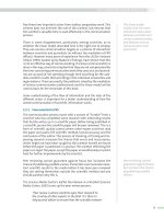
Figure 2A.1 Cross-sectional view of a low-level anti-coincidence beta-particle counter
A. Sample on a planchet. B. Thin window detector. C. Guard detector. Lead shielding
surrounds the entire detector system. Typical background count rates are about 1
count per minute for beta particles and 0.1 count per minute for alpha particles.
A sample mounted on a planchet (A) is placed below the thin window. When the
guard detector (C) is triggered by an extraneous radiation that penetrates the lead
shield, the sample detector (B) is inactivated. Immediately following, the detector (B)
responds to beta particles from the sample. For low-activity samples, the probability is
low that a particle from the sample registers a pulse at the same time that the counter
is inactivated.
www.pdfgrip.com
Radiation Detection Instrument Calibration and Quality Assurance
Figure 2A.2 Typical curve of beta-particle counting efficiency vs. energy for a thinwindow proportional counter.
geometric arrangement because of scattering from the sample support and
nearby solids. Back-scattering adds about 0.25 of the direct radiation, and
other scattering, about 0.02.
Beta particle calibration sources span energies from about 100 to 3,000
keV for proportional counters, and down to a few keV for liquid scintillation counters. In this experiment, a low-background, gas-flow, end-window
proportional counter with automatic sample changer for alpha- and betaparticle counting is calibrated. Beta-particles sources are counted with pulseheight discrimination to eliminate interference from alpha particles; the
discriminator may be turned off when no alpha particles are present.
The samples in this experiment are prepared for beta-particle counting by
pipetting a selected small volume of a radionuclide standard solution onto the
center of a planchet and then evaporating the solution to dryness. The planchet
is then placed to face the end-window of the detector for counting. For the
usual low-level samples, the planchet is placed as close to the window as
possible without damaging or contaminating the window, and is counted for
an extended period, typically for several thousand seconds. Blank planchets
also are counted – usually for even longer periods – to measure the radiation
background of the system. Considerable time is needed to count all samples,
hence the completion of this experiment will be delayed until all data are
accumulated and processed.
The counting efficiency ( ) of the proportional detector is calculated as the
ratio of the net count rate, in s−1 , to the activity (A), in Bq, of this standard
radionuclide solution. The net count rate is the standard’s gross count rate (RG )
minus the detector’s background count rate (RB ). The reported disintegration
rate (A) is the product of the radionuclide concentration, in Bq L−1 , and the
amount of counted sample, in L, adjusted for the radioactive decay of the
radionuclide between standardization and measurement. Equation 2A.1 is the
general form of this equation.
=
R G − RB
A
(2A.1)
An alternative unit to the Bq, or disintegration per second, is the curie (Ci),
which is 3.7 x 10¹⁰ dps (the activity of 1.0 gram of radium). Appendix 5 has
17
www.pdfgrip.com
18
Experiment 2
definitions for the two major units of radioactivity and the multiple units in
the international system (SI).
The counting efficiency is used to calculate the disintegration rates of
samples of known radionuclide identity but of unknown activity (to the
student) from their count rate and volume or mass. The counting efficiency
of radionuclides other than those used for calibration may be estimated by
interpolating efficiencies as a function of maximum beta-particle energy, and
adjusting for the decay fraction when multiple beta-particle groups are emitted
by a single radionuclide.
The beta-particle counting efficiency for a specific detector and radionuclide depends on numerous factors, notably intrinsic detector efficiency, the
source dimensions, distance from the detector, thickness and electron density,
support (backing) characteristics, scattering and attenuation environment, and
beta-particle energy distribution. Hence, the counting efficiency for other
radionuclides may differ somewhat from the interpolated estimate, and to a
greater degree for samples that are precipitated with different amounts and
types of carrier, or placed in different sample holders. The effect of sample
thickness is shown in Experiment 4. More discussion of counting efficiency
can be found in your Radioanalytical Chemistry textbook, Section 8.2.1.
Safety Reminders
• Follow the usual safety procedures when working in a radiological
laboratory.
• Caution should be exercised when preparing and working with corrosive
mineral acids.
• Liquids and solids are to be properly disposed of according to laboratory
rules and protocol.
• Standards must be returned to the designated place of storage.
Equipment and Supplies
• Low background gas-flow, end window proportional counter with automatic
sample changer for alpha and beta counting (or equivalent counting system)
• Steel planchets, 5-cm-dia., with 0.5-cm-high sides
• Pipettes, 100
Reagents
• Three certified standard low-level beta-particle solutions such as 60 Co
(Ebeta max 0.32 MeV, 99.9%), 204 Tl (0.76 MeV, 97.1%), and 89 Sr (1.50 MeV,
100%), each about 100 Bq/L
• Beta-particle solution of activity unknown to the student
Procedure
Step 1. Place 10 blank planchets in the proportional counter system and
count each for 50,000 s at settings (a) and (b) to determine the beta-particle
background count rate.
www.pdfgrip.com
Radiation Detection Instrument Calibration and Quality Assurance
a) beta particle, no alpha/beta pulse-height discrimination
b) beta particle with alpha/beta pulse-height discrimination
Record your measurements in Data Table 2A.1.
Step 2. Prepare sources in duplicate for the proportional counter by pipetting
the appropriate volume – typically 100 – specified by the instructor of each
of 3 standard solutions and the unknown solution onto the center of a separate
planchet. You will have a total of 8 planchets: 2 each for the beta-particle
standards and the unknown beta-particle sample. Dry under heat lamps placed
at sufficient distance over the planchets for slow drying.
Step 3. Place in the counter sample changing system the planchets with the
the beta-particle standard sources and the unknown beta-particle sample. Add
two background planchets. Set the time control to count each sample for a
time period specified by the instructor so that each accumulated count is at
least 1,000 counts (typically, 500 s per sample, 50,000 s per background).
Count each of the samples at settings (a) and (b). Repeat the count. Record
your measurements in Data Table 2A.2.
Step 4. Count one of the beta-particle standard sources 10 times, using
setting (a). Record your measurements in Data Table 2A.3.
Data Table 2A.1 Background Data for Step 1
Planchet #
Beta Count Rate
under Setting (a)
Beta Count Rate
under Setting (b)
1
2
3
4
5
6
7
8
9
10
Data Table 2A.2 Sample Counting Data for Step 3
Planchet ID
Std 1
Std 1
Std 2
Std 2
Std 3
Std 3
Unk 1
Unk 2
Bkg 1
Bkg 2
Beta Count Rate
under Setting (a)
Beta Count Rate
under Setting (b)
19