Advanced ESR methods in polymer research
Bạn đang xem bản rút gọn của tài liệu. Xem và tải ngay bản đầy đủ của tài liệu tại đây (5.35 MB, 359 trang )
0471450677.ffirs.qxd
8/16/2006
11:40 AM
Page i
www.pdfgrip.com
ADVANCED ESR
METHODS IN POLYMER
RESEARCH
0471450677.ffirs.qxd
8/16/2006
11:40 AM
Page iii
www.pdfgrip.com
ADVANCED ESR
METHODS IN POLYMER
RESEARCH
Edited by
SHULAMITH SCHLICK
University of Detroit Mercy
Detroit, Michigan
A John Wiley & Sons, Inc., Publication
0471450677.ffirs.qxd
8/16/2006
11:40 AM
Page iv
www.pdfgrip.com
Copyright © 2006 by John Wiley & Sons, Inc. All rights reserved
Published by John Wiley & Sons, Inc., Hoboken, New Jersey
Published simultaneously in Canada
No part of this publication may be reproduced, stored in a retrieval system, or transmitted in any form or by
any means, electronic, mechanical, photocopying, recording, scanning, or otherwise, except as permitted
under Section 107 or 108 of the 1976 United States Copyright Act, without either the prior written
permission of the Publisher, or authorization through payment of the appropriate per-copy fee to the
Copyright Clearance Center, Inc., 222 Rosewood Drive, Danvers, MA 01923, (978) 750-8400, fax (978)
750-4470, or on the web at www.copyright.com. Requests to the Publisher for permission should be
addressed to the Permissions Department, John Wiley & Sons, Inc., 111 River Street, Hoboken, NJ 07030,
(201) 748-6011, fax (201) 748-6008, or online at />Limit of Liability/Disclaimer of Warranty: While the publisher and author have used their best efforts in
preparing this book, they make no representations or warranties with respect to the accuracy or
completeness of the contents of this book and specifically disclaim any implied warranties of
merchantability or fitness for a particular purpose. No warranty may be created or extended by sales
representatives or written sales materials. The advice and strategies contained herein may not be suitable
for your situation. You should consult with a professional where appropriate. Neither the publisher nor
author shall be liable for any loss of profit or any other commercial damages, including but not limited to
special, incidental, consequential, or other damages.
For general information on our other products and services or for technical support, please contact our
Customer Care Department within the United States at (877) 762-2974, outside the United States at (317)
572-3993 or fax (317) 572-4002.
Wiley also publishes its books in a variety of electronic formats. Some content that appears in print may not
be available in electronic formats. For more information about Wiley products, visit our web site at
www.wiley.com.
Library of Congress Cataloging-in-Publication Data:
Advanced ESR methods in polymer research/edited by Shulamith Schlick.
p.cm.
Includes bibliographical references and index.
ISBN-13: 978-0-471-73189-4
ISBN-10: 0-471-73189-7
1. Electron paramagnetic resonance––Research. 2. Polymers––Research. I. Schlick,
Shulamith.
QC763.A32.2006
547'.7046––dc22
2006044267
Printed in the United States of America
10 9 8 7 6 5 4 3 2 1
0471450677.ffirs.qxd
8/16/2006
11:40 AM
Page v
www.pdfgrip.com
DEDICATION
My experience and understanding of ESR methodologies have benefited greatly
from interactions with my co-workers, who joined my lab and shared with me their
ambitions, knowledge, creativity, and technical skills. Over the years these coworkers became my professional family. To them this book is dedicated.
0471450677.ftoc.qxd
8/16/2006
11:43 AM
Page vii
www.pdfgrip.com
CONTENTS
PREFACE
ix
ABOUT THE EDITOR
xi
CONTRIBUTORS
PART I
1
ESR FUNDAMENTALS
Continuous-Wave and Pulsed ESR Methods
xiii
1
3
Gunnar Jeschke and Shulamith Schlick
2
Double Resonance ESR Methods
25
Gunnar Jeschke
3
Calculating Slow-Motion ESR Spectra of Spin-Labeled
Polymers
53
Keith A. Earle and David E. Budil
4
ESR Imaging
85
Shulamith Schlick
PART II
5
ESR APPLICATIONS
ESR Study of Radicals in Conventional Radical
Polymerization Using Radical Precursors Prepared by
Atom Transfer Radical Polymerization
99
101
Atsushi Kajiwara and Krzysztof Matyjaszewski
6
Local Dynamics of Polymers in Solution by Spin-Label ESR
133
Jan PilarB
vii
0471450677.ftoc.qxd
8/16/2006
11:43 AM
Page viii
www.pdfgrip.com
viii
7
CONTENTS
Site-Specific Information on Macromolecular Materials by
Combining CW and Pulsed ESR on Spin Probes
165
Gunnar Jeschke
8
ESR Methods for Assessing the Stability of Polymer
Membranes Used in Fuel Cells
197
Emil Roduner and Shulamith Schlick
9
Spatially Resolved Degradation in Heterophasic Polymers
From 1D and 2D Spectral–Spatial ESR Imaging Experiments
229
Shulamith Schlick and Krzysztof Kruczala
10
ESR Studies of Photooxidation and Stabilization of
Polymer Coatings
255
David R. Bauer and John L. Gerlock
11
Characterization of Dendrimer Structures by ESR Techniques
279
M. Francesca Ottaviani and Nicholas J. Turro
12
High-Field ESR Spectroscopy of Conductive Polymers
307
Victor I. Krinichnyi
INDEX
339
0471450677.fpref.qxd
8/16/2006
11:45 AM
Page ix
www.pdfgrip.com
PREFACE
In May 1994, I visited Professor Bengt Rånby at the Royal Institute of Technology in
Stockholm, Sweden. Professor Rånby, at that time Emeritus, was enthusiastic about his
numerous projects, including collaborations with Chinese scientists. On that occasion, I
mentioned to him how useful his 1977 book entitled ESR Spectroscopy in Polymer
Research, which he wrote together with J.F. Rabek, had been to me and many of my colleagues over the years. Professor Rånby confided that he planned a sequel, which “would
be published sometime soon.” I was hopeful, and expectant, but this was not to be.
So, what to do with all the excitement in the electron spin resonance (ESR) community over the extraordinary advances in ESR techniques in the last 20 years, techniques that have been used in Polymer Science? The pulsed, high field, double
resonance, and DEER experiments, ESR imaging, simulations? Someone must tell
the story, and I took the challenge.
In the winter of 2004, I was on sabbatical at the Max Planck Institute for Polymer
Research in Mainz, Germany, shared an office with Gunnar Jeschke, and worked
with him on the ESR chapter for the Encyclopedia of Polymer Science and
Technology (EPST).* Jacqueline I. Kroschwitz, the editor of EPST, encouraged me
to enlarge the chapter into a full volume. In all planning and writing stages, I benefited greatly from numerous discussions with Gunnar, who has enriched the book by
the three chapters that he contributed.
The final content of this book evolved during many talks with students and coworkers at UDM and colleagues at other institutions, and during long walks in my
neighborhood. It took the talent, dedication, and patience of the contributors to travel
*
Schlick, S.; Jeschke, G. Electron Spin Resonance, In Encyclopedia of Polymer Science and Engineering,
Kroschwitz, J.I., Ed.; Wiley-Interscience: New York, NY, 2004; Chap. 9, pp. 614–651 (web and hardcopy
editions).
ix
0471450677.fpref.qxd
8/16/2006
11:45 AM
Page x
www.pdfgrip.com
x
PREFACE
through the seemingly endless revisions and to arrive at the published volume. I am
grateful to Arza Seidel and her team at Wiley for guidance during all stages of this
project.
Part I of the present volume includes the fundamentals and developments of the
ESR experimental and simulations techniques. This part could be a valuable introduction to students interested in ESR, or in the ESR of polymers. Part II describes the
wide range of applications to polymeric systems, from living radical polymerization
to block copolymers, polymer solutions, ion-containing polymers, polymer lattices,
membranes in fuel cells, degradation, polymer coatings, dendrimers, and conductive
polymers: a world of ESR cum polymers. It is my hope that the wide range of ESR
techniques and applications will be of interest to students and mature polymer scientists and will encourage them to apply ESR methods more widely to polymeric materials. And I extend an invitation to ESR specialists, to apply their talents to polymers.
SHULAMITH SCHLICK
February 2006
0471450677.flast.qxd
8/16/2006
11:47 AM
Page xi
www.pdfgrip.com
ABOUT THE EDITOR
Shulamith Schlick, D.Sc., is Professor of Physical and Polymer Chemistry in the
Department of Chemistry and Biochemistry, University of Detroit Mercy in Detroit,
Michigan.
Dr. Schlick received her undergraduate degree in Chemical Engineering at the
Technion, Israel Institute of Technology in Haifa, Israel. At the same institution, she
also obtained her M.Sc. in Polymer Chemistry and her D.Sc. degree in Molecular
Spectroscopy. She taught at the Technion, Wayne State University, and the University
of Windsor. In 1983, she assumed her present position at UDM. In recent years, she
held Visiting Professorships at the Department of Chemistry, University of Florence,
Italy, at the Department of Chemistry, University of Bologna, Italy, and at the MaxPlanck Institute for Polymer Research, Mainz, Germany. She spent sabbatical leaves
at the Centre d’Études Nucléaires de Grenoble, in Grenoble, France; as Varon
Visiting Professor at the Weizmann Institute of Science, Rehovot, Israel; at the
Department of Polymer Chemistry, Tokyo Institute of Technology; at the University
of Bologna; and at MPI, Mainz, Germany.
Current research interests of the editor are morphology, phase separation, and
self-assembling in ionomers and nonionic polymeric surfactants; electron spin resonance imaging (ESRI) of transport processes in polymer solutions and swollen gels;
dynamical processes in disordered systems using electron spin probes and 2H NMR;
ESR and ESRI of degradation and stabilization processes in thermally-treated and
UV-irradiated polymers; study of the stability of polymeric membranes used in fuel
cells; and DFT calculations of the geometry and electronic structure of organic radicals, with emphasis on fluorinated radicals. Her research has resulted in more than
200 publications and has been supported by NSF, DOD, PRF, NATO, AAUW, Ford
Motor Company, Dow Chemical Company, and the Fuel Cell Activity Center of
xi
0471450677.flast.qxd
8/16/2006
11:47 AM
Page xii
www.pdfgrip.com
xii
ABOUT THE EDITOR
General Motors. Dr. Schlick was the recipient of two Creativity Awards from the
Polymer Program of the National Science Foundation, and of an Honorary Doctorate
(Doctor Honoris Causa) from Linköping University, Sweden, in May 2003.
Dr. Schlick is a member of the American Chemical Society, American Physical
Society, American Association for the Advancement of Science, American
Association of University Women, and International ESR Society.
0471450677.flast.qxd
8/16/2006
11:47 AM
Page xiii
www.pdfgrip.com
CONTRIBUTORS
David R. Bauer, Research and Advanced Engineering, Ford Motor Company,
Dearborn, Michigan, ESR Studies of Photooxidation and Stabilization of Polymer
Coatings (Chapter 10).
David E. Budil, Department of Chemistry, Northeastern University, Boston,
Massachusetts, Calculating Slow-Motion ESR Spectra of Spin-Labeled Polymers
(Chapter 3).
Keith A. Earle, Department of Physics, University of Albany (SUNY), Albany,
New York, Calculating Slow-Motion ESR Spectra of Spin-Labeled Polymers
(Chapter 3).
John L. Gerlock, Ford Motor Company (retired), ESR Studies of Photooxidation
and Stabilization of Polymer Coatings (Chapter 10).
Gunnar Jeschke, MPI for Polymer Research, Mainz, Germany, Continuous-Wave
and Pulsed ESR Methods (Chapter 1), Double Resonance ESR Methods (Chapter 2),
Site-Specific Information on Macromolecular Materials by Combining CW and
Pulsed ESR on Spin Probes (Chapter 7).
Astushi Kajiwara, Nara University of Education, Nara, Japan, ESR Study of
Radicals in Conventional Radical Polymerization Using Radical Precursors
Prepared by Atom Transfer Radical Polymerization (Chapter 5).
Victor I. Krinichnyi, Institute of Problems of Chemical Physics, Chernogolovka,
Moscow Region, Russia, High-Field ESR Spectroscopy of Conductive Polymers
(Chapter 12).
xiii
0471450677.flast.qxd
8/16/2006
11:47 AM
Page xiv
www.pdfgrip.com
xiv
CONTRIBUTORS
Krzysztof Kruczala, Faculty of Chemistry, Jagiellonian University, Cracow, Poland,
Spatially Resolved Degradation in Heterophasic Polymers From 1D and 2D
Spectral–Spatial ESR Imaging Experiments (Chapter 9) .
Krzysztof Matyjaszewski, Department of Chemistry, Carnegie Mellon University,
Pittsburgh, Pennsylvania, ESR Study of Radicals in Conventional Radical
Polymerization Using Radical Precursors Prepared by Atom Transfer Radical
Polymerization (Chapter 5).
M. Francesca Ottaviani, Institute of Chemical Sciences, University of Urbino,
Urbino, Italy, Characterization of Dendrimer Structures by ESR Techniques
(Chapter 11).
Jan Pilar, Institute of Macromolecular Chemistry, Academy of Sciences of the
Czech Republic, Prague, Czech Republic, Local Dynamics of Polymers in
Solution by Spin-Label ESR (Chapter 6).
Emil Roduner, Institute of Physical Chemistry, University of Stuttgart, Stuttgart,
Germany, ESR Methods for Assessing the Stability of Polymer Membranes Used
in Fuel Cells (Chapter 8).
Shulamith Schlick, Department of Chemistry and Biochemistry, University of
Detroit Mercy, Detroit, Michigan, Continuous-Wave and Pulsed ESR Methods
(Chapter 1), ESR Imaging (Chapter 4), ESR Methods for Assessing the Stability
of Polymer Membranes Used in Fuel Cells (Chapter 8), Spatially Resolved
Degradation in Heterophasic Polymers From 1D and 2D Spectral–Spatial ESR
Imaging Experiments (Chapter 9).
Nicholas J. Turro, Department of Chemistry, Columbia University, New York,
Characterization of Dendrimer Structures by ESR Techniques (Chapter 11).
JWUS_ESR-Schlick_Ch001.qxd
8/8/2006
1:20 PM
Page 1
www.pdfgrip.com
PART I
ESR FUNDAMENTALS
JWUS_ESR-Schlick_Ch001.qxd
8/8/2006
1:20 PM
Page 3
www.pdfgrip.com
1
CONTINUOUS-WAVE AND PULSED
ESR METHODS
GUNNAR JESCHKE
Max Planck Institute for Polymer Research, Mainz, Germany
SHULAMITH SCHLICK
University of Detroit Mercy, Detroit, Michigan
Contents
1. Introduction
2. Fundamentals of Electron Spin Resonance Spectroscopy
2.1. Basic Principles
2.2. Anisotropic Hyperfine Interaction and g-Tensor
2.3. Isotropic Hyperfine Analysis
2.4. Environmental Effects on g- and Hyperfine Interaction
2.5. Accessibility to Paramagnetic Quenchers
2.6. Line Shape Analysis for Tumbling Nitroxide Radicals
3. Multifrequency and High-Field ESR
4. Pulsed ESR Methods
Acknowledgments
References
3
4
4
10
12
12
13
15
16
18
22
22
1. INTRODUCTION
Electron spin resonance (ESR) is a spectroscopic technique that detects the transitions induced by electromagnetic radiation between the energy levels of electron
Advanced ESR Methods in Polymer Research, edited by Shulamith Schlick.
Copyright © 2006 John Wiley & Sons, Inc.
3
JWUS_ESR-Schlick_Ch001.qxd
8/8/2006
1:20 PM
Page 4
www.pdfgrip.com
4
CONTINUOUS-WAVE AND PULSED ESR METHODS
spins in the presence of a static magnetic field. The method can be applied to the
study of species containing one or more unpaired electron spins; examples include
organic and inorganic radicals, triplet states, and complexes of paramagnetic ions.
Spectral features, such as resonance frequencies, splittings, line shapes, and line
widths, are sensitive to the electronic distribution, molecular orientations, nature of
the environment, and molecular motions. Theoretical and experimental aspects of
ESR have been covered in a number of books,1–8 and reviewed regularly.9–11
Currently available textbooks and monographs are written for students and scientists that specialize in the development of ESR technique and its application to a broad
range of samples. Nowadays, however, research groups are interested in a specific
field of applications, such as polymer science, and apply more than one characterization method to the materials of interest. An introduction to ESR that targets such an
audience needs to be shorter, less mathematical, and focused on application rather
than methodological issues. This chapter is an attempt to provide such a short introduction on the application of ESR spectroscopy to problems in polymer science.
Organic radicals occur in polymers as intermediates in chain-growth and depolymerization reactions,12–15 or as a result of high-energy irradiation (γ, electron
beams).13,14 Paramagnetic transition metal ions are present in a number of functional
polymer materials, such as catalysts and photovoltaic devices.16 However, much of
the modern ESR work in polymer science focuses on diamagnetic materials that are
either doped with stable radicals as “spin probes”, or labeled by covalent attachment
of such radicals as “spin labels” to polymer chains.9,17–22 This chapter therefore treats
the basic concepts that are required to understand ESR spectra of a broad range of
organic radicals and transition metal ions, and describes more advanced concepts as
applied to the most popular class of spin probes and labels: nitroxide radicals.
2. FUNDAMENTALS OF ELECTRON SPIN RESONANCE
SPECTROSCOPY
2.1. Basic Principles
Spins are magnetic moments that are associated with angular momentum; they interact with external magnetic fields (Zeeman interaction) and with each other (couplings). In most cases, the Zeeman interaction of the electron spin is the largest
interaction in the spin system (high–field limit). The electron Zeeman (EZ) interaction can generally be described by the Hamiltonian below,
HEZ ϭ βeB0gS
(1)
where S is the spin vector operator, B0 is the transposed magnetic field vector in gauss
(G) or tesla (1 T ϭ 104 G), βe is the Bohr magneton equal to 9.274 ϫ 10Ϫ21 ergGϪ1 (or
9.274 ϫ 10Ϫ24 JTϪ1), and g is the g tensor. For a free electron, g is simply the number
ge ϭ 2.002319. The transition energy is then ∆E ϭ hνmw ϭ geβeB0, where B0 is the
magnitude of the magnetic field. Typical values are B0 ≈ 0.34 T (3400 G) corresponding to microwave (mw) frequencies of Ϸ9.6 GHz (X band), or B0 ≈ 3.35 T corresponding to mw frequencies of Ϸ94 GHz (W band).
JWUS_ESR-Schlick_Ch001.qxd
8/8/2006
1:20 PM
Page 5
www.pdfgrip.com
FUNDAMENTALS OF ELECTRON SPIN RESONANCE SPECTROSCOPY
5
The g-value of a bound electron generally exhibits some deviation from ge that is
mainly due to interaction of the spin with orbital angular momentum of the unpaired
electron (spin–orbit coupling). Spin–orbit coupling is a relativistic effect that tends to
increase with increasing atomic number of the nuclei that contribute atomic orbitals
to the singly occupied molecular orbital. Therefore, g-values deviate more strongly
from ge for transition metal complexes than for organic radicals. As the orbital angular momentum is quenched in the ground state of molecules, spin–orbit coupling
comes about only by admixture of excited orbitals. Such admixture is stronger for
low–lying excited states, which are relevant, for example, if the unpaired electron has
high density at an oxygen atom. Oxygen-centered organic radicals thus tend to have
higher g-values than carbon-centered ones.
As the orbital angular momentum relates to a molecular coordinate frame and the
spin is quantized along the magnetic field (z axis of the laboratory frame), the g-value
depends on the orientation of the molecule with respect to the field. This anisotropy
can be described by a second rank tensor with three principal values, gx, gy, and gz.
The corresponding principal axes define the molecular frame. In fluid solutions,
molecules tumble with a rotational diffusion rate that is much higher than the differences of the electron Zeeman frequencies between different orientations. In this
situation, the g-value is orientationally averaged and only its isotropic value
giso ϭ (gx ϩ gy ϩ gz)/3 can be measured. A good overview of isotropic g-values of
organic radicals can be found in Ref. 23; Ref. 5 collects information on g tensors for
transition metal complexes.
The real power of ESR spectroscopy for structural studies is based on the interaction of the unpaired electron spin with nuclear spins. This hyperfine interaction splits
each energy level into sublevels and often allows the determination of the atomic or
molecular structure of species containing unpaired electrons, and of the ligation
scheme around paramagnetic transition metal ions. For a system with m nuclear spins
(identified by index k) and a single electron spin, which may be larger than one-half
as explained below, the hyperfine Hamiltonian is given in Eq. 2,
Hhfi ϭ h⌺ S·Ak·Ik
(2)
where the Ik are nuclear spin vector operators and the Ak are hyperfine tensors in
frequency units (Hz). Each hyperfine tensor is characterized by three principal
values Ax, Ay, and Az and by the relative orientation of its principal axes system
with respect to the molecular frame defined by the g-tensor. This relative orientation is most easily defined by three Euler angles α, β, γ, which correspond to a
sequence of rotations about the z axis (by angle α), the new y' axis (by angle β),
and the final z'' axis (by angle γ); these rotations transform the principal axes
frame of the hyperfine tensor into that of the g-tensor. The relative orientation is
often given as direction cosines, which are the coordinates of unit vectors along
the directions of the hyperfine principal axes given in the coordinate frame of the
g-tensor.
Only the isotropic value Aiso ϭ (Ax ϩ Ay ϩ Az)/3 can be measured in fluid solutions, and is due to the Fermi contact interactions of electrons that reside in an s
orbital of the nucleus under consideration. The contribution of a single orbital is
JWUS_ESR-Schlick_Ch001.qxd
8/8/2006
1:20 PM
Page 6
www.pdfgrip.com
6
CONTINUOUS-WAVE AND PULSED ESR METHODS
proportional to the spin population (spin density) in that orbital, to the probability
density |ψ0|2 of the orbital wave function at its center (inside the nucleus), and to the
nuclear g-value, gn. To a very good approximation, the hyperfine couplings for different isotopes of the same element thus have the same ratio as the gn values.
Purely anisotropic contributions (Ax ϩ Ay ϩ Az ϭ 0) to the hyperfine coupling
result from spin density in p, d, or f orbitals on the nucleus and from the
dipole–dipole interaction T between the electron and nuclear spin. If the electron
spin is confined to a region that is much smaller than the electron–nuclear distance
ren, both spins can be treated as point dipoles and the magnitude of T is proportional
to renϪ3. In this case, T has axial symmetry and its principal values are given by
Tx ϭ Ty ϭ Ϫ T and Tz ϭ 2T. Furthermore, if the spin density in p, d, and f orbitals on
that nucleus is negligible, as is the case for protons (1H), the measurement of the
hyperfine anisotropy can provide the electron–nuclear distance ren. Any spin density
at the nucleus under consideration is negligible if this nucleus is located in a neighboring molecule and does not interact (by van der Waals or hydrogen bonding) with
a nucleus on which much spin density is located. Intermolecular distances larger than
Ϸ 0.3 nm can thus be inferred from hyperfine couplings.
For nuclei with significant hyperfine interaction, the other interactions of the
nuclear spin also need to be considered. The nuclear Zeeman (NZ) interaction of
these spins with the external magnetic field is described in Eq. 3.
HNZ ϭ Ϫ⌺ gn,k nB0 Ik
(3)
Nuclear spins with I > ᎏ12 have an electric quadrupole moment that interacts with
the quadrupole moment of the charge distribution around the nucleus. The
Hamiltonian for this nuclear quadrupole (NQ) interaction is given in Eq. 4,
HNQ ϭ h⌺ Ik Qk Ik
(4)
where Qk are the traceless (Qx ϩ Qy ϩ Qz ϭ 0) nuclear quadrupole tensors. Because
the tensor is traceless, this interaction is not detected in fluid media.
Both the nuclear Zeeman and nuclear quadrupole interaction do not depend on the
magnetic quantum number mS of the electron spin. As the selection rule for ESR transitions is given by Eq. 5,
⌬mS ϭ Ϯ1
and
⌬mI ϭ 0
(5)
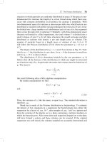
where mI is the nuclear spin quantum number, these interactions do not make a firstorder contribution to the ESR spectrum. In many cases, they can thus be neglected
in spectrum analysis. This situation is illustrated in Fig. 1 for a nitroxide in which
the nuclear spin I ϭ 1 of the 14N atom is coupled to the electron spin S ϭ ᎏ12ᎏ that
resides mainly in the pz orbitals on the N and O atom. The hyperfine coupling
causes a splitting of each of the electron spin levels (mS ϭ Ϫ ᎏ12ᎏ and mS ϭ ϩ ᎏ12ᎏ) into
three sublevels. When a constant microwave frequency νmw is irradiated and the
magnetic field is swept, three resonance transitions are observed (Fig. 1a). The
JWUS_ESR-Schlick_Ch001.qxd
8/8/2006
1:20 PM
Page 7
www.pdfgrip.com
FUNDAMENTALS OF ELECTRON SPIN RESONANCE SPECTROSCOPY
7
nuclear Zeeman interaction shifts both mI ϭ ϩ1 sublevels to lower and both
mI ϭ Ϫ1 sublevels to higher energy, but does not influence the resonance fields
where the splitting between the levels with different mS and the same mI matches the
energy of the mw quantum (Fig. 1b).
More generally, the higher sensitivity of ESR experiments can be used for the
detection of NMR frequencies by applying both resonant mw and resonant radio frequency (rf) irradiation to the spin system. Such electron nuclear double-resonance
(ENDOR) experiments are discussed in Chapter 2.
Transition metal ions can have several unpaired electrons when they are in their
high- spin state; examples are Cr(III) (3d3 configuration, S ϭ ᎏ32ᎏ), Mn(II) (3d 5, S ϭ ᎏ52ᎏ),
(a)
+1
E
0 −1
mS
+1/2
+1
0
−1
mI
h νmw
−1/2
−1
0
+1
B0
( b)
+1
E
0 −1
mS
+1
0
−1
mI
−1/2
−1/2
h νmw
−1
0
+1
B0
Fig. 1. Energy level schemes and ESR spectrum for a spin system of an electron spin S ϭ ᎏ1ᎏ
2
coupled to a nuclear spin I ϭ 1 (e.g., 14N in a nitroxide). (a) Only the electron Zeeman and
hyperfine interactions are considered. (b) The electron Zeeman, hyperfine, and nuclear
Zeeman interactions are considered. Note that the splittings match the microwave quantum at
the same resonance fields as in part a.
JWUS_ESR-Schlick_Ch001.qxd
8/8/2006
1:20 PM
Page 8
www.pdfgrip.com
8
CONTINUOUS-WAVE AND PULSED ESR METHODS
and Fe(III) (3d5, S ϭ ᎏ52ᎏ). The spins of these electrons are tightly coupled and have to
be considered as a single group spin S Ͼ ᎏ12ᎏ. Such an electron group spin also has an
electric quadrupole moment. For historical reasons, the electron spin analog of the
nuclear quadrupole interaction is termed zero-field splitting (ZFS) and is described
by Eq. 6,
HZFS ϭ h S D S
(6)
where D is a traceless tensor. Therefore, the ZFS can be characterized by two parameters, D ϭ 3Dz/2 and E ϭ (Dx Ϫ Dy)/2, rather than by giving all three principal values. For axial symmetry E ϭ 0, and for maximum nonaxiality E ϭ D/3.
With the exception of transition metal ions at a site with cubic symmetry, the ZFS
often exceeds the electron Zeeman interaction at magnetic fields Ͻ1 T, sometimes
even at the highest accessible fields (high-spin Fe(III)). In this situation, only the
lowest lying doublet of spin states may be populated and only transitions within this
doublet can be observed. It is convenient to describe such a doublet by an effective
spin S ' ϭ ᎏ12ᎏ. The ZFS of the group spin S Ͼ ᎏ12ᎏ then contributes to the effective g-tensor of the spin S' ϭ ᎏ12ᎏ. For example, X-band ESR spectra of high-spin Fe(III) in a
situation with maximum nonaxiality of the ZFS (E ϭ D/3) exhibit a sharp feature at
g ϭ 4.3. Note that unlike the normal g-tensor, the effective g-tensor may depend on
the applied magnetic field.
For low concentrations of the paramagnetic centers, the electron spins can be considered isolated from each other, and only a single electron spin S appears in the
Hamiltonian. In systems with a high concentration of paramagnetic transition metal
ions, this situation can be achieved by diamagnetic dilution with transition ions of the
same charge and similar radius and coordination chemistry. However, there are a
number of systems that feature coupled electron spins, for example, binuclear metal
complexes and biradicals. Any pair of electron spins Sk and Sl in such a system interacts through space by dipole–dipole coupling, which is analogous to the dipolar part
T of the hyperfine coupling. The Hamiltonian of the electronic dipole–dipole (DD)
coupling is given by Eq. 7,
HDD ϭ h ΣSk Dkl Sl
(7)
where the Dkl are the traceless dipole–dipole tensors. If the two electron spins are far
apart, the coupling can be described by a point-dipole approximation in which Dkl is
an axial tensor with principal values Dz,kl ϭ 2d and Dx,kl ϭ Dy,kl ϭ Ϫd. As d is
inversely proportional to the cube of the distance rkl between the two spins, a measurement of this coupling can thus yield the spin–spin distance. Such measurements
are discussed in more detail in Chapter 2.
The two electrons can exchange if their wave functions overlap. Even for localized electrons, such an exchange is significant at a distance rkl Ͻ 1.5 nm. For an antibonding overlap of the two orbitals, the exchange interaction J is negative and the
triplet state of the pair has lower energy than the singlet state. This is called a ferromagnetic exchange coupling. Consequently, bonding overlap leads to a positive J, a
JWUS_ESR-Schlick_Ch001.qxd
8/8/2006
1:20 PM
Page 9
www.pdfgrip.com
9
FUNDAMENTALS OF ELECTRON SPIN RESONANCE SPECTROSCOPY
lower lying singlet state, and antiferromagnetic coupling. The exchange coupling is
not strictly isotropic, but except for electron spins at distances Ͻ 0.5 nm, the
anisotropic contribution can usually be neglected. For a purely isotropic exchange
coupling, the Hamiltonian is written in Eq. 8.
Hex ϭ h ΣJklSkSl
(8)
Unlike the dipole–dipole coupling between the electron spins, the exchange coupling
can thus be detected in fluid solutions.
The ESR spectra of monoradicals and mononuclear transition ion complexes
can also be influenced by spin exchange, because the wave functions of the electrons overlap for a short time during diffusional collisions of paramagnetic
species.24 At moderate concentrations (1 M or larger), the collisions are so frequent
that line broadening and a decrease of the hyperfine splitting can be observed. In
macromolecular and supramolecular systems, this effect is sometimes perceptible
at lower bulk concentrations, as diffusion may be restricted or local concentrations
of some species strongly exceed their bulk concentration. Examples are discussed
in Chapter 7.
When the various spin interactions can be separated experimentally or by spectral
analysis, ESR spectra become a rich source of information not only on chemical
structure of the paramagnetic species, but also on the structure and dynamics of their
environment. Figure 2 provides an overview of time scales and length scales that can
be accessed in this way. T1 and T2 are the longitudinal and transverse relaxation times,
respectively.
100 kHz
1 MHz
100 MHz
NMR
frequency bands
X
ENDOR
energy
10 GHz
S
1 mJ mol−1
thermal energy
W
Q
1 J mol−1
1 mK
1 THz
100 J mol−1
1 K 4.2 K
50 K
electron−electron distance
8
4
2
1 nm
electron−proton distance
8
4
2
1Å
T1 (typical)
T2 (typical)
10 ms
1 ms
10 ns
slow tumbling
100 ps
1 ps
Fig. 2. Frequencies, time scales, energies, and length scales in ESR experiments.
JWUS_ESR-Schlick_Ch001.qxd
8/8/2006
1:20 PM
Page 10
www.pdfgrip.com
10
CONTINUOUS-WAVE AND PULSED ESR METHODS
2.2. Anisotropic Hyperfine Interaction and g-Tensor
Before considering the analysis of anisotropic solid-state ESR spectra in general, we
discuss the orientation dependence of spin interactions of the nitroxide radical as an
example. The ESR spectrum of a nitroxide is dominated by the hyperfine interaction
of the electron spin with the nuclear spin of the 14N atom and by g-shifts due to
spin–orbit coupling mainly in the 2pz orbital of the lone pair on the oxygen atom. The
14
N hyperfine coupling contains a sizeable isotropic contribution due to Fermi contact interaction in the 2s orbital on the nitrogen. An anisotropic contribution comes
from the spin density in the nitrogen 2pz orbital whose lobes are displayed in Fig. 3a.
If the external magnetic field B0 is parallel to these lobes (z axis of the molecular
frame), the hyperfine interaction and thus the splitting within the triplet is large; if it
is perpendicular to the lobes, the splitting is small. Conversely, g-shifts are small
when the lobes of the orbital under consideration (here the 2pz orbital on the oxygen)
are parallel to the field and large when they are perpendicular. In the case of a nitroxide, the strongest shift is observed when the field is parallel to the N–O bond, which
defines the x axis of the molecular frame. Hence, the triplets of lines at different orientations of the molecule with respect to the field do not only have different splittings, but their centers are also shifted with respect to each other.
In a macroscopically isotropic sample (all molecular orientations have the same
probability), the spectrum consists of contributions from all orientations when the
rotational motion is frozen on the time scale of the experiment. As ESR lines are
derivative absorption lines, negative and positive contributions from neighboring orientations cancel. Powder spectra are thus dominated by contributions at the minimum and maximum resonance fields, and by contributions at resonance fields that
are common to many spins. The latter contribution provides the center line in the
nitroxide powder spectrum (Fig. 3b). It corresponds mainly to molecules with
nuclear magnetic quantum number mI ϭ 0 (center line of all triplets, only g-shift).
The detailed shape of this powder spectrum can be simulated, but interpretation is not
easy, mainly because hyperfine and g anisotropy are of similar magnitude.
If one of the two interactions dominates, the spectra can be analyzed more easily.
For dominating g anisotropy (Fig. 4a), signals in the CW ESR spectrum are observed
at resonant fields corresponding to the principal values of the g- tensor: gz (low-field
edge), gy, and gx (high-field edge). For a g-tensor with axial symmetry (wave function of the unpaired electron has at least one symmetry axis Cn with n Ն 3), the intermediate feature coincides with one of the edges (Fig. 4b). For a dominating hyperfine
interaction with a nuclear spin I ϭ ᎏ12ᎏ the spectrum consists of two of these powder
patterns with mirror symmetry about the center of the spectrum (Fig. 4c).
When samples are available as single crystals, spectra corresponding to specific
orientations of the paramagnetic center with respect to the external field can be measured separately. The orientation dependence of the spectrum can then be studied systematically and the principal axes frames of the A- and g-tensors can be related to the
crystal frame. In polymer applications, samples are usually macroscopically
isotropic, so that only the principal values of the interactions, and in favorable cases
the relative orientations of their principal axes frames, can be obtained from spectral
simulations. How these frames are related to the molecular geometry then needs to be
JWUS_ESR-Schlick_Ch001.qxd
8/8/2006
1:20 PM
Page 11
www.pdfgrip.com
11
FUNDAMENTALS OF ELECTRON SPIN RESONANCE SPECTROSCOPY
(a)
2Azz (14N)
2Ayy (14N)
z
z
y
y
N
O
∆B =
x
h νmw
µ B∆ g
x
R
H
(b)
Fig. 3. Anisotropic interactions for a nitroxide radical. (a) Molecular frame of the nitroxide
molecule and single-crystal ESR spectra along the principal axes of this frame. (b) Powder
spectrum resulting from a superposition of the single-crystal spectra at all orientations of the
molecule with respect to the external magnetic field.
CW
Echo-detected
(a )
gy
gz
gy
gx
gz
gx
(b )
g⊥
g||
g||
g⊥
(c )
A⊥
A⊥
A||
A||
Fig. 4. Powder line shapes in continuous wave (CW) ESR (derivative absorption spectra) and
echo-detected ESR (absorption spectra). (a) Rhombic g-tensor. (b) Axial g-tensor. (c) Axial
hyperfine coupling tensor with dominating isotropic contribution.
JWUS_ESR-Schlick_Ch001.qxd
8/8/2006
1:20 PM
Page 12
www.pdfgrip.com
12
CONTINUOUS-WAVE AND PULSED ESR METHODS
established by theoretical considerations or by quantum chemical computations of
the interaction tensors.
2.3. Isotropic Hyperfine Analysis
Anisotropic line broadening in solids often leads to a situation in which only one
dominant hyperfine interaction is resolved, the one for the atom at which the spin
is localized. In fluid media, however, anisotropic contributions average, lines are
narrower, and a multitude of hyperfine interactions may be resolved. This situation is frequently observed for proton couplings in π radicals, where the electron
spin is distributed throughout a network of conjugated bonds. Examples can be
found in Ref. 23.
In isotropic ESR spectra, a single nucleus with spin Ik causes a splitting into 2Ik ϩ 1
lines corresponding to the magnetic quantum numbers mI ϭ ϪIk, ϪIk ϩ 1, … Ik. For a
group of nk equivalent nuclei (same isotropic hyperfine coupling), the number of lines
is 2nkIk ϩ 1. For groups of nonequivalent spins, the number of lines (multiplicities)
increases, and the total number of lines in the ESR spectrum is given in Eq. 9.
NESR ϭ ∏ (2nkIk ϩ 1)
(9)
An example is shown in Fig. 5, where the spectrum for an electron spin coupled to
four protons (I ϭ ᎏ12ᎏ) exhibits a regular pattern of 16 lines. In complicated spectra consisting of multiple interacting nuclei, some of the smaller hyperfine couplings cannot
be resolved. In such cases, ENDOR spectra are often easier to interpret, because each
proton contributes only two lines; this technique is described in Chapter 2.
2.4. Environmental Effects on g- and Hyperfine Interaction
Self-assembly of polymer chains is due to noncovalent interactions: hydrogen bonding, π stacking, and electrostatic and van der Waals interactions. The high sensitivity
of the NMR chemical shift of protons to π stacking (through ring currents) and
hydrogen bonding provides one way for their characterization.25 Since the magnetic
A3
A4
A2
A1
B res =
h νmw
µ Bg
magnetic field
Fig. 5. Isotropic ESR spectrum for a system consisting of four nuclear spins Ik ϭ ᎏ1ᎏ coupled to
2
a single electron spin S ϭ ᎏ1ᎏ.
2
JWUS_ESR-Schlick_Ch001.qxd
8/8/2006
1:20 PM
Page 13
www.pdfgrip.com
FUNDAMENTALS OF ELECTRON SPIN RESONANCE SPECTROSCOPY
13
parameters of paramagnetic probes are also sensitive to such interactions, ESR spectroscopy can confirm and complement the information obtained by NMR.
The hyperfine interaction is influenced by any environmental effect that can perturb the spin density distribution. For example, in nitroxide radicals the unpaired
electron is distributed between the nitrogen (Ϸ 40%) and oxygen atom (Ϸ 60%) in
the polar N–O bond (Fig. 6). This distribution can change in the vicinity of a polar
molecule (polar solvent or ion). Generally, a more polar solvent (higher dielectric
constant) leads to a higher spin density ρN on the nitrogen atom and thus to a larger
observed hyperfine coupling.26 The spin density distribution is also influenced by
hydrogen bonding to the oxygen atom, which also increases the hyperfine coupling.
The same interactions affect the deviation of gx from the free electron value ge, but
in the opposite direction, since the extent of spin–orbit coupling is proportional to the
spin density ρO on the oxygen atom. However, the effect on gx also depends on the
lone-pair energy, whose lowering causes stronger spin–orbit coupling. The lone-pair
energy in turn is more affected by hydrogen bonding than by the local polarity, so that
compared to Az, gx is more sensitive to hydrogen bonding than to polarity. Correlation
of gx to Az thus enable the separation of polarity and hydrogen-bonding effects.26 In
principle, the same effects scaled by a factor of one-third can be seen in the isotropic
values Aiso and giso, as the other principal values of the tensors are much less affected.
As a rule, measurements of Az and of gx in solid samples at high field (W band) are
much more precise than measurements of Aiso and giso at X-band frequencies.
2.5. Accessibility to Paramagnetic Quenchers
Spin exchange due to collision of paramagnetic species (see Section 2.1) can be used to
check whether a spin-labeled site in a macromolecule is accessible by the solvent. To
this end, a paramagnetic quencher is added to the solvent, and the effect on the spectrum
or relaxation time of the spin label is measured. The quencher is a fast relaxing paramagnetic species, usually a molecule or transition ion complex with spin S Ͼ ᎏ12ᎏ. The situation is illustrated in Fig. 7 for oxygen as the quencher (S ϭ 1, triplet ground state),
which is soluble in nonpolar solvents and only moderately soluble in water. We can
assume, without loss of generality, that at a certain time oxygen is in the TϪ1 triplet
z
y
+
δ
−
N
O
δ
x
H
Fig. 6. Effects of the local polarity and hydrogen bonding on the nitroxide radical. The distribution of the unpaired electron between the two 2pz orbitals on nitrogen and oxygen is
affected.