Tài liệu Báo cáo Y học: Human and Drosophila UDP-galactose transporters transport UDP-N-acetylgalactosamine in addition to UDP-galactose doc
Bạn đang xem bản rút gọn của tài liệu. Xem và tải ngay bản đầy đủ của tài liệu tại đây (742.4 KB, 11 trang )
Human and
Drosophila
UDP-galactose transporters transport
UDP-
N
-acetylgalactosamine in addition to UDP-galactose
Hiroaki Segawa*, Masao Kawakita and Nobuhiro Ishida
Department of Physiological Chemistry, The Tokyo Metropolitan Institute of Medical Science (Rinshoken), Honkomagome,
Bunkyo-ku, Tokyo, Japan
A putative Dros ophila nucleotide sugar transpor ter w as
characterized and shown to be the Drosophila homologue o f
the human UDP-Gal transporter (hUGT). When the
Drosophila melanogaster UDP-Gal transporter (DmUGT)
was expressed in mammalian cells, the transporter protein
was localized in the Golgi membranes and complemented
the UDP-Gal transport de®ciency of Lec8 cells but not the
CMP-Sia transport de®ciency of Lec2 cells. DmUGT and
hUGT were expressed in Saccharomyces cerevisiae c ells in
functionally active forms. Using microsomal v esicles isolated
from Saccharomyces cerevisiae expressing these transporters,
we unexpectedly found that both hUGT and DmUGT
could transport UDP-GalNAc as well as UDP-Gal. W hen
amino-acid residues that are conserved among human,
murine, ®ssion yeast a nd Drosophila UGTs, but are distinct
from corresponding ones conserved among CMP-Sia
transporters (CSTs), were substituted by those found in
CST, the mutant t ransporters were still active in transporting
UDP-Gal. One of these mutants in w hich Asn47 was sub-
stituted by Ala showed aberrant intracellular distribution
with concomitant destabilization of the protein product.
However, this mutation was suppressed by an Ile51 to Thr
second-site mutation. Both residues were localized within the
®rst transmembrane helix, suggesting that t he structure o f
the helix contributes to the stabilization and substrate rec-
ognition of the UGT molecule.
Keywords: UDP-galactose transporter; UDP-galactose;
UDP-N-acetylgalactosamine; nucleotide sugar transporter;
site-directed mutagenesis.
Oligosaccharide chains of secretory and membrane-bound
glycoproteins and glycolipids p lay important roles in
various biological processes. Two major groups of proteins,
nucleotide sugar transporters (NSTs) and glycosyltransfe-
rases, contribute to oligosaccharide synthesis. Nucleotide
sugar transporters carry speci®c nucleotide sugars that are
produced outside the Golgi apparatus and ER into these
organelles, where they s erve as the substrates for the
elongation of carbohydrate chains by appropriate glyco-
syltransf erase s. Changes in the activities of NSTs may
affect the structure of oligosaccharide chains by affecting
the availability of substrates for glycosyltransferases [1].
In fact, in organisms such as Drosophila melanogaster and
Caenorhabditis elegans, de ®ciencies i n e nzymes involved in
oligosaccharide biosynthesis and putative nucleotide sugar
transporters lead to abnormal d evelopment of these orga-
nisms [2,3]. However, the regulation of glycoconjugate
structure t hrough the availability of nucleotide sugar
substrates remains unclear, b ecause much less attention
has been paid so far to NSTs than to glycosyltransferases
and because the molecular d etail of NST structures has not
been determined until quite recently.
Several NST genes have been isolated recently from
organisms including yeasts [4±7], protozoa [8], worms [9],
and mammals [10±16]. These genes encode structurally
related hydrophobic membrane proteins. The UDP-Gal
transporter (UGT), UDP-GlcNAc transporter (UGlcN-
AcT) and CMP-Sia transporter (CST) show considerable
similarity with each other, but have distinct substrate
speci®cities. The mechanisms underlying the speci®c sub-
strate reco gnition are intriguing, but remain obscure.
Alignment o f new members o f the NST f amily with other
family members m ay offer clues about the mechanisms of
substrate recognition by NSTs.
In this communication, we d escribe the m olecular
cloning and characterization of a Drosophila homologue
of mammalian NST (DmNST), which we found in the
D. melanogaster expressed sequence tag (EST) database.
The deduced amino-acid sequence of DmNST showed
moderate similarity to hUGT, hUGlcNAcT and hCST, and
heterologous expression in yeast allowed us to identify the
Correspondence to M. Kawakita, Department of Applied Chemistry,
Kogakuin University, 1-24-2 Nishi-Shinjuku, Shinjuku-ku, Tokyo
163-8677, Japan. Fax: + 81 3 3340 0147, Tel.: + 81 3 3340 2731,
E-mail:
Abbreviations: NST, nucleotide sugar transporter; UGT, UDP-
galactose transporter; UGlcNAcT, UDP-N-acetylglucosamine trans-
porter; CST, CMP-sialic acid transporter; UDP-Gal, UDP-galactose;
UDP-GlcNAc, UDP-N-acety lglucosamine; CMP-Sia, CMP-sialic
acid; UDP-GalNAc, UDP-N-acetylgalactosamine; DmNST,
Drosophila melanogaster NST; EST, expressed sequence tag; hUGT,
human UDP-galactose transporter; hCST, human CMP-sialic acid
transporter; hUGlcNAcT, human UDP-N-acetylglucosamine trans-
porter; HA, in¯uenza virus hemagglutinin; FITC, ¯uorescein
isothiocyanate; GS-II, Grionia simplicifolia lectin II; PNA, peanut
agglutinin.
*Present address: Department of Biochemistry, University of Ken-
tucky Medical Center, College of Medicine, Lexington, KY, USA.
Present address: Department of Applied Chemistry, Kogakuin
University, Nishi-Shinjuku, Shinjuku-ku, Japan.
Note: the nucleotide sequence for DmUGT reported in this paper has
been submitted to the GenBank/EMBL/DDBJ under accession
number AB055493.
(Received 31 August 2001, accepted 24 October 2001)
Eur. J. Biochem. 269, 128±138 (2002) Ó FEBS 2002
new NST as the Drosophila homologue of hUGT
(DmUGT). Detailed analysis of substrate speci®city
revealed that both DmUGT and hUGT were able to
transport UDP-GalNAc in a ddition to UDP-Gal.
MATERIALS AND METHODS
Materials
Drosophila melanogaster cDNA clone GH12865 was
obtained from the BDGP/HHMI Drosophila EST project
through Research Genetics Inc. (Huntsville, AL, USA).
The radioactive substrates UDP-[6-
3
H]Gal (60 Ciámmol
)1
),
UDP-[6-
3
H]GalNAc (10 Ciámmol
)1
), UDP-[1-
3
H]Glc (15
Ciámmol
)1
), UDP-[6±
3
H(N)]GlcNAc (60 Ciámmol
)1
),
UDP-[1-
3
H]GlcA (15 C iámmol
)1
), UDP-[
14
C]Xyl (238
mCiámmol
)1
), CMP-[9-
3
H]Sia (15 Ci ámmol
)1
), and GDP-
[2-
3
H]Man (15 Ciámmol
)1
), were purchased from American
Radiolabeled Chemicals Inc. (St Louis, MO, USA).
Cells and transfection
Lec8 (ATCC CRL1737) and Lec2 (ATCC CRL1736) cells
were maintained in minimum essential medium a (MEM-a)
(Life Technologies, Gaithersberg, MD, USA) supplemented
with 10% fetal bovine serum. Transfection of expression
plasmids wascarriedoutusingL ipofectAMINEreagent(Life
Technologies), following the manufacturer's instructions.
Antibodies
A rat mon oclonal anti-HA Ig (clone 3F10) was purchased
from Roche Diagnostics (Basel, Switzerland). An Alexa594-
conjugated goat anti-(rat IgG) Ig (Molecular Probes,
Eugene, OR, USA) and a horseradish peroxidase (HRP)-
conjugated goat anti-(rat IgG) Ig (Santa Cruz Biotechnol-
ogy I nc., Santa Cruz, CA) w ere u sed as s econdary
antibodies in indirect immuno¯uorescence and Western
blot analysis, respectively.
Site-directed mutagenesis and insertion
of an hemagglutinin tag
We utilized the megaprimer method [17] to obtain a mino-
acid substitution mutants. Mutagenic primers listed in
Table 1 were used. The ®rst PCR was carried out using an
appropriate mutagenic primer and an upstream or a
downstream primer. The primer s ets are listed in Table 1.
Each PCR cycle consisted of denaturation at 98 °Cfor10 s,
annealing at 55 °C for 30 s, and extension at 72 °Cfor60s,
and this reaction cycle was repeated 30 times. Th e product
of the ®rst PCR was isolated by 1% agarose gel electro-
phoresis, a nd then used as the primer ( megaprimer) in the
second PCR. The ®nal PCR product was digested with PstI
and EcoRI or NotI, and u sed to replace the corresponding
fragment of p MKIT-neo-hUGT1-cHA. An in¯uenza virus
hemagglutinin (HA) epitope tag encoding the sequence
Table 1. Oligonucleotides used in mutagenesis i n this study. Bold letters indicate mismatched bases.
Mutagenic PCR primer set 1
Primers for the ®rst PCR:
Upstream primer:
NI254 : 5¢-GTCTTTGTTTCGTTTTCTGTTCTG-3¢
Downstream primer: one of the following mutagenic primers
V45L: 5¢-GGCATTCTGGAGCACCAGCA-3¢
N47A: 5¢-GGCAGCCTGGACCACCAGCA-3¢
I51T: 5¢-TGCTGAGGGTGAGGGAGGC-3¢
Q89E: 5¢-ACCCCTCTTCTCTGCGAAGAGC-3¢
Q129A: 5¢-GGCAACATACGCGAGGTTATT-3¢
L174M: 5¢-GCTGCAGTGGGCCTCCCTGCTGATGCTCTTCACTGG-3¢
Primers for the second PCR:
Upstream primer:
mega primers obtained from the ®rst PCR
Downstream primer:
NI255: 5¢-TGCCAGGCCTGCCCCAGGGTTCTG-3¢
Mutagenic PCR primer set 2
Primers for the ®rst PCR:
Upstream primer: one of the following mutagenic primers,
I181L: 5¢-GGCGTCGCCCTTGTCCAGGCAC-3¢
Q185K: 5¢-AGGCAAAGCAAGCCGGTGGG-3¢
F265Y: 5¢-GGTTTCTTTTATGGGTACACACCTGC-3¢
V286T: 5¢-CGGCGGGCTACTGACGGCTGTGGTTGTCA-3¢
Downstream primer:
11±5: 5¢-ACCCTTTAAGCCCCGCCCCATTTA-3¢
Primers for the second PCR:
Upstream primer:
NI335: 5¢-CTGGTTCTCTTCCTCCATGAG-3¢
Downstream primer:
megaprimers obtained from the ®rst PCRs
Ó FEBS 2002 UDP-Gal transporter transports UDP-GlcNAc (Eur. J. Biochem. 269) 129
YPYDPDYA was i ntroduced to the C-terminus of
DmNST by PCR using 5¢-DmNST (5¢-TAGAATTCTA
GCACCATGAATAGC-3¢)and3¢-DmNST-HA (5¢-CCG
CGGCCGCTCATGCGTAATCCGGAACGTCGTAG
GGGTAGACGCGCGGCAGCAG-3¢) as primers and
clone GH2865 as the template. Nucleotide sequences of
all the constructs were con®rmed before their use in
transfection experiments.
DNA sequencing
Nucleotide sequences of both strands of PCR products were
determined by the dideoxy chain termination method using
a Thermo Sequenase II Dye terminator cycle sequencing kit
(Amersham Pharmacia Biotech) with an ABI Prism A377
sequencer (PerkinElmer Applied Biosystems).
Yeast strains and transformations
To obtain expression of the product of a given cDNA
in yeast cells, the copper-inducible expression vector
pYEX-BX (Clontech Laboratories, Palo Alto, CA, USA)
was utilized. The plasmid was digested with EcoRI and then
treated with T4 DNA polymerase. The blunt-ended plasmid
was further digested with BamHI, and then a synthetic
oligonucleotide adapter [HS-16 (5¢-GATCCGAATTCC
CGGGCGGCCGC-3¢) annealed with HS-17 (5¢-GCGGC
CGCCCGGGAATTCG-3¢)]wasinsertedto®llthegap
between the BamHI and blunted Ec oRI sites. A plasmid
that had a multicloning site with four restriction sites
(BamHI, EcoRI, SmaIandNot I) was generated in this way.
The modi®ed plasmid, pYEX-BESN, was utilized to
construct pYEX-hUGT-cHA and pYEX-DmNST-cHA,
in which HA-tagged hUGT1 and HA-tagged DmUGT
cDNAs, respectively, were inserted into the EcoRI±NotI
site. S. cerevisiae YPH500 cells (MATa ura3-52 lys2-801
ade2-101 trp1-D63 his3-D200 leu2-D1) were transformed
with these expression plasmids by the lithium acetate
method [18].
Subcellular fractionation and nucleotide sugar
transport assay
The subcellular fractionation and transport assay were
performed as described previously [19,20]. The membrane
fractions obtained by centrifugation at 10 000 g and
100 000 g were combined and used in the transport
assay. Microsomes (50 lgofprotein)wereincubatedin
0.1 mL of TSM buffer [10 m
M
Tris/HCl (pH 7.0), 0.8
M
glucitol, 1 m
M
MgCl
2
,50m
M
dimercaptopropanol] con-
taining 1 l
M
radioactive substrate (6400 Ciámol
)1
unless
otherwise s peci®ed) at 30 °Cforthetimeperiod
indicated in each ® gure legend. To determine GDP-
Man transport, 30 lg of microsomal protein and GDP-
Man (3200 Ciámol
)1
) were used. The UDP-Xyl transport
assay was carried out using UDP-[
14
C]Xyl with a speci®c
radioactivity of 640 Ciámol
)1
. The reaction was terminat-
ed by 10-fold d ilution with ice-cold TSM buffer
containing 10 l
M
nonradioactive substrates. The radio-
active material incorporated into microsomes was
trapped on a nitrocellulose ®lter (Millipore, Be dford,
MA, USA) and the radioactivity retained on the ®lter
was measured.
Staining with lectin and antibody
Lectin staining and indirect immuno¯uorescence staining
were carried out as described previously [6]. Brie¯y, the cells
were ®xed with 3.7% formaldehyde in sodium phosphate
buffer, and permeabilized with 0.1% Triton X-100 in
phosphate buffered s aline. Then the cells were stained with
¯uorescein isothiocyanate (FITC)-conjugated Grionia
simplicifolia lectin II (GSII) or peanut agglutinin (PNA)
(EY L aboratories, San Mateo, CA, USA), and further
incubated with monoclonal anti-HA Ig to detect the
transporter protein expressed in the cells. T he cells were
then incubated with the secondary antibody, Alexa594-
conjugated anti-(rat IgG) Ig. Fluorescence labeling w as
visualized under a Carl Zeiss laser scanning confocal
microscope LSM510.
Western blot analysis
Western blot analysis was carried out as described previ-
ously [14]. Brie¯y, transfected cells were lysed in an
extraction buffer [10 m
M
Tris/Hepes (pH 7.4), 10 m
M
KCl, 1 m
M
EDTA, 0 .2% Nonidet P-40, 2 mgámL
)1
of
aprotinin, 2 mgámL
)1
of pepstatin A, 2 mgámL
)1
of
leupeptin, 0.5 m
M
phenylmethanesulfonyl¯uoride], and the
samples were fractionated b y electrophoresis on a 12%
SDS/polyacrylamide gel. The separated polypeptides were
electotransferred to a poly(vinylydene di¯uoride) mem-
brane, and the transporter proteins were d etected with a
monoclonal anti-HA Ig using a Renaissance Western Blot
Chemiluminescence Reagent Plus Kit (NEN Life Science
Products, Boston, MA). Luminescence was detected using a
Kodak IS440CF image analysis system (NEN Life Sciences).
RESULTS
Cloning and characterization of
D. melanogaster
nucleotide sugar transporter
We found a putative nucleotide sugar transporter gene
showing considerable similarity to human UGT,
UGlcNAcT and CST through a
BLAST
search of the
D. melanogaster EST database. We tentatively named the
gene ÔD . melanogaster nucleotide sugar transporterÕ
(DmNST). The gene turned out to be the D. melanogaster
UDP-Gal transporter, and was renamed Dm UGT,as
described later in this paper.
The cDNA clone GH12865, from which the pertinent
EST sequence was derived, was obtained from the BDGP/
HHMI Drosophila EST project (it¯y.org/
EST), and the nucleotide sequence was determined. The
nucleotide and deduced amino-acid sequ ences are shown in
Fig. 1A. When the nucleotide sequence obtained was
compared with the genomic DNA data, the DmNST
mRNA was revealed t o be composed of th ree exons. The
exon that coded for the N-terminal portion was different
from the one predicted by the
GENEFINDER
program in
1998 (SPTREMBL accession number O76865), but coin-
cided with the prediction made in 2000 (SPTREMBL
accession number: O9W4W6). The cDNA clone contained
an ORF encoding 357 amino acids with a calculated
molecular mass of 38 635.3 Da. The putative product was
very hydrophobic and the hydropathy pro®le resembled
130 H. Segawa et al. (Eur. J. Biochem. 269) Ó FEBS 2002
Fig. 1. Sequence analysis of the DmNST/DmUGT. (A) Nucleotide and deduced amino-acid sequences of DmNST/DmUGT. The GenBank/
EMBL/DDBJ accession number of the nucleotide sequence is AB055493. T he putative exon junctions were deduced from comparison with
genomic DNA data (accession numbers O76865 and O9W4W6) and i ndicated by the arrowheads. The s ymbol ÔVÕ indicates a po ten tial N -
glycosylation site. A putative polyadenylation signal is enclosed by a box. (B) Hydrophobicity plot of DmNST/DmUGT. The plot was calculated
with a window size of 10 amino acids using the hydrophobicity values of Kyte & Doolittle [29].
Ó FEBS 2002 UDP-Gal transporter transports UDP-GlcNAc (Eur. J. Biochem. 269) 131
those of other NSTs (Fig. 1B). As shown in Fig. 2,
comparison of the amino-acid sequence of DmNST with
those of human NSTs indicated that DmNST is equally
similar to those three transporters. DmNST had 74 residues
in common with UGT, 69 residues with UglcNAcT, and 40
residues with CST, in addition to 90 residues conserved
Fig. 2. Alignment of DmNST/DmUGT and human NST sequences. hUGT1, human UDP-Gal transporter 1 (GenBank accession number D84454)
[10]; h CST, human CMP-Sia transporter (D87969) [11]; h UGlcNAcT, human UDP-GlcNAc transporter (AB021981) [15]. Thick bars, putative
transmembrane helices as proposed by Eckhardt et al. [27]. Asterisks indicate the Ôsubstrate speci®cÕ residues described previously [15]. The solid
asterisks indicate ÔUGT-speci®cÕ residues conserved in DmUGT. Underlining indicates a potential glycosylation site of DmUGT.
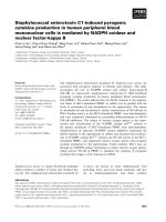
Fig. 3. Expression of DmNST/DmUGT in Lec2 and Lec8 cells. (A) Lec2 a nd Lec8 cells were transfected with appropriate plasmids as speci®ed
below, and CST and UGT activities of cDNA product s were assessed using FITC-labeled lectins as described in Materials and methods. a, pMKIT-
neo; b, pMKIT-neo-hCS T-cHA; c, pMKIT -neo-DmNST-c HA; d, pMKIT-neo ; e, pMKIT- neo-hUGT-cH A; f, pMK IT-neo-DmNS T-cHA. Bar,
10 lm (B) Western blot analysis of DmNST/DmUGT protein expressed in Lec2 (lanes 1 and 2) and Lec8 (lanes 3 and 4) cells. Cell extracts were
prepared from cells transfected with appropriate plasmids as speci®ed below, and were s ubjected to Western blot analysis. Lanes 1 and 3, pMKIT-
neo; lanes 2 and 4, pMKIT-neo-DmNST-cHA.
132 H. Segawa et al. (Eur. J. Biochem. 269) Ó FEBS 2002
among all these transporters. Accordingly, this sequence
comparison alone did not give us suf®cient i nformation to
infer the substrate speci®city of this fruit ¯y nucleotide sugar
transporter, but rather raised a possibility that the fru it ¯y
transporter could transport all the three nucleotide sugars.
To identify the transport substrate of DmNST, a DNA
fragment containing the entire c oding region was ampli®ed
by PCR and inserted into the expression vector pMKIT-neo
utilizing the EcoRI and NotI sites. An HA tag sequence was
added at the 3¢ end of the coding sequence to facilitate the
detection of the cDNA product. The expression plasmid,
pMKIT-neo-DmNST-cHA, was introduced into Lec2 and
Lec8 cells.
CST-de®cient Lec2 cells bind PNA, which recognizes the
terminal Gal residues on their defective surface glycocon-
jugates, while UGT-de®cient Lec8 cells bind GS-II, which
recognizes terminal GlcNAc residues [21]. If DmNST
complements the genetic d efects of these cells, the lectins
lose af®nity to the cells. This enabled us to asse ss the CMP-
Sia and UDP-Gal transport activities of the cDNA product
using FITC-labeled lectins. We are also able to examine the
expression of the protein products using an a nti-HA Ig at
the same time in the same specimen. Figure 3A shows that
DmNST was expressed in both Lec2 and Lec8 cells, but
only the genetic d efect of the latter, namely UDP-Gal
transport de®ciency, was complemented. This indicates that
DmNST has UDP-Gal transport activity but not CMP-Sia
transport activity. DmNST was located in the Golgi region,
as was hUGT1 (panels e and f).
In Western blot analysis, the DmNST was detected as a
broad band with an apparent molecular mass ranging from
30 to 36 kDa (Fig. 3B, lanes 2 and 4). The broadening of the
bands might be due to N-linked glycosylation at Asn311
(Fig. 1A), as this broadening was not observed with human
nucleotide sugar transporters (data not shown) that lack the
glycosylation motif at the corresponding sites (Fig. 2). The
DmNST expressed in Lec8 migrated slightly slower than
that expressed in Lec2 (Fig. 3B, lanes 2 and 4). This may be
explained by the fact that expression of DmNST comple-
mented the defect in UDP -Gal transp ort of Lec8 cells, and
that this would lead to the formation of f ully processed
oligosaccharide chains attached to t he protein.
hUGT and DmUGT both transport UDP-Gal
and UDP-GalNAc
To examine the substrate speci®city of DmUGT more
extensively, we utilized a yeast expression system.
HA-tagged DmUGT cDNA was inserted into the copper-
inducible yeast expression vector pYEX-BESN and trans-
fected into S. cerevisiae YPH500, and a t ransformant was
obtained. We prepared the microsomes from the transfor-
mant, and analyzed them for the presence of the DmNST
protein by Western blot analysis using anti-HA Ig (Fig. 4).
DmUGT-cHA migrated as a broad band with an apparent
molecular mass ranging from 28 to 36 kDa (lane 4).
Microsomes were prepared from transformants carrying
vectors with and without the DmUGT insert, and investi-
gated for their activity to transport nucleotide sugars
(Fig. 5). We also examined the substrate speci®city of
human UDP-Gal transporter extensively using microsomal
membranes obtained from an hUGT1-transformant of
S. cerevisiae YPH500. As expected from the results shown
in Fig. 3, UDP-Gal but not CMP-Sia was incorporated into
the m icrosomes expressing DmUGT. An unexpected ®nd-
ing was that these microsomal vesicles also incorporated
UDP-GalNAc ef®ciently. Figure 5 clearly shows that
hUGT1, which had been considered to be highly speci®c
for UDP-Gal, was also able to transport UDP-GalNAc in
addition to UDP-Gal. DmUGT and hUGT1 did not
transport either UDP-glucuronic acid (GlcA) or UDP-
xylose (Xyl). Furthermore, they did not seem to transport
UDP-GlcNAc, UDP-glucose (Glc), or GDP-mannose
(Man), although this is not certain due to interference by
the endogenous nucleotide sugar transport activity of
S. cerevisiae microsomal membranes. The apparent K
m
valuesofDmUGTandhUGT1wereestimatedtobe3.5 l
M
and 2.5 l
M
for UDP-Gal and 4.1 l
M
and 2.5 l
M
for UDP-
GalNAc, respectively (Fig. 6).
These results clearly indicate that DmUGT is the hUGT
homologue of D. melanogaster showing that both trans-
porters have exactly the same speci®city for substrates so far
examined (Fig. 5).
Mutagenesis of hUGT1 cDNA and assessment
of expression and NST activities of mutant proteins
DmUGT indicated signi®cant similarity to both hCST and
hUGlcNAcT c omparable with that to hUGT. Its substrate
speci®city w as, how ever, e xactly the same with that of
Fig. 4. Expression of DmNST/DmUGT in mammalian and yeast
microsomal membranes. Microsomes were prepared from Lec8 or yeast
cells expressing DmNST/DmUGT, and samples containing 30 lgof
protein were subjected to Western blot analysis. Lane 1, pMKIT-neo-
transfected Lec8; lane2, pMKIT-neo-DmNST-cHA-transfected Lec8;
lane 3, pYEX-BESN-transformed YPH500; lane 4, pYEX-BESN-
DmNST-cHA-transformed YPH500.
Ó FEBS 2002 UDP-Gal transporter transports UDP-GlcNAc (Eur. J. Biochem. 269) 133
hUGT as far as examined. In one of our previous
communications we noted that 10 amino-acid residues
seemed to be Ôsubstrate speci®cÕ in that they we re conserved
among transporters with identical substrates, but were
different b etween those speci®c for different substrates [15].
As shown in Fig. 2, a mong these 10 residues, only three, one
and three residues were shared by DmNST and hUGT,
hUGlcNAcT, or hCST, respectively. The three remaining
residues were not conserved between these transporters. To
see if these few conserved residues may be critical in
discriminating between speci®c substrates, we have chosen
the hUGT molecule as the representative of UDP-Gal
transporters, and altered the ÔUGT-speci®cÕ residues of
hUGT to their corresponding ÔCST-speci®cÕ residues by site-
directed mutagenesis. We paid particular attention to N47,
L174 and V285, which were shared by DmUGT and
hUGT.
HA-tagged single-site-mutant constructs, hUGT1-
(V45L)-cHA, hUGT1(N47A)-cHA, hUGT1(I51T)-cHA,
hUGT1(Q89E)-cHA, hUGT1(Q129A)-cHA, hUGT1-
(L174M)-cHA, hUGT1(I181L)-cHA, hUGT1(Q185K)-
cHA, hUGT1(F265Y)-cHA, hUGT1(V285T)-cHA, and
multiple-site-mutant constructs, hUGT1(N5aaCST)-cHA,
hUGT1(C5aaCST)-cHA, and hUGT1(10aaCST)-cHA
Fig. 5. Nucleotide sugar transport activity of
DmNST/DmUGT. M icrosomes were pre-
pared from pYEX-BESN-tra nsform ed
YPH500, pYEX-BESN-DmNST-cHA-trans-
formed YPH500, and pYEX-hUGT-cHA-
transformed YPH500. Microsomal vesicles
(50 lg protein/assay, or in GDP-Man trans-
port assay, 30 lg protein/assay) were incu-
bated at 30 °C for 30 s in the presence of
various radioactive nucleotide sugars as indi-
cated below the bars indicating the transport
activities. Uptake of substrates i nto microso-
mal vesicles was determined as described in
Materials and methods.
Fig. 6. Substrate concentration dependence of
UDP-Gal and UDP-GalNAc transport into
yeast microsomal membrane vesicles expressing
DmNST/DmUGT and hUGT1. Microsomes
(50 lg protein per a ssay) were incubated a t
30 °C for 1 min with various concentratio ns
of UDP-Gal (A) or UDP-GalNAc (C), and
the transport activity was determined as
described under Materials and methods. The
radioactivities trapped by vector control
microsomes were subtracted as background
values from corresponding experimental
values. The double reciprocal plot of the data
obtained in (A) and (C) and the results of
linear regression analyses are shown in (B) and
(D), respectively.
134 H. Segawa et al. (Eur. J. Biochem. 269) Ó FEBS 2002
were introduced into Lec2 and Lec8 cells. The hUGT1
(N5aaCST)-cHA mutant carried V45L, N47A, I51T, Q89E,
and Q129A substitutions, and hUGT1(C5aaCST)-cHA
carried L174M, I181L, Q 185K, F265Y, and V285T substi-
tutions. In the hUGT1(10aaCST)-cHA mutant all of the
ÔUGT-speci®c Õ residues were replaced by the corresponding
ÔCST-sp eci®cÕ ones [15].
The expression and the transport activities of each mutant
were assessed by immuno¯uorescence and FITC-labeled
lectin binding as in Fig. 3. Figure 7 shows t hat N47A,
L174M and V285T mutants retained UDP-Gal transport
activity but were unable to transport CMP-Sia. Other single
substitution mutants as well as three multiple substitution
mutants gave essentially the same results as V45L (Figs 7d,l)
and hUGT1(10aaCST) (Figs 7h,p), and were active in UDP-
Gal transport, but not in CMP-Sia transport (data not
shown). Most of the mutant proteins, except h UGT1
(N47A)-cHA, were localized in the Golgi apparatus as was
the wild-type p rotein. The hUGT1(N47A)-cHA mutant
protein was not con®ned to the Golgi region, but was dis-
tributed more diffusely in the perinuclear region. This mutant
showed UDP-Gal transport activity, but the frequency of
the cells expressing the mutant w as low (Figs 7e,m).
The amounts of wild-type and mutant UGT proteins
expressed in the transfected cells were analyzed by Western
blotting. Most o f the mutant proteins were detected in
roughly the same amounts as hUGT1-cHA, but the amount
of hUGT1(N47A)-cHA was much lower than the amounts
of the others (Fig. 8, lane 4), suggesting metabolic instability
of the m utant protein. It is noted, however, that hUGT1-
(N5aaCST)-cHA (lane 13) and hUGT1(10aaCST)-cHA
(lane 15) were expressed as e f®ciently as wild-type hUGT-
cHA, although they carry the N47A m utation. This implies
that the d estabilizing effect of the N47A mutation was
suppressed by one of the additional mutations introduced
into the hUGT1(N5aaCST)-cHA mutant. To identify the
second mutation responsible for the suppression of the
N47A ph enotype, we ®rst constructed a three-site mutant,
hUGT1(V45L, N47A, I51T)-cHA, and found that the
mutant protein was expressed ef®ciently and distributed
normally in the cell. We then constructed hUGT1(V45L,
N47A)-cHA, hUGT1(N47A, I 51T)-cHA, and h UGT1
(V45L, I51T)-cHA, and examined the expression levels of
these mutant proteins and their intracellular distribution. As
shown in Fig. 9, introduction of the I51T mutation
suppressed the N47A mutation and resulted in the normal
Fig. 7. Assessment of CMP-Sia transport and
UDP-Gal transport activities of mutant
hUGT1s. cDNA constructs coding for
HA-tagged transporter proteins, including
mutant proteins as speci®ed below, were
expressed in CST-de®cient Lec2 (panels a±h),
or UGT-de®cient Lec8 (panels i±p) cells. Lec2
cells were stained with FITC-labeled PNA,
and Lec8 cells with FITC-labeled GS-II to
assess CST and UGT activities of the trans-
porters and mutants. The expression of pro-
teins was detected by immunostaining with
anti-HA Ig, which was visualized by using
Alexa596-conjugated anti-(rat IgG) Ig.
(a and i), pMKIT-neo; b and j, pMKIT-neo-
hCST-cHA; (c and k), pMKIT-neo-hUGT1-
cHA; (d and l), pMKIT-neo-hUGT1(V45L)-
cHA; e a nd m, pMKIT-neo-hUGT1(N47A)-
cHA; (f and n), pMKIT-neo-
hUGT1(L174M)-cHA; (g and o), pMKIT-
neo-hUGT1(V285T)-cHA; (h and p),
pMKIT-neo-hUGT1(10aaCST)-cHA. Bar,
10 lm.
Ó FEBS 2002 UDP-Gal transporter transports UDP-GlcNAc (Eur. J. Biochem. 269) 135
intracellular distribution (Fig. 9A, panel e) and expression
level (Fig. 9B, lane e) of the double-mutant protein.
DISCUSSION
In this study we determined the primary structure of a
putative nucleotide sugar transporter of D. melanogaster,
and identi®ed it as the D. melanogaster homologue
(DmUGT) of human UDP-Gal transporter (hUGT). The
cDNA complemented the genetic defect of UGT-de®cient
Lec8 ce lls, and its product was detected in the Golgi region
of the transfected cells. Heterologous expression of the
cDNA in S. cerevisiae cells allowed us to demonstrate
directly that the cDNA product was able to transport UDP-
Gal and UDP-GalNAc across the microsomal membranes
(Figs 5 and 6).
Fig. 8. Western blot analysis of the expression of mutant hUGT1s in Lec2 cells. Lec2 cells transfected with an appropriate plasmid as speci®ed
below were incubated for 48 h, then lysed and subjected t o Western blot analysis. Proteins were detec ted by immunostaining with a nti-HA Ig.
1, pMKIT-ne o; 2, pMKIT-neo-hUGT1-c HA; 3, pMKIT-neo-hUGT1(V45L)-c HA; 4, pMKIT-neo-hUGT1(N47A) -cHA; 5, pMKIT-neo-
hUGT1(I51T)-cHA; 6, pMKIT-neo-hUGT1(Q89E)-cHA; 7, pMKIT-neo-hUGT1(Q129A)-cHA; 8, pMKIT-neo-hUGT1(L174M)-cHA; 9,
pMKIT-neo-hUGT1(I181L)-cHA; 10, pMKIT-neo-hUGT1(Q185K)-cHA; 11, pMKIT-neo-hUGT1(F265Y)-cHA; 12, pMKIT-neo-
hUGT1(V285T)-cHA; 13, pMKIT-neo-hUGT1(N5aaCST)-cHA; 14, pMKIT-neo-hUGT1(C5aaCST)-cHA; 15, pMKIT-neo-hUGT1(10aaCST)-
cHA; 16, pMKIT-neo-hCST-cHA.
Fig. 9. Suppression of N47A mutation by I51T second-site mutation. Lec8 cells transfected with an appropriate plasmid as speci®ed below were
stained with FITC-GS-II lectin and anti-HA a ntibody as in Fig. 7 in (A); and cell extracts were subjected to Western blot analysis as in Fig. 8 in (B).
a, pMKIT-neo; b, pMKIT-neo-hUGT1-cHA; c, pMKIT-neo-hUGT1(N47A)-cHA; d, pMKIT-neo-hUGT1(V45L, N47A)-cHA; e, pMKIT-neo-
hUGT1(N47A, I51T)-cHA; f, pMKIT-neo-h UGT1(V45L, N47A, I51T)-cHA; g, pMK IT-neo-hUG T1(N5aaCST)-cHA; h, pMKIT -neo-hUGT1
(10aaCST)-cHA. Bar, 10 lm.
136 H. Segawa et al. (Eur. J. Biochem. 269) Ó FEBS 2002
Nucleotide sugar transporters (NSTs), including hUGT,
have long been thought to be highly substrate speci®c [22].
Very recently, however, Muraoka et al. reported a new
member of the NST family, hUGTrel7, and showed that it
transports both UDP-GlcA an d UDP-GalNAc [16]. H ong
et al. also demonstrated that Leishmania GDP-Man trans-
porter, LPG2, can transport GDP-arabinose and GDP-
fucose in addition to GDP-Man [23]. Speci®c recognition of
two or more substrates by an NST may be more common
than has b een assumed until recently. The molecular
mechanisms underlying the multiple substrate recognition
are intriguing while remaining o bscure.
Various glycoconjugates are detected in a position and
stage-speci®c manner during Drosophila development [24].
Heparin-like glycosaminoglycans that contain galactose and
N-acetylgalactosamine residues are involved in the wingless
signaling [25]. The DmUGT protein was localized in the
GolgiregionwhenthecDNAwasexpressedinLec2andLec8
cells, and transported both UDP-Gal and UDP-GalNAc.
The subcellular localization and its substrate speci®city are
consistent with its possible involvement in this process. RNA
interference experiments [26] may help to answer this
intriguing question about the physiological role of DmUGT.
We found a single possible N-glycosylation site in the
DmUGT during analysis on the primary structure of the
fruit ¯y NST (Fig. 1A). Based on the 10-segment trans-
membrane model proposed by Eckhardt et al.[27],the
N-glycosylation site resides at the boundary between th e
ninth and tenth putative transmembrane regions (Fig. 2).
Eckhardt et al. were not able to decide whether these
hydrophobic regions (Fig. 2; Hxs9 and 10) traverse the
membrane, are enbedded in the membrane without being
exposed to the lumen side, or are just tightly membrane
associated, as anti-HA epitope antibodies failed to detect an
HA epitope introduced to this boundary region [27]. The
expressed DmUGT proteins were glycosylated in both
CHO (Fig. 3A ) and S. cerevisiae (Fig. 4) cells indicating
that the N-glycosylation site found is faced to Golgi lumen
and accessible to glycosyl transferases. These results suggest
that both the ninth and tenth hydrophobic regions form
discrete membrane-spanning domains.
Three amino-acid residues of hUGT, namely N47, L174,
and V285, are conserved among human, murine, ®ssion
yeast, and Drosophila UGTs, but are distinct from the
corresponding residues conserved among CMP-Sia trans-
porters and UDP-GlcNAc transporters, respectively, from
several species [15]. These Ôsubstrate-speci®cÕ residues as well
as several others were replaced by corresponding residues of
CMP-Sia transporter, to see whether these residues con-
tribute to the recognition of speci®c substrates, but the
switching of the substrate from UDP-Gal to CMP-Sia was
not observed with any of the substitution mutants tested.
This unexpected result is rather consistent with the r esults
recently obtained in analyses of UGT/CST chimeras,
indicating that different submolecular regions are critically
involved in the recognition of UDP-Gal and CMP-Sia
[21,28].
The N47A mutation of hUGT led to aberrant
intracellular distribution and destabilization of the mutated
transporter protein. The mutant phenotype of N47A was
suppressed by a second mutation, I51T. As N47 and I51
are predicted to be close to each o ther on the same side of
the ®rst transmembrane helix based on the 10-segment
transmembrane model of the transporter [27], it seems
that the intrahelical side-chain interaction between these
two residues is important for the conformational stability
of the protein and its proper interaction with the
membrane protein-sorting machinery. The importance of
helix 1 in stabilizing the UGT protein may also be inferred
from the instability of a truncated Schizosaccharomyces
pombe UGT that lacks the exon coding for the ® rst
transmembrane helix [6]. Aoki et al. also d emonstrated
that the ®rst helix from UGT is necessary for chimeric
constructs to transport UDP-Gal [21]. Further analysis of
the effects of mutations introduced in helix 1 may provide
clues to investigate th e mechanisms of integration, sorting
and substrate-recognition of this polytopic membrane
protein.
ACKNOWLEDGEMENTS
This work was supported in part by Grants-in-Aid for Scienti®c
Research no. 11480172, Grants-in-Aid for Scien ti®c Research on
Priority Area no . 12033222 f rom the Ministry of Education, Science,
Sports and Culture of Japan and a Grant from Mizutani Foundation
for Glycoscienc e.
REFERENCES
1. Kawakita, M., Ishida, N ., Miura, N., Sun-Wada, G H. &
Yoshioka, S. (1998) Nucleotide sugar transporters: elucidation of
their molecular identity and its implication f or future studies.
J. Biochem. (Tokyo). 12 3, 777±785.
2. Seppo, A. & Tiemeyer, M. (2000) Function and structure of
Drosophila glycans. Glycobiol. 10, 751±760.
3. Herman, T. & Horvitz, H.R. (1999) Three proteins involved in
Caenorhabditis elegans vulval invagination are similar to compo-
nents of a glycosylation pathway. Pr oc. Natl A cad. Sci. USA 96,
974±979.
4. Abeijon,C.,Robbins,P.W.&Hirschberg,C.B.(1996)Molecular
cloning of the Golgi apparat us uridine diphosphate-N-acetyl-
glucosamine transporter from Kluyveromyces lactis. Proc. Natl
Acad.Sci.USA93, 5963±5968.
5. Dean, N., Zhang, Y.B. & Poster, J.B. (1997) The VRG4 gene is
required for GDP-mannose transport into the lumen of the Golgi
in the yeast, Saccharomyces cerevisiae. J. Biol. Chem. 272, 31908±
31914.
6. Segawa, H., Ishida, N., Takegawa, K. & Kawakita, M. (1999)
Schizosaccharomyces pombe UDP-galactose transporter: identi®-
cation of its fun ctional form through cDNA cloning and expres-
sion in mammalian cells. FEBS Lett. 451, 295±298.
7. Roy, S.K., Chiba, Y., Takeuchi, M. & Jigami, Y. (2000) Char-
acterization of Yeast Yea4p, a uridine diphosphate-N-acetyl-
glucosamine transporter localized in the endoplasmic reticulum
and required for chitin synthesis. J. Biol. Chem. 275, 13580±
13587.
8. Ma, D.Q., Russell, D.G., Beverley, S.M. & Turco, S.J. (1997)
Golgi GDP-mannose uptake requires Leishmania LPG2: a mem-
ber of a eukaryotic family of putative nucleotide-sugar trans-
porters. J. Biol. Chem. 272, 3799±3805.
9. Herman, T., Hartwieg, E. & Horvitz, H.R. (1999) sqv mutants of
Caenorhabditis elegans are defective in vulval epithelial invagin-
ation. Proc. Natl Acad. Sci. USA 96, 967±973.
10. Miura, N., Ishida, N., Hoshino, M., Yamauchi, M., Hara, T.,
Ayusawa, D. & Kawakita, M. (1996) Human UDP-galactose
translocator: molecular cloning of a complementary DNA
that complements the genetic defect of a mutant cell line de®cient
in UDP-galactose translocator. J. Biochem. (Tokyo). 120,
236±241.
Ó FEBS 2002 UDP-Gal transporter transports UDP-GlcNAc (Eur. J. Biochem. 269) 137
11. Ishida, N., Miura, N., Yoshioka, S. & Kawakita, M. (1996)
Molecular cloning and characterization of a novel isoform of the
human UDP-galactose transporter, and of related complementary
DNAs belonging to the nucleotide-sugar transporter gene family.
J. Biochem. (Tokyo). 120, 1074±1078.
12. Eckhardt, M., Mulenho, M., Bethe, A. & Gerardy-Schahn, R.
(1996) Expression cloning of the Golgi CMP-sialic acid trans-
porter. Proc. Natl Acad. Sci. USA 93, 7572±7576.
13. Eckhardt, M. & Gerardy-Schahn, R. (1997) Molecular cloning of
the hamster CMP-sialic acid tran sporter. Eur. J. Biochem. 24 8,
187±192.
14. Ishida, N., I to, M., Yoshiok a, S., Sun-Wada,G H. & Kawakita, M.
(1998) Functional expression of human Golgi CMP-sialic acid
transporter in the Golgi complex of a transporter-de®cient Chinese
hamster ovary cell mutant. J. Biochem. (Tokyo). 124, 171±178.
15. Ishida, N., Yoshioka, S., Chiba, Y., Takeuchi, M. & Kawakita,
M. (1999) Molecular cloning and functional expression of the
human Golgi UDP-N-acetylglucosamine transporter. J. Biochem.
(Tokyo) . 126, 68±77.
16. Muraoka, M., Kawakita, M. & Ishida, N. (2001) Molecular
characterization of hum an UDP-glucuro nic acid/UDP- N-acety-
lgalactosamine transporter, a novel nucleotide sugar transporter
with dual substrate speci®city. FEBS Lett. 495, 89±93.
17. Sarker, G. & Sommer, S.S. (1990) The Ômegaprimer Õ method of
site-directed mutagenesis. Biotechniques 4, 404±407.
18. Ito, H., Fukuda, Y., Murata, K. & Kimura, A. (1983) Transfor-
mation o f intact yeast cells treated w ith alkali cations. J. Bacteriol.
153, 163±168.
19. Sun-Wada, G H., Yoshioka, S., Ishida, N. & Kawakita, M.
(1998) Functional expression of the human UDP-galactose
transporters in the yeast Saccharomyces cerevisiae. J. Biochem.
(Tokyo) . 123, 912±917.
20. Yoshioka, S., Sun-Wada, G H., Ishida, N. & Kawakita, M.
(1997) Expression of the human UDP-galactose transporter in the
Golgi membranes of murine Had-1 cells that lack the endogenous
transporter. J. Biochem. (Tokyo). 122 , 691±695.
21. Aoki, K., Ishida, N. & Kawakita, M. (2001) Substrate recognition
by UDP-galactose and CMP-sialic acid transporters: dierent sets
of transmembrane helices are utilized for the speci®c recognition of
UDP-galactose and CMP-sialic acid. J. Biol. Chem. 276, 21555±
21561.
22. Hirschberg, C.B., Robbins, P.W. & Abeijon, C. (1998) Trans-
porters of nucleotide sugars, AT P, and nucleotide sulfate in the
endoplasmic reticulum and Golgi apparatus. Annu.Rev.Biochem.
67, 49±69.
23.Hong,K.,Ma,D.,Beverley,S.M.&Turco,S.J.(2000)The
Leishmania GDP-mannose transporter is an autonom ous, multi-
speci®c, hexameric comple x of LPG2 subunits. B i och em ist ry 39 ,
2013±2022.
24. D'Amico, P. & Jacobs, J.R. (1995) Lectin histochemistry of the
Drosophila embryo. Tissue Cell 27, 23±30.
25. Binari, R .C., Staveley, B.E., Johnson, W.A., Godavarti, R.,
Sasisekharan, R. & Manoukian, A.S. (1997) Genetic evidence that
heparin-like glycosaminoglycans are involved in wingless signal-
ing. Development 124, 2623±2632.
26. Kennerdell, J.R. & Carthew, R.W. (1998) Use of dsRNA-
mediated genetic interference to demonstrate that frizzled and
frizzled 2 act in the wingless pathway. Cell 95, 1017±1026.
27. Eckhardt, M., Gotza, B. & Gerardy-Schahn, R. (1999) Membrane
topology of the m ammalian CMP-sialic acid transporter. J. Biol.
Chem. 274, 8779±8787.
28. Aoki, K., Sun-Wada, G H., Se gawa, H., Yoshioka, S., Ishida, N.
& Kawakita, M. (1999) Expression and activity of chimeric mol-
ecules between human UDP-galactose transporter and CMP-sialic
acid transporter. J. Biochem. (Tokyo). 126, 940±950.
29. Kyte, J. & Doolittle, R.F. (198 2) A simple method for display-
ing the hydropathic character of a protein. J. Mol. Biol. 157,
105±132.
138 H. Segawa et al. (Eur. J. Biochem. 269) Ó FEBS 2002