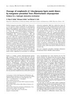
Tài liệu Báo cáo Y học: Binding of gelsolin domain 2 to actin An actin interface distinct from that of gelsolin domain 1 and from ADF/cofilin pptx
Bạn đang xem bản rút gọn của tài liệu. Xem và tải ngay bản đầy đủ của tài liệu tại đây (960.26 KB, 11 trang )
Binding of gelsolin domain 2 to actin
An actin interface distinct from that of gelsolin domain 1 and from ADF/cofilin
Celine Renoult
1
, Laurence Blondin
1
, Abdellatif Fattoum
2
, Diane Ternent
3
, Sutherland K. Maciver
3
,
Fabrice Raynaud
1
, Yves Benyamin
1
and Claude Roustan
1
1
UMR 5539 (CNRS) Laboratoire de Motilite
´
Cellulaire (Ecole Pratique des Hautes Etudes), Universite
´
de Montpellier, France;
2
Centre de Recherches de Biochimie Macromole
´
culaire, Montpellier, France;
3
Genes and Development Group,
Department of Biomedical Sciences, University of Edinburgh, Scotland
It is generally assumed that of the six domains that comprise
gelsolin, domain 2 is primarily responsible for the initial
contact with the actin filament that will ultimately result in
the filament being severed. Other actin-binding regions
within domains 1 and 4 are involved in gelsolin’s severing
and subsequent capping activity. The overall fold of all
gelsolin repeated domains are similar to the actin
depolymerizing factor (ADF)/cofilin family of actin-binding
proteins and it has been proposed that there is a similarity in
the actin-binding interface. Gelsolin domains 1 and 4 bind
G-actin in a similar manner and compete with each other,
whereas domain 2 binds F-actin at physiological salt
concentrations, and does not compete with domain 1. Here
we investigate the domain 2 : actin interface and compare
this to our recent studies of the cofilin : actin interface. We
conclude that important differences exist between the
interfaces of actin with gelsolin domains 1 and 2, and with
ADF/cofilin. We present a model for F-actin binding of
domain 2 with respect to the F-actin severing and capping
activity of the whole gelsolin molecule.
Keywords: actin; actin-binding proteins; cofilin; gelsolin.
The organization of the actin microfilaments in cells is
dynamic and is quickly rearranged in response to extra-
cellular signals. Gelsolin is one of the members of a family
of proteins (e.g. severin, villin), that is essential for
microfilament remodelling [1–3]. There are two forms of
gelsolin which differ in their N-terminal extremities. One is
specifically located in the blood and acts with vitamin
D-binding protein to accelerate clearing of actin from the
circulation [4], while the other form is intracellular. In vitro,
gelsolin interacts with G- and F-actins, promotes nucleation
and both severs and caps actin filaments. Cofilin belongs to
another family of actin-binding proteins that also severs
actin filaments and increases polymerization dynamics [5].
Despite a lack of sequence homology between the cofilin
and gelsolin families the fold adopted by each of gelsolin’s
130 amino-acid subdomains [2] is similar to the actin
depolymerizing factor (ADF)/cofilin family fold [6]. In
contrast with cofilin, gelsolin does not appear to be essential
for viability in the organisms where this has been tested,
probably due to the expression of related genes such as
adseverin/scinderin [7], but gelsolin is specifically required
for rapid movement of various dynamic cells [8]. Thus,
gelsolin over-expression in fibroblasts leads to enhanced cell
motility [9,10].
Domains 1–3 (S1–3) are sufficient for capping and
severing, while the C-terminal half of the molecule is
directly implicated in calcium regulation. In particular,
gelsolin domain 1 (S1) interacts both with monomeric actin,
and with the barbed end of the actin filaments inhibiting
polymerization.
S2, in contrast, preferably binds to the side of the actin
filament. Severing activity seems to require the binding of
S2 to the filament, followed by interaction of S1 between
two adjacent actins along the filament axis [11].
The tertiary structure of whole gelsolin in the inactive
Ca
21
free state has been determined [2], as has S1 in
complex with actin [11], gelsolin S4–6 [12], severin
domain 2 [13,14] and villin domain 2 [15]. The structure of
each gelsolin domain shows a surprising similarity to the
cofilin fold [6]. Therefore it is possible to hypothesize that
S2 binds actin in the same manner as cofilin [16].
The solution of the gelsolin structure [2], showed that
when S1 is in position according to the G-actin–S1 model
[11], S2 is not in contact with actin. This might suggest a
reorientation of S1 : S2 interfaces so that S2 could contact
both of the binding sites on the same actin unit in the
filament to which S1 is joined [12]. In addition, by studying
the S2–6 interaction with F-actin, McGough et al. [17]
showed that the S2 –3 position on F-actin is similar to the
actin-binding domain of a-actinin. Robinson et al. [12]
presented a model for gelsolin interaction based on the
Note: web pages are available at , and
/>Note: A gelsolin amino-acid numbering system based on the plasma
human gelsolin [1], in which S1 is defined as extending from Pro39 to
Tyr133 and S2 as being Gly137 to Leu247 [2], is used is this report.
Correspondence to C. Roustan, UMR 5539(CNRS) UM2 CC107,
Place E. Bataillon 34095 Montpellier Cedex 5, France.
Fax: 133 04 67 14 49 27,
E-mail:
(Received 14 June 2001, revised 28 September 2001, accepted
4 October 2001)
Abbreviations: S1–6, the six repeated segments of gelsolin; ADF,
Actin depolymerizing factor; 1,5-I-AEDANS, N,-iodoacetyl-N
0
-(sulfo-
1-naphthyl)-ethylenediamine; ELISA, enzyme-linked immunosorbant
assay; FITC, fluorescein 5-isothiocyanate; G-actin, monomeric actin;
F-actin, filamentous actin; EEDQ, N-ethoxycarbonyl-2-ethoxy-
1,2-dihydroquinoline.
Eur. J. Biochem. 268, 6165–6175 (2001) q FEBS 2001
determination of the S4–6 actin structure. They suggested
that changes in the structure of S1 –3 must occur to allow S2
to interact with the side of actin filament. Finally from
mutagenesis and structural data, Puius et al. [14] proposed a
model for S2 interaction in which 168RRV170 and 210RLK
212 are determinant in F-actin binding.
In this report, we investigated the gelsolin S2 : actin
interface. In particular, we focused on the comparison
between respective locations of gelsolin and cofilin on actin
filament and evidenced major differences in the interfaces.
MATERIALS AND METHODS
Proteins and peptides
Rabbit skeletal muscle actin was isolated from acetone
powder [18], and stored in buffer G (2 m
M Tris, 0.1 mM
CaCl
2
0.1 mM ATP pH 7.5). Actin was selectively cleaved
by Staphylcoccus aureus V8 protease [19] and thrombin
[20] and the fragments obtained were isolated by
electroelution as described previously [19]. Human gelsolin
domain 2 (S2) was produced in Escherichia coli,
BL21(pLysS) carrying a vector containing a cDNA encod-
ing residues including 151 – 266, the S2 repeat [21]. The
bacteria were grown in 1-L flasks with 2 Â TY medium with
ampicillin (150 mg
:
mL
21
) and induced with isopropyl thio-
b-
D-galactoside (final concentration 1 mM) when the culture
reached D
600
¼ 0.5. Cultures were then grown for a further
4hat378C and the cells collected by centrifugation. The
bacteria were lysed by repeated freeze–thaw cycles with
sonication. Supernatant containing the S2 protein was
applied to a DE52 column and purified further by
hydroxyapatite chromatography. The concentration of S2
was determined spectrophotometrically assuming 1
A
280
¼ 79 mM [21].
Antibodies directed towards gelsolin fragments 159–193
and 203–225 or actin sequences 75–105 and 285–375 were
elicited in rabbits [22]. The antibodies directed to the actin
sequences were selectively purified by affinity chromato-
graphy [23]. Anti-IgG antibodies labelled with alkaline
phosphatase were from Sigma.
Synthetic peptides derived from actin and gelsolin
sequences were prepared on a solid phase support using a
9050 Milligen PepSynthesizer (Millipore) according to the
Fmoc/tBu system. The crude peptides were deprotected and
purified thoroughly by preparative reverse-phase HPLC.
The purified peptides were shown to be homogenous by
analytical HPLC. Electrospray mass spectra, carried out
in the positive ion mode using a Trio 2000 VG Biotech
mass spectrometer (Altrincham, UK), were in line with the
expected structures.
Peptides were labelled at the cysteine residue with
N-iodoacetyl-N
0
-(sulfo-1-naphthyl)-ethylenediamine (1,5-I-
AEDANS) or at amino groups by fluorescein 5-isothio-
cyanate (FITC) [24,25]. Excess reagent was eliminated
by sieving through a Biogel P2 column equilibrated with
0.05
M NH
4
HCO
3
buffer pH 8.0. Actin and gelsolin S2
domain were labelled by FITC as described elsewhere [25].
Excess reagent was eliminated by chromatography on a
PD10 column (Pharmacia) in 0.1
M NaHCO
3
buffer pH 8.6.
Actin was specifically labelled at cysteine 374 by 1,5-I-
AEDANS [24].
Cross-linking experiments
Actin (1 mg
:
mL
21
) and gelsolin fragment 159–193
(0.1 mg
:
mL
21
) were incubated with 2.5 mM N-ethoxy-
carbonyl-2-ethoxy-1,2-dihydroquinoline (EEDQ; Sigma) in
100 m
M Mops pH 7.0 at 22 8C. The cross-linking reaction
was allowed to proceed for 45 min, and stopped by the
addition of 100 m
M 2-mercaptoethanol. The cross-linked
species were separated and analysed by SDS/PAGE and
immunoblotting.
Immunological techniques
The enzyme-linked immunosorbent assay (ELISA) [26],
that was previously described in detail [27], was used to
monitor interaction of ligands with coated peptides or actin.
Actin (0.5 mg
:
mL
21
) or peptides (5 mg
:
mL
21
)in50mM
NaHCO
3
/Na
2
CO
3
pH 9.5, were immobilized on plastic
microtiter wells. The plate was then saturated with 0.5%
gelatin/3% gelatin hydrolysate in 140 m
M NaCl/50 mM
Tris buffer pH 7.5. Experiments with coated peptides
were performed in 0.15
M NaCl, 10 mM phosphate
pH 7.5. Binding was monitored at 405 nm using alkaline
phosphatase-labelled anti-IgG antibodies (1 : 1000) or
alkaline phosphatase-labelled streptavidin (1 : 1000). Con-
trol assays were carried out in wells saturated with gelatin
and gelatin hydrolysate used alone. Each assay was con-
ducted in triplicate and the mean value plotted after sub-
traction of nonspecific absorption. The binding parameters
(apparent dissociation constant K
d
and the maximal binding
A
max
) were determined by nonlinear fitting A ¼ A
max
[L]/
(K
d
1 [L]) where A is the absorbance at 405 nm and [L] is
the ligand concentration, by using the
CURVE FIT software
developed by K. Raner Software (Victoria, Australia).
Additional details on the different experimental conditions
are given in the figure legends.
Western immunoblots were performed as described
previously [28]. The immunoblots were revealed using
alkaline phosphatase.
Fourier transform IR measurements
Fourier transform IR spectra were recorded using an IFS 28
Bruker spectrometer. Samples were placed in a horizontal
ATR plate and the spectra recorded at room temperature.
The peptide was analysed at a concentration of 5 mg
:
mL
21
in 10 mM phosphate buffer pH 7.5. A total of 500 scans
were accumulated in the 1800–1500 cm
21
range. The
Bruker
OPUS/IR 2 program was used for spectrum analysis
(second derivative).
Fluorescence measurements
Fluorescence experiments were conducted using a LS 50
Perkin-Elmer luminescence spectrometer. Spectra for 1,5-I-
AEDANS or FITC were obtained in 50 m
M Tris/HCl buffer
pH 7.5, with the excitation wavelength set at 340 and
470 nm, respectively. Fluorescence changes were deduced
from the area of the emission spectra of FITC between 480
and 500 nm. The parameters K
d
(apparent dissociation
constant) and A
max
(maximum effect) were calculated by
nonlinear fitting of the experimental data points.
The number of binding sited (n ) and the affinity constant
K
a
were also determined by another approach [29,30]. The
6166 C. Renoult et al. (Eur. J. Biochem. 268) q FEBS 2001
following relationship was then used:
1/ð1 2 XÞ¼K
a
ðC/ ðXEÞ 2 nÞð1Þ
where C and E are total concentrations of peptide and actin,
respectively, and X is the relative fluorescence change
A/A
max
(corresponding to the fraction of peptide bound to
actin).
A plot of 1/(1 – X) ¼ vs. C/(XE) (Eqn 1) was drawn. The
plot gives the number of binding sites which is the value
of C/(XE) for 1/(1–X) ¼ 0. The slope of the same curve
directly gives the value of the affinity constant.
Analytical methods
Protein concentrations were determined by UV absorbency
using a Varian MS 100 spectrophotometer. Electrophoresis
was carried out on 12.5% (w/v) polyacrylamide slab gels
(SDS/PAGE 12.5%) according to Laemmli [31] and stained
with Coomassie blue. The 14–97 kDa molecular weight
marker kits were from Biorad.
RESULTS
Several investigations [32–35] have suggested the parti-
cipation of gelsolin S2 sequences (and homologue S2
equivalents) within residues 197 – 226 (including the long
helix of the domain) and within residues 161–172
(including the A strand and the AB loop in S2) in the
interaction with F-actin. We have investigated subdomain 1
of actin, which possess accessible sequences in F-actin in
order to delimit the interface of gelsolin S2 with the actin
filament.
In an initial experiment, we tested the ability of a
sequence covering the helix of S2 to interact with actin.
The conformation of the synthesized peptide (sequence
203–225) was checked in aqueous solution by Fourier
transform IR. The IR spectrum of the peptide is charac-
terized by the presence of a band at 1645 cm-1 (data not
shown) suggesting an unordered conformation [36]. A
similar result has already been observed for the corre-
sponding peptide in cofilin [16]. Binding was tested by
fluorescence measurements. In a first experiment, we have
labelled actin at Cys374 with dansyl and the 203–225
peptide with FITC. The excitation was fixed at 340 nm and
the fluorescence emission monitored between 460 and
480 nm. The fluorescence was corrected for the contribution
of the FITC-labelled peptide alone. In this experiment, we
observed a quenching of dansyl fluorescence emission (data
not shown) that could be interpreted by energy transfer
between the two chromophores and/or changes in the
environment of Cys374 occurring during actin–peptide
complex formation. In a second approach, FITC-labelled
actin was incubated in the presence of increasing
concentrations of 203 – 225 peptide (0–19 m
M). The results
shown in Fig. 1 indicate change in the FITC fluorescence
induced by complex formation. The shape of the curve
shows that the binding takes place in a saturable manner
with an apparent K
d
of 5 mM. These experiments confirm the
results of van Troys et al. [33] which implicate the sequence
197–226 in the gelsolin : actin interface.
A second gelsolin S2 : actin interface is located in the
N-terminal part of S2. In order to delimit the footprint of this
gelsolin part on the actin structure, a peptide covering the
159–193 sequence was synthesized. Its conformation in an
aqueous solution was studied by IR in the amide 1 region.
The second derivative of the spectrum (Fig. 2), character-
ized by a major band at 1629
:
cm
21
associated with a band at
1680
:
cm
21
suggests the presence of an antiparallel beta
sheet structure [36]. In the corresponding region of gelsolin
S2, crystallographic data reported the occurrence of three
antiparallel strands [37].
The interaction of the 159–193 peptide with actin was
documented by three independent assays. Actin was treated
with EEDQ in the presence of peptide 159–193 and
analysed by gel electrophoresis. A typical protein band
pattern is shown in Fig. 3. The EEDQ treatment yields a
new product with an apparent molecular weight of 47 kDa,
which is not present with actin alone. This cross-linked
product corresponds to the actin–peptide complex.
Fig. 2. Structure of the synthetic gelsolin peptide 159 –193. Second
derivative of the IR Fourier transform spectrum. Amide I region of the
IR spectrum was observed in 10 m
M phosphate, pH 7.5.
Fig. 1. Binding of FITC-labelled G-actin with gelsolin 203 –225
peptide. Interaction of FITC-labelled G-actin at Lys61 (0.7 m
M) with
gelsolin 203–225 peptide was monitored by fluorescence. Changes in
the intensity of the fluorescence emission spectra of FITC were
recorded at various peptide concentrations in buffer G pH 7.5.
Excitation was fixed at 470 nm and emission between 510 and 530 nm.
q FEBS 2001 The actin gelsolin domain22 interface (Eur. J. Biochem. 268) 6167
The binding of the peptide to actin was also tested by
ELISA. Its interaction with coated G-actin was revealed by
using specific antibodies to sequence 159–193 within
gelsolin S2. The results presented in Fig. 4A show that the
peptide binds to G-actin with an apparent K
d
of < 2 mM.
These data were confirmed in solution using fluorescence
experiments. G-actin labelled with FITC was incubated in
the presence of increasing 159– 193 peptide concentration
and the changes in fluorescence were monitored. The
saturation curve observed suggests a specific interaction
with a K
d
of 2 mM. A stoichiometry of < 1 mole peptide per
mole G-actin was also estimated (Fig. 4C). A similar
experiment was performed with dansylated F-actin at
Cys374 (Fig. 4B inset). The interaction induces a
fluorescence quenching of the chromophore (K
d
¼ 2 mM).
Determination of the 159–193 peptide/actin interface
Two approaches were used for identification of large
fragments of actin to which gelsolin 159–193 peptide could
be cross-linked by EEDQ. They involved the electrophoretic
and immunological analysis of the cross-linked products
formed either on proteolysis of the complex by V8 protease
or on cross-linking of the 159–193 peptide to actin after
digestion by thrombin. Digestion of actin by V8 protease
gives two major fragments [19] of 31 and 16 kDa (1 – 225
and 226–375 sequence, respectively). As shown in Fig. 5,
digestion of the cross-linked actin peptide complex reveals
two faint bands at 33 kDa and 46 kDa which are missing
from the controls. They can be stained by both anti-actin
(directed towards sequence 75–105) and anti-gelsolin
Fig. 4. Binding of gelsolin fragment 159 –193 with actin. (A)
Interaction of gelsolin fragment monitored by ELISA. Coated G-actin
was reacted with the gelsolin fragment at the concentrations indicated.
Binding was monitored at 405 nm, using specific anti-gelsolin
antibodies. (B) Interaction of FITC-labelled actin (0.7 m
M) with
gelsolin fragment was monitored by fluorescence. Changes in the
intensity of the fluorescence emission spectra of FITC were recorded at
various peptide concentrations (0–2.5 m
M) in buffer G pH 7.5
supplemented with 50 m
M KCl. Inset, Binding of gelsolin peptide to
dansylated F-actin (1 m
M) determined by fluorescence. The experiment
was carried out in buffer F pH 7.5. (C) Quantitative analysis of the data
in (B) for the interaction between G-actin and 159 –193 peptide was
performedbyplotting1/(12 X) vs. C/(XE) where C is the
concentration of peptide expressed in m
M and E is the concentration
of G-actin fixed at 0.7 m
M. X, the binding ratio, was determined as
described in Materials and methods.
Fig. 3. Cross-linking pattern of actin –gelsolin peptide 159– 193
complex by EEDQ. The cross-linking reactions are performed as
reported in Materials and methods. SDS/PAGE was carried out on a
12.5% acrylamide gel and then stained with Coomassie blue. Molecular
mass markers (lane 1), actin alone (lane 2) and actin–peptide complex
(lane 3) treated by EEDQ.
6168 C. Renoult et al. (Eur. J. Biochem. 268) q FEBS 2001
antibodies. These results show that the 159– 173 peptide is
cross-linked to the 1–225 fragment of actin.
The EEDQ-induced covalent complex between gelsolin
peptide and thrombic digest of actin produces a 32-kDa
band resulting from the covalent association between the
27 kDa fragment of actin (114–375 sequence) and the
gelsolin fragment. This conclusion is supported by the fact
that this band can be revealed by anti-gelsolin and anti-actin
antibodies (directed towards 285–375 sequence) (Fig. 6)
These results reveal that the cross-linking reactions impli-
cate the residues within the 114–225 sequence of actin.
Two large purified actin fragments [19,20] derived
from thrombic and V8 protease digestion of actin
(114–375 and 226 – 375 fragments) were tested for their
possible interaction with 159–193 peptide by ELISA. The
results shown in Fig. 7 indicate that both large fragments
interacted with the gelsolin peptide. However binding to
the 114–375 fragment was of higher affinity (apparent
K
d
¼ 1.8 mM) that binding to the 226–375 fragment
(apparent K
d
¼ 10 mM). Therefore, these results locate the
actin site in central and C-terminal parts of actin.
Identification of amino acid sequences implicated in the
interfaces between actin and 159–193 fragment
In the N-terminal extremity, the sequence 18–28 was
previously show to be involved in gelsolin S2–3 domains
binding [38]. We tested here the ability of the sequence to
interact with the 159 –193 peptide. ELISA experiments in
which 18–28 peptide was coated to plastic showed no
Fig. 5. Analysis of the cross-linking between the gelsolin fragment 159–193 and actin with EEDQ after protease V8 digestion. The cross-
linking reactions followed by a limited digestion by the V8 protease were carried out as described in Material and methods. Proteolysed material was
analysed by 15% SDS/PAGE. Molecular mass markers (lane 1), actin treated by EEDQ (lane 2), gelsolin fragment 159–193-actin complex treated by
EEDQ (lane 3). (A) SDS/PAGE stained by Coomassie blue. (B) Immunoblot revealed by specific antigelsolin antibodies. (C) Immunoblot revealed by
specific anti-actin antibodies directed towards 75–105 sequence.
Fig. 6. Analysis of the cross-linking between gelsolin fragment 159–193 and a thrombin digest of actin with EEDQ. After digestion of actin by
thrombin, the cross-linking reaction with EEDQ was conducted as described in Material and methods. Proteolysed material was analysed by 17%
SDS/PAGE. Molecular mass markers (lane 1), thrombic digest of actin (lane 2), thrombic digest of actin treated by EEDQ (lane 3), mixture of gelsolin
fragment 159–193 and thrombic digest of actin treated by EEDQ (lane 4) and gelsolin fragment 159–193 treated by EEDQ (lane 5). (A) SDS/PAGE
stained by Coomassie blue. (B) Immunoblot revealed by specific anti-gelsolin antibodies. (C) Immunoblot revealed by specific anti-actin antibodies
directed towards 285–375 sequence.
q FEBS 2001 The actin gelsolin domain22 interface (Eur. J. Biochem. 268) 6169
binding of the gelsolin fragment (Fig. 8A). The results
indicate that this N-terminal part of actin may bind to
another regions of S2. In addition, no interaction can be
detected with the 85–103 sequence located near the 18–28
sequence at the surface of actin subdomain 1.
A first interface was determined in a central region of
actin as evidenced by cross-linking experiments (sequence
114–225). Two peptides belonging both to the 114–225
sequence and exposed regions of subdomain 1 were tested
(112 – 125 and 119–132 peptides). Interaction of the
159–193 fragment with the coated peptides was revealed
using specific anti-gelsolin antibodies. The results reported
in Fig. 8A indicate that only peptide 119–132 interacted
with a K
d
of 2.9 mM.
The binding of the 119 –132 peptide was confirmed in
solution. To perform such an experiment gelsolin 159–193
fragment was labelled with FITC and mixed with 112–125
and 119–132 actin peptides. As shown in Fig. 8B, the
119–132 peptide does not perturb FITC. In contrast
the 112–125 peptide induces a fluorescence decrease of
the label, but the corresponding binding is very weak
(K
d
. 50 mM). Therefore, to test the 119–132 sequence,
corresponding peptide was synthesized with an extra
cysteine at the N-terminal extremity, then labelled with
1,5-I-AEDANS. The binding of the gelsolin fragment
increases the dansyl fluorescence (Fig. 8C). Analysis of the
saturation curve shows binding parameters which confirm
the ELISA results (K
d
¼ 2 mM).
A second interface was then evidenced in the C-terminal
part of actin. The more accessible sequences in this region
were first investigated by ELISA. One corresponds to the
helix 338–348, and the other to two helices and one turn
located in the 356–375 sequence. The corresponding
peptides (339–349, 347–365, 356–375 and 360–372) were
coated to plastic. We observed (Fig. 8A and Table 1) that
only peptides 356–375 and 347–365 interacted signi-
ficantly with the gelsolin fragment. The activities of
overlapping peptides within the C-terminal of actin towards
Fig. 7. Interaction of gelsolin peptide 159–193 with two large
C-terminal fragments of actin. Actin (0.5 mg
:
mL
21
)(W) or two actin
fragments (0.5 mg
:
mL
21
) derived by protease v8 digestion (B) (actin
sequence 226 –375) or thrombic digestion (X) (actin sequence
114–375) were coated to plastic. Increasing concentrations of gelsolin
fragment 159–193 were added in buffer containing 0.15
M NaCl, 1%
BSA 10 m
M phosphate pH 7.5, 0.1 mM dithiothreitol. Binding was
detected by using anti-gelsolin antibodies and was monitored at
405 nm.
Fig. 8. Determination of actin sequences involved in the gelsolin
fragment 159–193 : actin interface. (A) Interaction of the gelsolin
fragment with various actin synthetic peptides monitored by ELISA.
Actin peptides [sequences 18–28 (O), 112 –125 (A), 119 –132 (W),
339–349 (B) and 356–375 (X)] were coated to plastic at a
concentration of 5 mg
:
mL
21
. ELISA was carried out as in Fig. 7. (B)
Interaction of FITC-labelled gelsolin fragment 159–193 with actin
synthetic peptides of sequences 84–103 (O), 112–125 (W), 347–365
(A), 360–372 (B) and 356–375 (X). Experiments were carried out with
FITC-labelled peptide (2 m
M)in50mM Tris buffer pH 7.6. (C)
Interaction of dansylated synthetic peptides derived from actin
sequence to gelsolin fragment 159 –193. In creasing concentrations of
gelsolin fragment were added to peptide 119–132 (X), 347–365 (W)
and 360–372 (B) in 0.05
M Tris buffer pH 7.6.
6170 C. Renoult et al. (Eur. J. Biochem. 268) q FEBS 2001
FITC-labelled gelsolin fragment were finally tested by
fluorescence. As shown in Fig. 8B, only 356 –375 peptide
interaction can be characterized by this method. Finally,
dansylated peptides 347–365 and 360–372 were tested. The
peptide interaction of 348–365 peptide with the gelsolin
fragment was evidenced (Fig. 8C). All of these facts
suggested corresponding interfaces to be located in the
C-terminal part of actin.
Competition between the N-terminal part of gelsolin S2
and cofilin
Van Troys and colleagues [16] have proposed that cofilin
and gelsolin S2 share a similar target site on the filament. To
show the overlapping of these two proteins on the actin
surface, competition between cofilin and the gelsolin
159–193 fragment was studied by ELISA. G-actin was
coated to plastic and increasing concentrations of gelsolin
peptide were added to a fixed concentration of cofilin
(0.8 m
M). The binding of the ligand used at a fixed con-
centration was monitored using the corresponding cofilin-
specific antibodies. The results presented in Fig. 9 indicate
that a ternary complex actin–cofilin–gelsolin peptide might
occur as the binding of cofilin decreases only partially to
< 45% as the gelsolin peptide concentration is increased.
Footprint of gelsolin S2 on actin
To confirm the ability of sequences 119–132, 18 –28 and
356–375 to bind gelsolin, experiments were performed with
the entire domain 2. First, although this domain appears
to bind preferentially to F-actin, we tested the possible
interaction with G-actin. For this purpose, gelsolin domain 2
was labelled with FITC and increasing concentrations of
Fig. 10. Binding of gelsolin domain S2 to actin. (A) FITC labelled
gelsolin domain S2 (0.26 m
M) was mixed with 0 –4 mM G-actin in
G-buffer pH 7.5. Changes in the emission spectra were reported vs.
actin concentrations. (B) Increasing concentrations (0–7 m
M)of
gelsolin domain S2 were incubated with several fluorescent peptides
derived from actin sequence [dansylated peptide 18–28 (B), dansylated
peptide 119–132 (X) and FITC-labelled peptide 356–375 (W)].
Fluorescence changes were reported vs. S2 concentrations. (C) Effect of
short actin peptide on the interaction of gelsolin S2 to G-actin. FITC-
labelled gelsolin S2 (0.26 m
M) was mixed with increasing concentration
of G-actin (final concentration, 2.5 m
M) in G-buffer pH 7.5 and the
spectrum was recorded between 510 and 530 nm. Then, increasing
concentrations of actin peptides [peptide 1–10 (A), 18 –28 (B),
119–132 (W), 339–349 (O) and 356–375 (X)] were added and
corresponding spectra were recorded. The binding is expressed as
binding relative to that without peptides.
Fig. 9. Competition binding study between gelsolin fragment
159–193 and cofilin. The binding of cofilin (0.8 m
M) to coated
G-actin in 150 m
M NaCl, 10 mM phosphate buffer pH 7.5 supple-
mented with 1% BSA and 0.1 m
M dithiothreitol was performed in the
presence of increasing gelsolin fragment concentrations (0–24 m
M).
Binding was detected by using anti-cofilin antibodies and was
monitored at 405 nm.
q FEBS 2001 The actin gelsolin domain22 interface (Eur. J. Biochem. 268) 6171
actin were added. As shown in Fig. 10A, we observed
changes in the fluorescence intensity. Analysis of these data
give an apparent K
d
of < 5 mM. The interactions evidenced
for gelsolin domain 2 with the three actin peptides (peptides
18–28, 119 –132 and 356–375 [39]) labelled, either with
dansyl or FITC (Fig. 10B), are in agreement with the above
results.
Finally competitions for the binding of S2 and actin
peptides to G-actin were also performed (Fig. 10C). We
observed the dissociation of actin–S2 complex by peptides
119–132 and 356–375. However peptides 18–28 and
338–348 had no effect.
DISCUSSION
The actin-binding site on S2
S2 (137–247) contains gelsolin’s initial F-actin binding
site prior to severing/capping microfilaments [40], but the
orientation of contacting residues and to a lesser extent
the identity of these residues within S2 is less certain. The
first 10 residues of S2 in addition to S1 is the minimal
requirement for filament severing [41]. The standard
explanation for this is that a very weak F-actin binding
region exists within these 10 residues; however, additional
F-actin affinity afforded by other residues of S2 is necessary
for full severing [42]. A peptide derived from villin equi-
valent to residues 159–171 of human gelsolin S2 was found
to bind F-actin and to bundle it if an extra cysteine residue
was placed at the C terminus of the peptide allowing
dimerization by disulfide cross-linkage [35]. A similar
peptide (159–174) from gelsolin itself has also been shown
to bind F-actin [34], with a K
d
of 4 mM. Residues 198–227
of human gelsolin bind actin, cross-link to F-actin and
compete with S2–3 for binding to F-actin [33]. A deletion
study [32] concluded that a mutant 173–266 was not able to
bind actin. Additional data on the actin-binding site on S2
comes from mutational studies in which the importance of
two sites (168–171 and 210–213) were highlighted [14]. It
is possible that the introduction of such mutations may alter
binding by subtle disruption of the structure and so a more
convincing approach has been taken by Southwick [43] in
which the non-actin binding S2 equivalent of the gelsolin-
related protein CapG was transformed into an actin binding
region by the substitution of gelsolin 108 Leu2114 Gly, in
the equivalent position of CapG thus indicating their likely
importance in actin-binding. We too have confirmed the
importance of the NH
2
-terminal portion of S2 in actin
binding and report that gelsolin 203–225 peptide binds
actin, as does 159–193. The comparatively weak binding
and short length of peptide 203–225 has made the deter-
mination of its binding site on actin and the stochiometry of
Fig. 11. A model for the interaction of gelsolin with the actin
filament. Our data suggest that S1 and S2 bind to the same actin
monomer exposed at the barbed end of the filament after severing. S3
acts as a spacer connecting S2 to S4 which binds either to the diagonally
opposed actin monomer ‘a’ or monomer ‘b’. We prefer monomer ‘a’ as
this affords the shortest distance across the filament. S4 binds the actin
monomer with a similar interface as S1. S5 and S6 do not, as far as is
known, bind actin and may stick out from the filament as illustrated.
Table 1. Summary of binding of gelsolin peptide 159–193 and cofilin to various parts of actin and whole actin in the F- and G-form by similar
methods. Note that no K
d
value is given for cofilin binding to F-actin as the co-operativity of the interactions precludes this. ND, Not determined.
Tested
sequences
Peptide
159–193
K
d
ELISA
Peptide
159–193
K
d
fluorescence
Cofilin
K
d
Reference
for cofilin
Actin G 1.3 m
M 2.0 mM 1.5 mM [51]
Actin F ND 2.0 m
M ?–
1–10 No binding ND No binding [51]
18–28 No binding ND 3 m
M
a
84–103 No binding No binding 1–2 mM
a
112–125 No binding 50 mM 4 mM [51]
119–132 2.9 m
M 2.0 mM 12 mM [51]
347–365 2 m
M Binding 1 15 mM
a
338–348 40 mM ND 4 mM
a
360–372 . 30 mM No binding 2 mM
a
355–375 2 mM 4 mM 2 mM
a
a
L. Blondin, C. Renoult, Y. Bemyamin & C. Roustan, unpublished data.
6172 C. Renoult et al. (Eur. J. Biochem. 268) q FEBS 2001
interaction uncertain; however, we have no reason to believe
that this is not simply 1 : 1. Furthermore we have located the
site of interaction on the actin molecule.
The S2-binding site on actin
Van Troy and colleagues [33] have used sequence-specific
actin antibodies to localize the site cross-linked to S2
peptide 198–227 and found that they could exclude residues
12–44, 228–257 and 354–375 from being the site of
peptide binding. Our adjacent peptide S2 159–193 did not
bind the C terminus of actin (360–372) either but we did
measure a reasonable binding (K
d
3–5 mM) to actin peptide
355–375 and to 347–365 (K
d
2 mM) (Table 1). It is possible
that both S2 and the antibody used by this group [33] are
able to bind 355–375 of actin simultaneously. We measured
tight binding (K
d
1.8 mM) to 114–375 and weaker binding
(K
d
10 mM) to 226–375 of actin. As the affinity for S2 to the
entire actin molecule is within this range (K
d
1.4–7.9 mM)
[14,32] perhaps there is no other region on the surface of the
actin molecule that binds actin. This is not compatible with
Puius et al. [14] who postulated a second monomer interface
with the DNase1 binding loop of actin in subdomain 2. Pope
et al. [44] have shown that DNase1 does not interfere with
the binding of S2–3, but perhaps binding can occur through
the first actin binding site in S2 [14].
The S2 actin interface compared with ADF/cofilin
The similarity in structural fold between all gelsolin
domains and between these and the ADF/cofilin fold has
enticed some to compare the latter to both S1 [6,45] and S2
[16], despite the fact that important differences exist in
the manner in which S1 and S2 bind actin and that they
bind different, nonoverlapping sites [44]. S1 binds G-actin
primarily, S2 binds F-actin exclusively (in salts) and the
ADF/cofilins bind both G- and F-actin but in a significantly
different manner to either gelsolin family domain. The
main feature of ADF/cofilin is the extreme co-operativity
in F-actin binding [46] (a Hill coefficient of 3 has been
measured). This is in marked contrast with the situation with
S2 where no evidence for co-operation in F-actin binding
was observed by many other studies [47]. Binding of S2,
S2–3 found by Scatchard analysis to bind F-actin with a K
d
value of 1.44 mM [47]. S2 competes with a-actinin for
actin binding [21,48]; however, only slight competition is
evident between cofilin and a-actinin [49]. McGough and
colleagues [50] suggest that a-actinin binds between two
longitudinally associated actin monomers in the filament in
line with the model proposed by Puius et al. [14] who
suggest that S2 binds actin via two faces, one S1-like, the
other encompassing 38–62 includes the DNase1 binding
loop and 92–95. In this respect S2 is like ADF/cofilin as we
have postulated a similar two-site scheme [51].
The fact that the gelsolin fold is so similar to the ADF
fold with only tentative suggestions of homology [52,53] is
all the more remarkable because despite the structural
similarity and the fact that both gelsolin and ADF/cofilin are
actin-binding proteins, the fold seems to form at least three
quite distinct actin binding interfaces. Ultimately, structural
solutions of both S2-decorated and ADF/cofilin-decorated
F-actin will be required to establish the exact F-actin
binding characteristics of these different protein families
and how similar or otherwise they truly are.
The orientation of S2 with respect to actin, and
implications for gelsolin on the microfilamen
t
How the six gelsolin domains arrange themselves around
the actin filament to sever and cap it remain controversial.
We have characterized an S2-binding site on subdomain 1
of actin adjacent to but not overlapping that of the S1 site
between subdomains 1 and 3 [11]. S1 plus a short peptide
(Phe134–Gln160) running into S2 is sufficient for severing
[41]. As this is likely to be brought about by weak F-actin
binding by the peptide, and this region is so close to S1 it is
probable that the N terminus of S2 binds subdomain1 of
actin. We now report that 159–193 of S2 binds to regions
within 119 –132 and 347 –375 of actin both towards the
outer surface of the filament on subdomain 1. The actin
monomer is generally flat, and in the standard orientation
the actin monomer has it flat face presented. We have
determined that S2 binds subdomain 1 on the lower edge
and even perhaps ‘behind’ this flat face surface. This
placement would explain the capping activity observed in
S2 [32] as binding in this region would prevent monomer
addition at the barbed end by blocking the longitudinal
binding site between subdomain 1 and the DNase1 site of
the incoming monomer. However our model is not easily
reconcilable with that proposed by Puius et al. [39] who
predict that S2 binds actin with a similar interface as S1 in
addition to binding around the DNAse1 binding site in
subdomain 2 of actin.
The Puius model is attractive in that proposing two actin-
binding sites explains why S2 causes oligomerization of
actin [39], why S1 –3 binds two actin monomers [54], and
fits a reconstruction of the S2–6 decorated filament. S2
produces oligomerization of actin monomers [39], and the
peptide 159 –174 from S2 increases the rate of spontaneous
actin polymerization [34] but does not increase elongation
or the extent of final polymerization. One possible inter-
pretation of these facts is that S2 binds two longitudinally
associated monomers; however, it is also possible that S2
binding induces a change in the conformation of actin [55]
to that of the F-monomer accounting for the tendency for
oligomerization and polymerization.
Our model (Fig. 11) is similar to that proposed previously
by Pope et al. [44] in that we also propose that S3 connects
S2 to S4 the ‘long-way around’ the microfilament (so that S4
binds the diagonally opposite actin monomer) and that both
models place S1 and S2 on the same actin monomer. Where
the present model differs is that we place S2 beside S1 on
subdomain 1 of actin instead of on subdomain 2 and this
requires S3 or parts of it to be more extended than the other
domains. In the crystallographic solution of gelsolin, it is
clear that S2 is connected to S3 by a relatively long linker
region which in the absence of Ca
21
connects S2 to S3 by
wrapping around S1. The position of S2 at the edge of
subdomain 1 shortens the distance that S3 has to straddle S2
and S4.
Major rearrangements between the domains must occur
between the Ca
21
-free and Ca
21
-bound gelsolin [2,12].
There is presently little data to distinguish if S4, which binds
actin [56] in a manner to S1 [12], binds the actin monomer
(a) as shown (Fig. 11) or the monomer that would have been
q FEBS 2001 The actin gelsolin domain22 interface (Eur. J. Biochem. 268) 6173
placed immediately under it (b); however, we prefer the
model as shown as it seems that this would be the shortest
route given how the backbone is positioned at the C terminus
of S2. The positions of S5 and S6 relative to the capped
filament are not known with any precision but are shown
‘sticking out’ from the filament as electron microscopic data
from gelsolin S2–6-decorated microfilaments [17] indicate
that this is possible.
ACKNOWLEDGEMENT
We thank P. McLaughlin for many valuable comments on the work.
REFERENCES
1. Kwiatkowski, D.J., Stossel, T.P., Orkin, S.H., Mole, J.E., Colten,
H.R. & Yin, H.L. (1986) Plasma and cytoplasmic gelsolins are
encoded by a single gene and contain a duplicated actin-binding
domain. Nature 323, 455 –458.
2. Burtnick, L.D., Koepf, E.K., Grimes, J., Jones, E.Y., Stuart, D.I.,
McLaughlin, P.J. & Robinson, R.C. (1997) The crystal structure of
plasma gelsolin: Implications for actin severing, capping and
nucleation. Cell 90, 661–670.
3. Yin, H.L. (1988) Gelsolin: calcium and polyphosphoinositide-
regulated actin-modulating protein. Bioessays 7, 176–179.
4. Haddad, J.G., Harper, K.D., Guoth, M., Pietra, G.G. & Sanger, J.W.
(1990) Angiogenic consequences of saturating the plasma
scavenger system for actin. Proc. Natl Acad. Sci USA 87,
1381–1385.
5. Bamburg, J.R. (1999) Proteins of the ADF/cofiln family: Essential
regulators of actin dynamics. Ann. Rev. Cell Dev. Biol. 15,
185–230.
6. Hatanaka, H., Ogura, K., Moriyama, M., Ichikawa, S., Yahara, I. &
Inagaki, F. (1996) Tertiary structure of destrin and structural
similarity between two actin-regulating protein families. Cell 85,
1047–1055.
7. Tchakarov, L., Vitale, M L., Jeyapragasan, M., Castillo, A.R.D. &
Trifaro, J M. (1990) Expression of scinderin, an actin filament-
severing protein, in different tissues. FEBS Lett. 268, 209– 212.
8. Witke, W., Sharpe, A.H.H., Artwig, J.H., Azuma, T., Stossel, T.P. &
Kwiatkowski, D.J. (1995) Hemostatic, inflammatory and fibroblast
responses are blunted in mice lacking gelsolin. Cell 81, 41–51.
9. Arora, P.D. & Mcculloch, C. (1996) Dependence of fibroblast
migration on actin severing activity of gelsolin. J. Biol. Chem. 271,
20516–20523.
10. Cunningham, C.C., Stossel, T.P. & Kwiatkowski, D.J. (1991)
Enhanced motility in NIH 3T3 fibroblasts that overexpress gelsolin.
Science 251, 1233–1236.
11. McLaughlin, P.J., Gooch, J.T., Mannherz, H.G. & Weeds, A.G.
(1993) Structure of gelsolin segment-1-actin complex and the
mechanism of filament severing. Nature 364, 685– 692.
12. Robinson, R.C., Mejillano, M., Le, V.P., Burtnick, L.D., Yin, H.L.
& Choe, S. (1999) Domain movement in gelsolin: a calcium-
activated switch. Science 286, 1939–1942.
13. Schnuchel, A., Wiltscheck, R., Eichinger, L, Schleicher, M. &
Holak, T.A. (1995) Structure of severin domain 2 in solution. J. Mol.
Biol. 247, 21 –27.
14. Puius, Y.A., Fedorov, E.V., Eichinger, L., Schleicher, M. & Almo,
S.C. (2000) Mapping the functional surface of domain 2 in the
gelsolin superfamily. Biochemistry 39, 5322 –5331.
15. Markus, M.A., Nakayama, T., Matsudaira, P. & Wagner, G. (1994)
Solution structure of villin 14T, a domain conserved among actin-
severing proteins. Protein Sci. 3, 70–81.
16. Van Troys, M., Dewitte, D., Verschelde, J L., Goethals, M.,
Vandekerckhove, J. & Ampe, C. (1997) Analogous F-actin binding
by cofilin and gelsolin segment 2 substantiates their structural
relationship. J. Biol. Chem. 272, 32750–32758.
17. McGough, A., Chiu, W. & Way, M. (1998) Determination of the
gelsolin binding site on F-actin: Implications for severing and
capping. Biophys. J. 74, 764–772.
18. Spudich, J.A. & Watt, S. (1971) The regulation of rabbit skeletal
muscle contraction. Biochemical studies of the interaction of the
tropomyosin-troponin complex with actin and the proteolytic
fragments of myosin. J. Biol. Chem. 246, 4866–4871.
19. Roustan, C., Benyamin, Y., Boyer, M., Bertrand, R., Audemard, E.
& Jauregui-Adell, J. (1985) Conformational changes induced by
Mg
21
on actin monomers. An immunologic attempt to localize the
affected region. FEBS Lett. 181, 119–123.
20. Muszbek, L. & Laki, K. (1974) Cleavage of actin by thrombin.
Proc. Natl Acad. Sci. USA 71, 2208 –2211.
21. Way, M., Pope, B. & Weeds, A.G. (1992) Evidence for functional
homology in the F-actin binding domains of gelsolin and alpha-
actinin: implications for the requirements of severing and capping.
J. Cell Biol. 119, 835 –842.
22. Me
´
jean, C., Hue, H.K., Pons, F., Roustan, C. & Benyamin, Y.
(1988) Cation binding sites on actin: a structural relationship
between antigenic epitopes and cation exchange. Biochem.
Biophys. Res. Commun. 152, 368– 375.
23. Benyamin, Y., Roustan, C. & Boyer, M. (1986) Anti-actin
antibodies. Chemical modification allows the selective production
of antibodies to the N-terminal region. J. Immunol. Methods 86,
21–29.
24. Takashi, R. (1979) Fluorescence energy transfer between
subfragment-1 and actin points in the rigor complex of
actosubfragment-1. Biochemistry 18, 5164–5169.
25. Miki, M., dos Remedios, C.G. & Barden, J.A. (1987) Spatial
relationship between the nucleotide-binding site, Lys-61 and
Cys-374 in actin and a conformational change induced by myosin
subfragment-1 binding. Eur. J. Biochem. 168, 339–345.
26. Engvall, E. (1980) Enzyme immunoassay ELISA and EMIT.
Methods Enzymol 70, 419–439.
27. Me
´
jean, C., Lebart, M.C., Boyer, M., Roustan, C. & Benyamin, Y.
(1992) Localization and identification of actin structures involved
in the filamin –actin interaction. Eur. J. Biochem. 209, 555– 562.
28. Astier, C., Labbe
´
, J.P., Roustan, C. & Benyamin, Y. (1993) Effects
of different enzymic treatments on the release of titin fragments
from rabbit skeletal myofibrils. Purification of an 800 kDa titin
polypeptide. Biochem. J. 290, 331–344.
29. Reddy, S.R.R., Houmeida, A., Benyamin, Y. & Roustan, C. (1992)
Interaction in vitro of scallop muscle arginine kinase with
filamentous actin. Eur. J. Biochem. 206, 251–257.
30. Valentin-Ranc, C., Combeau, C., Carlier, M.F. & Pantaloni, D.
(1991) Myosin subfragment-1 interacts with two G-actin molecules
in the absence of ATP. J. Biol. Chem. 266, 17872–17879.
31. Laemmli, U.K. (1970) Cleavage of structural proteins during the
assembly of the head of bacteriophage T4. Nature 227, 680– 685.
32. Sun, H.Q., Wooten, D.C., Janmey, P.A. & Yin, H.L. (1994) The
actin side-binding domain of gelsolin also caps actin filaments.
Implications for actin filament severing. J. Biol. Chem. 269,
9473–9479.
33. Van Troys, M., Dewitte, D., Goethals, M., Vandekerckhove, J. &
Ampe, C. (1996) Evidence for an actin binding helix in gelsolin
segment 2; have homologous sequences in segments 1 and 2 of
gelsolin evolved to divergent actin binding functions? FEBS Lett.
397, 191–196.
34. Feinberg, J., Kwiatek, O., Astier, C., Diennet, S., Mery, J., Heitz, F.,
Benyamin, Y. & Roustan, C. (1998) Capping and dynamic relation
between domains 1 and 2 of gelsolin. J. Peptide Sci. 4, 116–127.
35. deArruda, M.V., Bazari, H., Wallek, M. & Matsudaira, P. (1992) An
actin footprint on villin. Single site substitutions in a cluster of
basic residues inhibit the actin severing but not capping activity of
villin. J. Biol. Chem. 267, 13079–13085.
6174 C. Renoult et al. (Eur. J. Biochem. 268) q FEBS 2001
36. Jackson, M. & Mantsch, H.H. (1995) The use and misuse of FTIR
spectroscopy in the determination of protein structure. Crit Rev.
Biochem. Mol Biol. 30, 95–120.
37. Burtnick, L.D., Koepf, E.K., Grimes, J., Jones, E.Y., Stuart, D.I.,
McLaughlin, P.J. & Robinson, R.C. (1997) The crystal structure of
plasma gelsolin: implications for actin severing, capping and
nucleation. Cell 90, 661–670.
38. Feinberg, J., Benyamin, Y. & Roustan, C. (1995) Definition of an
interface implicated in gelsolin binding to the sites of actin
filaments. Biochem. Biophys. Res. Comm. 209, 426–432.
39. Bryan, J. (1988) Gelsolin has three actin-binding sites. J. Cell Biol.
106, 1553–1562.
40. Yin, H.L., Iida, K. & Janmey, P.A. (1988) Identification of a
polyphospho-inositide-modulated domain in gelsolin which binds
to the sides of actin filaments. J. Cell Biol. 106, 805– 812.
41. Kwiatkowski, D.J., Janmey, P.A. & Yin, H.L. (1989) Identification
of critical functional and regulatory domains in gelsolin. J. Cell
Biol. 108, 1717–1726.
42. Weeds, A.G., Gooch, J., Hawkins, M., Pope, B. & Way, M. (1991)
Role of actin-binding proteins in cytoskeletal dynamics. Biochem.
Soc. Trans. 19, 1016–1020.
43. Southwick, F.S. (1995) Gain-of-function mutations conferring
actin-severing activity to human macrophage Cap G. J. Biol. Chem.
270, 45–48.
44. Pope, B., Way, M. & Weeds, A.G. (1991) Two of the three actin-
binding domains of gelsolin bind to the same subdomain of actin.
FEBS Lett. 280, 70–74.
45. Wriggers, W., Tang, Z.X., Azuma, T., Marks, P.W. & Janmey, P.A.
(1998) Cofilin and gelsolin segment-1: molecular dynamics
simulation and biochemical analysis predict a similar binding
mode. J. Mol. Biol. 282, 921–932.
46. Hawkins, M., Pope, B., Maciver, S.K. & Weeds, A.G. (1993)
Human actin depolymerizing factor mediates a pH-sensitive
destruction of actin filaments. Biochemistry 32, 9985–9993.
47. Fujita, H., Allen, P.G., Janmey, P.A., Azuma, T., Kwiatkowski, D.J.,
Stossel, T.P., Furu-uchi, K. & Kuzumaki, N. (1997) Characteriz-
ation of gelsolin truncates that inhibit actin polymerization by
severing activity of gelsolin and cofilin. Eur. J. Biochem. 248,
834–839.
48. Pope, B., Way, M., Matsudaira, P.T. & Weeds, A.G. (1994)
Characterization of the F-actin binding domains of villin:
classification of F-actin binding proteins into two groups according
to their binding sites on actin. FEBS Lett. 338, 58–62.
49. Yonezawa, N., Nishida, E., Maekawa, S. & Sakai, H. (1988)
Studies on the interaction between actin and cofilin purified by a
new method. Biochem. J. 251, 121– 127.
50. McGough, A., Way, M. & DeRosier, D. (1994) Determination of
the a-actinin-binding site on actin filaments by cryoelectron
microscopy and image analysis. J. Cell Biol. 126, 433–443.
51. Renoult, C., Ternent, D., Maciver, S.K., Fattoum, A., Astier, C.,
Benyamin, Y. & Roustan, C. (1999) The identification of a
second cofilin binding site on actin suggests a novel, intercalated
arrangment of F-actin binding. J. Biol. Chem. 274,
28893–28899.
52. Eichinger, L., Noegel, A.A. & Schleicher, M. (1991) Domain
structure in actin-binding proteins: expression and functional
characterization of truncated severin. J. Cell Biol. 112, 665–676.
53. Way, M., Pope, B. & Weeds, A.G. (1992) Are the conserved
sequences in segment 1 of gelsolin important for binding actin?
J. Cell Biol. 116, 1135 –1143.
54. Way, M., Gooch, J., Pope, B. & Weeds, A.G. (1989) Expression of
human plasma gelsolin in E. coli and dissection of actin binding
sites by segmental deletion mutagenesis. J. Cell Biol. 109,
593–605.
55. Khaitlina, S. & Hinssen, H. (1997) Conformational changes in actin
induced by its interaction with gelsolin. Biophys. J. 73, 929–937.
56. Pope, B., Maciver, S. & Weeds, A.G. (1995) Localization of the
calcium-sensitive actin monomer binding site in gelsolin to
segment 4 and identification of calcium-binding sites. Biochemistry
34, 1583–1588.
q FEBS 2001 The actin gelsolin domain22 interface (Eur. J. Biochem. 268) 6175