Tài liệu Báo cáo khoa học: TMPRSS13, a type II transmembrane serine protease, is inhibited by hepatocyte growth factor activator inhibitor type 1 and activates pro-hepatocyte growth factor pdf
Bạn đang xem bản rút gọn của tài liệu. Xem và tải ngay bản đầy đủ của tài liệu tại đây (411.65 KB, 13 trang )
TMPRSS13, a type II transmembrane serine protease, is
inhibited by hepatocyte growth factor activator inhibitor
type 1 and activates pro-hepatocyte growth factor
Tomio Hashimoto
1
, Minoru Kato
2
, Takeshi Shimomura
2
and Naomi Kitamura
1
1 Department of Biological Sciences, Graduate School of Bioscience and Biotechnology, Tokyo Institute of Technology, Nagatsuta,
Midori-ku, Yokohama, Japan
2 Advanced Medical Research Laboratory, Mitsubishi Tanabe Pharma Corporation, Kamoshida-cho, Aoba-ku, Yokohama, Japan
Introduction
Type II transmembrane serine proteases (TTSPs) are
structurally defined by the presence of a short N-termi-
nal cytoplasmic domain, a transmembrane domain
located near the N-terminus, and a C-terminal extra-
cellular serine protease domain. In addition, TTSPs
possess a stem region that may contain a diverse array
Keywords
activation of pro-hepatocyte growth factor;
hepatocyte growth factor activator inhibitor
type 1 (HAI-1); Kunitz-type inhibitor;
TMPRSS13; type II transmembrane serine
protease (TTSP)
Correspondence
N. Kitamura, Department of Biological
Sciences, Graduate School of Bioscience
and Biotechnology, Tokyo Institute of
Technology, Nagatsuta, Midori-ku,
Yokohama 226-8501, Japan
Fax: +81 45 924 5771
Tel: +81 45 924 5701
E-mail:
(Received 31 May 2010, revised 26 August
2010, accepted 24 September 2010)
doi:10.1111/j.1742-4658.2010.07894.x
Type II transmembrane serine proteases (TTSPs) are structurally defined
by the presence of a transmembrane domain located near the N-terminus
and a C-terminal extracellular serine protease domain. The human TTSP
family consists of 17 members. Some members of the family have pivotal
functions in development and homeostasis, and are involved in tumorigene-
sis and viral infections. The activities of TTSPs are regulated by endoge-
nous protease inhibitors. However, protease inhibitors of most TTSPs have
not yet been identified. In this study, we investigated the inhibitory effect
of hepatocyte growth factor activator inhibitor type 1 (HAI-1), a Kunitz-
type serine protease inhibitor, on several members of the TTSP family. We
found that the protease activity of a member, TMPRSS13, was inhibited
by HAI-1. A detailed analysis revealed that a soluble form of HAI-1 with
one Kunitz domain (NK1) more strongly inhibited TMPRSS13 than
another soluble form of HAI-1 with two Kunitz domains (NK1LK2). In
addition, an in vitro protein binding assay showed that NK1 formed com-
plexes with TMPRSS13, but NK1LK2 did not. TMPRSS13 converted
single-chain pro-hepatocyte growth factor (pro-HGF) to a two-chain form
in vitro, and the pro-HGF converting activity of TMPRSS13 was inhibited
by NK1. The two-chain form of HGF exhibited biological activity,
assessed by phosphorylation of the HGF receptor (c-Met) and extracellular
signal-regulated kinase, and scattered morphology in human hepatocellular
carcinoma cell line HepG2. These results suggest that TMPRSS13
functions as an HGF-converting protease, the activity of which may be
regulated by HAI-1.
Abbreviations
BSA, bovine serum albumin; ERK, extracellular signal-regulated kinase; HA, haemagglutinin; HAI-1, hepatocyte growth factor activator
inhibitor type 1; HAI-2, hepatocyte growth factor activator inhibitor type 2; HGF, hepatocyte growth factor; HGFA, hepatocyte growth factor
activator; HPAI, highly pathogenic avian influenza; IC
50,
the concentration of inhibitor that inhibited the enzymatic activity by 50% compared
with the uninhibited control; LDL, low-density lipoprotein; MSPL, mosaic serine protease large form; PBS, phosphate-buffered saline;
TTSP, type II transmembrane serine protease.
4888 FEBS Journal 277 (2010) 4888–4900 ª 2010 The Authors Journal compilation ª 2010 FEBS
of protein domains [1,2]. The human TTSP family
consists of 17 members, which are classified into four
subfamilies [2]. TTSPs are synthesized as inactive sin-
gle-chain pro-enzymes, the proteolytic cleavage of
which is required for the enzymes to exert their activity
[2]. Several members of the TTSP family have been
shown to have pivotal functions in development and
homeostasis [1,2]. Moreover, recent studies revealed
that some members are involved in tumorigenesis
and viral infections [3]. However, the physiological and
pathological functions of most members of the TTSP
family remain to be investigated.
The activities of some members of the TTSP family
are regulated by endogenous protease inhibitors, which
include Kunitz-type inhibitors and serpins [2]. Hepato-
cyte growth factor activator inhibitor type 1 (HAI-1),
a Kunitz-type serine protease inhibitor, is implicated in
the inhibition of two members of the TTSP family,
matriptase and hepsin. HAI-1 was originally identified
as a potent inhibitor of hepatocyte growth factor acti-
vator (HGFA), a blood coagulation factor XII-like
serine protease that converts pro-hepatocyte growth
factor (pro-HGF) to the active form [4]. HAI-1
was also isolated from human milk in a complex with
matriptase, and potentially inhibits the protease activ-
ity of matriptase [5]. The physiological role of the inhi-
bition of matriptase by HAI-1 was determined by
analysing knockout mice. The homozygous deletion of
HAI-1 resulted in embryonic lethality due to impaired
formation of the placental labyrinth layer [6,7],
whereas matriptase ⁄ HAI-1 double-deficient mice
formed the placental labyrinth and developed to term,
indicating an essential role of the inhibition of matrip-
tase by HAI-1 during placental development in the
mouse embryo [8]. Hepsin has an ability to convert
pro-HGF to the active form with an activity com-
parable with HGFA. The HGF-converting activity is
inhibited by HAI-1 [9,10].
The protease inhibitors that regulate the activities of
most TTSPs have not been identified yet. Because
HAI-1 is a potent inhibitor of matriptase and hepsin,
it might also inhibit the protease activities of other
TTSPs. To test this possibility, we have searched for
TTSPs targeted by HAI-1, and found that the activity
of TMPRSS13 is potentially inhibited by HAI-1.
TMPRSS13 is a splice variant of mosaic serine pro-
tease large form (MSPL), and belongs to the hep-
sin ⁄ TMPRSS subfamily of the TTSP family. MSPL
and TMPRSS13 were isolated by a PCR-based screen-
ing from a human lung cDNA library using degenerate
primers designed on the basis of the conserved cata-
lytic motif of known trypsin-type serine proteases
[11,12]. The amino acid sequence of TMPRSS13 is
identical to that of MSPL except for an insertion of
five amino acids in the N-terminal cytoplasmic region
and the C-terminal end following the protease domain,
in which TMPRSS13 has eight amino acids and MSPL
has a different 27 amino acids [12]. MSPL and
TMPRSS13 preferentially recognize cleavage sites con-
sisting of paired basic amino acid residues [12].
Recently, MSPL and TMPRSS13 have been shown to
be candidates for haemagglutinin (HA)-processing pro-
teases of highly pathogenic avian influenza (HPAI)
viruses. Namely, a full-length recombinant HA of an
HPAI virus was efficiently converted to mature HA
subunits with membrane-fused giant cell formation in
MSPL- or TMPRSS13-transfected cells, but not in
untransfected cells. Furthermore, infection and multi-
plication of the HPAI virus were detected in the trans-
fected cells [13]. MSPL and TMPRSS13 are expressed
in a variety of tissues, and predominantly in lung,
placenta, pancreas and prostate [12]. Therefore, in
addition to the function in HA processing, MSPL and
TMPRSS13 may have physiological functions in these
tissues that remain to be explored.
Here, we characterize in detail the inhibitory effect
of HAI-1 on TMPRSS13. Moreover, we demonstrate
a possible physiological function of TMPRSS13, that
is its HGF-converting activity.
Results
Search for TTSPs targeted by HAI-1
To search for targets of HAI-1, we constructed Escheri-
chia coli expression vectors encoding protease domains
with short pro-sequences of six members of the TTSP
family. These proteases have been shown to be co-
expressed with HAI-1 in various tissues (database of
BioExpress System, Gene Logic Inc., Gaithersburg,
MD, USA). Then, the putative activation cleavage
sequences were replaced with the enterokinase recogni-
tion sequence (DDDDK) for activation in vitro.
Escherichia coli cells were transformed with the expres-
sion vectors, and expressed proteins were purified from
cell lysate. The purified proteins were treated with
enterokinase, and protease activity was measured using
synthetic substrates suitable for each TTSP. TMPRSS3
and TMPRSS4 expressed in this system did not show
protease activity. Thus, other TTSPs that did show
activity were tested for the inhibitory activity of HAI-1
using the first Kunitz domain of HAI-1 (HAI-1–K1).
Among these TTSPs, TMPRSS11A, HAT-like 4 and
HAT-like 5 were not inhibited by HAI-1–K1. By
contrast, the protease activity of TMPRSS13 was
potentially inhibited by HAI-1–K1. We therefore
T. Hashimoto et al. Protease TMPRSS13 is inhibited by HAI-1
FEBS Journal 277 (2010) 4888–4900 ª 2010 The Authors Journal compilation ª 2010 FEBS 4889
characterized the inhibitory activity of HAI-1 against
TMPRSS13 in detail.
Preparation and activation of a secreted form of
pro-TMPRSS13 expressed in mammalian cells
TMPRSS13 expressed in E. coli showed weak protease
activity, probably because of incorrect protein folding.
We therefore expressed pro-TMPRSS13 in mammalian
cells. To obtain pro-TMPRSS13 from conditioned
medium of mammalian cells, we constructed an expres-
sion vector encoding a secreted form of this protein
that lacked the cytoplasmic and transmembrane
domains. In addition, the putative activation cleavage
sequence (AMTGR325) was replaced with the entero-
kinase recognition sequence (DDDDK) for activation
in vitro, and the protein was tagged at the C-terminus
with myc-His for purification and immunoblot analysis
(Fig. 1A). COS-7 cells were transiently transfected with
the expression vector. The protein was purified from
the conditioned medium of the transfected cells. The
immunoblot analysis of the purified protein using
an anti-c-Myc IgG showed a band of 63 kDa under
reducing and nonreducing conditions (Fig. 1B,C), indi-
cating that pro-TMPRSS13 was highly expressed in
this system.
To activate pro-TMPRSS13, we treated the protein
with enterokinase. The immunoblot analysis of the
reaction product using the anti-c-Myc IgG showed a
band of 37 kDa under reducing conditions (Fig. 1B),
and that of 67 kDa under nonreducing conditions
(Fig. 1C). The 37 kDa band probably corresponded to
the protease domain of TMPRSS13, suggesting the
proteolytic activation of the pro-protein. Detection of
the 67 kDa band suggests that the pro-protein was
cleaved at a single site, and the cleaved protein is a
two-chain form linked by a disulfide bond. The prote-
ase domain of TMPRSS13 was quantified by scanning
densitometry of the immunoblot, using the protease
domain of the TMPRSS13 expressed in E. coli as a
standard. The protease activity of the enterokinase-
treated pro-TMPRSS13 was measured using a synthetic
substrate (Pyr–RTKR–MCA), which has been shown as
an efficient substrate of the protease [13]. This substrate
was not cleaved by enterokinase itself, or by the
untreated pro-TMPRSS13. The enterokinase-treated
N
C
TM
LDLA SRCR
SPD
SS
Pro-TMPRSS13 (wild-type)
AMTGR
VGG
325
I
326
N
myc-His-
C
SS
DDDDKIVGG
Recombinant Pro-TMPRSS13
N
myc-His-
C
SS
Enterokinase
(kDa)
100
50
37
25
IB: anti-c-Myc
63
250
12
IB: anti-c-Myc
Reduced
Nonreduced
1 2
150
75
(kDa)
100
50
37
25
63
250
150
75
A
BC
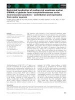
Fig. 1. Production and activation of the recombinant pro-TMPRSS13. (A) Schematic representation of the structure of pro-TMPRSS13 (wild-
type), the recombinant pro-TMPRSS13 and the enterokinase-cleaved pro-TMPRSS13. The wild-type pro-TMPRSS13 comprises 567 amino
acids. The amino acid numbering starts from the putative N-terminus of the protein. The domain structures are indicated in pro-TMPRSS13
(wild-type). TM, transmembrane domain; LDLA, LDL receptor class A domain; SRCR, scavenger receptor cysteine-rich domain; SPD, serine
protease domain. The predicted disulfide linkage is shown as SS. The putative activation cleavage site (indicated by an arrow) and its sur-
rounding sequence are shown in pro-TMPRSS13 (wild-type). The recombinant pro-TMPRSS13 is a secreted form in which the cytoplasmic
domain and transmembrane domain (Met1-Gln186) are replaced with the mouse immunoglobulin j-chain signal peptide. In addition,
AMTGR325 in the wild-type protein is replaced with the enterokinase recognition sequence (DDDDK, underlined) for cleavage in vitro before
Ile326 (activation cleavage). The recombinant pro-TMPRSS13 is tagged at the C-terminus with myc-His. The enterokinase-cleaved recombi-
nant pro-TMPRSS13, the disulfide-linked two-chain form, is illustrated at the bottom. (B, C) Immunoblot analysis of the recombinant pro-
TMPPRSS13 produced in COS-7 cells, and its enterokinase-treated product. Samples of pro-TMPRSS13 (lane 1) and enterokinase-treated
pro-TMPRSS13 (lane 2) were separated by SDS ⁄ PAGE under reducing conditions (B) or under nonreducing conditions (C), and analysed
by immunoblotting with the anti-c-Myc IgG. The protease domain of TMPRSS13 was quantified by scanning densitometry of the immunoblot
(B, lane 2) with NIH
IMAGEJ software using the protease domain of TMPRSS13, which was expressed in E. coli, as a standard.
Protease TMPRSS13 is inhibited by HAI-1 T. Hashimoto et al.
4890 FEBS Journal 277 (2010) 4888–4900 ª 2010 The Authors Journal compilation ª 2010 FEBS
pro-TMPRSS13 efficiently cleaved the substrate, and
thus was used for an assay of inhibition by HAI-1.
Inhibition of TMPRSS13 protease activity by
soluble HAI-1
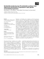
Inhibition of the protease activity of TMPRSS13 was
assessed using recombinant soluble forms of HAI-1,
HAI-1–NK1 and HAI-1–NK1LK2. HAI-1 is first pro-
duced as a 66 kDa transmembrane form, and subse-
quent ectodomain shedding releases two major soluble
forms of 40 and 58 kDa from the cell surface into the
extracellular space [14]. HAI-1–NK1, which corre-
sponds to the 40 kDa form, consists of the N-terminal
region (N) and one Kunitz domain (K1), whereas
HAI-1–NK1LK2, corresponding to the 58 kDa form,
consists of the N-terminal region (N), two Kunitz
domains (K1 and K2), and the low-density lipoprotein
(LDL) receptor class A domain (L) between the
Kunitz domains (Fig. 2A). Inhibition by aprotinin was
compared with that by HAI-1, because aprotinin has
been shown to efficiently inhibit the protease activity
of TMPRSS13 [12]. TMPRSS13 (100 pm) was incu-
bated with various concentrations of HAI-1–NK1,
HAI-1–NK1LK2 and aprotinin, and protease activity
was measured using the synthetic substrate. Figure 2C
shows the dose dependence of the inhibitory activities.
HAI-1–NK1 had the most potent inhibitory effect
(IC
50
= 2.18 ± 0.18 nm). HAI-1–NK1LK2 and apro-
tinin showed much weaker inhibitory activity than
HAI-1–NK1.
Hepatocyte growth factor activator inhibitor type 2
(HAI-2), also known as placental bikunin, is also a
transmembrane Kunitz-type serine protease inhibitor
[15,16]. HAI-2 has been shown to inhibit matriptase
and hepsin [9,10,17]. Thus, we examined the effect of a
soluble form of HAI-2 (Fig. 2B) on the protease activ-
ity of TMPRSS13. HAI-2 inhibited TMPRSS13
(IC
50
= 1.54 ± 0.01 nm) (Fig. 2C), and the IC
50
was
similar to that of HAI-1–NK1. However, the inhibi-
tion curves were quite different: the inhibition curve of
HAI-2 was sigmoidal, whereas that of HAI-1–NK1
was not (Fig. 2C).
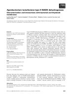
Formation of complexes of TMPRSS13 and
HAI-1–NK1
To confirm the inhibitory effect of HAI-1–NK1 on
TMPRSS13, we examined the formation of complexes
by the protease–inhibitor pair. HAI-1–NK1 and HAI-
1–NK1LK2 were incubated with the activated
TMPRSS13 at different molar ratios. The samples
were boiled or not boiled, and subjected to an immu-
noblot analysis. Immunoblotting with an anti-HAI-1
IgG showed that increasing concentrations of
TMPRSS13 shifted the HAI-1–NK1 band (40 kDa) to
a higher molecular mass species (70 kDa) when the
samples were not boiled (Fig. 3A). This shift was con-
firmed by an immunoblot analysis with an anti-
TMPRSS13 IgG (Fig. 3B). When samples were boiled,
the band did not shift (Fig. 3A,B). These results indi-
cate the formation of TMPRSS13ÆHAI-1–NK1 com-
plexes. On the other hand, the HAI-1–NK1LK2 band
(58 kDa) did not shift to a high molecular mass species
even in the presence of a high concentration of
B
A
Hepatocyte growth factor activator inhibitor type-1 (HAI-1)
Hepatocyte growth factor activator inhibitor Type-2 (HAI-2)
(1)
C
SP
K1
LDLA
N
K2 TM
(2)
myc-His-
N
N
C
(3)
myc-His-
C
myc-His-
C
SP
K1
N
TMPRSS13 activity (%)
Inhibitor concentration (nM)
.
.
.
.
.
.
.
.
.
.
.
.
.
.
.
.
.
.
.
.
.
.
.
.
.
.
.
.
.
.
.
.
.
.
.
.
.
.
.
.
.
.
.
.
.
.
.
.
.
.
.
.
.
.
.
.
.
.
.
.
.
.
.
.
.
.
.
.
.
.
.
.
.
.
.
.
.
.
.
.
.
.
.
.
.
.
.
.
.
.
.
.
.
.
.
.
.
.
.
.
.
.
.
.
.
.
.
.
.
.
.
.
.
.
.
.
.
.
.
.
.
.
.
.
.
.
.
.
SP
K1
LDLA
N
K2
N
C
HAI-2
NK1
Aprotinin
NK1LK2
(2)
N
.
.
.
.
.
.
.
.
.
.
.
.
.
.
.
.
.
.
.
.
.
.
.
.
.
.
.
.
.
.
.
.
.
.
.
.
.
.
.
.
.
.
SP
K1 K2
(1)
C
N
.
.
.
.
.
.
.
.
.
.
.
.
.
.
.
.
.
.
.
.
.
.
.
.
.
.
.
.
.
.
.
.
.
.
.
.
.
.
.
.
.
.
SP
K1 K2
TM
0
10
20
30
40
50
60
70
80
90
100
0.01 0.1 1 10 100 1000
Fig. 2. Dose dependence of the inhibitory activity of soluble
forms of HAI-1 and HAI-2 against the protease activity of
TMPRSS13. (A) Schematic representation of the structure of the
full-length HAI-1 (1) and soluble forms of HAI-1, HAI-1–NK1LK2
(2) and HAI-1–NK1 (3), tagged at the C-terminus with myc-His.
SP, signal peptide; N, N-terminal region; K1, Kunitz domain 1;
LDLA, LDL receptor class A domain; K2, Kunitz domain 2; TM,
transmembrane domain. (B) Schematic representation of the
structure of the full-length HAI-2 (1) and a soluble form of HAI-2
tagged at the C-terminals with myc-His (2). (C) Dose dependence
of the inhibitory activity of soluble forms of HAI-1 and HAI-2
against the protease activity of TMPRSS13. TMPRSS13 was
incubated with various concentrations of HAI-1–NK1 (
•
), HAI-1–
NK1LK2 (j), aprotinin (m) or HAI-2 (r). Then, Pyr-RTKR-MCA
was added, and after further incubation, the fluorescence of the
reaction mixtures was measured. Data show the mean ± stan-
dard deviation for three separate experiments and are expressed
as a percentage of TMPRSS13 activity.
T. Hashimoto et al. Protease TMPRSS13 is inhibited by HAI-1
FEBS Journal 277 (2010) 4888–4900 ª 2010 The Authors Journal compilation ª 2010 FEBS 4891
TMPRSS13 (Fig. 3A,B), which is consistent with data
showing weak inhibitory activity of HAI-1–NK1LK2
against the protease activity of TMPRSS13.
Proteolytic activation of pro-HGF by TMPRSS13
Pro-HGF is proteolytically activated by matriptase
and hepsin, and the protease activity is inhibited by
HAI-1 [9,10,18]. Therefore, we examined whether
TMPRSS13 also functions as an HGF-converting pro-
tease. The single-chain pro-HGF (2 lm) was incubated
with various concentrations of TMPRSS13. The reac-
tion products were separated by SDS⁄ PAGE under
reducing conditions and stained with Coomassie Bril-
liant Blue. The incubation generated two main bands
of 60 and 32 kDa (Fig. 4A). The sizes corresponded
to the heavy chain and light chain of activated HGF,
suggesting that pro-HGF is activated by TMPRSS13.
The intensity of the pro-HGF band on the gel was
quantified by scanning densitometry, and the percent-
age of HGF processed was calculated. Pro-HGF was
almost completely converted to the two-chain form by
54 nm TMPRSS13 (Fig. 4B).
We then analysed the effect of HAI-1–NK1 on the
pro-HGF converting activity of TMPRSS13. The sin-
gle-chain pro-HGF (2 lm) was incubated with
TMPRSS13 (54 nm) pretreated with or without HAI-1–
NK1 (5 lm). The pretreatment of TMPRSS13 with
HAI-1–NK1 did not generate the 60 and 32 kDa bands
(Fig. 4C), indicating that HAI-1–NK1 inhibits the
pro-HGF converting activity of TMPRSS13.
A
TMPRSS13 (nM) TMPRSS13 (nM)
50
37
75
(kDa)
50
37
75
(kDa)
50
75
100
(kDa)
50
75
100
(kDa)
NK1 (n
M)
Not boiled
Boiled Boiled
NK1LK2 (nM)
Not boiled
2 20 200
20
2 20 200
20 20 20 20 20 20 20
TMPRSS13 (n
M) TMPRSS13 (nM)
NK1 (n
M) NK1LK2 (nM)
2 20 200
20
2 20 200
20 20 20 20 20 20 20
B
TMPRSS13 (nM) TMPRSS13 (nM)
50
37
75
(kDa)
(kDa)
50
37
75
100
NK1 (n
M)
Not boiled
NK1LK2 (nM)
Not boiled
2 20 200
20
2 20 200
20 20 20 20 20 20 20
TMPRSS13 (n
M) TMPRSS13 (nM)
50
37
75
(kDa)
50
37
75
100
NK1 (n
M)
Boiled
NK1LK2 (nM)
Boiled
2 20 200
20
2 20 200
20 20 20 20 20 20 20
(kDa)
IB: HAI-1
IB: TMPRSS13
*
**
*
Fig. 3. TMPRSS13 forms complexes with HAI-1–NK1. TMPRSS13
at the indicated concentrations was incubated with 20 n
M HAI-1–
NK1 and HAI-1–NK1LK2 at 37 °C for 2 h. After the addition of SDS
sample buffer with 100 m
M dithiothreitol, each sample was boiled
or not boiled (as indicated). Samples were separated by SDS ⁄ PAGE
under reducing conditions, and analysed by immunoblotting with
anti-HAI-1 IgG (A) or anti-TMPRSS13 IgG (B). The asterisks indicate
complexes of TMPRSS13 and HAI-1–NK1.
A
TMPRSS13 (nM)
0
3.4 6.8 13.5 27
54
100
75
50
37
25
(kDa)
100
75
50
37
25
(kDa)
Pro-HGF
HGF heavy-chain
HGF light-chain
Pro-HGF
HGF heavy-chain
HGF light-chain
HAI-1-NK1
B
HGF processing (%)
Protease concentration (nM)
0
20
40
60
80
100
3.4 6.8 13.5 27 54
C
HAI-1-NK1 (µM)
TMPRSS13 (n
M)
54 54
5
0
00
Fig. 4. Proteolytic conversion of pro-HGF by TMPRSS13 and its inhi-
bition by HAI-1–NK1. (A) Pro-HGF (2 l
M) was incubated with various
concentrations of TMPRSS13. The reaction mixtures were separated
by SDS ⁄ PAGE under reducing conditions. The gel was stained with
Coomassie Brilliant Blue. (B) The intensity of the band of pro-HGF
was quantified with NIH
IMAGEJ software and the percentage of HGF
processed was calculated. (C) Pro-HGF (2 l
M) was incubated with
TMPRSS13 (54 n
M) pretreated with or without HAI-1–NK1 (5 lM).
The reaction mixtures were analysed as described in (A).
Protease TMPRSS13 is inhibited by HAI-1 T. Hashimoto et al.
4892 FEBS Journal 277 (2010) 4888–4900 ª 2010 The Authors Journal compilation ª 2010 FEBS
Biological activities of HGF converted by
TMPRSS13
To examine the biological activities of the HGF con-
verted by TMPRSS13, we used the human hepatocellu-
lar carcinoma cell line HepG2. HGF induces a
scattering of cell colonies and inhibition of serum-
dependent proliferation in HepG2 cells [19]. These bio-
logical responses to HGF are transduced through the
activation of a high affinity receptor, the c-met proto-
oncogene product (c-Met), and also require strong
activation of the extracellular signal-regulated kinase
(ERK) [20]. Therefore, we first analysed the activation
of c-Met by assessing its tyrosine phosphorylation.
HepG2 cells were treated with the TMPRSS13-cleaved
pro-HGF, and tyrosine phosphorylation of c-Met was
analysed by immunoblotting using an anti-phospho-c-
Met IgG. The tyrosine phosphorylation was induced in
HepG2 cells treated with the TMPRSS13-cleaved pro-
HGF at a level comparable with that in cells treated
with the purified active HGF, whereas it was not
induced in HepG2 cells treated with the uncleaved
pro-HGF (Fig. 5A). Treatment of the cells with
TMPRSS13 itself did not induce the phosphorylation
(Fig. 5A).
We then analysed the activation of ERK by assess-
ing its phosphorylation. Immunoblotting using an
anti-phospho-ERK1 ⁄ 2 IgG showed that the phosphor-
ylation of ERK1 ⁄ 2 was more enhanced in HepG2 cells
treated with the TMPRSS13-cleaved pro-HGF than in
HepG2 cells treated with the uncleaved pro-HGF or
with TMPRSS13 (Fig. 5B). Finally, we analysed the
biological response of HepG2 cells by observing their
scattering phenotype. Treatment with the TMPRSS13-
cleaved pro-HGF induced a scattering of cell colonies,
whereas no scattering was observed in the cells treated
with the uncleaved pro-HGF or with TMPRSS13
(Fig. 5C). These results indicate that TMPRSS13 con-
verts the inactive pro-HGF into the active two-chain
form of HGF.
Co-expression of TMPRSS13 and HAI-1 mRNA in
cultured cell lines
Because TMPRSS13 and HAI-1 are both transmem-
brane proteins, HAI-1 is probably co-expressed with
TMPRSS13 in the same cells to function as a physio-
logical inhibitor of the protease. We examined the
co-expression of TMPRSS13 and HAI-1 mRNA in
cultured cell lines by RT-PCR. We analysed five
human carcinoma cell lines: a lung carcinoma cell line
A549, a colon carcinoma cell line LoVo, stomach car-
cinoma cell lines MKN45 and MKN74, and HepG2.
A549 and LoVo cells have been shown to express
TMPRSS13 mRNA [13]. MKN45 cells were used for
identification of HAI-1 proteins [4]. MKN74 and
HepG2 cells have been shown to respond to HGF [20].
TMPRSS13 mRNA was detected in MKN45 and
MKN74 cells, but not in A549, LoVo and HepG2
cells. On the other hand, HAI-1 mRNA was detected
in LoVo, MKN45, MKN74 and HepG2 cells (Fig. 6).
These results indicate that HAI-1 mRNA is
co-expressed with TMPRSS13 mRNA in MKN45 and
MKN74 cells.
A
B
C
Pro-HGF
Active HGF
TMPRSS13
IB: Phospho-c-Met
IB: Phospho-ERK1/2
IB: ERK1/2
IB: c-Met
(Tyr1234/1235)
Pro-HGF + TMPRSS13
Pro-HGF
Active HGF
TMPRSS13 TMPRSS13
Pro-HGF
+
Pro-HGF
Active HGF
TMPRSS13 TMPRSS13
Pro-HGF
+
Fig. 5. Biological activity of HGF converted by TMPRSS13. Cells
were treated with reaction mixtures of pro-HGF alone (Pro-HGF),
TMPRSS13 alone (TMPRSS13) or pro-HGF and 2000 ngÆmL
)1
TMPRSS13 (Pro-HGF + TMPRSS13) at 50 ngÆmL
)1
pro-HGF. Cells
were also treated with purified active HGF at 50 ngÆmL
)1
(Active
HGF). (A) Cells were cultured for 5 min. Lysate of the cells was
immunoblotted with the anti-phospho-c-Met IgG (upper panel) and
anti-c-Met IgG (lower panel). (B) Cells were cultured for 5 min.
Lysate of the cells was immunoblotted with the anti-phospho-
ERK1 ⁄ 2 IgG (upper panel) and anti-ERK1 ⁄ 2 IgG (lower panel).
(C) Cells were cultured for 4 days. The morphology of the cells
was analysed by light microscopy.
T. Hashimoto et al. Protease TMPRSS13 is inhibited by HAI-1
FEBS Journal 277 (2010) 4888–4900 ª 2010 The Authors Journal compilation ª 2010 FEBS 4893
Discussion
In this study, we tested the inhibitory effect of HAI-1
on the protease activity of several members of the
TTSP family using enzymes expressed in E. coli.We
found that the protease activity of TMPRSS13 was
inhibited by HAI-1, but that of TMPRSS11A, HAT-
like 4 and HAT-like 5 was not. TMPRSS11A, HAT-
like 4, and HAT-like 5 belong to the HAT ⁄ DESC
subfamily [2]. Mouse DESC1, also of the HAT ⁄ DESC
subfamily, forms stable inhibitory complexes with plas-
minogen activator inhibitor-1 and protein C inhibitor
[21]. Thus, these serpins might be endogenous inhibi-
tors of TMPRSS11A, HAT-like 4 and HAT-like 5.
The protease activity of TMPRSS13 expressed in
E. coli was weak, probably because of incorrect pro-
tein folding. Thus, we expressed the enzyme in mam-
malian cells. To obtain an active TMPRSS13 in
mammalian cells, we constructed an expression vector
encoding a recombinant protein with two modifica-
tions, and transfected COS-7 cells with the vector. One
modification was that we deleted the N-terminal cyto-
plasmic and transmembrane domains and tagged the
C-terminus with six His sequences, to simply purify
the protein from the conditioned medium of the trans-
fected cells by one-step column chromatography. The
other modification was that we replaced the putative
activation cleavage sequence with the enterokinase rec-
ognition sequence, because the molecular mechanism
of the proteolytic activation of pro-TMPRSS13 is
unknown. The purified pro-enzyme did not show any
protease activity, and the enterokinase treatment gen-
erated an active enzyme (Fig. 1). Using this active
TMPRSS13, we demonstrated that HAI-1–NK1 had
inhibitory activity against the protease (Fig. 2). The
activity was much stronger than that of aprotinin,
which was previously described as an inhibitor of
TMPRSS13 [12]. The inhibitory activity of HAI-1–
NK1 against TMPRSS13 was confirmed by in vitro
binding assays. HAI-1–NK1 formed complexes with
the active TMPRSS13 (Fig. 3). HAI-1–NK1 consists
of the N-terminal region and the first Kunitz domain,
and corresponds to the 40 kDa form of HAI-1 gener-
ated from a transmembrane form by extracellular
shedding [4]. TMPRSS13 mRNA is expressed in a
variety of human adult tissues, and predominantly in
lung, placenta, pancreas and prostate [12]. HAI-1
mRNA is also highly expressed in placenta, pancreas
and prostate [4]. Thus, the 40 kDa form of HAI-1
could function as an endogenous regulator of
TMPRSS13 in these tissues.
HAI-1–NK1LK2 had a much weaker inhibitory
effect against TMPRSS13 than HAI-1–NK1 (Fig. 2).
Moreover, no complex of HAI-1–NK1LK2 and
TMPRSS13 was detected in the in vitro binding assays
(Fig. 3). These results indicate that HAI-1–NK1LK2
only weakly associates with TMPRSS13. HAI-1–
NK1LK2 consists of the N-terminal region, the first
Kunitz domain, the LDL receptor class A domain,
and the second Kunitz domain, and corresponds to the
58 kDa form of HAI-1 identified in the conditioned
medium of cultured carcinoma cells [14]. Weaker
inhibitory activity of HAI-1–NK1LK2 against HGFA
and matriptase was also observed, and an idea that the
second Kunitz domain may obstruct the protease-bind-
ing site of the first Kunitz domain was proposed
[22,23]. The present results indicate that this idea may
also apply to TMPRSS13. The weaker inhibitory activ-
ity of HAI-1–NK1LK2 was prominent against
TMPRSS13, compared with that against HGFA and
matriptase. Thus, the presence of the second Kunitz
domain may more strongly affect the binding of the
first Kunitz domain to TMPRSS13.
A soluble form of HAI-2, another Kunitz-type
inhibitor, also inhibited the protease activity of
TMPRSS13, with an IC
50
similar to that of HAI-1–
NK1 (Fig. 2C). HAI-2 mRNA is highly expressed in
various human adult tissues [15], some of which also
express TMPRSS13 mRNA, suggesting that HAI-2
could be an endogenous inhibitor of TMPRSS13 in
these tissues.
The inhibition curve of HAI-1–NK1 was not sigmoi-
dal, which is unusual, compared with the sigmoidal
curve of HAI-2. Moreover, a high concentration of
HAI-1–NK1 was needed for full inhibition of the pro-
tease activity of TMPRSS13 (Fig. 2C). These results
suggest the characteristic association of HAI-1–NK1
with TMPRSS13, the mechanism of which remains to
be investigated. The in vitro binding assays showed
that only small portions of HAI-1–NK1 and
TMPRSS13
HAI-1
GAPDH
HAI-1B
HAI-1
No template control
A549
LoVo
MKN45
MKN74
HepG2
Fig. 6. RT-PCR analysis of TMPRSS13 and HAI-1 mRNA in human
carcinoma cell lines. Total RNA was isolated from cultured A549,
LoVo, MKN45, MKN74 and HepG2 cells, and subjected to RT-PCR
analysis. The primers for HAI-1 generate two PCR products of HAI-1
and its splice variant (HAI-1B) [35]. GAPDH mRNA was used as an
internal control.
Protease TMPRSS13 is inhibited by HAI-1 T. Hashimoto et al.
4894 FEBS Journal 277 (2010) 4888–4900 ª 2010 The Authors Journal compilation ª 2010 FEBS
TMPRSS13 formed complexes (Fig. 3). The weak
complex formation may be related to the characteristic
association of the protease–inhibitor pair.
In the present study we have shown that
TMPRSS13 converted the single-chain pro-HGF to a
two-chain form in vitro (Fig. 4). We proved that the
two-chain form of HGF is biologically active, by three
assessments. Its treatment of HepG2 cells induced the
tyrosine phosphorylation of c-Met, enhanced the phos-
phorylation of ERK, and induced the scattering
phenotype (Fig. 5). Thus, the proteolytic cleavage of
pro-HGF by TMPRSS13 generates a biologically
active HGF. The concentration for half-maximal activ-
ity of TMPRSS13 was 15 nm (Fig. 4B). This value was
0.17 nm for HGFA under similar reaction conditions
[24]. Thus, the specific activity of TMPRSS13 is
approximately 90-fold lower than that of HGFA.
TMPRSS13 preferentially recognizes cleavage sites
consisting of paired basic amino acid residues (RR or
KR at positions P2 and P1). In addition, the presence
of a basic amino acid residue (R or K) at position P4
enhances the efficiency of cleavage [13]. The HA protein
of an HPAI virus strain with the KKKR motif at the
cleavage site was efficiently converted to mature HA
subunits in TMPRSS13-transfected cells [13], suppor-
ting the preference for the cleavage sequences in sub-
strates of TMPRSS13. Pro-HGF has the KQLR motif
at the cleavage site [25]. Thus, the nonbasic amino acid
residue at position P2 may cause the low specific activity
of TMPRSS13 for the conversion of pro-HGF.
HGF is a pleiotropic factor that functions as a mito-
gen, motogen and morphogen for a variety of cells,
particularly epithelial cells [25,26]. HGF is thought to
play a crucial role in the regeneration of various
tissues following injury [27]. HGF is a mesenchymal
cell-derived heparin-binding glycoprotein that is
secreted as an inactive single-chain precursor. The
secreted HGF normally remains inactive, probably
associated with the extracellular matrix in the tissues
producing it. In response to tissue injury, such as
hepatic and renal injury, the inactive single-chain HGF
is converted to a two-chain form exclusively in
the injured tissue. This conversion is mediated by ser-
ine protease activity, which is induced in the injured
tissue [28]. The two-chain form is required for the bio-
logical activity of HGF [29,30]. Thus, the biological
effects of HGF in injured tissue are regulated through
proteolytic processing by a serine protease. HGFA is a
serum-derived serine protease that efficiently converts
the single-chain HGF to the biologically active two-
chain form in vitro [31]. The role of HGFA in the pro-
teolytic activation of HGF in vivo was determined by
analysing knockout mice. In HGFA-deficient mice,
regeneration of the injured intestinal mucosa and the
activation of HGF were impaired, but the injured liver
was completely regenerated, suggesting that HGFA is
responsible for the activation of HGF in the injured
intestinal mucosa, but not in other injured tissues [32].
Thus, other serine proteases are probably involved in
the activation of HGF in these tissues.
Several serine proteases have been shown to convert
pro-HGF to the active form in vitro. They include serine
proteases involved in blood coagulation, such as plasma
kallikrein, and coagulation factors XIa and XIIa
[24,33]. These serine proteases might be responsible for
the activation of HGF in injured tissues. Matriptase
and hepsin, members of the TTSP family, also convert
pro-HGF to the active form [9,10,15]. Thus, it is possi-
ble that these TTSPs function as HGF-converting pro-
teases in injured tissue. A two-step model for the
activation of HGF in injured tissues has been proposed.
When tissue injury occurs, circulating plasma serine
proteases, such as HGFA, are activated in response to
the activation of the coagulation cascade and inflamma-
tion. The activated proteases convert pro-HGF to the
active form (the first step). Subsequently, the activated
HGF functions as a mitogen for the epithelial cells. The
proliferating epithelial cells produce TTSPs, such as
matriptase. The TTSPs convert pro-HGF to the active
form (the second step). The activated HGF is involved
in further proliferation of the epithelial cells [32].
TMPRSS13 might also function as an HGF-converting
protease in the second step, because it appears to be
expressed in epithelial cells [13]. The specific activity of
the HGF conversion of TMPRSS13 is much lower than
that of HGFA as described above. However,
TMPRSS13 localizes to the cell surface, and thus could
function in the pericellular activation of HGF.
The pro-HGF converting activity of TMPRSS13
was inhibited by HAI-1–NK1 (Fig. 4C), suggesting
that HAI-1 functions as a regulator for the activation
of HGF in injured tissues. RT-PCR analysis showed
that TMPRSS13 mRNA is co-expressed with HAI-1
mRNA in MKN45 and MKN74 carcinoma cells
(Fig. 6). Thus, the pericellular activation of HGF by
TMPRSS13 could be regulated by HAI-1 produced in
the same cells. Further characterization is required to
clarify the roles of TMPRSS13 and HAI-1 in regulat-
ing the activation of HGF in vivo.
Experimental procedures
DNA constructs
The cDNA clones for the protease domains with short pro-
sequences of TTSPs were obtained from appropriate human
T. Hashimoto et al. Protease TMPRSS13 is inhibited by HAI-1
FEBS Journal 277 (2010) 4888–4900 ª 2010 The Authors Journal compilation ª 2010 FEBS 4895
cDNA libraries (Takara, Kyoto, Japan) by PCR, and
inserted into an E. coli expression vector, pMAL-c2X (New
England BioLabs, Ipswich, MA, USA). The putative acti-
vation cleavage sequences were replaced with the enteroki-
nase recognition sequence (DDDDK) using a QuikChange
site-directed mutagenesis kit (Stratagene, La Jolla, CA,
USA).
The cDNA clone for the full-length TMPRSS13 was
obtained from a human placenta cDNA library (Takara)
by PCR. The PCR product was further amplified by PCR
using a primer containing an EcoRI restriction site and a
primer containing an XbaI site, which also had a point
mutation replacing the stop codon with a Leu codon. The
PCR product was subcloned into a mammalian expression
vector, p3xFLAG-CMV14 (Sigma, St. Louis, MO, USA).
To construct an expression vector encoding pro-
TMPRSS13 lacking the cytoplasmic and transmembrane
domains, a cDNA sequence encoding amino acid residues
187–567 was amplified by PCR using p3xFLAG-CMV14-
TMPRSS13 as a template. The PCR product was
subcloned into the EcoRI and PstI sites of a mammalian
expression vector, pSecTag2C (Invitrogen, Carlsbad, CA,
USA). The activation cleavage site (A321MTGR325) was
replaced with the enterokinase recognition sequence as
described above.
To construct an E. coli expression vector encoding pro-
TMPRSS13, the cDNA sequence was excised by digestion
with HindIII and XbaI from p3xFLAG-CMV14-
TMPRSS13, and subcloned into an expression vector,
pcDNA3.1 ⁄ myc-His-A (Invitrogen). The activation cleavage
site was replaced with the enterokinase recognition
sequence as described above. A cDNA sequence encoding
amino acid residues 315–567 with the C-terminally tagged
myc-His sequence was amplified by PCR, and subcloned
into the EcoRI and PstI sites of an E. coli expression
vector, pMAL-c2X.
To construct an E. coli expression vector encoding HAI-
1–K1, the cDNA sequence encoding amino acid residues
241–305 was amplified by PCR using cDNA of HAI-1 [4]
as a template. The PCR product was subcloned into the
BamHI and XbaI sites of the vector, pcDNA3.1 ⁄ myc-His-
A. The cDNA sequence encoding HAI-1–K1 with the
C-terminally tagged myc-His sequence was amplified by
PCR, and subcloned into the NdeI and NotI sites of an
E. coli expression vector, pET30a (EMD Chemicals, Gibbs-
town, NJ, USA).
To construct expression vectors encoding HAI-1–NK1
and HAI-1–NK1LK2, cDNA sequences encoding amino
acid residues 1–314 and 1–436 were amplified by PCR using
cDNA of HAI-1 [4] as a template. The PCR products were
subcloned into the HindIII and XbaI sites of pcDNA3.1 ⁄
myc-His-A.
To construct an expression vector encoding HAI-2, the
cDNA sequence encoding amino acid residues 1–194 was
amplified by PCR using cDNA of HAI-2 [15] as a template.
The PCR product was subcloned into the HindIII and XbaI
sites of pcDNA3.1 ⁄ myc-His-A.
Preparation and activation of pro-TTSPs
expressed in E. coli
Escherichia coli cells were transformed with the expression
vectors encoding pro-TTSPs. The cells were lysed by soni-
cation, and the lysate was applied to an amylose resin
(New England BioLabs). After the resin was washed with
phosphate-buffered saline (PBS), bound proteins were
eluted with 1 mm maltose in PBS. The eluted fraction was
treated overnight with enterokinase (EMD Chemicals) at
2 unitsÆ100 lL
)1
.
Cell culture
COS-7 cells, A549 cells and HepG2 cells were cultured in
Dulbecco’s modified Eagle’s medium, CHO cells and Lovo
cells were cultured in Ham’s F12 medium, and MKN45
cells and MKN74 cells were cultured in RPMI1640 med-
ium, supplemented with 10% fetal bovine serum, 100
unitsÆmL
)1
penicillin and 100 lgÆmL
)1
streptomycin at
37 °C in a humidified atmosphere containing 5% CO
2
.
Preparation and activation of pro-TMPRSS13
expressed in COS-7 cells
Cells were seeded on eight 100 mm collagen-coated plates
(Iwaki, Chiba, Japan) at a density of 1·10
6
cellsÆplate
)1
.
The cells were transfected with the expression vector encod-
ing the secreted form of pro-TMPRSS13 at 6 lgÆplate
)1
using the FuGENE-6 reagent (Roche Diagnostics, India-
napolis, IN, USA). After 24 h, the medium was replaced
with serum-free medium, and cells were further cultured for
3 days. The conditioned medium was applied to a nickel
nitrilotriacetic acid resin (EMD Chemicals), and the proteins
bound to the resin were eluted with nickel nitrilotriacetic acid
buffer (EMD Chemicals). The eluted fraction was treated
overnight with enterokinase at 2 unitsÆ100 lL
)1
.
Quantification of TMPRSS13
The enterokinase-treated pro-TMPRSS13 was quantified by
immunoblotting using the protein expressed in E. coli as a
standard. The protease domain with its short pro-sequence
and the enterokinase recognition sequence of TMPRSS13
fused at the N-terminus to maltose-binding protein and
tagged at the C-terminus with myc-His was expressed in
E. coli. Preparation of the cell lysate, purification of the
proteins, and treatment with enterokinase were carried out
as described above for TTSPs expressed in E. coli. The
enterokinase-treated pro-TMPRSS13 was separated by
SDS ⁄ PAGE under reducing conditions, and stained by
Protease TMPRSS13 is inhibited by HAI-1 T. Hashimoto et al.
4896 FEBS Journal 277 (2010) 4888–4900 ª 2010 The Authors Journal compilation ª 2010 FEBS
Coomassie Brilliant Blue. The intensity of the band of the
protease domain was quantified using bovine serum albu-
min (BSA) as a standard.
The enterokinase-treated pro-TMPRSS13 obtained from
COS-7 cells was separated by SDS ⁄ PAGE under reducing
conditions. In parallel, various amounts of the enteroki-
nase-treated pro-TMPRSS13 obtained from E. coli were
separated by SDS ⁄ PAGE. After electrophoresis, the sam-
ples were subjected to an immunoblot analysis with the
anti-c-Myc IgG. The intensity of the band of the protease
domain was quantified using the protease domain of the
protein obtained from E. coli .
Enzyme inhibition assay
HAI-1–K1 was prepared as follows. Escherichia coli cells
were transformed with the expression vector encoding HAI-
1–K1. The cells were lysed by sonication. The lysate was
centrifuged, and the pellet was dissolved in urea (6 m). To
refold proteins, glutathione (oxidized form, 5 mm), glutathi-
one (reduced form, 1 mm) and arginine (100 mm) were
added to the solution, and the final concentration of urea
was adjusted to 0.5 m. The refolded HAI-1–K1 was purified
by column chromatography using a nickel nitrilotriacetic
acid resin, followed by dialysis against PBS. The enteroki-
nase-treated pro-TTSPs were mixed with HAI-1–K1
(0.67 lm) and incubated in the assay buffer (50 mm
Tris ⁄ HCl pH 7.5, 150 mm NaCl, and 0.05% Brij 35) for
10 min at 37 °C. Then each substrate was added to the
mixture at a final concentration of 100 lm. After incuba-
tion for 3 h at 37 °C, the amount of 7-amino-4-methyl-
coumarin liberated from the substrate was determined
fluorimetrically with excitation and emission wavelengths of
355 and 460 nm, respectively, using a fluorometer (1420
ARVOsx; Perkin Elmer Life Science, Boston, MA, USA).
HAI-1–NK1, HAI-1–NK1LK2 and HAI-2 were prepared
as follows. The expression vectors encoding HAI-1–NK1,
HAI-1–NK1LK2 and HAI-2 were introduced into CHO
cells using Superfect transfection reagent (Qiagen, Hilden,
Germany). Transfected cells were cultured at 37 °C over-
night. The medium was replaced with fresh medium con-
taining Geneticin (G418). Neomycin-resistant colonies were
selected and further cultured in a roller bottle. When the
cells became confluent, the medium was replaced with
serum-free medium, and the cells were further cultured for
5 days. The proteins were purified from the conditioned
medium by column chromatography using nickel nitrilotri-
acetic acid and anti-c-Myc IgG resins. Aprotinin was
obtained from Nakarai Tesque (Kyoto, Japan). The entero-
kinase-treated pro-TMPRSS13 (100 pm) and a series of
concentrations of inhibitors were mixed and incubated in
the assay buffer (50 mm Tris ⁄ HCl pH 8.0, 150 mm NaCl,
and 0.05% Brij 35) for 10 min at 37 °C. Then, Pyr-RTKR-
MCA (Peptide Institute, Osaka, Japan) was added to the
mixture at a final concentration of 100 lm. The final
volume of each mixture was 200 lL. After incubation for
1 h at 37 °C, the amount of 7-amino-4-methylcoumarin lib-
erated from the substrate was determined as described
above. The enzymatic activity without inhibitors was used
as an uninhibited control. The IC
50
was defined as the con-
centration of inhibitor that inhibited the enzymatic activity
by 50% compared with the uninhibited control. The per-
centage value relative to the uninhibited control was plotted
against the log of inhibitor concentrations. The IC
50
value
was calculated using the graphpad prism software (Graph-
Pad Software, San Diego, CA, USA).
Binding assay
HAI-1–NK1 or HAI-1–NK1LK2 was mixed with various
concentrations of TMPRSS13 in the assay buffer. The mix-
ture was incubated at 37 °C for 2 h and SDS sample
buffer (20 mm Tris ⁄ HCl pH 6.8, 0.5% SDS, 5% glycerol
and 0.002% bromophenol blue) with 100 mm dithiothreitol
was added. Some of the samples were boiled for 5 min.
Twenty microlitres of each sample was analysed by
immunoblotting.
HGF-converting activity of TMPRSS13
The recombinant pro-HGF was prepared as described pre-
viously [34]. Pro-HGF (2 lm) was mixed with various con-
centrations of TMPRSS13 in 20 lLof20mm sodium
phosphate (pH 7.3) containing 100 mm NaCl and 0.01%
Chaps and incubated at 37 °C for 2 h. The reaction mixture
was separated by SDS ⁄ PAGE under reducing conditions.
Proteins in the gel were stained with Coomassie Brilliant
Blue. The intensity of the pro-HGF band was quantified by
scanning densitometry using NIH imagej software.
To examine the inhibitory effect of HAI-1–NK1 on the
HGF-converting activity of TMPRSS13, TMPRSS13
(54 nm) was incubated with HAI-1–NK1 (5 lm)in20mm
sodium phosphate (pH 7.3) containing 100 mm NaCl and
0.01% Chaps at 37 °C for 10 min. Then, pro-HGF (2 lm)
was added to the mixture. The final volume of the mixture
was 20 l L. After incubation at 37 °C for 2 h, the reaction
mixture was analysed by SDS ⁄ PAGE, as described above.
Preparation of cell lysate
HepG2 cells were seeded at 1·10
6
cellsÆ100 mmÆplate
)1
.
They were treated with reaction mixtures of the assay for
HGF-converting activity of TMPRSS13 or with purified
active HGF (provided by the Research Center of Mitsubi-
shi Chemical Corp., Yokohama, Japan) for 5 min. The cells
were washed twice with ice-cold PBS, and lysed with lysis
buffer (137 mm NaCl, 8.1 mm Na
2
HPO
4
Æ12H
2
O, 2.68 mm
KCl, 1.47 mm KH
2
PO
4
,1mm Na
3
VO
4
,5mm EDTA,
1% Nonidet-P40, 0.5% sodium deoxycholate, 1 lgÆmL
)1
T. Hashimoto et al. Protease TMPRSS13 is inhibited by HAI-1
FEBS Journal 277 (2010) 4888–4900 ª 2010 The Authors Journal compilation ª 2010 FEBS 4897
leupeptine, 1 lgÆmL
)1
pepstatin A, 1 lgÆmL
)1
aprotinin
and 1 mm phenylmethylsulfonyl fluoride). The cell lysate
was cleared by centrifugation, and the protein concentra-
tion of the cleared lysate was determined with the BCA
protein assay reagent (Thermo Fisher Scientific, Rockford,
IL, USA).
Antibodies and immunoblotting
Antibodies were obtained as follows: anti-TMPRSS13 IgG
(ab59865), which recognizes the catalytic domain of
TMPRSS13, from Abcam (Cambridge, MA, USA); anti-
human HAI-1 ectodomain IgG from R&D systems (Minne-
apolis, MN, USA); anti-phospho-c-Met (Try1234 ⁄ 1235)
IgG, anti-phospho-p44 ⁄ 42 mitogen-activated protein kinase
(ERK1 ⁄ 2) (Thr202 ⁄ Tyr204) IgG and anti-p44 ⁄ 42 mitogen-
activated protein kinase (ERK1 ⁄ 2) IgG from Cell Signaling
Technology (Beverly, MA, USA); anti-c-Met IgG (c-28)
and horseradish peroxidase-conjugated anti-goat IgG
(sc-2020) from Santa Cruz Biotechnology (Santa Cruz, CA,
USA); and horseradish peroxidase-conjugated anti-rabbit
and anti-mouse IgG from GE Healthcare UK (Bucking-
hamshire, UK). The anti-c-Myc IgG was prepared as fol-
lows. An anti-c-Myc IgG hybridoma cell line (9E10) was
purchased from ATCC (Manassas, VA, USA) and cultured
in RPMI 1640 medium supplemented with 10% fetal
bovine serum. The anti-c-Myc IgG was purified from condi-
tioned medium by column chromatography using protein A
sepharose (GE Healthcare UK).
Equal amounts of protein in the cell lysate were sepa-
rated by SDS ⁄ PAGE. The proteins in the gel were trans-
ferred electrophoretically to a poly(vinylidene difluoride)
membrane (Pall Corporation, Port Washington, NY,
USA). For the detection of HAI-1 and ERK1 ⁄ 2, the
blotted membrane was treated with BSA blocking buffer
(5% BSA, 20 mm Tris ⁄ HCl pH 7.4, 100 mm NaCl,
0.05% Tween20 and 0.02% sodium azide). For the detec-
tion of other proteins, the membrane was treated with
skim milk blocking buffer (5% skim milk, 20 mm
Tris ⁄ HCl pH 7.4, 150 mm NaCl, 0.05% Tween20 and
0.02% sodium azide). The membranes were incubated
with the primary antibody overnight at 4 °C and then
with horseradish peroxidase-conjugated secondary anti-
body for 1 h at room temperature. Immunoreactive pro-
teins were visualized with an enhanced chemiluminescence
western blotting detection system (ECL; GE Healthcare
UK).
Cell scattering assay
HepG2 cells were seeded at 2.5·10
5
cellsÆ100 mmÆplate
)1
.
They were treated with reaction mixtures of the assay for
HGF-converting activity of TMPRSS13 or with purified
active HGF, and cultured for 4 days. The morphology of
the cells was analysed by light microscopy.
RT-PCR
Total RNA was purified from cultured cells with Isogen
(Nippon Gene Co., Tokyo, Japan) followed by treatment
with RNase-free DNase I (Takara). The total RNA (2 lg)
was subjected to a RT reaction (20 lL) using oligo(dT)
primers and Superscript II RT (Invitrogen). To remove
RNA complementary to the cDNA, the RT reaction mix-
ture was incubated with RNase H (1 lL). The RT reaction
product (1 lL) was amplified by PCR using the following
gene-specific primer sets: 5¢-TCCCATCTGTAGCAGCA
ACT-3¢ and 5 ¢-GGATTTTCTGAATCGCACCT-3¢ for
TMPRSS13 (34 cycles), and 5¢-ATGGAGGCTGCTTGG
GCAACA-3¢ and 5¢-ACAGGCAGCCTCGTCGGAGG-3¢
for HAI-1 (26 cycles). The GAPDH-specific primer set,
5¢-AGGTGAAGGTCGGAGTCAAC-3¢ and 5¢-TACTCC
TTGGGAGGCCATGTG-3¢, was used for control reac-
tions (20 cycles). The PCR products were run on a 1% (for
TMPRSS13 and GAPDH) or 2.5% (for HAI-1) agarose gel
and stained with ethidium bromide.
Acknowledgement
We thank Mrs M. Kamizono for excellent technical
assistance.
References
1 Szabo R, Wu Q, Dickson RB, Netzel-Arnett S,
Antalis TM & Bugge TH (2003) Type II transmem-
brane serine proteases. Thromb Haemost 90, 185–193.
2 Bugge TH, Antalis TM & Wu Q (2009) Type II trans-
membrane serine proteases. J Biol Chem 284, 23177–
23181.
3 Choi SY, Bertram S, Glowacka I, Park YW & Pohlmann
S (2009) Type II transmembrane serine proteases in can-
cer and viral infections. Trends Mol Med 15, 303–312.
4 Shimomura T, Denda K, Kitamura A, Kawaguchi T,
Kito M, Kondo J, Kagaya S, Qin L, Takata H, Miyaz-
awa K et al. (1997) Hepatocyte growth factor activator
inhibitor, a novel Kunitz-type serine protease inhibitor.
J Biol Chem 272, 6370–6376.
5 Lin C-Y, Anders J, Johnson M & Dickson RB (1999)
Purification and characterization of a complex containing
matriptase and a Kunitz-type serine protease inhibitor
from human milk. J Biol Chem 274, 18237–18242.
6 Tanaka H, Nagaike K, Takeda N, Itoh H, Kohama K,
Fukushima T, Miyata S, Uchiyama S, Uchinokura S,
Shimomura T et al. (2005) Hepatocyte growth factor
activator inhibitor type 1 (HAI-1) is required for
branching morphogenesis in the chorioallantoic pla-
centa. Mol Cell Biol 25, 5687–5698.
7 Fan B, Brennan J, Grant D, Peale F, Rangell L &
Kirchhofer D (2007) Hepatocyte growth factor activator
Protease TMPRSS13 is inhibited by HAI-1 T. Hashimoto et al.
4898 FEBS Journal 277 (2010) 4888–4900 ª 2010 The Authors Journal compilation ª 2010 FEBS
inhibitor-1 (HAI-1) is essential for the integrity of base-
ment membranes in the developing placental labyrinth.
Dev Biol 303, 222–230.
8 Szabo R, Molinolo A, List K & Bugge TH (2007)
Matriptase inhibition by hepatocyte growth factor
activator inhibitor-1 is essential for placental develop-
ment. Oncogene 26, 1546–1556.
9 Kirchhofer D, Peek M, Lipari T, Billeci K, Fan B &
Moran P (2005) Hepsin activates pro-hepatocyte growth
factor and is inhibited by hepatocyte growth factor acti-
vator inhibitor-1B (HAI-1B) and HAI-2. FEBS Lett
579, 1945–1950.
10 Herter S, Piper DE, Aaron W, Gabriele T, Cuter G,
Cao P, Bhatt AS, Choe Y, Craik CS, Walker N et al.
(2005) Hepatocyte growth factor is a preferred in vitro
substrate for human hepsin, a membrane-anchored ser-
ine protease implicated in prostate and ovarian cancers.
Biochem J 390, 125–136.
11 Kim DR, Sharmin S, Inoue M & Kido H (2001)
Cloning and expression of novel mosaic serine proteases
with and without a transmembrane domain from
human lung. Biochim Biophys Acta 1518, 204–209.
12 Kido H & Okumura Y (2008) MSPL ⁄ TMPRSS13.
Front Biosci 13, 754–758.
13 Okumura Y, Takahashi E, Yano M, Ohuchi M,
Daidoji T, Nakaya T, Bottcher E, Garten W, Klenk
HD & Kido H (2010) Novel type II transmembrane
serine proteases, MSPL and TMPRSS13, proteolytically
activate membrane fusion activity of hemagglutinin of
highly pathogenic avian influenza viruses and induce
their multicycle replication. J Virol 84, 5089–5096.
14 Shimomura T, Denda K, Kawaguchi T, Matsumoto
K, Miyazawa K & Kitamura N (1999) Multiple sites
of proteolytic cleavage to release soluble forms of
hepatocyte growth factor activator inhibitor type 1
from a transmembrane form. J Biochem 126, 821–
828.
15 Kawaguchi T, Qin L, Shimomura T, Kondo J,
Matsumoto K, Denda K & Kitamura N (1997)
Purification and cloning of hepatocyte growth factor
activator inhibitor type 2, a Kunitz-type serine protease
inhibitor. J Biol Chem 272 , 27558–27564.
16 Marlor CM, Delaria KA, Davis G, Muller DK,
Greve JM & Tamburini PP (1997) Identification and
cloning of human placenta bikunin, a novel serine
protease inhibitor containing two Kunitz domains.
J Biol Chem 272, 12202–12208.
17 Szabo R, Hobson JP, List K, Molinolo A, Lin C-Y &
Bugge TH (2008) Potent inhibition and global co-locali-
zation implicate the transmembrane Kunitz-type serine
protease inhibitor hepatocyte growth factor activator
inhibitor-2 in the regulation of epithelial matriptase
activity. J Biol Chem 283, 29495–29504.
18 Lee SL, Dickson RB & Lin CY (2000) Activation of
hepatocyte growth factor and urokinase ⁄ plasminogen
activator by matriptase, an epithelial membrane serine
protease. J Biol Chem 275, 36720–36725.
19 Kondo A, Hirayama N, Sugito Y, Shono M, Tanaka T
& Kitamura N (2008) Coupling of Grb2 to Gab1 medi-
ates hepatocyte growth factor-induced high intensity
ERK signal required for inhibition of HepG2 hepatoma
cell proliferation. J Biol Chem 283, 1428–1436.
20 Tsukada Y, Miyazawa K & Kitamura N (2001)
High-intensity ERK signal mediates hepatocyte growth
factor-induced proliferation inhibition of the human
hepatocellular carcinoma cell line HepG2. J Biol Chem
276, 40968–40976.
21 Hobson JP, Netzel-Arnett S, Szabo R, Rehault SM,
Church FC, Strickland DK, Lawrence DA,
Antalis TM & Bugge TH (2004) Mouse DESC1 is
located within a cluster of seven DESC1-like genes
and encodes a type II transmembrane serine protease
that forms serpin inhibitory complexes. J Biol Chem
279, 46981–46994.
22 Denda K, Shimomura T, Kawaguchi T, Miyazawa K &
Kitamura N (2002) Functional characterization of
Kunitz domains in hepatocyte growth factor activator
inhibitor type 1. J Biol Chem 277, 14053–14059.
23 Kojima K, Tsuzuki S, Fushiki T & Inouye K (2008)
Roles of functional and structural domains of
hepatocyte growth factor activator inhibitor type 1 in
the inhibition of matriptase. J Biol Chem 283, 2478–
2487.
24 Shimomura T, Miyazawa K, Komiyama Y, Hiraoka H,
Naka D, Morimoto Y & Kitamura N (1995) Activation
of hepatocyte growth factor by two homologous prote-
ases, blood coagulation factor XIIa and hepatocyte
growth factor activator. Eur J Biochem 229, 257–261.
25 Miyazawa K, Tsubouchi H, Naka D, Takahashi K,
Okigaki M, Arakaki N, Nakayama H, Hirono S, Sakiy-
ama O, Takahashi K et al. (1989) Molecular cloning
and sequence analysis of cDNA for human hepatocyte
growth factor. Biochem Biophys Res Commun 163, 967–
973.
26 Brinkmann V, Foroutan H, Sachs H, Weidner KM &
Birchmeier W (1995) Hepatocyte growth factor ⁄ scatter
factor induces a variety of tissue specific morphogenic
programs in epithelial cells. J Cell Biol 131, 1573–1586.
27 Matsumoto K & Nakamura T (1996) Emerging multi-
potent aspects of hepatocyte growth factor. J Biochem
119, 591–600.
28 Miyazawa K, Shimomura T, Naka D & Kitamura N
(1994) Proteolytic activation of hepatocyte growth
factor in response to tissue injury. J Biol Chem 269,
8966–8970.
29 Naka D, Ishii T, Yoshiyama Y, Miyazawa K, Hara H,
Hishida T & Kitamura N (1992) Activation of
hepatocyte growth factor by proteolytic conversion of a
single-chain form to a heterodimer. J Biol Chem 267,
20114–20119.
T. Hashimoto et al. Protease TMPRSS13 is inhibited by HAI-1
FEBS Journal 277 (2010) 4888–4900 ª 2010 The Authors Journal compilation ª 2010 FEBS 4899
30 Gak E, Taylor WG, Chan AM-L & Rubin JS (1992)
Processing of hepatocyte growth factor to the heterodi-
meric form is required for biological activity. FEBS Lett
311, 17–21.
31 Miyazawa K, Shimomura T, Kitamura A, Kondo J,
Morimoto Y & Kitamura N (1993) Molecular cloning
and sequence analysis of the cDNA for a human serine
protease responsible for activation of hepatocyte growth
factor – structural similarity of the protease precursor
to blood coagulation factor XII. J Biol Chem 268,
10024–10028.
32 Itoh H, Naganuma S, Takeda N, Miyata S, Uchinok-
ura S, Fukushima T, Uchiyama S, Tanaka H, Nagaike
K, Shimomura T et al. (2004) Regeneration of injured
intestinal mucosa is impaired in hepatocyte growth
factor activator-deficient mice. Gastroenterology 127,
1423–1435.
33 Peek M, Moran P, Mendoza N, Wichramasinghe D
& Kichhofer D (2002) Unusual proteolytic activation
of pro-hepatocyte growth factor by plasma kallikrein
and coagulation factor XIa. J Biol Chem 277, 47804–
47809.
34 Shimomura T, Ochiai M, Kondo J & Morimoto Y
(1992) A novel protease obtained from FBS-containing
culture supernatant, that processes single-chain form
hepatocyte growth factor to two chain form in serum-
free culture. Cytotechnology 8, 219–229.
35 Kirchhofer D, Peek M, Li W, Stamos J, Eigenbrot C,
Kadkhodayan S, Elliott JM, Corpuz T, Lazarus RA
& Moran P (2003) Tissue expression, protease speci-
ficity, and Kunitz domain functions of hepatocyte
growth factor activator inhibitor-1B (HAI-1B), a
new splice variant of HAI-1. J Biol Chem 278,
36341–36349.
Protease TMPRSS13 is inhibited by HAI-1 T. Hashimoto et al.
4900 FEBS Journal 277 (2010) 4888–4900 ª 2010 The Authors Journal compilation ª 2010 FEBS