Tài liệu Báo cáo Y học: Importance of the amino-acid composition of the shutter region of plasminogen activator inhibitor-1 for its transitions to latent and substrate forms pdf
Bạn đang xem bản rút gọn của tài liệu. Xem và tải ngay bản đầy đủ của tài liệu tại đây (1.17 MB, 10 trang )
Importance of the amino-acid composition of the shutter region of
plasminogen activator inhibitor-1 for its transitions to latent and
substrate forms
Martin Hansen, Marta N. Busse and Peter A. Andreasen
Laboratory of Cellular Protein Science, Department of Molecular and Structural Biology, University of Aarhus, Denmark
The serpins are of general protein chemical interest due to
their ability to undergo a large conformational change
consisting of the insertion of the reactive centre loop (RCL)
as strand 4 of the central b sheet A. To make space for the
incoming RCL, the ‘shutter region’ opens by the b strands
3A and 5A sliding apart over the underlying a helix B. Loop
insertion occurs during the formation of complexes of
serpins with their target serine proteinases and during
latency transition. This type of loop insertion is unique to
plasminogen activator inhibitor-1 (PAI-1). We report here
that amino-acid substitutions in a buried cluster of three
residues forming a hydrogen bonding network in the shutter
region drastically accelerate a PAI-1 latency transition; that
the rate was in all cases normalized by the PAI-1 binding
protein vitronectin; and that substitution of an adjacent b
strand 5A Lys residue, believed to anchor b strand 5A to
other secondary structural elements, had differential effects
on the rates of latency transition in the absence and the
presence of vitronectin, respectively. An overlapping, but
not identical set of substitutions resulted in an increased
tendency to substrate behaviour of PAI-1 at reaction with its
target proteinases. These findings show that vitronectin
regulates the movements of the RCL through conformation-
al changes of the shutter region and b strand 5A, are in
agreement with RCL insertion proceeding by different
routes during latency transition and complex formation, and
contribute to the biochemical basis for the potential use of
PAI-1 as a therapeutic target in cancer and cardiovascular
diseases.
Keywords: cancer; extracellular proteolysis; fibrinolysis;
proteinase inhibitors; serine proteinases.
The serpins constitute a protein family of which the best
characterized members are serine proteinase inhibitors,
including antithrombin III, a
1
-antitrypsin, and plasminogen
activator inhibitor-1 (PAI-1). The serpins are globular
proteins consisting of nine a helices and three b sheets
(reviewed in [1–3]). Serpins are of general protein chemical
interest due to their ability to undergo a large confor-
mational change with the insertion of the surface-exposed
reactive centre loop (RCL) as strand 4 of the large central b
sheet A as the main event (Fig. 1). The RCL insertion
results in a considerable stabilization compared to the native
serpin structure, and is often referred to as the stressed-to-
relaxed transition (for a review, see [2]). This stabilization
forms the basis for the mechanism behind the inhibitory
function of serpins. After cleavage of the P
1
–P
1
0
peptide
bond in the RCL, the active site serine of the proteinase
remains attached to the carboxyl group of the P
1
residue by
an ester bond [4–6]. The subsequent RCL insertion into
b sheet A therefore results in an < 7-nm translocation of
the proteinase from the position of its initial encounter with
the RCL to the other pole of the serpin [7– 10]. The
translocation results in distortion of the proteinase [11] and
inactivation of the enzymatic machinery [10]. Delayed RCL
insertion results in hydrolysis of the ester bond, the serpin
thus behaving as an ordinary substrate [12]. The stabil-
ization caused by RCL insertion also underlies the unique
conversion of active PAI-1 to the latent state, in which the
N-terminal part of the intact RCL is inserted as b strand 4A
without cleavage of any peptide bonds, and the C-terminal
part is stretched along the surface of the molecule [13]
(Fig. 1).
In order to make space for the incoming new strand
during RCL insertion, a fragment of the structure consisting
of b strands 1A, 2A, 3A, and a helix F (the small serpin
fragment) must slide away from the rest of the structure (the
large serpin fragment). During the b sheet opening, the
region around a helices D and E forms a flexible joint, and
b strands 3A and 5A slide apart in a shutter-like manner over
the underlying a helix B [14]. The central part of b strands
3A and 5A and the N-terminal part of a helix B is therefore
referred to as the shutter region [2]. By high resolution X-ray
crystal structure analysis of the native form of the serpin
plasminogen activator inhibitor-2 (PAI-2) and the P
1
–P
1
0
cleaved form of horse leukocyte elastase inhibitor, a buried
Enzymes: Urokinase-type plasminogen activator (EC 3.4.21.73).
Note: plasminogen activator inhibitor-1 and vitronectin have the NCBI
accession numbers P05121 and P04004, respectively.
Note: a website is available at
Correspondence to M. Hansen, Laboratory of Cellular Protein Science,
Department of Molecular and Structural Biology, University of Aarhus,
10C Gustav Wieds Vej, 8000 Aarhus C, Denmark.
Fax: þ 45 86123178, Tel.: þ 45 89425079,
E-mail:
(Received 16 July 2001, revised 5 October 2001, accepted
8 October 2001)
Abbreviations: HEK293T, the human embryonic kidney cell line 293T;
LMW-uPA, low M
r
uPA; PAI-1, plasminogen activator inhibitor-1;
PAI-2, plasminogen activator inhibitor-2; RCL, reactive centre loop;
S-2444,
L-5-pyroglutamyl-glycyl-L-arginine-p-nitroaniline; uPA,
urokinase-type plasminogen activator.
Eur. J. Biochem. 268, 6274–6283 (2001) q FEBS 2001
cluster with a complicated hydrogen bonding network was
seen to be present in the shutter region, although differently
organized, in both the stressed and the relaxed confor-
mations [15]. The network involves the side chains of the
amino acids in positions 53 and 56 in a helix B, 186 in b
strand 3A, and position 334 in b strand 5A (Fig. 1; the
numbering of amino acids in PAI-1 is according to the
a
1
-antitrypsin template numbering scheme [1,3]). Sequence
alignments of 219 serpins showed that residue 53 is a Ser in
92% of the cases; residue 56 is a Ser in 74% of the cases;
residue 186 an Asn in 87% of the cases; and residue 334 a
His in 80% of the cases [3]. In addition, residue 54 is a Pro in
89% of the cases. The importance of the identity of the
residues present in these and adjacent positions are
supported by the clustering of disease-causing mutations
in the shutter region [16,17].
PAI-1 differs from most other serpins with respect to the
identity of the residues in the buried cluster in the shutter
region, having a Gly in position 56 and a Gln in position
334 (Fig. 1). This composition of amino acids in
positions 53/56/334 is present in only 5% of the serpins,
for example PN-1, RASP-1, TSA2004, and the viral serpins
SPI-1, M2L, and H14-B [3]. A few previous studies have
addressed the importance of the shutter region for the
movements of the RCL in PAI-1. Berkenpas et al. [18]
demonstrated that Ser and Thr substitutions of Pro54
delayed latency transition. We showed that a Q334H
substitution accelerated latency transition [19]. We also
implicated the region of b strand 5A overlying the buried
cluster in RCL movements by demonstrating that increased
proteolytic susceptibility of the peptide bonds Gln331–
Ala332, Ala332–Leu333, and Lys335–Val336 accom-
panied a transition to substrate behaviour in detergent-
containing buffers at low temperatures [20,21]; and that a
K335A substitution potentiated activity-neutralization of
PAI-1 by some monoclonal antibodies [22]. Substitutions of
Lys335 in a
1
-antitrypsin, a
1
-antichymotrypsin, and anti-
thrombin III resulted in an increased conformational
stability and a decreased specific inhibitory activity
[23,24]. Lys335, localized in b strand 5A, points outward
from the hydrophobic core and is conserved in 66% of
serpins, the remaining serpins having Gln (10%), Ala (5%),
and Arg (5%) in this position [3].
In order to investigate the importance of the shutter region
for the unique types of RCL insertion in PAI-1, we have now
undertaken a number of substitutions in the shutter region
and b strand 5A of PAI-1 and studied their effect on the
transition to latent and substrate forms and on the stabilizing
effect of vitronectin, a flexible joint region-binding a
cofactor known to delay PAI-1 latency transition (reviewed
in [25,26]). Both transitions to latent and substrate forms
were strongly but differently influenced by the amino-acid
composition of the shutter region. Surprisingly, we found
that substitution of Lys335 to Ala affected the rate of latency
transition differently in the absence and presence of
vitronectin.
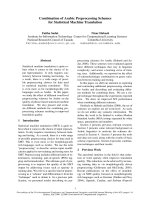
Fig. 1. The buried cluster and Lys335 in the
shutter region of PAI-1. The top panel shows
ribbon diagrams of active (left) and latent
PAI-1 (right). Secondary structure elements are
indicated as follows: blue, b sheet A; red,
a helix B; green, gate region; yellow, RCL and
b strand 1C in active PAI-1 and RCL inserted
as b strand 4A in latent PAI-1. The P
1
Arg is
displayed as a stick. The lower panel shows the
three-dimensional structure of the shutter
region of active PAI-1 (left) and latent PAI-1
(right). The molecules were rotated < 908
around a horizontal axis compared to the top
panel. The colour code for secondary structure
elements are as in the top panel. Presented
amino-acid residues are: green, shutter region
residues Ser53, Gly56, and Gln334; grey,
Asn186 in b strand 3A; yellow, Lys335; purple,
potential interaction partners for Lys335, i.e.
Glu294 in b strand 6A and the backbone of
Asn171 in the a helix F/b strand 3A loop.
Note:
SWISSPDB VIEWER uses the same
signature for a helices and the short 3
10
-helix
found in the a helix F/b strand 3A loop of
active PAI-1.
q FEBS 2001 PAI-1 shutter region mutations (Eur. J. Biochem. 268) 6275
MATERIALS AND METHODS
PAI-1
In order to generate recombinant wild-type and mutated
PAI-1, PAI-1 cDNA [27] was cloned into the expression
vector pcDNA3.1(–) (Invitrogen) by use of standard
techniques. The generated expression plasmid was denoted
pcDNA3.1( –)PAI-1. Relevant fragments of the PAI-1
sequence were transferred to the mutagenesis vector
LITMUS 28 (New England Biolabs). Point mutations
were introduced into the PAI-1 cDNA fragment inserted into
LITMUS 28 by use of the PCR-based QuickChangee Site-
Directed Mutagenesis kit (Stratagene). The mutagenesis
primers were from DNA Technology (Aarhus, Denmark),
had a melting point above 60 8C, and were designed with the
desired mutation(s) in the middle of their sequence. After
mutagenesis, the fragments were moved back into
pcDNA3.1(–)PAI-1 by the use of unique restriction sites.
All mutations were verified by DNA sequencing of both
strands of the PCR produced fragment after transfer back
to pcDNA3.1(– )PAI-1, by use of either the Thermo
Sequenasee II dye terminator cycle sequencing kit
(Amersham Pharmacia Biotech AB) or the ABI PRISMe
dye terminator cycle sequencing ready reaction kit
(PerkinElmer).
Recombinant PAI-1 variants were expressed in human
embryonic kidney 293T cells (phenotype 293tsA1609neo)
[28], grown in Dulbecco’s modified Eagle’s medium, by
transient transfection using the calcium/phosphate precipi-
tation technique [28]. Briefly, 1 h prior to transfection, new
medium with 10% fetal bovine serum and 25 m
M
chloroquine was added to cells grown to 90% confluence
in a 15-cm culture dish. Transfection was carried out by
mixing 30 mgDNA(H
2
O added to a total of 1752 mL),
248 mL2
M CaCl
2
, and 2 mL 42 mM Hepes, pH 7.05,
274 m
M NaCl, 10 mM KCl, 1.5 mM Na
2
HPO
4
,11mM
D
-(þ )-glucose. After 1–2 min, this mixture was added
dropwise to the cell medium and carefully distributed. Fresh
medium without fetal bovine serum and chloroquine was
added after 9–11 h of incubation. The conditioned medium
was harvested after 48 and 96 h. Nontransfected or mock
transfected HEK293T cells were shown not to express either
PAI-1 or uPA by standard ELISA with monoclonal and
polyclonal antibodies as capture and detection antibodies,
respectively. Recombinant PAI-1 variants were purified
from serum-free conditioned medium of the transfected
cells by immunoaffinity chromatography in one step
[29,30]. After purification, the variants were dialysed
against NaCl/P
i
(0.01 M NaH
2
PO
4
, pH 7.4, 0.14 M NaCl)
and concentrated to < 1 mg·mL
21
.
Other proteins and miscellaneous materials
The following materials were purchased from the indicated
sources: BSA (Sigma); media components for HEK293T
culturing (Life Technology); Qiaquick gel extraction kit
(Qiagen); Rapid DNA ligation kit (Boehringer Mannheim);
restriction enzymes (New England Biolabs Inc.; or
Amersham Pharmacia Biotech AB; or Boehringer
Mannheim);
L-5-oxopropyl-glycyl-L-arginine-p-nitro-
anilide (S-2444, Chromogenix AB); SDS (Serva); human
urokinase-type plasminogen activator (uPA; Wakamoto
Pharmaceutical Co.); vitronectin (Becton Dickinson; or
Haemochrom AB). All other chemicals and reagents were of
the highest quality commercially available.
Activation of latent PAI-1
Unless otherwise indicated, latent PAI-1 was converted to
the active conformation by denaturation with 0.1% SDS for
1 h at room temperature and refolding by a . 50-fold
dilution in 0.1
M Tris, pH 8.1 (37 8C), containing either 1%
BSA or 0.2% Triton X-100. Alternatively, latent PAI-1 was
reactivated by denaturation with guanidinium chloride and
refolded by dialysis against NaCl/P
i
.
Assays for measuring specific inhibitory activity of PAI-1
The specific inhibitory activity of the reactivated PAI-1
variants was measured by titration against uPA in a direct
peptidyl anilide assay at 37 8C, in the presence or absence of
a slight excess of vitronectin over PAI-1 [30]. A twofold
dilution series of PAI-1, with or without vitronectin, was
made immediately after refolding, to avoid loss of activity
due to fast latency transition. The dilution series of
denatured and refolded PAI-1 (0–20 mg·mL
21
, 0–370 nM)
were quickly (in less than 1 min) mixed with an equal
volume (100 mL) of 0.25 mg·mL
21
(4.3 nM)uPA,0.1M
Tris, pH 8.1, 1% BSA or 0.2% Triton X-100. The final
concentrations of uPA was 0.125 mg·mL
21
(2.15 nM), of
PAI-1 in the range 0–10 mg·mL
21
(0–185 nM), and of
vitronectin in the range 0 –15 mg·mL
21
(0–200 nM). Upon
completion of the uPA inhibition reaction (. 5 min), the
remaining uPA activity in the reaction mixture was
determined by use of
L-5-oxopropyl-glycyl-L-arginine-
p-nitroanilide (S-2444), a chromogenic peptidyl anilide
substrate for uPA. The amount of active PAI-1, and thus the
specific inhibitory activity, was calculated from the total
amount of PAI-1 that had to be present to inhibit half of the
uPA activity in the assays.
PAI-1 latency transition assay
Denatured and refolded PAI-1 wild-type and variants, in a
concentration of 20 mg·mL
21
(370 nM), were incubated at
37 8C in 0.1
M Tris, pH 8.1, 1% BSA in the presence or
absence of 30 mg·mL
21
(400 nM) vitronectin. Following
incubation for different time periods, the specific inhibitory
activity of PAI-1 wild-type and variants were determined as
described above, and the functional half-lives of the variants
were calculated.
Analysis of functional behaviour of PAI-1 by reaction with
low
M
r
uPA (LMW-uPA) and SDS/PAGE
PAI-1 portions (30 mg each) were denatured with 3 mL1%
SDS, refolded by dilution to 1200 mL with 0.1
M Tris,
pH 8.1, 1% BSA and incubated at 37 8C. At various time
points, samples of 5 mg PAI-1 were mixed with 7.5 mg
LMW-uPA and incubated for at least 2 min at 37 8C. BSA
was then removed by the following procedure, performed at
room temperature unless otherwise indicated: One-hundred
micrograms of monoclonal murine anti-(PAI-1) IgG from
hybridoma clone 2 [31], coupled to Sepharose-4B, was
transferred to Ultrafreew-MC 0.22-mm filter units for
6276 M. Hansen et al. (Eur. J. Biochem. 268) q FEBS 2001
centrifugal filtration (Millipore, USA) and washed twice
with 0.1
M Tris, pH 8.1. The sample (PAI-1, LMW-uPA,
BSA) was then added and incubated for at least 30 min
followed by four washes with 0.1
M Tris, 1 M NaCl, pH 8.1,
and one wash with 0.1
M Tris, pH 8.1. PAI-1 was eluted by
incubation with 400 mL3
M ammoniumthiocyanat
(NH
4
SCN) for at least 30 min at 37 8C before centrifugation
at 16 500 g for 10 min. The samples were precipitated
with trichloroacetic acid, and subjected to 6–16% gradient
SDS/PAGE.
Determination of second order rate constants for the
reaction between PAI-1 and uPA
The second order rate constants were determined as
described previously [32]. The calculation of the second
order rate constants is based on the assumption that the
concentration of active PAI-1 is unchanged during the assay.
As most of these variants have significantly shorter
functional half-lives (see below) than wild-type, the
calculated second order rate constants for the variants
were expected to be somewhat lower than their real values.
Gel filtration
Thirty-microgram portions of PAI-1 wild-type and variants
were analysed by FPLC gel filtration on a Superdex 200 HR
10/30 column (Pharmacia) in 0.1
M Tris, pH 8.1, 0.5 M
NaCl at 4 8C, using a flow rate of 0.3 or 0.4 mL·min
21
. The
following marker proteins were used: BSA (M
r
67 000),
murine IgG (M
r
150 000), and b-galactosidase (M
r
540 000).
Molecular graphics
SWISSPDB VIEWER [33] was used to display the three-
dimensional X-ray structure of active [34] and latent [13]
PAI-1.
Statistical analysis
Data were evaluated by Student’s t-test.
Fig. 2. Gel filtration of purified recombinant PAI-1 from HEK293T
cells. As representative gel filtration profiles are shown those of PAI-1
wild-type, G56S/Q334H, and PAI-1 G56S/Q334H/K335A. All other
variants also showed a single peak in the position expected for
monomeric PAI-1. V
0
, void volume. The migration of the marker
proteins BSA (M
r
67 000), murine IgG (M
r
150 000), and
b-galactosidase (M
r
540 000) are indicated by arrows above the
profiles.
Table 1. Specific inhibitory activity of PAI-1 variants towards uPA. The most common amino-acid composition of the buried polar cluster
(positions 53/56/334) in serpins is S/S/H. The composition S/S/Q is identical to that of alaserpin, S/G/H is identical to that of CP-9, A/G/H is identical
to that of heparin cofactor II, while the S/A/S composition is present in angiotensinogen [1,3]. The investigated residues according to the PAI-1
numbering (1Ser-Ala-Val-His-His-) are 37/40/324/325 [27]. Means ^ SD (numbers of assays are indicated). *, Significantly different from wild-type
(P , 0.005). †, Significantly different from the value without vitronectin (P , 0.005).
PAI-1 variant
Composition of
positions 53/56/334
PAI-1 activity
(% of theoretical max)
Vitronectin effect
(fold increase)– Vitronectin þ Vitronectin
Wild-type S/G/Q 87.1 ^ 22.2 (20) 113.7 ^ 28.0 (10) † 1.3
K335A S/G/Q 114.9 ^ 21.5 (5) 129.5 ^ 21.1 (3) 1.1
S53A A/G/Q 71.6 ^ 9.4 (8) 84.9 ^ 5.9 (3) 1.2
S53A/K335A A/G/Q 84.3 ^ 15.5 (4) 91.0 ^ 13.3 (3) 1.1
G56A S/A/Q 59.6 ^ 7.8 (6) * 56.1 ^ 6.3 (3) * 0.9
G56S S/S/Q 73.1 ^ 10.6 (5) 127.1 ^ 6.2 (3) † 1.7
G56S/K335A S/S/Q 120.0 ^ 1.6 (3) 129.6 ^ 2.5 (3) † 1.1
Q334A S/G/A 58.1 ^ 7.5 (6) * 73.1 ^ 5.7 (3) 1.3
Q334A/K335A S/G/A 62.8 ^ 4.7 (4) 57.8 ^ 5.3 (4) * 0.9
Q334H S/G/H 61.9 ^ 13.5 (6) 90.7 ^ 5.8 (3) 1.5
Q334H/K335A S/G/H 72.5 ^ 24.0 (6) 87.3 ^ 15.5 (3) 1.2
Q334S S/G/S 75.9 ^ 20.6 (7) 104.8 ^ 5.9 (3) 1.4
S53A/Q334H A/G/H 48.9 ^ 10.0 (9) * 74.5 ^ 9.1 (3) † 1.5
G56A/Q334S S/A/S 34.5 ^ 4.4 (7) * 23.8 ^ 2.1 (3) *† 0.7
G56S/Q334H S/S/H 64.7 ^ 14.2 (9) * 101.0 ^ 5.2 (3) † 1.6
G56S/Q334H/K335A S/S/H 83.8 ^ 14.2 (6) 81.6 ^ 9.8 (3) 1.0
q FEBS 2001 PAI-1 shutter region mutations (Eur. J. Biochem. 268) 6277
RESULTS
Expression and purification of recombinant PAI-1 in
HEK293T cells
PAI-1 wild-type and the substitution variants were expressed
in HEK293T cells and purified from their conditioned
medium by immunoaffinity chromatography. The yields of
purified protein were 1.4 –11.4 mg protein per L of
conditioned medium. PAI-1 wild-type, K335A, S53A/
K335A, G56S/K335A, and Q334H/K335A were obtained
in the greatest yields. All variant preparations were . 95%
pure, as evaluated by SDS/PAGE, and migrated as a single
sharp peak in the position expected for monomeric PAI-1 in
gel filtration (Fig. 2). N-Terminal sequencing of the
produced PAI-1 showed two distinct sequences in almost
equal amounts, SAVHHPPS and VHHPPSYV, in agreement
with the previously reported N-terminal heterogeneity of
natural PAI-1 [27]. The purified recombinant PAI-1 was in
the latent form, but could be reactivated by denaturation and
refolding, either by SDS and refolding by dilution into a
buffer with 1% BSA, or by guanidinium chloride and
refolding by dialysis against NaCl/P
i
. In this study, PAI-1
was routinely reactivated using SDS, as some of the variants
had a very fast latency transition (see below) and would
therefore lose all activity during the dialysis used for
refolding after guanidinium chloride denaturation. The
specific inhibitory activities of most PAI-1 variants, when
denatured with SDS and refolded in BSA-containing buffer,
were 60– 80% of the theoretical maximum, and thus
indistinguishable from recombinant PAI-1 wild-type
(Table 1). However, the recombinant variants G56A,
Q334A, S53A/Q334H, G56A/Q334S, and G56S/Q334H
showed a small, but statistically significant (P , 0.005)
reduction in specific inhibitory activity as compared to wild-
type. All variants except the variant G56A/Q334S had a
second order rate constant differing less than 2.5-fold from
that of wild-type (data not shown). The second order rate
constant for G56A/Q334S was 3.8-fold lower than that of
the wild-type, but this can be ascribed to a fast decrease in
inhibitory activity of this variant during the experiment (see
below). Vitronectin caused a small, but statistically
significant increase of the specific inhibitory activity of
the PAI-1 wild-type and the variants G56S, S53A/Q334H,
G56A/Q334S, G56S/Q334H, and G56S/K335A. Interest-
ingly, the specific inhibitory activity of G56A/Q334S was
slightly decreased by vitronectin.
Latency transition of PAI-1 wild-type and PAI-1 variants in
the absence and the presence of vitronectin
To estimate the functional stability of the variants, their
specific inhibitory activities were measured after different
times of incubation at 37 8C. The rate of activity loss was
determined in the absence or the presence of vitronectin.
Representative examples are given in Fig. 3 and a summary
of all experiments is given in Table 2.
All variants with substitutions in the buried cluster, except
S53A, had significantly reduced functional half-lives. The
K335A substitution caused at most a slight (, 1.4-fold)
delay of latency transition when introduced into wild-type,
S53A, G56S, and Q334A, but caused considerable delays
Fig. 3. Determination of functional stability of PAI-1 variants. The
specific inhibitory activity of the indicated PAI-1 variants was determined
after the indicated time periods of incubation at 37 8C in the absence (closed
circles) or presence (open circles) of vitronectin. The relative specific
inhibitory activity was plotted as a function of time. The functional half-lives
of the shown variants in the absence and presence of vitronectin, respectively,
in the experiments shown were: wild-type, 48.6 and 65.2 min; K335A, 78.9
and 41.8 min; S53A, 69.4 and 109.9 min; S53A/K335A, 99.2 and
43.5 min; G56S/Q334H, 5.3 and 59.8 min; G56S/Q334H/K335A, 26.5
and 35.1 min. A summary of all experiments is given in Table 2.
6278 M. Hansen et al. (Eur. J. Biochem. 268) q FEBS 2001
(2.9-fold and 4.6-fold, respectively) of the very unstable
variants Q334H and G56S/Q334H.
At the routinely used pH of 8.1, there is only a slight
effect of vitronectin on the rate of latency transition of PAI-1
wild-type, and the use of this pH therefore allowed
optimization of the difference in the effect of vitronectin
on wild-type and the unstable variants. The presence of
vitronectin during the incubations had a stabilizing effect on
all PAI-1 variants with substitutions in the buried cluster of
the shutter region. The most pronounced effect was
observed on the most unstable variants, so that their
functional half-lives in the presence of vitronectin increased
towards that of PAI-1 wild-type (Table 2). Surprisingly, the
stabilizing effect of vitronectin was abolished by the K335A
substitution. None of the variants with this substitution had
longer half-lives in the presence of vitronectin, and with
most of them, vitronectin even accelerated the activity loss.
Hence, while the K335A substitution had a stabilizing
effect in the absence of vitronectin, it had a destabilizing
effect in the presence of vitronectin. Importantly, the
K335A substitution did not result in altered affinity of PAI-1
to vitronectin (T. Wind, & P.A. Andreasen, Department of
Molecular and Structural Biology, Aarhus University,
Denmark, personal communication).
Although the assays were routinely performed in a buffer
of 0.1
M Tris, pH 8.1, similar results were obtained with a
buffer of 0.1
M Tris, pH 7.4. In addition, when examining
the results obtained with SDS-activated PAI-1 vs.
guanidinium chloride-activated PAI-1 with wild-type and
two of the most stable variants (S53A and K335A), no
distinguishable difference was observed.
In order to ensure that the loss of activity during the
incubations at 37 8C was due to latency transition, PAI-1,
that had been incubated for various time periods at 37 8C,
was reacted with an excess of LMW-uPA, and the reaction
products were analysed by SDS/PAGE. Representative
experiments are shown in Fig. 4. Nonincubated wild-type
reacted to form the expected < 80 000-Da LMW-uPA–
PAI-1 complex. A fraction of wild-type reacted in a
substrate-manner, giving rise to the < 50 000-Da
N-terminal fragment resulting from P
1
–P
1
0
cleavage, the
< 4000-Da C-terminal fragment not being recovered by
the gel system used here. During incubation at 37 8C, the
amount of a PAI-1 form inert to reaction with LMW-uPA
increased with time, and the complex formation and
substrate reaction decreased, in agreement with an
increasing fraction of PAI-1 being in the latent state. The
same was true for the PAI-1 variants, except that the
accumulation of inert PAI-1 occurred faster, in agreement
with the activity measurements. It should also be noted that
a fraction of all tested variants seemed to be present in a
stable substrate form [30,35,36], the amount of which did
not decrease during the incubations at 37 8C. On this basis,
we concluded that the loss of activity during incubations at
37 8C was caused by latency transition, and that the effects
of the substitutions and of vitronectin on the functional
half-lives was caused by changes in the rate of latency
transition.
Effect of shutter region substitutions on PAI-1 active to
substrate transition
Nonionic detergents induce substrate behaviour in glycosy-
lated PAI-1 at 0 8C, but much less so at 37 8C [20,21], and
induce substrate behaviour in nonglycosylated PAI-1 at
37 8C as well as at 0 8C [37]. Therefore, to study the effect
of shutter region substitutions on the transition of PAI-1 to a
substrate form, we replaced the BSA in the assay buffer with
0.2% Triton X-100. The variants with a S53A, Q334A, or
Q334S substitution all had significantly reduced specific
inhibitory activity in Triton X-100 containing buffer at
37 8C as compared to PAI-1 wild-type (Table 3). Analysis of
Table 2. Stability of specific inhibitory activity of PAI-1 variants at 37 8C. Means, SDs, and numbers of experiments are indicated. *, Significantly
different from wild-type (P , 0.005). †, Significantly different from the value without vitronectin (P , 0.005). ‡, Significantly different from the
corresponding variants without the K335A substitution (P , 0.02).
PAI-1 variant
Functional half-lives
(min)
Vitronectin effect
(fold increase)– Vitronectin þ Vitronectin
Wild-type 54.7 ^ 13.5 (16) 63.4 ^ 11.6 (10) 1.2
K335A 76.9 ^ 11.6 (4) *‡ 35.3 ^ 6.7 (3) *†‡ 0.5
S53A 64.0 ^ 8.6 (5) 100.7 ^ 9.4 (3) *† 1.6
S53A/K335A 85.1 ^ 20.5 (4) * 32.0 ^ 10.0 (3) *†‡ 0.4
G56A 26.1 ^ 4.4 (4) * 36.5 ^ 6.8 (3) * 1.4
G56S 19.7 ^ 1.7 (4) * 54.9 ^ 4.1 (3) † 2.8
G56S/K335A 25.5 ^ 6.3 (3) * 12.3 ^ 2.1 (3) *‡ 0.5
Q334A 10.9 ^ 1.5 (4) * 39.9 ^ 10.2 (3) *† 3.7
Q334A/K335A 12.9 ^ 2.9 (3) * 14.1 ^ 0.9 (3) *‡ 1.1
Q334H 10.9 ^ 1.4 (4) * 52.3 ^ 6.0 (3) † 4.8
Q334H/K335A 32.1 ^ 2.7 (5) *‡ 29.4 ^ 1.2 (3) *‡ 0.9
Q334S 23.4 ^ 2.5 (5) * 75.2 ^ 15.8 (3) † 3.2
S53A/Q334H 18.5 ^ 3.2 (7) * 78.3 ^ 12.5 (3) † 4.2
G56A/Q334S 9.7 ^ 1.6 (4) * 72.9 ^ 17.7 (3) † 7.5
G56S/Q334H 6.1 ^ 1.5 (7) * 62.4 ^ 15.6 (4) † 10.2
G56S/Q334H/K335A 27.8 ^ 2.6 (5) *‡ 34.3 ^ 2.3 (3) *‡ 1.2
q FEBS 2001 PAI-1 shutter region mutations (Eur. J. Biochem. 268) 6279
the distribution of PAI-1 between different functional forms
by the use of reaction with LMW-uPA and SDS/PAGE
confirmed that the decreased specific inhibitory activity was
caused by an increased tendency to substrate behaviour
and not the generation of an inert form, as shown by the
representative experiments shown in Fig. 5. The K335A
substitution did not counteract the increased tendency to
substrate behaviour (Table 3).
DISCUSSION
In this report, we show that the combination of amino acids
in positions 53, 56, and 334 in the shutter region of PAI-1 is
an important determinant of the latency transition rate.
Except one, all tested deviations from the wild-type
combination of amino acids in these positions resulted in
an accelerated latency transition. Substitution of the Lys
residue in position 335 counteracted the accelerating effect
of some of the substitutions in positions 53, 56, and 334. The
substitutions also had specific effects on the vitronectin and
Triton X-100 induced changes in PAI-1 latency transition
and specific inhibitory activity, respectively.
Based on our observations we propose that the
substitutions in positions 53, 56, and 334 affect the latency
transition by changing the local conformation of these
residues, including their hydrogen bonds. As there is no
P
1
–P
1
0
cleavage during latency transition, strand insertion
must imply the passage of the intact RCL through the ‘gate
region’, which is situated between (a) the turn between b
strands 3C and 4C (residues 204–219) and (b) the turn
between b strands 3B and a helix G (residues 257–259)
[13,38,39] (Fig. 1). Because of steric reasons, it is not very
likely that the RCL can surround the turn between b strands
3C and 4C without having a completely stretched-out
conformation. Only after the RCL has passed this turn can
the final insertion into b sheet A proceed [39]. Considering
that RCL insertion into b sheet A is several orders of
magnitude faster during complex formation than during
latency transition, it seems reasonable to presume that the
passage of the RCL through the gate region is rate limiting
for latency transition. This presumption is supported by the
observation that substitutions of basic residues in the turn
between b strands 3C and 4C with acidic residues accelerate
latency transition [40,41]. On this basis, we reach the
conclusion that the substitutions in the shutter region affect
the rate of latency transition by affecting the rate of passage
of the RCL through the gate region. Based on the amino-
acid sequence of the RCL and b strand 5A being directly
continuous, it may be proposed that movements of the RCL
during passage through the gate region are coupled to
movements of b strand 5A and therefore sensitive to the
interactions of b strand 5Awith the underlying structure. An
alternative, but with the presently available information, a
less likely explanation is that passage of the RCL through
the gate region is rapid and reversible, and that it is the b
sheet A opening and the final insertion of RCL as b strand
4A that is rate limiting for latency transition.
Two facts complicate the interpretation of our results
on the basis of detailed structural considerations. First,
the three-dimensional structure available for active PAI-1
[34,42] is that of a mutant with a strongly delayed latency
transition. The stabilizing mutations may well have affected
Fig. 4. Analysis of the functional behavior of PAI-1 by SDS/PAGE.
PAI-1 wild-type or Q334H (25 mg·mL
21
) were SDS-denatured and
incubated in a buffer of 0.1
M Tris, 1% BSA, pH 8.1 at 37 8C. After the
indicated time periods, samples corresponding to 5 mg PAI-1 were
incubated with 7.5 mg LMW-uPA for at least 2 min at 37 8C at a PAI-1
concentration of 20 mg·mL
21
and an LMW-uPA concentration of
30 mg·mL
21
before inert PAI-1, reactive center-cleaved PAI-1, and
LMW-uPA–PAI-1 complex were isolated from the reaction mixture by
immunoaffinity chromatography, removing most of the BSA and most
of the excess of LMW-uPA. The samples were then precipitated with
trichloroacetic acid and subjected to SDS/PAGE in gradient gels with
6–16% polyacrylamide. The positions of LMW-uPA, reactive centre-
cleaved PAI-1 (RCC PAI-1), native/inert PAI-1, BSA, and LMW-uPA-
PAI-1 complex (complex) are indicated to the right. N, PAI-1 incubated
for at least 3 h at 37 8C without LMW-uPA added. The apparently low
fraction of nonincubated PAI-1 forming a complex is related to a
somewhat higher tendency to substrate behavior at the high PAI-1 and
LMW-uPA concentrations used in this assay [22].
Table 3. Effect of 0.2% Triton X-100 on specific inhibitory activity
of PAI-1 at 37 8C. The specific inhibitory activity of each variant is
given as a fraction of the specific inhibitory activity of the same variant
in 1% BSA. Means, SDs, and numbers of experiments are indicated. *,
Significantly different from wild-type (P , 0.005).
PAI-1 variant Specific inhibitory activity
Wild-type 0.87 ^ 0.12 (7)
K335A 0.95 ^ 0.09 (3)
S53A 0.20 ^ 0.02 (3)*
S53A/K335A 0.17 ^ 0.02 (3)*
G56A 0.65 ^ 0.11 (3)
G56S 1.17 ^ 0.16 (3)
G56S/K335A 1.06 ^ 0.03 (3)
Q334A 0.12 ^ 0.01 (3)*
Q334A/K335A 0.18 ^ 0.03 (3)*
Q334H 0.61 ^ 0.08 (3)*
Q334H/K335A 0.77 ^ 0.01 (3)
Q334S 0.21 ^ 0.05 (3)*
S53A/Q334H 0.13 ^ 0.01 (3)*
G56A/Q334S 0.10 ^ 0.02 (3)*
G56S/Q334H 0.45 ^ 0.03 (3)*
G56S/Q334H/K335A 0.74 ^ 0.09 (3)
6280 M. Hansen et al. (Eur. J. Biochem. 268) q FEBS 2001
the conformation of the shutter region. Second, the rate of
strand insertion during latency transition may be affected
not only through a change in the conformation of the active
form, but also by a change in the conformation of a
transition state with an unknown three-dimensional
structure. Nevertheless, it seems reasonable to conclude
that the hydrogen bonds from the side chain of S53A have
little influence on the rate of latency transition, as the S53A
substitution resulted only in a slightly decreased rate of
latency transition. Also, the Q334A substitution, removing
the hydrogen bonding ability of the side chain in position
334, resulted in a half-life similar to that of the Q334H
substitution, indicating that the specific hydrogen bonds
formed by Gln334, absent in Q334A and apparently not
substituted by any possible hydrogen bonds formed by a His
in this position, play a pivotal role. Furthermore, the
accelerating effect of substitutions of Gly56 is likely to be
caused by a slight reorganization of the region introduced by
the larger side chains.
The variant with the amino-acid combination Ser53/
Ser56/His334, identical to that of 63% of serpins [3], has the
shortest half-life of all variants tested here, and in fact one of
the shortest half-lives reported for a PAI-1 variant. Thus, the
reason for the tendency of PAI-1 to undergo latency
transition is to be sought outside the shutter region, in
agreement with previous results [18,40,41]. On the other
hand, if PAI-1 had possessed an amino-acid composition in
the shutter region identical to that of most other serpins, its
tendency to latency transition would have given it an
extremely short half-life, probably incompatible with its
physiological functions.
Irrespective of the substitutions introduced into positions
53, 56, and 334, vitronectin brought the latency transition
rate back to values close to that of PAI-1 wild-type. The
most plausible explanation of this observation is that
vitronectin, from its binding site in the flexible joint region
[43], directs the movements of the RCL, almost totally
over-ruling the effect of the local hydrogen bonding network
of the residues in positions 53, 56, and 334.
The K335A substitution delayed the latency transition
when introduced in some of the variants, and most strongly
when introduced into the very unstable variants Q334H and
G56S/Q334H. The side chain of Lys335 points away from
the buried cluster in positions 53/56/334 (Fig. 1). On the
basis of the available three-dimensional structures, several
intramolecular interactions of Lys335 may be suggested.
The possible interactions include a connection to the loop
between a helix F and b strand 3A by a hydrogen bond to
the carbonyl oxygen atom of the backbone of Asn171
[19,22], by hydrophobic interactions with residues in that
loop [23], or by participation in formation of a chloride
binding site together with residues in that loop and Lys337
[44]. The possible interactions also include a salt bridge to
Glu294 in b strand 6A (Fig. 1). It therefore seems likely that
the constraints caused by the interactions of Lys335
contribute to maintaining the RCL in a state with a
relatively facilitated passage through the gate region during
latency transition, via an effect on the conformation of
b strand 5A and of the buried cluster in positions 53, 56, and
334.
In contrast, in the presence of vitronectin, the K335A
substitution caused a twofold to fivefold acceleration of
latency transition compared to wild-type. In fact, vitronectin
did not delay latency transition of any of the variants
harbouring the K335A substitution. On the basis of the
opposite effects of the K335A substitution in the absence
and presence of vitronectin, we propose that the
conformational change of PAI-1 following the binding of
vitronectin implicates a reorientation of the side chain of
Lys335 relative to its surroundings, allowing it to make new
contacts, concerted rearrangements of the shutter region and
changes in the movements of the RCL.
We observed an increased tendency to substrate
behaviour in Triton X-100 at 37 8C in a set of mutations
overlapping with, but not identical to that giving an
increased rate of latency transition. Previously, Triton X-100
was found to induce substrate behaviour in nonglycosylated
PAI-1 at 0 and 37 8C [37] and to induce substrate behaviour
Fig. 5. Effect of Triton X-100 on the distribution of PAI-1 between different functional forms. The indicated concentrations of the indicated
PAI-1 variants, were incubated in 0.1
M Tris, 0.2% Triton X-100, pH 8.1 at 37 8C with LMW-uPA at a concentration of 0.175 mg·mL
21
for at least
15 min prior to determination of the remaining LMM-uPA activity with S-2444. Inset: portions of 3 mg of the PAI-1 variants were diluted in 0.1
M
Tris, 0.2% Triton X-100, pH 8.1 to 0.16 mg·mL
21
followed by incubation at 37 8C with 0.175 mg·mL
21
LMM-uPA for 15 min. The samples were
precipitated with trichloroacetic acid and subjected to SDS/PAGE in gradient gels with 6–16% polyacrylamide. The positions of LMW-uPA, reactive
centre-cleaved PAI-1 (RCC PAI-1), native/inert PAI-1, and LMW-uPA–PAI-1 complex are indicated to the left.
q FEBS 2001 PAI-1 shutter region mutations (Eur. J. Biochem. 268) 6281
in glycosylated PAI-1 at 0 8C, but not at 37 8C [20,21]. On
the basis of our present findings, we propose that Triton
X-100 acts by destabilizing the shutter region, this
happening more readily with less perfect interactions
between the side chains, resulting in a delay in strand
insertion during reaction with the target proteinase. On the
other hand, the Triton X-100-induced substrate behaviour
did not seem to implicate the interactions of the Lys335 side
chain, in contrast to antibody-induced substrate behaviour
that was potentiated by the K335A substitution [19,22]. The
observation of latency transition and complex formation
being affected differently by mutations in the shutter region
and b strand 5A is in agreement with RCL insertion
following different routes in the two cases.
PAI-1 is a potential target for antithrombotic [45] and
anticancer therapy [46,47]. The biochemical mechanism of
action of a few PAI-1 neutralisers has been characterized,
including monoclonal antibodies and organochemical
compounds. These compounds neutralize PAI-1 either by
steric hindrance, by inducing conversion to the latent state,
by inducing substrate behaviour, and/or by inducing
conversion to inert polymers [48– 52]. The present results
prompt further studies into the role of the shutter region and
b strand 5A in PAI-1 in conformational changes leading to
neutralization.
ACKNOWLEDGEMENTS
Dr Kees Rodenburg is thanked for fruitful discussions in the early phase
of this work. Dr Claus Oxvig is acknowledged for providing the
HEK293T cell line. This work was supported financially by the Danish
Cancer Society, the Danish Research Agency, the Danish Heart
Foundation, the NOVO-Nordisk Foundation, and the Danish Cancer
Foundation.
REFERENCES
1. Huber, R. & Carrell, R.W. (1989) Implications of the three-
dimensional structure of a
1
-antitrypsin for structure and function of
serpins. Biochemistry 28, 8951–8966.
2. Carrell, R.W. & Stein, P.E. (1996) The biostructural pathology of
the serpins: critical function of sheet opening mechanism. Biol.
Chem. Hoppe Seyler 377, 1–17.
3. Irving, J.A., Pike, R.N., Lesk, A.M. & Whisstock, J.C. (2000)
Phylogeny of the serpin superfamily: implications of patterns of
amino acid conservation for structure and function. Genome Res.
10, 1845–1864.
4. Lawrence, D.A., Ginsburg, D., Day, D.E., Berkenpas, M.B.,
Verhamme, I.M., Kvassman, J.O. & Shore, J.D. (1995) Serpin-
protease complexes are trapped as stable acyl-enzyme inter-
mediates. J. Biol. Chem. 270, 25309–25312.
5. Wilczynska, M., Fa, M., Ohlsson, P.I. & Ny, T. (1995) The
inhibition mechanism of serpins. J. Biol. Chem. 270,
29652–29655.
6. Egelund, R., Rodenburg, K.W., Andreasen, P.A., Rasmussen, M.S.,
Guldberg, R.E. & Petersen, T.E. (1998) An ester bond linking a
fragment of a serine proteinase to its serpin inhibitor. Biochemistry
37, 6375–6379.
7. Shore, J.D., Day, D.E., Francis-Chmura, A.M., Verhamme, I.,
Kvassman, J., Lawrence, D.A. & Ginsburg, D. (1995) A fluorescent
probe study of plasminogen activator inhibitor-1. Evidence for
reactive center loop insertion and its role in the inhibitory
mechanism. J. Biol. Chem. 270, 5395–5398.
8. Stratikos, E. & Gettins, P.G. (1999) Formation of the covalent
serpin-proteinase complex involves translocation of the proteinase
by more than 70A
˚
and full insertion of the reactive center loop into
b-sheet A. Proc. Natl Acad. Sci. USA 96, 4808–4813.
9. Fa, M., Bergstrom, F., Hagglof, P., Wilczynska, M., Johansson,
L.B. & Ny, T. (2000) The structure of a serpin–protease complex
revealed by intramolecular distance measurements using donor–
donor energy migration and mapping of interaction sites. Struct.
Fold Des. 8, 397–405.
10. Huntington, J.A., Read, R.J. & Carrell, R.W. (2000) Structure of a
serpin–protease complex shows inhibition by deformation. Nature
407, 923–926.
11. Egelund, R., Petersen, T.E. & Andreasen, P.A. (2001) A serpin-
induced extensive proteolytic susceptibility of urokinase-type
plasminogen activator implicates distortion of the proteinase
substrate-binding pocket and oxyanion hole in the serpin inhibitory
mechanism. Eur. J. Biochem. 268, 673 – 685.
12. Lawrence, D.A., Olson, S.T., Muhammad, S., Day, D.E., Kvass-
man, J.O., Ginsburg, D. & Shore, J.D. (2000) Partitioning of serpin-
proteinase reactions between stable inhibition and substrate
cleavage is regulated by the rate of serpin reactive center loop
insertion into b-sheet A. J. Biol. Chem. 275, 5839–5844.
13. Mottonen, J., Strand, A., Symersky, J., Sweet, R.M., Danley, D.E.,
Geoghegan, K.F., Gerard, R.D. & Goldsmith, E.J. (1992) Structural
basis of latency in plasminogen activator inhibitor-1. Nature 355,
270–273.
14. Stein, P. & Chothia, C. (1991) Serpin tertiary structure
transformation. J. Mol. Biol. 221, 615–621.
15. Harrop, S.J., Jankova, L., Coles, M., Jardine, D., Whittaker, J.S.,
Gould, A.R., Meister, A., King, G.C., Mabbutt, B.C. & Curmi, P.M.
(1999) The crystal structure of plasminogen activator inhibitor 2 at
2.0A
˚
resolution: implications for serpin function. Structure 7,
43–54.
16. Stein, P.E. & Carrell, R.W. (1995) What do dysfunctional serpins
tell us about molecular mobility and disease? Nat. Struct. Biol. 2,
96–113.
17. Davis, R.L., Shrimpton, A.E., Holohan, P.D., Bradshaw, C., Feiglin,
D., Collins, G.H., Sonderegger, P., Kinter, J., Becker, L.M.,
Lacbawan, F., Krasnewich, D., Muenke, M., Lawrence, D.A.,
Yerby, M.S., Shaw, C.M., Gooptu, B., Elliott, P.R., Finch, J.T.,
Carrell, R.W. & Lomas, D.A. (1999) Familial dementia caused by
polymerization of mutant neuroserpin. Nature 401, 376 –379.
18. Berkenpas, M.B., Lawrence, D.A. & Ginsburg, D. (1995)
Molecular evolution of plasminogen activator inhibitor-1 func-
tional stability. EMBO J. 14, 2969–2977.
19. Kirkegaard, T., Jensen, S., Schousboe, S.L., Petersen, H.H.,
Egelund, R., Andreasen, P.A. & Rodenburg, K.W. (1999)
Engineering of conformations of plasminogen activator inhibitor-
1. A crucial role of b-strand 5A residues in the transition of active
form to latent and substrate forms. Eur. J. Biochem. 263, 577–586.
20. Kjøller, L., Martensen, P.M., Sottrup-Jensen, L., Justesen, J.,
Rodenburg, K.W. & Andreasen, P.A. (1996) Conformational
changes of the reactive-centre loop and b-strand 5A accompany
temperature-dependent inhibitor-substrate transition of plasmino-
gen-activator inhibitor 1. Eur. J. Biochem. 241, 38–46.
21. Andreasen, P.A., Egelund, R., Jensen, S. & Rodenburg, K.W.
(1999) Solvent effects on activity and conformation of plasminogen
activator inhibitor-1. Thromb. Haemost. 81, 407–414.
22. Schousboe, S.L., Egelund, R., Kirkegaard, T., Preissner, K.T.,
Rodenburg, K.W. & Andreasen, P.A. (2000) Vitronectin and
substitution of a b-strand 5A lysine residue potentiate activity-
neutralization of PA inhibitor-1 by monoclonal antibodies against
a-helix F. Thromb. Haemost. 83, 742–751.
23. Im, H. & Yu, M.H. (2000) Role of Lys335 in the metastability and
function of inhibitory serpins. Protein Sci. 9, 934–941.
24. Im, H., Seo, E.J. & Yu, M.H. (1999) Metastability in the inhibitory
mechanism of human a
1
-antitrypsin. J. Biol. Chem. 274,
11072–11077.
6282 M. Hansen et al. (Eur. J. Biochem. 268) q FEBS 2001
25. Preissner, K.T. & Jenne, D. (1991) Structure of vitronectin and its
biological role in haemostasis. Thromb. Haemost. 66, 123–132.
26. Deng, G., Royle, G., Seiffert, D. & Loskutoff, D.J. (1995) The PAI-
1/vitronectin interaction: two cats in a bag? Thromb. Haemost. 74,
66–70.
27. Andreasen, P.A., Riccio, A., Welinder, K.G., Douglas, R., Sartorio,
R., Nielsen, L.S., Oppenheimer, C., Blasi, F. & Danø, K. (1986)
Plasminogen activator inhibitor type-1: reactive center and amino-
terminal heterogeneity determined by protein and cDNA sequen-
cing. FEBS Lett. 209, 213–218.
28. DuBridge, R.B., Tang, P., Hsia, H.C., Leong, P.M., Miller, J.H. &
Calos, M.P. (1987) Analysis of mutation in human cells by using an
Epstein–Barr virus shuttle system. Mol. Cell. Biol. 7, 379–387.
29. Munch, M., Heegaard, C., Jensen, P.H. & Andreasen, P.A. (1991)
Type-1 inhibitor of plasminogen activators. Distinction between
latent, activated and reactive centre-cleaved forms with thermal
stability and monoclonal antibodies. FEBS Lett. 295, 102–106.
30. Munch, M., Heegaard, C.W. & Andreasen, P.A. (1993)
Interconversions between active, inert and substrate forms of
denatured/refolded type-1 plasminogen activator inhibitor. Bio-
chim. Biophys. Acta 1202, 29–37.
31. Nielsen, L.S., Hansen, J.G., Andreasen, P.A., Skriver, L., Danø, K.
& Zeuthen, J. (1983) Monoclonal antibody to human 66,000
molecular wieght plasminogen activator from melanoma cells.
Specific enzyme inhibition and one-step affinity purification.
EMBO J. 2, 115–119.
32. Petersen, H.H., Hansen, M., Schousboe, S.L. & P.A.A. (2001)
Localisation of epitopes of monoclonal anti-urokinase-type
plasminogen activator antibodies. Relationship to antibody effects
on molecular interactions of the enzyme. Eur. J. Biochem. 268,
4430–4439.
33. Guex, N. & Peitsch, M.C. (1997) SWISS-MODEL and the Swiss-
PdbViewer: an environment for comparative protein modeling.
Electrophoresis 18, 2714–2723.
34. Nar, H., Bauer, M., Stassen, J.M., Lang, D., Gils, A. & Declerck,
P.J. (2000) Plasminogen activator inhibitor 1. Structure of the
native serpin, comparison to its other conformers and implications
for serpin inactivation. J. Mol. Biol. 297, 683–695.
35. Declerck, P.J., De Mol, M., Vaughan, D.E. & Collen, D. (1992)
Identification of a conformationally distinct form of plasminogen
activator inhibitor-1, acting as a noninhibitory substrate for tissue-
type plasminogen activator. J. Biol. Chem. 267, 11693–11696.
36. Urano, T., Strandberg, L., Johansson, L.B. & Ny, T. (1992) A
substrate-like form of plasminogen-activator-inhibitor type 1.
Conversions between different forms by sodium dodecyl sulphate.
Eur. J. Biochem. 209, 985–992.
37. Gils, A. & Declerck, P.J. (1998) Modulation of plasminogen
activator inhibitor 1 by Triton X-100-identification of two
consecutive conformational transitions. Thromb. Haemost. 80,
286–291.
38. Aertgeerts, K., De Bondt, H.L., De Ranter, C.J. & Declerck, P.J.
(1995) Mechanisms contributing to the conformational and
functional flexibility of plasminogen activator inhibitor-1. Nat.
Struct. Biol. 2, 891–897.
39. Kru
¨
ger, P., Verheyden, S., Declerck, P.J. & Engelborghs, Y. (2001)
Extending the capabilities of targeted molecular dynamics:
simulation of a large conformational transition in plasminogen
activator inhibitor 1. Protein Sci. 10, 798–808.
40. Gils, A., Lu, J., Aertgeerts, K., Knockaert, I. & Declerck, P.J.
(1997) Identification of positively charged residues contributing to
the stability of plasminogen activator inhibitor 1. FEBS Lett. 415,
192–195.
41. Vleugels, N., Leys, J., Knockaert, I. & Declerck, P.J. (2000) Effect
of stabilizing versus destabilizing interactions on plasminogen
activator inhibitor-1. Thromb. Haemost. 84, 871–875.
42. Sharp, A.M., Stein, P.E., Pannu, N.S., Carrell, R.W., Berkenpas,
M.B., Ginsburg, D., Lawrence, D.A. & Read, R.J. (1999) The
active conformation of plasminogen activator inhibitor 1, a target
for drugs to control fibrinolysis and cell adhesion. Structure 7,
111–118.
43. Lawrence, D.A., Berkenpas, M.B., Palaniappan, S. & Ginsburg, D.
(1994) Localization of vitronectin binding domain in plasminogen
activator inhibitor-1. J. Biol. Chem. 269, 15223–15228.
44. Stout, T.J., Graham, H., Buckley, D.I. & Matthews, D.J. (2000)
Structures of active and latent PAI-1: a possible stabilizing role for
chloride ions. Biochemistry 39, 8460–8469.
45. Juhan-Vague, I. & Alessi, M.C. (1996) Fibrinolysis and risk of
coronary artery disease. Fibrinolysis 10, 127–136.
46. Andreasen, P.A., Kjøller, L., Christensen, L. & Duffy, M.J. (1997)
The urokinase-type plasminogen activator system in cancer
metastasis: a review. Int. J. Cancer 72, 1–22.
47. Andreasen, P.A., Egelund, R. & Petersen, H.H. (2000) The
plasminogen activation system in tumor growth, invasion, and
metastasis. Cell. Mol. Life Sci. 57, 25–40.
48. Debrock, S. & Declerck, P.J. (1997) Neutralization of plasminogen
activator inhibitor-1 inhibitory properties: identification of two
different mechanisms. Biochim. Biophys. Acta 1337, 257–266.
49. Bjo
¨
rquist, P., Ehnebom, J., Inghardt, T., Hansson, L., Lindberg, M.,
Linschoten, M., Stro
¨
mqvist, M. & Deinum, J. (1998) Identification
of the binding site for a low-molecular-weight inhibitor of
plasminogen activator inhibitor type 1 by site-directed mutagen-
esis. Biochemistry 37, 1227–1234.
50. Friederich, P.W., Levi, M., Biemond, B.J., Charlton, P., Templeton,
D., van Zonneveld, A.J., Bevan, P., Pannekoek, H. & ten Cate, J.W.
(1997) Novel low-molecular-weight inhibitor of PAI-1 (XR5118)
promotes endogenous fibrinolysis and reduces postthrombolysis
thrombus growth in rabbits. Circulation 96, 916 – 921.
51. Wind, T., Jensen, M.A. & Andreasen, P.A. (2001) Epitope mapping
for four monoclonal antibodies against human plasminogen
activator inhibitor type-1: implications for antibody-mediated
PAI-1-neutralization and vitronectin-binding. Eur. J. Biochem. 268,
1095–1106.
52. Egelund, R., Einholm, A.P., Pedersen, K.E., Nielsen, R.W.,
Christensen, A., Deinum, J. & Andreasen, P.A. (2001) A regulatory
hydrophobic area in the flexible joint region of plasminogen
activator inhibitor-1, defined with fluorescent activity-neutralizing
ligands. Ligand-induced serpin polymerization. J. Biol. Chem. 276,
13077–13086.
q FEBS 2001 PAI-1 shutter region mutations (Eur. J. Biochem. 268) 6283