Tài liệu Thai ngoài tử cung chưa vỡ docx
Bạn đang xem bản rút gọn của tài liệu. Xem và tải ngay bản đầy đủ của tài liệu tại đây (531.06 KB, 24 trang )
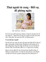
Thai ngoài tử cung chưa vỡ
NGUYỄN THIỆN HÙNG
KHOA SIÊU ÂM MEDIC
Nữ, 45 tuổi, Kampuchea.
Không trễ kinh hoặc đau bụng
Khám bệnh vì mệt mỏi, ăn kém.
BS lâm sàng gửi MEDIC khám siêu âm kiểm tra.
Mổ nội soi lấy túi thai cùng ngày tại BV
Triều An (BS Trương ngọc Thanh)
Túi thai có phôi, CRL=13,7mm, khỏang 7 tuần
Tubal Ring sign(+)
Nữ, 22tuổi, Q. Phú nhuận, trễ kinh hơn 30 ngày,
đau bụng 3 ngày, Hct=24%, Quickstick(+), chọc ổ
bụng không rút được dịch (BS T Tâm + BS Tùng)
Bv Saigon gửi khám siêu âm cấp cứu = Xuất huyết
nội + U nang buồng trứng T xoắn ?.
Lúc khám, bệnh nhân tỉnh, tiếp xúc tốt, da niêm
không tái, bớt đau bụng.
Ít dịch tụ ở hố chậu P
Dạng phôi bên P tử cung
Siêu âm (Bs T Hùng)= U nang buồng trứng T có
vách, còn phổ mạch máu. Dạng phôi bên P tử
cung, CRL=, 11mm, khỏang 7 w, không nhịp tim,
túi thai không rõ, dịch tụ hố chậu P ít .
TVS (BS T Thi)= Túi thai bên P tử cung, dịch túi
cùng ít + u nang buồng trứng T có vách.
U nang buồng trứng T có vách, còn phổ mạch máu
ECTOPIC PREGNANCY
= implantation outside the endometrial cavity
Incidence:
1.4% of all pregnancies (increasing); 9.9:10,000
women annually; 73,700 cases in 1986 in United
States; 4 - 15% of maternal deaths (decreasing);
coexistent with intrauterine pregnancy in 1:6,800 -
30,000 pregnancies (higher number of coexisting
ectopic with ovulation induction)
Risk of recurrence: 10%
Cause: delayed transit of the fertilized zygote
secondary to abnormal angulation of oviduct /
adhesions from inflammation / slowed tubal transit
Risk factors:
(1) Previous tubal surgery / ligation
(2) Previous PID (30 - 50%)
(3) Ovulation induction
(4) Endometriosis
(5) Previous ectopic pregnancy (10-fold increase in
risk, 25% chance of recurrence)
(6) IUD in place
+ If the pregnancy cannot be documented as
intrauterine, the patient should be considered at risk!
Time of manifestation: usually by 7th week of MA
CLASSIC TRIAD (<50%):
• abnormal vaginal bleeding (75 - 86%)
• pelvic pain (97%)
• palpable adnexal mass (30 - 41%)
• secondary amenorrhea (61%)
• cervical tenderness
• positive urinary pregnancy test (50%)
• b-HCG does not rise >66% within 48 hours (lower levels + slower rise
and decline compared with IUP)
+ Most ectopic pregnancies do not exhibit a b-HCG of >6500 mIU/mL (1st
IRP) prior to symptomatology!
+ A b-HCG level above the discriminatory zone with absence of IUP
suggests ectopic pregnancy!
Discriminatory zone of b-HCG (at which a normal IUP should be
visualized):
(a) by transabdominal scan:
³6500 mIU/mL (1st IRP) with 100% sensitivity + 96% specificity
(b) by endovaginal scan:
³2000 to 3000 mIU/mL (1st IRP)
Location:
(a) tubal (95%): (1) Ampullary ectopic
(2) Isthmic ectopic (92%)
(3) Interstitial ectopic (3%)
(b) other (5%): (1) Abdominal ectopic
(2) Ovarian ectopic
(3) Interligamentary ectopic
(4) Cervical ectopic (very rare)
Spectrum:
Type 1: unruptured live ectopic + heartbeat
Type 2: early embryonic demise without
rupture / embryonic structures / heartbeat
Type 3: ruptured ectopic with blood in pelvis
Type 4: no sonographic signs of ectopic
Transvesical US (usually less sensitive than transvaginal):
* absence of intrauterine pregnancy (beyond 6 weeks MA / with b-
HCG level >1,000 - 2,000 mIU/mL [IRP])
(a) no IUP by transvesical US = ectopic pregnancy in 43 - 46%
(b) no IUP by endovaginal US = ectopic pregnancy in 67%
* decidual cast = hyperechoic endometrial thickening (50%)
* pseudogestational sac = parietal decidual reaction + anechoic
fluid center from bleeding (10 - 20%)
* echogenic adnexal mass (42%) with small anechoic center =
gestational sac ± embryo ± heartbeat
* live embryo in adnexa (6 - 17%) = only specific sonographic
finding
* free abdominal fluid / hyperechoic clot in cul-de-sac
* hydro-/ hematosalpinx
* corpus luteum within ovary in >50% on side of ectopic pregnancy
Transvaginal US (5 - 26% false-negative rate):
* extrauterine mass of any type (84%)
* solid / complex adnexal mass = clotted blood free in
peritoneal cavity / hematosalpinx (36%)
* extrauterine gestational sac without live embryo / yolk sac
(35%)
* embryonic heartbeat (12 - 28%)
* free fluid (40 - 83%): echogenic / particulate fluid
(= hemoperitoneum) has 93% positive predictive value for
ectopic pregnancy (small amount of anechoic fluid found in 10 -
27% of IUP)
* decidual cast (21%)
* decidual cyst = 1- to 5-mm cyst in endometrium remote from
endometrial canal (14%)
Doppler-US (low diagnostic impact):
* high-velocity low-impedance flow around extrauterine
gestation in 54% (up to 4 kHz shift with 3 MHz transducer, 0.38
± 0.2 Pourcelot index)
* absence of peritrophoblastic flow after 36 days (<0.8 kHz
shift with 3 MHz transducer or <1.3 kHz shift with 5 MHz
transducer)
DDx of low-impedance flow:
corpus luteum cyst, tuboovarian abscess, fibroid
Probability of ectopic pregnancy in absence of IUP + clinical
symptoms of an ectopic pregnancy:
5% normal scan / simple cyst in adnexa
92% complex adnexal mass
95% tubal ring
100% live embryo outside uterus
Interstitial (cornual) ectopic (2 - 4%)
+ Often rupture late because of greater myometrial distensibility
compared with other parts of tube!
+ High likelihood of catastrophic hemorrhage + death due to abundant
blood supply by both ovarian + uterine arteries!
Increased risk: previous ipsilateral salpingectomy
* eccentric heterogeneous mass in cornual region (66%)
* eccentrically placed gestational sac (25%)
* thinning of myometrial mantle to <5 mm (33%)
* interstitial line sign = thin echogenic line extending directly up to the
center of ectopic pregnancy
(= endometrial canal / interstitial portion of Fallopian tube) in 92%
* large vascular channels with peritrophoblastic flow
•absence of double decidual sign
Prognosis:
massive bleeding from erosion of uterine arteries + veins (pregnancy
survives only 12 - 16 weeks GA);
2-fold mortality compared with other tubal ectopics
DDx: pregnancy within horn of bicornuate uterus; hydatidiform mole;
degenerating uterine fibroid
Abdominal ectopic (1:6000)
• bloating, abdominal pain (fetal movement / peritoneal
irritation due to adhesions)
• bleeding, hypotension, shock
* extrauterine location of fetus + placenta
* uterus compressed with visible endometrial cavity line
* absence of uterine wall between gestation + bladder /
abdominal wall
* anhydramnios
Cx: bowel obstruction / perforation; erosion of
pregnancy through abdominal wall
Lithopedion
= "stone child" = very rare obstetric complication
consisting of a dehydrated + calcified demised fetus in
an extrauterine pregnancy existing for >3 months
without infection
Types:
(1) Lithokelyphosis = fetal membranes calcified
(2) Lithokelyphopedion = fetus + membranes
calcified
(3) True lithopedion = only fetus calcified
Maternal age at discovery: 23 - 100 years of age;
within 4 - 20 years of fetal demise
Vôi hóa buồng trứng P, bnh nữ 70 tuổi
Vôi hóa buồng trứng P, bnh nữ 70 tuổi
Location:
most common in adnexae
* large densely calcified mass in lower
abdomen / upper pelvis
* CT scan reveals fetal skeleton
DDx:
uterine fibroid, calcified ovarian malignancy / cyst, sarcoma
Dx: (1) Laparoscopy (almost 100% accurate)
(2) Culdocentesis (high probability for ectopic with
aspiration of nonclotting blood with a hematocrit >15)
Cx: maternal death in 1:1,000; tubal rupture (10 -
15%)
DDx: (1) Hemorrhagic corpus luteum / hematoma
(2) Adnexal mass: hydrosalpinx, endometrioma,
ovarian cyst
(3) Fluid-containing small bowel loop
(4) Eccentrically placed GS in bicornuate /
retroflexed / fibroid uterus