Lab Exercises in Organismal and Molecular Microbiology_1 pot
Bạn đang xem bản rút gọn của tài liệu. Xem và tải ngay bản đầy đủ của tài liệu tại đây (23.52 MB, 188 trang )
ISBN: 0-07-248744-5
Description: ©2004 / Spiral Bound/Comb / 384 pages
Publication Date: March 2003
Overview
A modern general microbiology laboratory manual that combines the procedural details of a
laboratory manual with the photographic support of a laboratory atlas. The 46 class-tested
laboratory experiments are divided into 9 specialty areas, and the extensive four-color illustration
program includes 220 photos and micrographs plus 150 line drawings.
Features
• An extensive full-color art program integrated into the laboratory exercises allows students to not only conduct a
variety of laboratory exercises but also to interpret and confirm their results with the help of the large collection of color
photographs.
• Unique exercise!! Simulation of Infectious Disease Transmission (Lab Exercise 44). Developed in conjunction with
the pioneering program "The Biology Project" at the University of Arizona, this exercise allows class members to trade
simulated "body fluids" in a random pattern coordinated by the lab instructor. ELISA testing makes it clear to students
how easily the mock pathogen has passed through intermediaries to individuals in distant locations (across the lab).
• Emphasis on modern lab safety issues. Besides the usual safety advisories, this manual includes a table ranking the
Biosafety Level of every bacteria used in the lab exercises, specific guidelines for working with bacteria in each
Biosafety Level, and prominent icons throughout the lab exercises advising students of the Biosafety Level of the
bacteria in use. Safety Stops throughout the manual also remind students of particular hazards in each exercise. No
other lab manual on the market provides the Biosafety Level cautions and identification.
Alexander−Strete−Niles:
Lab Exercises in
Organismal and Molecular
Microbiology
Front Matter Preface
© The McGraw−Hill
Companies, 2003
ix
Preface
Organization
Our 46 exercises are organized into the following nine
sections:
Section I Survey of Microscopic Organisms
Section II Staining Techniques
Section III Bacterial Cultivation
Section IV Bacterial Identification
Section V Medical Microbiology
Section VI Controlling the Risk and Spread
of Bacterial Infections
Section VII Bacterial Genetics
Section VIII Viruses
Section IX Hematology and Serology
The standard presentation of each section makes it easy
for both students and lab managers to prepare for an
exercise. Each exercise:
1. Opens with a short background that conveys only
information relevant to the exercise.
2. Lists all needed materials, by category.
3. Presents procedures for the exercise in easy-to-
follow steps and includes special notes, hints, and
instructions to ensure success.
4. Integrates all photographs and line drawings into
the text of the exercise where they will provide
the student with the most support.
5. Includes a tear-out laboratory report conveniently
located at the end of the exercise.
Instructor Support Material
An Instructor Image Bank provides digital files in the
easy-to-use JPEG format for all of the photos and line
art included in this lab manual. They are organized by
section and placed in PowerPoint sets for easy access.
These may prove useful for lab preparation packets,
testing, or discussion sessions. Ask your McGraw-Hill
representative for further details.
When students move from the lecture hall to the micro-
biology laboratory, they need help bridging the
gap between the theory and the practice of what they are
learning. The equipment is unfamiliar, the procedures
are unfamiliar, and many of the materials they are han-
dling are unfamiliar. Linking the information from their
classroom lectures to the laboratory procedures is nec-
essary for their ultimate success. Our goal for this
laboratory manual is to provide the bridge that helps
students integrate their classroom lectures with their
laboratory experiences. This integrated approach is
the only way to ensure understanding and mastery in
microbiology.
Features
•
Class-tested experiments have been vetted in our
own courses and provide a thoughtful progression
of opportunities—from basic lab techniques, such
as Exercises 9–15 on various staining techniques,
to more challenging exercises, such as the simu-
lated epidemic in Exercise 44: “Enzyme-linked
Immunosorbent Assay (ELISA).” This building-
block approach allows students to develop
comfort and confidence in their laboratory skills.
•
Exceptional full-color art program includes over
250 of our own photographs created specifically
for these laboratory exercises, plus 150 line
drawings of equipment, procedures, and results.
Students can easily confirm their results and
procedures by referring to the illustrations.
•
Exceptional attention to safety issues is given
throughout the manual. A basic lab safety section
beginning on page xi includes a table identifying
the biosafety level of every organism used in the
experiments. The BSL 2 icon appears where
appropriate to remind students of the needed safety
precautions when working with pathogens. Caution
symbols appear throughout the lab manual to
provide students with additional safety warnings
as needed.
Alexander−Strete−Niles:
Lab Exercises in
Organismal and Molecular
Microbiology
Front Matter Preface
© The McGraw−Hill
Companies, 2003
Kristin M. Snow, Fox Valley Technical College
Carole Rehkugler, Cornell University
Paul E. Wanda, Southern Illinois University,
Edwardsville
Our gratitude is also extended to our publishing team at
McGraw-Hill:
Colin Wheatley, Publisher/Sponsoring Editor
Jean Sims Fornango, Senior Developmental Editor
Tami Petsche, Marketing Manager
Gloria Schiesl, Project Manager
Sandy Ludovissy, Production Supervisor
Wayne Harms, Designer
Carrie Burger, Photo Editor
x
The Instructor’s Manual for this set of labora-
tory exercises may be found online at:
www.mhhe.com/biosci/ap/labcentral/
It provides answers to lab report questions, tips for lab
exercise success, and other useful information.
Acknowledgments
In the end, our hope is that we have put together a man-
ual that will serve as a valuable teaching tool for the
microbiology laboratory. Our efforts were greatly aided
by the following reviewers, whom we gratefully
acknowledge:
Daniel R. Brown,
Sante Fe Community College
Kathy Buhrer, Tidewater Community College
Linda E. Fisher, University of Michigan, Dearborn
Georgia Ineichen, Hinds Community College
Hubert Ling, County College of Morris
Rita Moyes, Texas A&M University
Richard C. Renner, Laredo Community College
Ken Slater, Utah Valley State College
Alexander−Strete−Niles:
Lab Exercises in
Organismal and Molecular
Microbiology
Front Matter Safety Guidelines for the
Microbiology Laboratory
© The McGraw−Hill
Companies, 2003
xi
Safety Guidelines for the
Microbiology Laboratory
General Guidelines for Every Lab Session
1. Wear appropriate clothing and shoes to the laboratory. Shoes must completely cover the feet to
provide protection from broken glass and spills.
2. Place all books, backpacks, purses, etc., in an area designated by your laboratory instructor. Carry to
your work area only the items you will use in the lab.
3. Wash your hands thoroughly with antibacterial soap before beginning the lab session.
4. Wipe your work area with disinfectant, and allow to air-dry before beginning the lab session.
5. Do not perform activities in the lab until you are given instructions by your laboratory instructor.
6. Do not eat, drink, smoke, or apply makeup while working in the laboratory.
7. If you cut or burn yourself while working, report this immediately to your laboratory instructor.
8. Broken glassware should be immediately brought to the attention of your laboratory instructor. Bro-
ken glass should be placed in a special sharps container for disposal and not in the
trash container.
9. If using a Bunsen burner, tie back long hair. Do not lean over the countertop. When in use, always be
aware of the flame. Keep flammable items away from the flame. Turn off the burner when not in use.
10. Before leaving the lab, make sure all items have been returned to their appropriate location.
11. After your work area is clear, wipe down your countertop with disinfectant before leaving.
12. Wash your hands thoroughly with antibacterial soap before leaving the lab.
13. Do not remove any item from the lab unless you have been directed to do so by the laboratory
instructor.
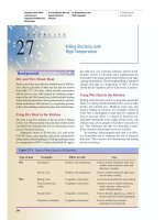
Guidelines for Working with Biosafety Level (BSL) 1 Bacteria
Handling live bacteria in the laboratory, even those considered nonpathogenic, requires special guidelines
beyond the general guidelines already mentioned. All bacteria are potentially pathogenic, especially if
they gain entry into the human body. So observe the following guidelines when handling the biosafety
level (BSL) 1 bacteria listed in the summary table.
1. Do not put anything into your mouth when working with cultures. Do not pipette by mouth; use a
pipette aid instead. Keep your hands, pencil, pen, etc., away from your mouth, eyes, and nose.
2. When inoculating cultures, sterilize the loop or needle before placing it on the counter.
3. Always keep tubes in test tube racks when working with liquid media. Do not stand them up or lay
them down on the countertop where they may spill.
4. If you accidentally spill a culture, cover the spill with a paper towel, flood it with disinfectant, and
notify your laboratory instructor.
5. Place all used culture media, paper towels, gloves, etc., into the waste container designated by your
laboratory instructor. A separate waste container for sharps (slides, pipettes, swabs, broken glass,
etc.) will also be provided. All this waste will be autoclaved before disposal or reuse. Do not throw
any of these items into the trash container.
6. If you have a burn or wound on one of your hands, cover it with a plastic strip and wear disposable
gloves for added protection.
Alexander−Strete−Niles:
Lab Exercises in
Organismal and Molecular
Microbiology
Front Matter Safety Guidelines for the
Microbiology Laboratory
© The McGraw−Hill
Companies, 2003
Guidelines for Working with Biosafety Level (BSL) 2 Bacteria
Handling pathogenic bacteria in the laboratory requires special guidelines beyond the general guidelines and
those for BSL 1 bacteria. The following additional guidelines apply when working with the biosafety level
(BSL) 2 bacteria listed in the summary table.
1. When handling pathogens, access to the laboratory must be restricted to only those working in
the lab.
2. Disposable gloves and a lab coat must be worn. The gloves should be disposed of in a container des-
ignated by the instructor. The lab coat must be removed before leaving and kept in a designated area
of the lab.
3. Avoid creating aerosols when working with pathogens. If there is a chance of creating tiny airborne
droplets, work under a safety hood.
xii
Biosafety level (BSL) Description of infectious agents Examples from this lab manual
1 Agents that typically do not cause Alcaligenes denitrificans
disease in healthy adults; they Alcaligenes faecalis
generally do not pose a disease Bacillus cereus
risk to humans. Bacillus subtilis
Corynebacterium pseudodiphtheriticum
Enterobacter aerogenes
Enterococcus faecalis
Escherichia coli
Micrococcus luteus
Neisseria sicca
Proteus vulgaris
Pseudomonas aeruginosa
Serratia marcescens
Staphylococcus epidermidis
Staphylococcus saprophyticus
2 Agents that can cause disease in Klebsiella pneumoniae
healthy adults; they pose Mycobacterium phlei
moderate disease risk to Salmonella typhimurium
humans. Shigella flexneri
Staphylococcus aureus
Streptococcus pneumoniae
Streptococcus pyogenes
3 Agents that can cause disease in None; these agents are not used in
healthy adults; they are airborne this lab manual.
and pose a more serious disease
risk to humans.
4 Agents that can cause disease in None; these agents are not used in
healthy adults; they pose a this lab manual.
lethal disease risk to humans;
no vaccines or therapy
available.
Summary of Biosafety Levels for Infectious Agents
Alexander−Strete−Niles:
Lab Exercises in
Organismal and Molecular
Microbiology
Front Matter Safety Guidelines for the
Microbiology Laboratory
© The McGraw−Hill
Companies, 2003
xiii
Name Date
Universal Precautions
All human blood and certain other body fluids are treated as if they are infectious for blood-borne pathogens,
such as human immunodeficiency virus (HIV), hepatitis B virus (HBV), and hepatitis C virus (HCV).
Such precautions are the rule among nurses, doctors, phlebotomists, and clinical laboratory personnel,
and are a critical component of infection control.
1. Wear gloves.
2. Change gloves when they are soiled or torn.
3. Remove gloves when you are finished handling a specimen, and before you touch other objects such
as drawer handles, door knobs, refrigerator handles, pens/pencils, and paper.
4. Wash hands thoroughly with soap and water after removing gloves.
5. Dispose of gloves and blood-contaminated materials in a biohazard receptacle.
Additional precautions that may not apply to this laboratory exercise:
6. Wear a lab coat when soiling with blood or body fluids is possible.
7. Wear a mask, goggles, or glasses with side shields if splashing of the face is possible.
Safety Commitment
I have read and understand the safety guidelines described above. I declare my commitment to safety in
the microbiology laboratory and promise to follow each rule during the course of the semester.
Alexander−Strete−Niles:
Lab Exercises in
Organismal and Molecular
Microbiology
I. Survey of Microscopic
Organisms
1. Structure, Function, and
Use of the Microscope
© The McGraw−Hill
Companies, 2003
2
Structure, Function,
and Use of the Microscope
EXERCISE
Part Function
1. Ocular (eyepiece) Magnifies image, usually 10x
2. Thumb wheel Adjusts distance between
oculars to match your eyes
3. Lock screw Secures head after rotation
4. Head Holds oculars
5. Arm Holds head and stage
6. Revolving Rotates objective lenses
nosepiece into viewing position
7. Objective Magnifies image, usually low
(4µ), medium (10µ), high dry
(40µ), and oil-immersion
(100µ)
8. Slide holder Fixed and movable parts
secure slide on stage
9. Mechanical Includes slide holder and is
stage used to locate specimen
10. Stage Holds slide
11. Stage aperture Admits light
12. Condenser Focuses light on specimen
and fills lens with light
13. Diaphragm lever Controls amount of light
entering stage aperture
14. Substage- Raises and lowers condenser
adjustment knob
15. Mechanical- Moves slide back and forth
stage control on stage
16. Light source Illuminates specimen
17. Coarse- Rapidly brings specimen into
adjustment knob focus
18. Fine-adjustment Slowly brings specimen into
knob sharp focus
19. Base Supports microscope
*Parts are listed in order from top to bottom, and their numbers
correspond to those in figure 1.1.
Table 1.1 Functions of the Parts of
the Light Microscope*
1
Background
The study of microscopic organisms is greatly aided by
the use of microscopes. The light microscope (LM) mag-
nifies objects up to 1,000 times (1,000µ) and can be used
to study cell size, shape, and arrangement. However, the
LM gives little information about internal cell structures.
The internal details of a cell are studied using a trans-
mission electron microscope (TEM), since useful mag-
nifications of up to 100,000µ are possible. The infection
of a cell by viruses or bacteria can also be studied using
a TEM. In addition, a three-dimensional view of cells in
their natural environment is possible with a scanning
electron microscope (SEM). Useful magnifications of up
to 20,000× are obtained with a SEM.
This exercise is designed to familiarize you with the
structure, function, and use of the light microscope. In
addition, TEM and SEM views of cells will be provided
for comparison.
Materials
Prepared slides (2)
Blood (human)
Budding yeast
Equipment
Microscope
Miscellaneous supplies
Immersion oil
Lens paper
Procedure
1. Familiarize yourself with the structure and
function of the light microscope by reviewing
the following: (a) the microscope in figure 1.1;
(b) the parts of the microscope and their
functions in table 1.1; and (c) the magnifications
obtained using different objectives in table 1.2.
Complete step 1 of the laboratory report.
Alexander−Strete−Niles:
Lab Exercises in
Organismal and Molecular
Microbiology
I. Survey of Microscopic
Organisms
1. Structure, Function, and
Use of the Microscope
© The McGraw−Hill
Companies, 2003
Table 1.2 Total Magnification Possible
with Different Objective Lenses
of the Light Microscope
Power Objective Ocular Total
lens lens magnification
Low 4µ 10µ 40µ
Medium 10µ 10µ 100µ
High dry 40µ 10µ 400µ
Oil- 100µ 10µ 1,000µ
immersion
2. Table 1.3 lists the steps for using the light
microscope. Follow these steps carefully as you
examine two slides: human blood and budding
yeast. Using figure 1.2 as a guide, identify as
many of the cell types and structures as you can.
For each slide, record in the laboratory report
what you see at 40µ, 100µ, 400µ, and 1,000µ.
3. Examine the photographs of the TEM
(figure 1.3) and the SEM (figure 1.4).
Also examine the images of cells that these
microscopes provide (figures 1.5–1.8).
How do these views of cells differ from
those provided by the light microscope?
(1) Ocular
(2) Thumb
wheel
(3) Lock screw
(4) Head
(5) Arm
(6) Revolving
nosepiece
(7) Objective
(8) Slide holder
(9) Mechanical
stage
(10) Stage
(12) Condenser
(13) Diaphragm
lever
(14) Substage–
adjustment knob
(15) Mechanical–
stage control
(16) Light source
(17) Coarse–
adjustment knob
(18) Fine–
adjustment knob
(19) Base
(11) Stage aperture
near center
Figure 1.1 The parts of the microscope.
Structure, Function, and Use of the Microscope E
XERCISE
1 3
Alexander−Strete−Niles:
Lab Exercises in
Organismal and Molecular
Microbiology
I. Survey of Microscopic
Organisms
1. Structure, Function, and
Use of the Microscope
© The McGraw−Hill
Companies, 2003
4 S
ECTION
I Survey of Microscopic Organisms
Table 1.3 Steps in the Use of the Light Microscope
Carry the microscope upright with two hands (figure 1.9, p.10). Place the microscope on the countertop, plug it in,
and turn on the light. Follow these steps as you examine the human blood and budding yeast slides:
1. Clip the slide into place on the stage using the slide holder.
2. Use the mechanical-stage control to move the slide so that the specimen is centered over the condenser.
3. Rotate the nosepiece to position the 4µ objective (figure 1.10a, p. 11). When this objective is in place over the
specimen, move the coarse-adjustment knob until the stage and objective are as close together as possible.
4. While looking through the oculars, move the coarse-adjustment knob to slowly increase the distance between the
stage and the objective. Stop when the specimen comes into focus.
5. Adjust the distance of the ocular lens by moving the thumb wheel until two images become one.
6. Close your left eye, and focus for the right eye using the fine-adjustment knob. Close your right eye, and focus for
the left eye using the focusing ring on the left ocular lens. Open both eyes and move the fine-adjustment knob
until a sharp image is obtained. You are now ready to make your observations at 40µ total magnification.
7. Center the specimen, and then rotate the nosepiece to position the 10µ objective (figure 1.10b, p. 11). Since most
microscopes are parfocal, the only adjustment that should be necessary is the fine adjustment. When the image is
sharp, make your observations at 100µ total magnification.
8. Rotate the nosepiece to position the 40µ objective (figure 1.10c, p. 11). Move the fine-adjustment knob, and make
your observations at 400µ total magnification.
9. Move the 40µ objective out of the way, and place a drop of immersion oil on top of the specimen. Position the
100µ oil-immersion objective (figure 1.10d, p. 11). Move only the fine-adjustment knob. You may need to open
the iris diaphragm with the diaphragm lever to allow more light to enter the objective lens. Make your
observations at 1,000µ total magnification.
10. When observations are complete, position the 4µ objective lens and wipe the oil off the oil-immersion objective
with a piece of lens paper. Remove the slide from the stage, and wipe off the oil if the specimen is covered by a
coverslip. If not, let the oil drain off by placing the slide upright in a slide box.
11. When finished, turn off the light, unplug the cord, and wrap it around the base. Return the microscope to the
storage cabinet.
Yeast cellsLymphocytes
Nuclei Parent cell
Buds
Lobed nucleus
Neutrophils
(b)(a)
Red blood cells
Figure 1.2 (a) Formed elements of human blood (1,000µ); (b) Yeast cells (1,000µ).
Alexander−Strete−Niles:
Lab Exercises in
Organismal and Molecular
Microbiology
I. Survey of Microscopic
Organisms
1. Structure, Function, and
Use of the Microscope
© The McGraw−Hill
Companies, 2003
Structure, Function, and Use of the Microscope E
XERCISE
1 5
Figure 1.3 Transmission electron microscope (TEM).
Figure 1.4 Scanning electron microscope (SEM).
Alexander−Strete−Niles:
Lab Exercises in
Organismal and Molecular
Microbiology
I. Survey of Microscopic
Organisms
1. Structure, Function, and
Use of the Microscope
© The McGraw−Hill
Companies, 2003
6 S
ECTION
I Survey of Microscopic Organisms
Figure 1.5 TEM view of white blood cells showing the internal structures characteristic of eucaryotic cells (12,000µ).
Alexander−Strete−Niles:
Lab Exercises in
Organismal and Molecular
Microbiology
I. Survey of Microscopic
Organisms
1. Structure, Function, and
Use of the Microscope
© The McGraw−Hill
Companies, 2003
Structure, Function, and Use of the Microscope E
XERCISE
1 7
Figure 1.6 TEM view of a virus-infected cell. Viruses are the circular particles with dark centers (20,000µ).
Alexander−Strete−Niles:
Lab Exercises in
Organismal and Molecular
Microbiology
I. Survey of Microscopic
Organisms
1. Structure, Function, and
Use of the Microscope
© The McGraw−Hill
Companies, 2003
8 S
ECTION
I Survey of Microscopic Organisms
Figure 1.7 TEM view of a Chlamydia-infected cell. Chlamydia bacteria are the numerous dark circles (3,000µ).
Alexander−Strete−Niles:
Lab Exercises in
Organismal and Molecular
Microbiology
I. Survey of Microscopic
Organisms
1. Structure, Function, and
Use of the Microscope
© The McGraw−Hill
Companies, 2003
Structure, Function, and Use of the Microscope E
XERCISE
1 9
Figure 1.8 SEM view of fungal hyphae on the surface of a potato leaf (5,000µ).
Alexander−Strete−Niles:
Lab Exercises in
Organismal and Molecular
Microbiology
I. Survey of Microscopic
Organisms
1. Structure, Function, and
Use of the Microscope
© The McGraw−Hill
Companies, 2003
10 S
ECTION
I Survey of Microscopic Organisms
Figure 1.9 Method used to carry the light microscope.
Alexander−Strete−Niles:
Lab Exercises in
Organismal and Molecular
Microbiology
I. Survey of Microscopic
Organisms
1. Structure, Function, and
Use of the Microscope
© The McGraw−Hill
Companies, 2003
Structure, Function, and Use of the Microscope E
XERCISE
1 11
(d)(c)
(b)(a)
Figure 1.10 Positions of light microscope objectives when viewing the specimen.
(a) 4µ objective
(b) 10µ objective
(c) 40µ objective
(d) 100µ oil-immersion objective
Alexander−Strete−Niles:
Lab Exercises in
Organismal and Molecular
Microbiology
I. Survey of Microscopic
Organisms
1. Structure, Function, and
Use of the Microscope
© The McGraw−Hill
Companies, 2003
Alexander−Strete−Niles:
Lab Exercises in
Organismal and Molecular
Microbiology
I. Survey of Microscopic
Organisms
1. Structure, Function, and
Use of the Microscope
© The McGraw−Hill
Companies, 2003
EXERCISE
1
L
ABORATORY
R
EPORT
N
AME
D
ATE
L
AB
S
ECTION
13
Part Function
d.
e.
f.
(a)
(b)
(f)
(e)
(d)
(c)
Part Function
a.
b.
c.
Structure, Function, and Use of the Microscope
1. Identify the parts (a–f) of the microscope below, and fill in their functions.
Alexander−Strete−Niles:
Lab Exercises in
Organismal and Molecular
Microbiology
I. Survey of Microscopic
Organisms
1. Structure, Function, and
Use of the Microscope
© The McGraw−Hill
Companies, 2003
b. Budding yeast
Draw and label parent
cells and buds you find.
2. Depict the morphology of a few representative cells at each total magnification. Try to draw the cells
at the size scale you observed.
a. Human blood
Draw and label the
cell types you find.
14 S
ECTION
I Survey of Microscopic Organisms
40µ 100µ
400µ 1,000µ
40µ 100µ
400µ 1,000µ
Alexander−Strete−Niles:
Lab Exercises in
Organismal and Molecular
Microbiology
I. Survey of Microscopic
Organisms
1. Structure, Function, and
Use of the Microscope
© The McGraw−Hill
Companies, 2003
Structure, Function, and Use of the Microscope E
XERCISE
1 15
3. Which microscope (LM, TEM, or SEM) would be most useful to study the following?
a. Size of cells
b. Whether or not a cell has a nucleus (i.e., is procaryotic or eucaryotic)
c. Whether or not a cell is infected with viruses
d. A three-dimensional view of cells attached to a surface
e. Cell shapes and arrangements
f. Cells infected with Chlamydia
4. Answer the following questions in the space provided.
a. (1) Give the general formula used to calculate the total magnification:
µ =total magnification
(2) What is the total magnification when using the 100µ oil-immersion objective lens?
b. In general, should the condenser be kept close to or far from the stage? Explain.
c. When increasing magnification from high dry to oil-immersion, should the iris diaphragm be
open or closed? How is this done? Does this adjustment increase or decrease the light reaching
the objective lens?
d. Explain why oil must be used with the oil-immersion lens.
e. Based on your observations of blood cells and yeast cells, which total magnification would you
recommend for best viewing? Explain.
Alexander−Strete−Niles:
Lab Exercises in
Organismal and Molecular
Microbiology
I. Survey of Microscopic
Organisms
2. Micro. Comparisons of
Microorganisms, Multi.
Parasites & Micro. Invert.
© The McGraw−Hill
Companies, 2003
Microscopic Comparisons of Microorganisms,
Multicellular Parasites, and Microscopic
Invertebrates
EXERCISE
2
17
two widely accepted classification systems for these
organisms. The Whittaker system, which consists of five
kingdoms, emphasizes differences in cellular traits and
nutrition, while the Woese system, which consists of
three domains, emphasizes differences in biochemical
traits. Neither system includes the viruses, due to their
unique makeup and method of replication.
In this exercise, you will use the microscope to
make comparisons of the microscopic organisms exam-
ined in Section I. You will learn to make size measure-
ments, and will measure a variety of microscopic
organisms. After you measure, be sure to note the mor-
phology of the microorganisms, multicellular parasites,
and microscopic invertebrates.
Materials
Prepared slides (8)
Select one slide from each category in
table 2.1.
Equipment
Light microscope
Miscellaneous supplies
Immersion oil
Lens paper
Ocular micrometer
Stage micrometer slide
Procedure
1. Clip the stage micrometer slide into position
on the stage, and position the scale over the
condenser (figure 2.2a, b). Focus on the scale
using the 4µ objective lens.
2. Align the ocular micrometer and stage
micrometer scales as depicted in figure 2.2c.
Now follow figure 2.2d to calibrate the ocular
micrometer for the 4µ objective lens.
Background
Microorganisms (bacteria, cyanobacteria, fungi, pro-
tozoans, and algae) and small animals (multicellular
parasites and microscopic invertebrates) display a vari-
ety of shapes and sizes (table 2.1). Figure 2.1 depicts
Kingdom AnimaliaKingdom FungiKingdom Plantae
Kingdom Monera
Kingdom Protista
Fungi
Plants
Protozoans
Extreme thermophiles,
halophiles, and methanogens
Other bacteria
Cyanobacteria
Animals
(Multicellular eucaryotes)
Bacteria and cyanobacteria
(unicellular procaryotes)
Protozoans and algae
(unicellular eucaryotes)
Nonphotosynthetic
(ingest food)
Photosynthetic
Eucaryotes
Archaebacteria
Eubacteria
(b) Woese system
(a) Whittaker system
Nonphotosynthetic
(absorb food)
Figure 2.1 Two classification systems recognized by
biologists and microbiologists: (a) the five-kingdom
classification system of R. H. Whittaker; (b) the
three-domain system of C. Woese.
Alexander−Strete−Niles:
Lab Exercises in
Organismal and Molecular
Microbiology
I. Survey of Microscopic
Organisms
2. Micro. Comparisons of
Microorganisms, Multi.
Parasites & Micro. Invert.
© The McGraw−Hill
Companies, 2003
18 S
ECTION
I Survey of Microscopic Organisms
4µ objective
0
0.5
1.0
mm
Ocular micrometer Stage micrometer
Sample calculation from (c):
Stage micrometer Ocular micrometer Calibration
40µ:
(1) 0.5 mm 20 ocular units (ou's) 0.025 mm/ou
(2) 1.0 mm 40 ocular units (ou's) 0.025 mm/ou
Average = 25 m/ou
0
0.5
1.0
mm
(a)
(b)
(c)
(d)
Figure 2.2 Calibration of the ocular micrometer.
Microscopic Size (in
Organism microns, m)
Bacteria
Bacillus 8
Escherichia coli 2-3
Spirillum 20
Staphylococcus 1
Treponema pallidum 15
Cyanobacteria
Oscillatoria (filament) 400
Yeasts (fungi)
Saccharomyces (with bud) 10
Molds (fungi)
Aspergillus (conidiophore) 1,200
Rhizopus (zygospore) 400
Protozoans
Amoeba proteus 300
Paramecium caudatum 200
Algae
Diatoms (centric) 100
Diatoms (pennate) 50
Dinoflagellates 100
Spirogyra (filament) 2,500
Volvox (colony) 200
Multicellular parasites
Clonorchis sinensis 7,500
(liver fluke)
Dipylidium caninum 2,500
(tapeworm proglottid)
Microscopic invertebrates
Cyclops 500
Daphnia 500
Nauplius larvae 600
Tick 2,500
Table 2.1 Typical Sizes of Selected
Microscopic Organisms
Alexander−Strete−Niles:
Lab Exercises in
Organismal and Molecular
Microbiology
I. Survey of Microscopic
Organisms
2. Micro. Comparisons of
Microorganisms, Multi.
Parasites & Micro. Invert.
© The McGraw−Hill
Companies, 2003
Microscopic Comparisons of Microorganisms, Multicellular Parasites, and Microscopic Invertebrates E
XERCISE
2 19
Table 2.2 Calculations in the Calibration of the Ocular Micrometer
Stage micrometer Ocular micrometer Calibration
a. 40µ
1.
2.
Average
b. 100µ
1.
2.
Average
c. 400µ
1.
2.
Average
d. 1,000µ
Calibration at 100µ /10
3. Repeat the calibration steps for the 10µ and 40µ
objectives. To calculate the calibration for the
100µ objective, take the calibration for the 10µ
objective and divide by 10. Record your ocular
calibration results in table 2.2 and in the
laboratory report.
4. Select one slide from each category in table 2.1
(eight total). Using your ocular calibration
results, calculate and record in the laboratory
report the size of each organism at the
appropriate magnification. When comparing your
results to those in table 2.1, do not expect results
for every organism to be exactly like those
shown, since the size of individual cells and
cell groupings may vary.
5. Also be sure to depict the morphology of
each organism in the circles provided in the
laboratory report.
Alexander−Strete−Niles:
Lab Exercises in
Organismal and Molecular
Microbiology
I. Survey of Microscopic
Organisms
2. Micro. Comparisons of
Microorganisms, Multi.
Parasites & Micro. Invert.
© The McGraw−Hill
Companies, 2003
Alexander−Strete−Niles:
Lab Exercises in
Organismal and Molecular
Microbiology
I. Survey of Microscopic
Organisms
2. Micro. Comparisons of
Microorganisms, Multi.
Parasites & Micro. Invert.
© The McGraw−Hill
Companies, 2003
EXERCISE
2
L
ABORATORY
R
EPORT
N
AME
D
ATE
L
AB
S
ECTION
21
Microscopic Comparisons of Microorganisms, Multicellular
Parasites, and Microscopic Invertebrates
1. Record your ocular calibration results from table 2.2.
40µ: µ/ocular unit (ou)
100µ: µ/ou
400µ: µ/ou
1,000µ: µ/ou
2. Determine the size of each of the eight selected organisms by multiplying the length you measured in
ocular units by the appropriate ocular calibration result recorded in question 1. Also sketch each
organism in the circle provided.
Bacteria Cyanobacteria
Organism Organism
Magnification Magnification
Length (in ou’s) Length (in ou’s)
Size ( ou’s! µ/ou)= µ Size ( ou’s! µ/ou)= µ