Proteogly cans Methods and Protocols doc
Bạn đang xem bản rút gọn của tài liệu. Xem và tải ngay bản đầy đủ của tài liệu tại đây (5.74 MB, 374 trang )
M
ETHODS
IN
M
OLECULAR
B
IOLOGY
™
Series Editor
John M. Walker
School of Life Sciences
University of Hertfordshire
Hatfield, Hertfordshire, AL10 9AB, UK
For further volumes:
/>
Proteoglycans
Methods and Protocols
Edited by
Françoise Rédini
INSERM UMR957-EA3822, Nantes, France
ISSN 1064-3745 e-ISSN 1940-6029
ISBN 978-1-61779-497-1 e-ISBN 978-1-61779-498-8
DOI 10.1007/978-1-61779-498-8
Springer New York Dordrecht Heidelberg London
Library of Congress Control Number: 2011944507
© Springer Science+Business Media, LLC 2012
All rights reserved. This work may not be translated or copied in whole or in part without the written permission of the
publisher (Humana Press, c/o Springer Science+Business Media, LLC, 233 Spring Street, New York, NY 10013, USA),
except for brief excerpts in connection with reviews or scholarly analysis. Use in connection with any form of information
storage and retrieval, electronic adaptation, computer software, or by similar or dissimilar methodology now known or
hereafter developed is forbidden.
The use in this publication of trade names, trademarks, service marks, and similar terms, even if they are not identified
as such, is not to be taken as an expression of opinion as to whether or not they are subject to proprietary rights.
Printed on acid-free paper
Humana Press is part of Springer Science+Business Media (www.springer.com)
Editor
Françoise Rédini, PhD
INSERM UMR957-EA3822
Nantes, France
Dedication
To Marc Padrines,
This book is the testimony of a friendly collaboration of more than ten years on proteoglycans
and bone. Thank you for the lively and fruitful discussions that we could share during this
period, and for your unwavering cheerfulness.
vii
Preface
Proteoglycans: Complex Diversity in Structure and Functions
Scientifi c interest and curiosity for proteoglycan research has dramatically increased over
the past decades, these molecules being no more considered as a scaffold for the cells of a
given tissue, but more as a reservoir for growth factors and cytokines modulating their
activation status and turnover. For example, heparin and other related heparan sulfate mol-
ecules are increasingly being recognized as important modulators of many signaling
pathways.
The evolution and accuracy of methodologies specifi c to their complex structure allow
a better knowledge of their structure together with a more precise defi nition of their func-
tions and involvement in physiology and pathologies. The huge increase of original research
articles on proteoglycans refl ects the increasing interest for these molecules.
There is no unifying structure for proteoglycans (PGs), such as collagen triple helix for
example, and they display a great diversity of protein forms. However, their basic structure
is defi ned as a protein portion and long unbranched polysaccharides (named glycosamino-
glycans or GAGs). PGs were initially grouped together because of the high negative charges
of their GAG chains; it makes them easily separable from other molecules by ion-exchange
chromatography. However, PGs are not that similar. The core protein size ranges from 10
to >500 kDa, and the number of GAG chains attached varies from 1 to >100. In addition,
several PGs carry GAG chains of more than one type (hybrid PGs: aggrecan, syndecans…)
and/or have additional N - or O -linked sugar modifi cations. Not all PGs are “full-time”
PGs. There are also a growing number of matrix molecules which may or may not be linked
with GAG chains, depending on the developmental stage or due to regulatory factors. They
are called “part-time” PGs, such as MHC class II invariant chain, thrombomodulin, CD44,
macrophage colony-stimulating factor, amyloid precursor protein, collagen type IX, XII,
XIV, and XVIII, and the transferring receptor, with alternatively spliced variants having
GAG-initiation sites. Some PGs such as versican or CD44 also occur as alternatively spliced
forms with varying sugar modifi cations. Versican can also be considered as part-time PGs
because a variant of versican without GAG attachment sites has been discovered.
The protein forms have complex modular structures with protein motifs that are of
similar sequence to those found in other protein families: several PGs thus contain dis-
tinct protein and carbohydrate domain structures that confer specifi c functional proper-
ties. The protein domains are often the products of separate exons. Recent studies have
identifi ed approximately 30 different PG protein cores; these cores are not only scaffolds
for GAGs but they also contain domains that have particular biological activities. Many
PGs are thus multifunctional molecules that engage in several different specifi c interac-
tions at the same time.
In addition, numerous variations also occur in GAG chain structure; GAGs are large
extended structures with highly charged sulfate and carbohydrate groups, and they domi-
nate the physical properties of the protein to which they are attached. PGs in the extracel-
lular matrix thus function physically as creators of a water-fi lled compartment. Their high
fi xed negative charge attracts counter ions, and the osmotic imbalance caused by a local
viii Preface
high concentration of ions draws water from the surrounding areas. PGs thus keep the
matrix hydrated and create a water compartment because they exclude other macromole-
cules while retaining permeability to low molecular weight solutes. This property increases
the concentration of the macromolecules and therefore may increase reaction rates and
promote all interactions that are concentration-dependent. Thus, PGs have important
physical effects on events in the concentrated milieu around the cells and in the extracellular
matrix.
The GAG side chains covalently linked to the core protein may be chondroitin sulfate
(CS), or its epimerized homolog dermatan sulfate (DS), or keratan sulfate (KS), heparan
sulfate (HS), or heparin (HP). Except KS, GAG synthesis is initiated by sequential addition
of four monosaccharides: xylose (xyl), galactose (Gal), galactose and glucuronic acid
(GlcUA). From this linker tetrasaccharide, the sugar chains are extended by addition of two
alternating monosaccharides: an aminosugar and GlcUA. In HP and HS, the aminosugar is
N -acetyl-glucosamine (GlcNAc) and in CS/DS, it is N -acetyl-galactosamine (GalNAc).
The extent of epimerization of GlcUA to iduronic acid (IdUA) and the sulfation pattern of
the disaccharide units distinguish HP from HS, and CS from DS. In KS, the GAGs are initi-
ated as N -linked or O -linked oligosaccharides and extended by addition of GlcNAc and
Gal. There is also regional variability to the epimerization and sulfation in each GAG chain.
Studies of these patterns have defi ned the motifs required for specifi c interactions with
growth factors, cytokines, matrix components, enzymes, and other proteins.
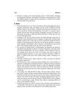
Divided into three categories, the volume fi rst covers issues of basic concepts and up to
date analysis methods for (I)proteoglycan and (II)glycosaminoglycan respectively at the
protein and saccharide levels. Then the multifunctional aspect of proteoglycans is high-
lighted through three relevant examples of proteoglycans with highly different structures:
serglycin, aggrecan, and heparin sulfate proteoglycans. The fi nal chapter describes proteo-
glycan involvement in the pathogenesis of various disorders (kidney, corneal epithelial
wound healing,…) and their potential therapeutic value in osteo-articular diseases.
Nantes, France Françoise Rédini, PhD
ix
Contents
Dedication . . . . . . . . . . . . . . . . . . . . . . . . . . . . . . . . . . . . . . . . . . . . . . . . . . . . . . . . . . . v
Preface. . . . . . . . . . . . . . . . . . . . . . . . . . . . . . . . . . . . . . . . . . . . . . . . . . . . . . . . . . vii
Contributors. . . . . . . . . . . . . . . . . . . . . . . . . . . . . . . . . . . . . . . . . . . . . . . . . . . . . . xi
P
ART I PROTEOGLYCANS
1 Proteoglycans: Gene Cloning . . . . . . . . . . . . . . . . . . . . . . . . . . . . . . . . . . . . . 3
Mauricio Cortes, James R. Mensch, Miriam Domowicz,
and Nancy B. Schwartz
2 Proteoglycan: Site Mapping and Site-Directed Mutagenesis. . . . . . . . . . . . . . . 23
Fred K. Hagen
3 Mapping of the Wnt/b-Catenin/TCF Response Elements in the Human
Versican Promoter. . . . . . . . . . . . . . . . . . . . . . . . . . . . . . . . . . . . . . . . . . . . . . 35
Maziar Rahmani, Jon M. Carthy, and Bruce M. McManus
4 Gene Silencing in Mouse Embryonic Stem Cells . . . . . . . . . . . . . . . . . . . . . . . 53
Norihiko Sasaki and Shoko Nishihara
5 A Novel Strategy for a Splice-Variant Selective Gene Ablation:
The Example of the Versican V0/V2 Knockout. . . . . . . . . . . . . . . . . . . . . . . . 63
María T. Dours-Zimmermann and Dieter R. Zimmermann
6 Detection of Neurocan in Cerebrospinal Fluid . . . . . . . . . . . . . . . . . . . . . . . . 87
Uwe Rauch
P
ART II GLYCOSAMINOGLYCANS ANALYSIS
7 Glycosaminoglycan Chain Analysis and Characterization
(Glycosylation/Epimerization) . . . . . . . . . . . . . . . . . . . . . . . . . . . . . . . . . . . . 99
Shuji Mizumoto and Kazuyuki Sugahara
8 Characterization of Glycosaminoglycans by Tandem Vibrational
Microspectroscopy and Multivariate Data Analysis. . . . . . . . . . . . . . . . . . . . . . 117
Nathalie Mainreck, Stéphane Brézillon, Ganesh D. Sockalingum,
François-Xavier Maquart, Michel Manfait, and Yanusz Wegrowski
9 Glycosaminoglycans: Oligosaccharide Analysis by Liquid Chromatography,
Capillary Electrophoresis, and Specific Labeling . . . . . . . . . . . . . . . . . . . . . . . 131
Derek J. Langeslay, Christopher J. Jones, Szabolcs Beni, and Cynthia K. Larive
10 Brain Chondroitin/Dermatan Sulfate, from Cerebral Tissue to Fine Structure:
Extraction, Preparation, and Fully Automated Chip-Electrospray Mass
Spectrometric Analysis . . . . . . . . . . . . . . . . . . . . . . . . . . . . . . . . . . . . . . . . . . 145
Alina D. Zamfir, Corina Flangea, Alina Serb, Eugen Sisu,
Leon Zagrean, Andreas Rizzi, and Daniela G. Seidler
x Contents
11 Use of Neutrons Reveals the Dynamics of Cell Surface
Glycosaminoglycans . . . . . . . . . . . . . . . . . . . . . . . . . . . . . . . . . . . . . . . . . . . . 161
Marion Jasnin
12 Following Protein–Glycosaminoglycan Polysaccharide Interactions
with Differential Scanning Fluorimetry . . . . . . . . . . . . . . . . . . . . . . . . . . . . . . 171
Katarzyna A. Uniewicz, Alessandro Ori, Timothy R. Rudd, Marco Guerrini,
Mark C. Wilkinson, David G. Fernig, and Edwin A. Yates
13 In Vivo Scintigraphic Imaging of Proteoglycans . . . . . . . . . . . . . . . . . . . . . . . 183
Elisabeth Miot-Noirault, Aurélien Vidal, Philippe Auzeloux,
Caroline Peyrode, Jean-Claude Madelmont, and Jean-Michel Chezal
P
ART III PGS: MULTIFUNCTIONAL CELL REGULATORS
14 Serglycin: The Master of the Mast Cell . . . . . . . . . . . . . . . . . . . . . . . . . . . . . . 201
Elin Rönnberg and Gunnar Pejler
15 Analysis of Aggrecan Catabolism by Immunoblotting
and Immunohistochemistry. . . . . . . . . . . . . . . . . . . . . . . . . . . . . . . . . . . . . . . 219
Peter J. Roughley and John S. Mort
16 Heparan Sulfate Proteoglycans as Multifunctional Cell Regulators:
Cell Surface Receptors . . . . . . . . . . . . . . . . . . . . . . . . . . . . . . . . . . . . . . . . . . 239
Jin-ping Li and Dorothe Spillmann
P
ART IV PROTEOGLYCANS INVOLVEMENT IN PATHOPHYSIOLOGY
17 Models for Studies of Proteoglycans in Kidney Pathophysiology . . . . . . . . . . . 259
Scott J. Harvey
18 Lumican Promotes Corneal Epithelial Wound Healing . . . . . . . . . . . . . . . . . . 285
Chia-Yang Liu and Winston Whei-Yang Kao
19 Shedding of Cell Membrane-Bound Proteoglycans . . . . . . . . . . . . . . . . . . . . . 291
Eon Jeong Nam and Pyong Woo Park
20 Modulatory Effects of Proteoglycans on Proteinase Activities . . . . . . . . . . . . . 307
Steven Georges, Dominique Heymann, and Marc Padrines
21 Proteoglycans and Osteolysis. . . . . . . . . . . . . . . . . . . . . . . . . . . . . . . . . . . . . . 323
Marc Baud’Huin, Céline Charrier, Gwenola Bougras, Régis Brion,
Frédéric Lezot, Marc Padrines, and Dominique Heymann
22 Proteoglycans and Cartilage Repair . . . . . . . . . . . . . . . . . . . . . . . . . . . . . . . . . 339
Mohamed Ouzzine, Narayanan Venkatesan, and Sylvie Fournel-Gigleux
Index. . . . . . . . . . . . . . . . . . . . . . . . . . . . . . . . . . . . . . . . . . . . . . . . . . . . . . . . . . . 357
xi
Contributors
PHILIPPE AUZELOUX
•
INSERM, UMR 990 , Clermont-Ferrand , France;
Imagerie moléculaire et thérapie vectorisée , Clermont Université,
Université d’Auvergne , Clermont-Ferrand , France
M
ARC BAUD’HUIN
•
INSERM, UMR 957 , Nantes , France; Physiopathologie de la
Résorption Osseuse et Thérapie des Tumeurs Osseuses Primitives , Université de Nantes,
Nantes Atlantique Universités , Nantes , France
S
ZABOLCS BENI
•
Department of Chemistry , University of California—Riverside ,
Riverside , CA , USA; Department of Pharmaceutical Chemistry ,
Semmelweis University , Budapest , Hungary
G
WENOLA BOUGRAS
•
INSERM, UMR 957 , Nantes , France; Physiopathologie de la
Résorption Osseuse et Thérapie des Tumeurs Osseuses Primitives , Université de Nantes,
Nantes Atlantique Universités , Nantes , France
S
TÉPHANE BRÉZILLON
•
Laboratoire de Biochimie Médicale et de Biologie Moléculaire,
CNRS UMR 6237—MEDyC , Université de Reims-Champagne-Ardenne ,
Reims , France
R
ÉGIS BRION
•
INSERM, UMR 957 , Nantes , France; Physiopathologie de la Résorption
Osseuse et Thérapie des Tumeurs Osseuses Primitives , Université de Nantes,
Nantes Atlantique Universités , Nantes , France
J
ON M. CARTHY
•
Department of Pathology and Laboratory Medicine, The James
Hogg iCAPTURE Centre for Cardiovascular and Pulmonary Research ,
Institute for Heart + Lung Health, University of British Columbia ,
Vancouver , BC , Canada
C
ÉLINE CHARRIER
•
INSERM, UMR 957 , Nantes , France; Physiopathologie de la
Résorption Osseuse et Thérapie des Tumeurs Osseuses Primitives , Université de Nantes,
Nantes Atlantique Universités , Nantes , France
J
EAN-MICHEL CHEZAL
•
INSERM, UMR 990 , Clermont-Ferrand , France;
Imagerie moléculaire et thérapie vectorisée , Clermont Université,
Université d’Auvergne , Clermont-Ferrand , France
M
AURICIO CORTES
•
Departments of Pediatrics , The University of Chicago ,
Chicago , IL , USA
M
IRIAM DOMOWICZ
•
Departments of Pediatrics , The University of Chicago ,
Chicago , IL , USA
M
ARÍA T. DOURS-ZIMMERMANN
•
Institute of Surgical Pathology , University Hospital
Zurich , Zurich , Switzerland
D
AVID G. FERNIG
•
Institute of Integrative Biology , University of Liverpool ,
Liverpool , UK
C
ORINA FLANGEA
•
Department of Chemical and Biological Sciences ,
“Aurel Vlaicu” University of Arad , Arad , Romania
S
YLVIE FOURNEL-GIGLEUX
•
UMR 7561 CNRS-Université Henri Poincaré Nancy I ,
Vandoeuvre-lès-Nancy , France
xii Contributors
STEVEN GEORGES
•
INSERM, U957 , Nantes , France; Laboratoire de Physiopathologie
de la Résorption Osseuse et Thérapie des Tumeurs Osseuses Primitives ,
Université de Nantes, Nantes Atlantique Universités , Nantes , France
M
ARCO GUERRINI
•
Ronzoni Institute for Chemical and Biochemical Research ,
Milan , Italy
F
RED K. HAGEN
•
Department of Biochemistry and Biophysics, Proteomics Center ,
University of Rochester Medical Center , Rochester , NY , USA
S
COTT J. HARVEY
•
INSERM Avenir U983 , Hôpital Necker-Enfants Malades ,
Paris , France
D
OMINIQUE HEYMANN
•
INSERM, U957, Laboratoire de Physiopathologie de la
Résorption Osseuse et Thérapie des Tumeurs Osseuses Primitives , Université de Nantes,
Nantes Atlantique Universités , Nantes , France; Centre Hospitalier Universitaire
de Nantes , Nantes , France
M
ARION JASNIN
•
Department of Molecular Structural Biology , Max Planck Institute
of Biochemistry , Martinsried , Germany
C
HRISTOPHER J. JONES
•
Department of Chemistry , University of California—
Riverside , Riverside , CA , USA
W
INSTON WHEI-YANG KAO
•
Department of Ophthalmology, College of Medicine,
Edith J. Crawley Vision Research Center , University of Cincinnati ,
Cincinnati , OH , USA
D
EREK J. LANGESLAY
•
Department of Chemistry , University of California—Riverside ,
Riverside , CA , USA
C
YNTHIA K. LARIVE
•
Department of Chemistry , University of California—Riverside ,
Riverside , CA , USA
F
RÉDÉRIC LEZOT
•
INSERM, UMR 957 , Nantes , France; Physiopathologie de la
Résorption Osseuse et Thérapie des Tumeurs Osseuses Primitives , Université de Nantes,
Nantes Atlantique Universités , Nantes , France
J
IN-PING LI
•
Department of Medical Biochemistry and Microbiology ,
Uppsala University , Uppsala , Sweden
C
HIA-YANG LIU
•
Department of Ophthalmology, College of Medicine,
Edith J. Crawley Vision Research Center , University of Cincinnati ,
Cincinnati , OH , USA
J
EAN-CLAUDE MADELMONT
•
INSERM, UMR 990 , Clermont-Ferrand , France;
Imagerie moléculaire et thérapie vectorisée , Clermont Université,
Université d’Auvergne , Clermont-Ferrand , France
N
ATHALIE MAINRECK
•
Laboratoire de Biochimie Médicale et de Biologie Moléculaire,
CNRS UMR 6237—MEDyC , Université de Reims-Champagne-Ardenne ,
Reims , France
M
ICHEL MANFAIT
•
Equipe MEDIAN, CNRS UMR 6237—MEDyC ,
Université de Reims-Champagne-Ardenne , Reims , France
F
RANÇOIS-XAVIER MAQUART
•
CHU de Reims, CNRS UMR 6237—MEDyC ,
Université de Reims-Champagne-Ardenne , Reims , France
B
RUCE M. MCMANUS
•
Department of Pathology and Laboratory Medicine,
The James Hogg iCAPTURE Centre for Cardiovascular and Pulmonary Research ,
Institute for Heart + Lung Health, University of British Columbia ,
Vancouver , BC , Canada
xiiiContributors
JAMES R. MENSCH
•
Departments of Pediatrics , The University of Chicago ,
Chicago , IL , USA
E
LISABETH MIOT-NOIRAULT
•
INSERM, UMR 990 , Clermont-Ferrand , France;
Imagerie moléculaire et thérapie vectorisée , Clermont Université, Université
d’Auvergne , Clermont-Ferrand , France
S
HUJI MIZUMOTO
•
Laboratory of Proteoglycan Signaling and Therapeutics ,
Frontier Research Center for Post-Genomic Science and Technology,
Hokkaido University , Sapporo , Japan
J
OHN S. MORT
•
Research Unit, Shriners Hospital for Children ,
Montreal , QC , Canada
E
ON JEONG NAM
•
Division of Respiratory Diseases, Children’s Hospital ,
Harvard Medical School , Boston , MA , USA
S
HOKO NISHIHARA
•
Department of Bioinformatics, Laboratory of Cell Biology ,
Soka University , Tokyo , Japan
A
LESSANDRO ORI
•
Structural and Computational Biology Unit, EMBL ,
Heidelberg , Germany
M
OHAMED OUZZINE
•
UMR 7561 CNRS-Université Henri Poincaré Nancy I ,
Vandoeuvre-lès-Nancy , France
M
ARC PADRINES
•
INSERM, U957 , Nantes , France; Laboratoire de Physiopathologie de
la Résorption Osseuse et Thérapie des Tumeurs Osseuses Primitives ,
Université de Nantes, Nantes Atlantique Universités , Nantes , France
P
YONG WOO PARK
•
Division of Respiratory Diseases, Children’s Hospital ,
Harvard Medical School , Boston , MA , USA
G
UNNAR PEJLER
•
Department of Anatomy, Physiology and Biochemistry ,
Swedish University of Agricultural Sciences , Uppsala , Sweden
C
AROLINE PEYRODE
•
INSERM, UMR 990 , Clermont-Ferrand , France;
Imagerie moléculaire et thérapie vectorisée , Clermont Université,
Université d’Auvergne , Clermont-Ferrand , France
M
AZIAR RAHMANI
•
Department of Pathology and Laboratory Medicine,
The James Hogg iCAPTURE Centre for Cardiovascular and Pulmonary Research ,
Institute for Heart + Lung Health, University of British Columbia ,
Vancouver , BC , Canada
U
WE RAUCH
•
Department of Vascular Wall Biology , Institute of Experimental
Medical Sciences, Lunds University , Lund , Sweden
A
NDREAS RIZZI
•
Institute of Analytical Chemistry and Food Chemistry ,
University of Vienna , Vienna , Austria
E
LIN RÖNNBERG
•
Department of Anatomy, Physiology and Biochemistry ,
Swedish University of Agricultural Sciences , Uppsala , Sweden
P
ETER J. ROUGHLEY
•
Research Unit, Shriners Hospital for Children ,
Montreal , QC , Canada
T
IMOTHY R. RUDD
•
Institute of Integrative Biology , University of Liverpool ,
Liverpool , UK; Ronzoni Institute for Chemical and Biochemical Research ,
Milan , Italy
N
ORIHIKO SASAKI
•
Department of Bioinformatics, Laboratory of Cell Biology ,
Soka University , Tokyo , Japan
xiv Contributors
NANCY B. SCHWARTZ
•
Departments of Pediatrics, and Biochemistry and Molecular
Biology , The University of Chicago , Chicago , IL , USA
D
ANIELA G. SEIDLER
•
Institute for Physiological Chemistry and Pathobiochemistry ,
University Hospital of Münster , Münster , Germany
A
LINA SERB
•
Department of Biochemistry , “Victor Babes” University of Medicine
and Pharmacy , Timisoara , Romania
E
UGEN SISU
•
Department of Biochemistry , “Victor Babes” University of Medicine
and Pharmacy , Timisoara , Romania
D
OROTHE SPILLMANN
•
Department of Medical Biochemistry and Microbiology ,
Uppsala University , Uppsala , Sweden
G
ANESH D. SOCKALINGUM
•
Equipe MEDIAN, CNRS UMR 6237—MEDyC ,
Université de Reims-Champagne-Ardenne , Reims , France
K
AZUYUKI SUGAHARA
•
Laboratory of Proteoglycan Signaling and Therapeutics ,
Frontier Research Center for Post-Genomic Science and Technology,
Hokkaido University , Sapporo , Japan
K
ATARZYNA A. UNIEWICZ
•
Institute of Integrative Biology , University of Liverpool ,
Liverpool , UK; PromoCell GmbH Sickingenstr, Heidelberg, Germany
N
ARAYANAN VENKATESAN
•
UMR 7561 CNRS-Université Henri Poincaré Nancy I ,
Vandoeuvre-lès-Nancy , France
A
URÉLIEN VIDAL
•
INSERM, UMR 990 , Clermont-Ferrand , France;
Clermont Université, Université d’Auvergne, Imagerie moléculaire et thérapie
vectorisée, BP 10448 , 63000 , Clermont-Ferrand , France
Y
ANUSZ WEGROWSKI
•
Laboratoire de Biochimie Médicale et de Biologie Moléculaire,
CNRS UMR 6237—MEDyC , Université de Reims-Champagne-Ardenne ,
Reims , France
M
ARK C. WILKINSON
•
Institute of Integrative Biology , University of Liverpool ,
Liverpool , UK
E
DWIN A. YATES
•
Institute of Integrative Biology , University of Liverpool ,
Liverpool , UK
L
EON ZAGREAN
•
Neuroscience Laboratory , “Carol Davila” University of Medicine
and Pharmacy , Bucharest , Romania
A
LINA D. ZAMFIR
•
Department of Chemical and Biological Sciences ,
“Aurel Vlaicu” University of Arad , Arad , Romania; Mass Spectrometry Laboratory ,
National Institute for Research and Development in Electrochemistry
and Condensed Matter , Timisoara , Romania
D
IETER R. ZIMMERMANN
•
Institute of Surgical Pathology , University Hospital Zurich ,
Zurich , Switzerland
PART I
PROTEOGLYCANS
sdfsdf
3
Françoise Rédini (ed.), Proteoglycans: Methods and Protocols, Methods in Molecular Biology, vol. 836,
DOI 10.1007/978-1-61779-498-8_1, © Springer Science+Business Media, LLC 2012
Chapter 1
Proteoglycans: Gene Cloning
Mauricio Cortes , James R. Mensch , Miriam Domowicz ,
and Nancy B. Schwartz
Abstract
Aggrecan is a large proteoglycan that plays roles in numerous tissues during vertebrate development and
adult life. The 6,327-nt chick aggrecan coding sequence had been determined from overlapping clones,
but a full-length cDNA, needed for use in transgenic expression studies, had not been constructed.
The strategy employed to do so was to generate two overlapping cDNA subfragments that shared a unique
restriction site in the overlap and then join them at that site. These subfragments were obtained and cloned
into the TOPO-TA vector pCR2.1. Digestion of the two constructs with the shared-site enzyme, XbaI,
produced vector/5 ¢ -cDNA and 3 ¢ -cDNA fragments with XbaI-ends; these were ligated to produce the
fi nal full-length cDNA.
Key words: Proteoglycan , Aggrecan , Cloning , Strategy , Full-length , cDNA
Aggrecan is a large molecule found in the extracellular matrix of
many vertebrate tissues, notably cartilage and brain (
1 ) . It plays a
variety of roles during development and in the adult; some are still
being elucidated. The proteoglycan aggrecan consists of a core
protein to which numerous carbohydrate chains are attached; these
include chondroitin sulfate, keratan sulfate, and various oligosac-
charides (see Fig.
1 ) ( 2 ) . Our laboratory has studied many aspects
of the physiological, biochemical, and structural properties of
aggrecan, with emphasis on its roles during development (
3– 11 ) .
Chicken was the initial organism employed, affording ease of
producing staged embryonic material and of experimental access
to live embryos; a lethal recessive mutation, nanomelia , provided a
natural aggrecan knock-out model (
12, 13 ) . Much work has also been
done in mice, which also have natural knock-out mutant models
1. Introduction
4 M. Cortes et al.
available: the cartilage matrix defi ciency ( cmd ) alleles ( 14, 15 ) .
These models and the ability to genetically manipulate mice
through controlled breeding and by creation of transgenic animals
have enabled the study of aggrecan function in a mammalian system.
We wanted to construct transgenic mice bearing the chick aggrecan
coding sequence, as an antibody, S103L, is available that would
permit differentiation of transgenically expressed chick aggrecan
from the endogenous mouse protein (
16– 18 ) . We had determined
that the aggrecan coding sequences expressed in chick brain and
cartilage are the products of a single gene from the sequence of
overlapping cDNA fragments ( see Fig.
1 ) ( 19 ) , but in order to
generate transgenic mice carrying the chick aggrecan coding region
we fi rst needed to obtain a full-length chick aggrecan cDNA, which
had not previously been done due to its large size, 6,327 bp.
The full-length cloning was accomplished by cloning two over-
lapping subfragments and then ligating them at a shared restriction
enzyme cleavage site in the overlap. First, oligo(dT) was used to
prime reverse transcription of chick brain total RNA to produce
fi rst-strand cDNA, and then primers chosen from the known chick
aggrecan mRNA sequence (GenBank Accession # U78555.1) were
used to amplify two aggrecan-specifi c subfragments with overlap-
ping sequences which shared a unique XbaI restriction site. The
subfragments were purifi ed by electrophoresis in and extraction
from agarose gels, separately treated with Taq polymerase to
add protruding 3 ¢ -dA single-nucleotide tails, then incorporated
into plasmids using the TOPO-TA cloning system with the vector
pCR2.1 from Invitrogen. Briefl y, the plasmid pCR2.1 is supplied
precleaved by Vaccinia virus topoisomerase I, leaving protruding
3 ¢ -dT ends with topoisomerases covalently attached via
3 ¢ -phosphodiester linkages to the Tyr-274 residues of the enzyme.
Importantly, the pCR2.1 plasmid has a unique XbaI restriction site
near the TA cloning site; the desired orientation for the aggrecan
Fig. 1. Schematic representations of the aggrecan cDNA domain structure and aggrecan
structure. The drawings indicate the approximate locations of the globular domains
(G1, G2 and G3) and the keratan sulfate (KS) and chondroitin sulfate (CS) attachment
regions. An arbitrary number of CS and KS chains is represented by vertical lines.
51 Proteoglycans: Gene Cloning
subfragments in the TA site is for their 5 ¢ →3 ¢ sense sequence to be
5 ¢ of this XbaI site. When the vector was mixed with one of the
aggrecan subfragments having single-nucleotide 3 ¢ -dA tails, the
complementary ends annealed and were ligated by the topoi-
somerases, which were released in the process, generating a circular
plasmid. The respective ligated products, designated pCRAgg5 ¢
and pCRAgg3 ¢ , were recovered by using them separately to trans-
form competent E. coli TOP10F ¢ cells (Invitrogen). Transformants
were selected by plasmid-conferred ampicillin resistance, and
insert-bearing plasmid clones were identifi ed via blue/white col-
ony screening. Insert-bearing clones were grown in liquid LB
medium, and plasmid DNA was extracted and purifi ed using the
QIAprep Spin Miniprep Kit (QIAGEN). Several putative clones
of each subfragment were screened for correct insert size by EcoRI
digestion and the 5 ¢ Agg inserts for correct orientation by XbaI
digestion. Positive candidates were sequenced to confi rm insert
integrity. Confi rmed pCRAgg5 ¢ and pCRAgg3 ¢ plasmids were then
separately digested with XbaI, the former yielding a near-complete
plasmid bearing the 5 ¢ portion of the aggrecan coding sequence
and having XbaI ends, and the latter releasing the 3 ¢ portion of the
cDNA plus a small part of the plasmid multiple cloning site, also
with XbaI ends. These two DNA fragments were gel-purifi ed as
before. The pCRAgg5 ¢ -XbaI fragment was treated with a
5 ¢ - phosphatase to avoid self-ligation and repurifi ed using the
QIAquick PCR Purifi cation Kit (QIAGEN). The two fragments
were mixed in a 3:1 ratio of Agg3 ¢ to pCRAgg5 ¢ and ligated with
T4 DNA ligase. Ligation products were used to transform TOP10F ¢
cells; insert-positive transformant clones were identifi ed as before
then screened for correct overall insert size and 3 ¢ -portion orienta-
tion by BamHI digestion. The fi nal full-length cDNA construct
chosen was sequenced to confi rm its integrity.
The chick aggrecan full-length cDNA was subsequently cloned
into various mammalian expression plasmids in one-step proce-
dures using conventional restriction enzyme-based cloning or
recombination-based technology for expression constructs larger
than 10 kb, e.g., tissue-specifi c transgenic plasmids.
As example, we constructed a plasmid with the chondrocyte-
specifi c Collagen IIa (Col2A) promoter (
20 ) and the resulting vec-
tor was named pBSCol2a. To test the specifi city of this plasmid, the
fl uorescent protein EGFP cDNA was cloned in to create the plasmid
pBSCol2aEGFP. Specifi city of the resulting plasmid was tested by
transfecting pBSCol2aEGFP into chicken primary chondrocytes,
which revealed distinct expression of EGFP only in chondrocytes
(small round cells) and absence in fi broblasts (fl at cells) (see Fig.
2a ).
For generating COL2A aggrecan transgenic mice, Col2aAggrecan
DNA was linearized, purifi ed, and injected into C57BL6 eggs, gen-
erating founder mice as determined by Southern blot analysis,
which then underwent germline transmission. An antibody (S103L)
6 M. Cortes et al.
against chick aggrecan revealed expression of the transgene in the
extracellular matrix of the developing growth plates of the trans-
genic mice (see Fig.
2b ).
In sum, we have successfully cloned the full-length aggrecan
coding sequence and have generated transgenic animals to study
aggrecan function in vivo . This methodology could be adapted to
cloning other cDNA sequences, including those of other large
proteoglycans.
General:
1.5-mL microcentrifuge tubes and 1–200- and 100–1,000- m L
pipettor tips were from USA Scientifi c (all are RNase/DNase/
pyrogen-free as supplied, autoclave if sterility is desired).
2. Materials
Fig. 2. ( a ) pBSCol2a chondrocyte specifi city. The EGFP cDNA sequence was cloned into the pBSCol2a plasmid. E14 chick
primary chondrocytes were nucleoporated with a pBSCol2aEGFP plasmid and the expression of EGFP was analyzed by
fl uorescence. EGFP expression was observed only in the small round cells, which is the characteristic morphology of
chondrocytes, and absent in the fl at cells which resemble fi broblasts. ( b ) Determination of the chick aggrecan transgene
expression in Col2Agc transgenic mice. Hind-limb sections of postnatal day 3 mice were treated with chondrotinase prior
to immunofl uorescence with the S103L chick aggrecan antibody. Immunostaining revealed chondrocyte-specifi c staining
of the transgene in the extracellular matrix.
71 Proteoglycans: Gene Cloning
Sterile 17 × 100-mm polypropylene snap-cap tubes (presumed to
be RNase-free) were from Sarstedt or Fisher.
10-mL sterile (presumed RNase-free), individually wrapped pipets
were from Falcon (Fisher).
– TRIzol RNA Extraction Reagent was from Invitrogen.
DEPC-treated water was prepared by adding 1 – m L diethylpy-
rocarbonate (Sigma) per mL to deionized-distilled water
(ddH
2
O), shaking the solution vigorously, allowing the
solution to stand overnight at room temperature, and then
autoclaving it. This treatment inactivates RNases.
Isopropanol (2-propanol) and chloroform were from Fisher. –
1.2 M NaCl/0.8 M sodium citrate (Fisher) was prepared using –
DEPC-treated water.
Formamide was from Sigma. –
– The SuperScript II First-Strand Synthesis System from
Invitrogen includes SuperScript II reverse transcriptase, 10× RT
reaction buffer, 0.5 m g/ m L oligo(dT)
12–18
primer, 10 mM
dNTP mix, 40 U/ m L RNaseOUT, 25 mM MgCl
2
, and 0.1 M
dithiothreitol (DTT) for performing reverse transcription from
mRNA templates; also includes 2 U/ m L RNase H for removal
of template RNA from fi rst-strand cDNA product.
DEPC-treated H –
2
O.
– Oligonucleotide primers for the subfragment PCRs (two pairs)
were ordered from Integrated DNA Technologies.
PfuUltra High-Fidelity polymerase for PCR of aggrecan cDNA –
subfragments (Stratagene), supplied with 10× reaction buffer.
10 mM dNTP solution from SuperScript kit. –
– Agarose was from Invitrogen. Preparation of a 0.75% agarose
gel is detailed in Subheading
3.4 .
Ethidium bromide was from Sigma. Stock solution is 10 mg/mL –
in ddH
2
O. (WARNING: This compound is toxic/mutagenic;
handle with care!)
EDTA (ethylenediaminetetracetic acid, disodium salt), UltraPure –
was from Invitrogen.
TAE gel buffer: 50× stock solution is 242 g Tris base (UltraPure, –
Invitrogen), 57.1 mL glacial acetic acid (Fisher), and 100 mL
of 0.5 M EDTA made to 1 L with ddH
2
O.
QIAquick Gel Extraction Kit (QIAGEN) for recovery of –
gel-purifi ed DNA fragments. Kit contains: Buffers QG, PE,
and EB; QIAquick spin columns and 2-mL collection tubes;
and 6× gel loading buffer.
2.1. RNA Extraction
from Embryonic
Chick Brain
2.2. Reverse
Transcription
for First-Strand
cDNA Synthesis
2.3. PCR of Aggrecan
cDNA Subfragments
2.4. Agarose
Gel Purifi cation
of PCR Products
8 M. Cortes et al.
Taq polymerase and 10 mM dATP were from Invitrogen.
TOPO-TA Cloning Kit (Invitrogen), containing linearized pCR2.1
vector with topoisomerase covalently attached, 1.2 M NaCl/0.06 M
MgCl
2
(Salt solution).
– TOP10F ¢ One Shot competent cells for recovering plasmid
constructs by transformation. S.O.C. medium for posttrans-
formation cell recovery is included with cells.
Sterile plastic Petri dishes (Falcon 1029) were from Fisher. –
Tryptone, yeast extract, and NaCl to prepare LB medium; agar –
for preparing LB plates (Fisher).
LB medium is 10 g Tryptone, 5 g yeast extract, and 10 g NaCl –
per liter in ddH
2
O, pH adjusted to 7.5–8 with 5 N NaOH
(Fisher), then autoclaved 25 min at ³ 121°C.
For LB agar (1.5% w/v), 15 g agar per liter of LB is added prior –
to autoclaving. Place the fl ask of LB agar in an autoclave-proof
tray (stainless-steel or polypropylene) to catch any overfl ow.
Antibiotic for selection of transformed bacterial clones: ampi- –
cillin sodium (Sigma). A 100 mg/mL stock solution (1,000×)
is prepared in ddH
2
O and sterilized by fi ltration through a
Millipore GV 0.22- m m syringe fi lter.
Ampicillin stock solution is added (1 mL/L of agar) after agar –
has cooled to ~45°C (or to liquid LB at room temperature).
The molten agar is swirled gently to mix (avoid foaming) then –
poured into sterile plastic Petri dishes (Fill dishes about half-way,
20–25 mL each for 100-mm diameter plates (Falcon 1029)).
IPTG (isopropyl- – b -
D -thiogalactopyranoside, Research Products
International) solution was made 100 mM in ddH
2
O
(23.8 mg/mL) and fi lter-sterilized.
X-gal (5-bromo-4-chloro-3-indolyl- – b -
D -galactopyranoside)
solution was made 40 mg/mL in dimethylformamide (DMF,
Sigma), solution need not be sterilized.
IPTG/X-gal solutions and plating aliquots of cells are spread –
on agar plates using fl ame-bent glass rods or Pasteur pipets, or
disposable plastic loops bent against the inside of the plate lid.
Glass spreaders are sterilized for each plate by dipping in etha-
nol and passing through a fl ame. Touch the spreader to the
agar surface to cool.
– Fisherbrand sterile plastic loops (Fisher)
Tube cultures: Pipet 5 mL of LB + 100 – m g/mL ampicillin into
sterile 17 × 100-mm snap-cap tubes.
2.5. Treatment
of Gel-Purifi ed
Fragments
with Taq Polymerase
2.6. TOPO-TA Insertion
of PCR Products
into pCR2.1 Plasmids
2.7. Transformation
of Competent Cells
and Subsequent
Plating
2.8. Picking
Transformants
and Growth of Small
Liquid Cultures
91 Proteoglycans: Gene Cloning
Plasmid Spin Miniprep Kit (QIAGEN) contains: Buffers P1, RNase
solution to add to P1, P2, N3, PE, and EB; QIAprep spin columns.
– EcoRI and XbaI restriction enzymes, associated 10× reaction
buffers, and 10 mg/mL bovine serum albumin (BSA) were
from New England Biolabs.
An agarose gel was prepared as before (see Subheading –
2.4 ).
– LB medium: 5 mL in tubes, 250 mL in 1-L fl asks (autoclaved)
with 100 m g/mL ampicillin added.
The QIAfi lter Plasmid Maxi Prep Kit (QIAGEN) was used; it –
contains: Buffers P1 (and RNase A to be added to P1), P2, P3,
QBT, QC, and QF; QIAfi lter cartridges; and QIAGEN-tips (500
size).
Isopropanol was from Fisher. –
TE buffer: 10 mM Tris–HCl/1 mM EDTA (pH 8) Tris solution –
is made from Tris base in ddH
2
O and pH is adjusted with HCl.
EDTA is added from 0.5 M stock solution
XbaI, 10× NEBuffer 4, and 10 mg/mL BSA were from New
England Biolabs.
– 0.75% agarose gel with EtBr (see Subheadings
2.4 and 3.4 ).
QIAquick Gel Extraction Kit (QIAGEN) was again used for –
recovery of gel-purifi ed DNA fragments.
Calf intestinal phosphatase and NEBuffer 3 were from New
England Biolabs.
QIAquick Nucleotide Removal Kit (QIAGEN) was used to purify
the CIP-treated DNA. The kit contains: Buffers PN, PE, and EB,
and QIAquick spin columns.
T4 DNA ligase and 10× T4 ligase reaction buffer were from New
England Biolabs.
2.9. Small-Scale
Plasmid DNA
Preparations
2.10. Screening
Recovered Plasmids
for Aggrecan cDNA
Subfragment
Constructs
2.11. Large-Scale
Preparations
of pCRAgg5 ¢
and pCRAgg3 ¢ DNA
2.12. Digestion
of Plasmid
Constructs with XbaI
2.13. Purifi cation
of XbaI-Ended DNAs
2.14. Calf Intestinal
Phosphatase (CIP)
Treatment of
pCRAgg5 ¢ -XbaI Ends
2.15. Removal of CIP
from 7.3-kb pCRAgg5 ¢
Fragment
2.16. Ligation
to Produce Full-Length
cDNA
10 M. Cortes et al.
The materials used were as described in Subheadings 2.7 – 2.9 .
BamHI, NEBuffer 3, and 10 mg/mL BSA were from New England
Biolabs.
An agarose gel was prepared as before (see Subheading
2.4 ).
The QIAfi lter Plasmid Maxi Prep Kit (QIAGEN) was again used
(see Subheading
2.11 ).
1. Fertilized chicken eggs were obtained from a commercial vendor
and placed in a Jamesway incubator cabinet with automatic turn-
ing, at 38°C. On day 14 of incubation (E14), an egg was opened,
the embryo extracted, and the brain dissected from the skull.
2. The brain was placed into a sterile 17 × 100-mm polypropylene
snap-cap tube containing 4 mL of TRIzol. (The TRIzol
amount varies according to the weight of the specimen: 1 mL
per 50–100 mg of tissue).
3. The tissue was disrupted with a Polytron homogenizer, and
then the homogenate was allowed to stand for 10 min at room
temperature.
4. The homogenate was then centrifuged at 12,500 × g
(11,000 rpm in a SA-600 Sorvall rotor) for 10 min at 4°C. The
resulting top layer of fat was removed, after which the clear
supernatant was collected into a fresh 17 × 100-mm tube,
avoiding the pelleted tissue debris.
5. In a fume hood, 0.4 mL of chloroform was added; the tube
was capped and shaken vigorously for 15 s, then maintained at
room temperature for 2.5 min.
6. The tube was centrifuged at 12,500 × g (11,000 rpm) in an
SA-600 Sorvall rotor at 4°C for 10 min to separate the aqueous
and organic phases.
7. The upper (aqueous) phase containing the extracted RNA was
transferred to a new sterile 17 × 100-mm polypropylene snap-
cap tube, 1 mL isopropanol and 1 mL of 1.2 M NaCl/0.8 M
sodium citrate were added, the contents mixed, and the tube
let stand for 10 min at room temperature (see Note 1 ).
2.17. Transformation
into Competent Cells
and Small-Scale
Plasmid DNA
Preparations
2.18. Screening
for Correct Full-Length
cDNA Constructs
2.19. Large-Scale
Preparation
of pCkAggFull DNA
3. Methods
3.1. Preparation
of Total RNA from
Embryonic Chick Brain