Báo cáo khoa học: Substrate preference and phosphatidylinositol monophosphate inhibition of the catalytic domain of the Per-Arnt-Sim domain kinase PASKIN ppt
Bạn đang xem bản rút gọn của tài liệu. Xem và tải ngay bản đầy đủ của tài liệu tại đây (1.29 MB, 12 trang )
Substrate preference and phosphatidylinositol
monophosphate inhibition of the catalytic domain of the
Per-Arnt-Sim domain kinase PASKIN
Philipp Schla
¨
fli*, Juliane Tro
¨
ger*,, Katrin Eckhardtà, Emanuela Borter§, Patrick Spielmann and
Roland H. Wenger
Institute of Physiology and Zu
¨
rich Center for Integrative Human Physiology, University of Zu
¨
rich, Switzerland
Keywords
metabolism; phospholipid; protein
translation; ribosomal protein S6;
sensory kinase
Correspondence
R. H. Wenger, Institute of
Physiology, University of Zu
¨
rich,
Winterthurerstrasse 190, CH-8057
Zu
¨
rich, Switzerland
Fax: +41 (0) 44 6356814
Tel: +41 (0) 44 6355065
E-mail:
Website:
*These authors contributed equally to this
work
Present addresses
Division of Digestive and Liver Diseases,
Department of Medicine, Columbia
University, New York, NY, USA
àInstitute of Cell Biology, ETH Zu
¨
rich,
Switzerland
§Biogen-Dompe
´
, Zug, Switzerland
(Received 25 October 2010, accepted 14
March 2011)
doi:10.1111/j.1742-4658.2011.08100.x
The Per-Arnt-Sim (PAS) domain serine ⁄ threonine kinase PASKIN, or PAS
kinase, links energy flux and protein synthesis in yeast, regulates glycogen
synthesis and protein translation in mammals, and might be involved in
insulin regulation in the pancreas. According to the current model, binding
of a putative ligand to the PAS domain disinhibits the kinase domain, lead-
ing to PASKIN autophosphorylation and increased kinase activity. To
date, only synthetic but no endogenous PASKIN ligands have been
reported. In the present study, we identified a number of novel PASKIN
kinase targets, including ribosomal protein S6. Together with our previous
identification of eukaryotic elongation factor 1A1, this suggests a role for
PASKIN in the regulation of mammalian protein translation. When
searching for endogenous PASKIN ligands, we found that various phos-
pholipids can bind PASKIN and stimulate its autophosphorylation. Inter-
estingly, the strongest binding and autophosphorylation was achieved with
monophosphorylated phosphatidylinositols. However, stimulated PASKIN
autophosphorylation did not correlate with ribosomal protein S6 and
eukaryotic elongation factor 1A1 target phosphorylation. Although auto-
phosphorylation was enhanced by monophosphorylated phosphat-
idylinositols, di- and tri-phosphorylated phosphatidylinositols inhibited
autophosphorylation. By contrast, target phosphorylation was always
inhibited, with the highest efficiency for di- and tri-phosphorylated phos-
phatidylinositols. Because phosphatidylinositol monophosphates were
found to interact with the kinase rather than with the PAS domain, these
data suggest a multiligand regulation of PASKIN activity, including a still
unknown PAS domain binding ⁄ activating ligand and kinase domain bind-
ing modulatory phosphatidylinositol phosphates.
Structured digital abstract
l
A list of the large number of protein-protein interactions described in this article is available
via the MINT article ID
MINT-8145255
Abbreviations
DAG, diacylglycerol; DOG, dioctanoylglycerol; eEF1A1, eukaryotic elongation factor 1A1; GST, glutathione S-transferase; MEF, mouse
embryonic fibroblast; mTOR, mammalian target of rapamycin; p70S6K, p70 S6 kinase; PA, phosphatidic acid; PAS, Per-Arnt-Sim; PC,
phosphatidylcholine; PE, phosphatidylethanolamine; PK, protein kinase; PL, phospholipase; PS, phosphatidylserine; PSK, protein Ser ⁄ Thr
kinase; PtdIns, phosphatidylinositol; S6K, S6 kinase; TOP, terminal oligopyrimidine.
FEBS Journal 278 (2011) 1757–1768 ª 2011 The Authors Journal compilation ª 2011 FEBS 1757
Introduction
In lower organisms, the Per-Arnt-Sim (PAS) domain is
found often in environmental protein sensors involved
in the perception of light intensity, oxygen partial pres-
sure, redox potentials, voltage and certain ligands [1].
In mammals, the PAS domain is mainly found as a
heterodimerization interface of transcription factors
involved in dioxin signalling, the circadian clock and
oxygen sensing [2–4]. We and others previously identi-
fied a novel mammalian PAS protein, alternatively
called PASKIN [5] or PAS kinase [6]. PASKIN con-
tains two PAS domains (PAS A and PAS B) and a
serine ⁄ threonine kinase domain that might be regulated
in cis by binding of so far unknown ligands to the PAS
domain [7]. PASKIN shows a striking structural simi-
larity to the bacterial oxygen sensor FixL, which con-
tains an oxygen-binding heme group within its PAS
domain [5]. Subsequent to de-repression by ligand bind-
ing, autophosphorylation in trans results in the ‘switch-on’
of the kinase domain of FixL. A similar mode of activa-
tion has been suggested also for PASKIN [6].
Protein Ser ⁄ Thr kinase (PSK)1 and PSK2, the bud-
ding yeast homologues of PASKIN, phosphorylate
three translation factors and two enzymes involved in
the regulation of glycogen and trehalose synthesis,
thereby coordinately controlling translation and sugar
flux [8]. Further experiments revealed that, under stress
conditions, yeast PSK regulates translocation of UDP-
glucose pyrophosphorylase 1 to the plasma membrane,
where it increases cell wall glucan synthesis at the
expense of glycogen storage. In the absence of PSKs,
glycogen rather than glucan is produced, affecting the
strength of the cell wall [9]. Two independent cell stres-
sors have been identified to activate PSKs in yeast.
Cell integrity stress (e.g. heat shock or SDS treatment)
required the Wsc1 membrane stress sensor, and growth
in nonglucose carbon sources (e.g. raffinose) required
the AMP-dependent kinase homologue, sucrose
nonfermenting 1. Although PSK2 was predominantly
activated by Wsc1, PSK1 was indispensable for func-
tioning of sucrose nonfermenting 1 [10].
In mammals, PASKIN-dependent phosphorylation
inhibits the activity of glycogen synthase [11]. PASKIN
has also been suggested to be required for glucose-
dependent transcriptional induction of preproinsulin
gene expression, which might be related to PASKIN-
dependent regulation of the nuclear import of pancre-
atic duodenal homeobox-1 transcription factor [12,13].
However, by generating PASKIN-deficient knockout
mice, we could not demonstrate any PASKIN-depen-
dent difference in insulin gene expression or glucose
tolerance [14,15]. Moreover, conflicting data were also
reported on the resistance of these Paskin knockout
mice towards high fat diet-induced metabolic
syndrome [16,17].
We previously found that the eukaryotic elongation
factor 1A1 (eEF1A1) is phosphorylated by PASKIN
at T432 [18]. However, the role of this modification in
translational control awaits further investigation. In
the present study, by screening for new PASKIN
kinase targets, we demonstrate that another crucial
translation factor, ribosomal protein S6, can be phos-
phorylated by PASKIN, suggesting that PASKIN
regulates protein translation not only in yeast, but also
in mammals. Moreover, we identified phospholipid
ligands binding to PASKIN and studied their effects
on PASKIN activity.
Results
Identification of novel PASKIN kinase targets
Two approaches were applied to search for novel
mammalian PASKIN targets: yeast two-hybrid and
phosphorylation of peptide arrays. By yeast two-
hybrid screening of a HeLa cell-derived library, we
previously identified eEF1A1 as a PASKIN target [18].
In addition to novel proteins interacting with
PASKIN, we also screened for novel proteins that can
be phosphorylated by PASKIN. Therefore, a peptide
microarray containing 1176 potential phosphoacceptor
peptides was incubated with recombinant PASKIN
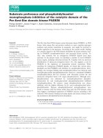
and radioactively labelled ATP. As shown in Fig. 1A,
distinct peptides were strongly phosphorylated by
PASKIN (for a list of the 75 most strongly phosphory-
lated peptides, see Table S1). The consensus phospho-
acceptor site of the 30 most strongly phosphorylated
peptides was found to be similar to protein kinase
(PK)A and C motifs (Fig. 1B). These data are sup-
ported by recent findings based on a combinatorial
peptide library, which demonstrated a strong prefer-
ence for arginine at position –3 [19]. Accordingly, from
the 75 strongest hits in our screening, 70 hits indeed
contain arginine three amino acids before the serine or
threonine phosphoacceptor site (Table S1). Several
proteins were identified more than once, either because
more than one phosphoacceptor site within the same
protein could be phosphorylated or because overlap-
ping peptides containing the same phosphoacceptor
site were present, or because the peptide was derived
from the same site but from distinct species. Seventeen
different pyruvate kinase-derived peptides, for exam-
ple, were identified in this way. One of the proteins
Targets and stimulation of PASKIN P. Schla
¨
fli et al.
1758 FEBS Journal 278 (2011) 1757–1768 ª 2011 The Authors Journal compilation ª 2011 FEBS
listed in Fig. 1B is glycogen synthase, which has previ-
ously been identified as a PASKIN kinase target [11].
Thus, glycogen synthase identification confirmed the
feasibility of our approach and was used as a reference
target protein for subsequent experiments.
To corroborate PASKIN-dependent phosphoryla-
tion of these rather short arrayed peptides, 11 of the
most strongly phosphorylated candidate PASKIN
kinase targets were synthesized as 20-mer peptides and
used for in vitro phosphorylation by recombinant
PASKIN (Table S2). As shown in Fig. 1C, six peptides
were significantly better phosphorylated by PASKIN
than the unrelated control peptide, and three of them
showed an even stronger phosphorylation than the
known PASKIN targets glycogen synthase and pan-
creatic duodenal homeobox-1 (i.e. 40S ribosomal
protein S6, phosphorylase kinase b and 6-phospho-
fructo-2-kinase ⁄ fructose 2,6-bisphosphatase).
Ribosomal protein S6 is phosphorylated by
PASKIN
Because a role for PASKIN in protein translation has
been reported previously [8,18], the finding that a
R
Consensus
AB
C
Fig. 1. Identification of novel PASKIN kinase targets. (A) Recombinant His
6
-PASKIN purified from SF9 insect cells was used for in vitro phos-
phorylation of a microarray of 1176 peptides in the presence of [c-
33
P]ATP. The magnified inset shows an example of the results obtained
after detection by phosphorimaging. (B) Peptide sequences of the 30 most phosphorylated targets and their similarities to PKA and PKC
consensus motifs. (C) Target validation. Biotinylated peptides of 20 amino acids in length were incubated together with recombinant PASKIN
in the presence of [c-
33
P]ATP, captured with streptavidin sepharose beads and quantified by liquid scintillation counting. The sequences were
normalized to a glycogen synthase-derived peptide, a known target for PASKIN. A PDX-1-derived peptide, another known PASKIN target,
served as second positive control. Mean ± SD values of three independent experiments are shown. Asterisks indicate statistically significant
differences compared to the unrelated negative control peptide derived from activating transcription factor ATF-4 (*P < 0.05; **P < 0.01;
paired t-test). Peptides were named: GYS, glycogen synthase; PDX1, pancreatic and duodenal homeobox 1; PIAS1, protein inhibitor of acti-
vated STAT 1; RAB11BP, RAB11-binding protein; FXN, frataxin; HERG, human ether-a-go-go related gene; CREB1, cAMP response element-
binding protein; NFATC4; nuclear factor of activated T-cells c4; 4E-T, eIF4E-transporter; S6, 40S ribosomal protein S6; PHKB, phosphorylase
kinase b; PFKFB, 6-phosphofructo-2-kinase ⁄ fructose 2,6-bisphosphatase. Note that some of the PASKIN target sequences, as shown in (B),
can be found in several distinct proteins, leading to the partially altered designations in (C), as outlined in Table S2.
P. Schla
¨
fli et al. Targets and stimulation of PASKIN
FEBS Journal 278 (2011) 1757–1768 ª 2011 The Authors Journal compilation ª 2011 FEBS 1759
S6-derived peptide was strongly phosphorylated by
PASKIN was further investigated. S6 is a target of the
mammalian target of rapamycin (mTOR) signalling
pathway that regulates nutrient-dependent protein
translation by p70 S6 kinase (p70S6K)-mediated phos-
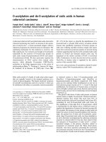
phorylation of S6 at Ser235 ⁄ 236 [20]. Therefore,
recombinant S6 was expressed and purified either as
wild-type, C-terminally truncated or Ser235 ⁄ 236Ala
double-mutant glutathione S-transferase (GST) fusion
protein (Fig . 2A). As shown in Fig. 2B, PASKIN
phosphorylated wild-type but not truncated or serine
double-mutant S6 in vitro, suggesting that PASKIN
also targets S6 at Ser235 ⁄ 236.
To analyze PASKIN-dependent phosphorylation of
endogenous S6 in vivo, we used mouse embryonic
fibroblasts (MEFs) derived from either Paskin
+ ⁄ +
wild-type or Paskin
) ⁄ )
knockout mice [14]. However,
as shown in Fig. 2C, no difference in constitutive
p70S6K or S6 phosphorylation could be detected in
these cells. Because basal S6 phosphorylation by
p70S6K might overcome subtle changes caused by
PASKIN, we next used MEFs deficient for both genes
encoding mouse p70S6K (S6K1
) ⁄ )
⁄ S6K2
) ⁄ )
) [21], and
transiently overexpressed full-length PASKIN or an
N-terminally truncated version preserving the kinase
domain in these cells. Whereas S6 total protein levels
remained unchanged, phosphorylated S6 was strongly
reduced in S6K1
) ⁄ )
⁄ S6K2
) ⁄ )
double-knockout MEFs
(Fig. 2D). Interestingly, overexpression of myc-tagged
PASKIN, or its kinase domain alone, led to increased
phosphorylation of S6 at Ser235 ⁄ 236 (Fig. 2D). In
summary, S6 is not only a new in vitro target, but
PASKIN can also phosphorylate S6 in vivo and might
even partially contribute to the residual S6 phosphory-
lation observed in p70S6K-deficient cells [22]. How-
ever, a more prominent S6 kinase function in vivo
probably awaits the identification of the endogenous
stimulus of PASKIN catalytic activity.
Autophosphorylation of recombinant PASKIN is
activated by phospholipids
A possible mechanism of PASKIN activation in vivo
might be the binding of a so far unknown ligand, as
suggested previously [7]. However, no endogenous
PASKIN ligand is known so far. By comparing the
activity of PASKIN with PKCd, we have obtained
first indication of a potential endogenous ligand. We
previously reported that both PASKIN and PKCd
phosphorylate eEF1A1 [18], and both kinases are
A
B
C
D
Input
Fig. 2. Ribosomal protein S6 is phosphory-
lated by PASKIN. (A) Sequence comparison
of the S6 peptides used in the microarray,
20-mer peptide used for the in vitro reac-
tions, and recombinant GST fusion proteins
purified from E. coli. (B) Phosphorylation
reactions in vitro using purified His
6
-PASKIN
and recombinant S6 in the presence of
[c-
33
P]ATP. Subsequent to SDS ⁄ PAGE, the
phosphorylated proteins were visualized by
phosphorimaging. Equal input was con-
trolled by immunoblotting against S6 and
the GST-tag. (C) Immunoblot analysis of the
phosphorylation status of p70S6K and S6 in
Paskin
+ ⁄ +
and Paskin
) ⁄ )
MEFs. (D) Immu-
noblot analysis of the phosphorylation status
of p70S6K and S6 in S6K1
) ⁄ )
⁄ S6K2
) ⁄ )
dou-
ble-knockout MEFs after overexpression of
a negative control (enhanced green fluores-
cent protein, EGFP), myc-PASKIN or myc-
KIN. Monoclonal antibodies against myc and
PASKIN were used to confirm PASKIN over-
expression.
Targets and stimulation of PASKIN P. Schla
¨
fli et al.
1760 FEBS Journal 278 (2011) 1757–1768 ª 2011 The Authors Journal compilation ª 2011 FEBS
known to autophosphorylate themselves [6,23].
Because PKCd kinase activity is known to be stimu-
lated by diacylglycerol (DAG) and phosphatidylserine
(PS) [24], we were interested in whether other similari-
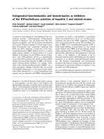
ties exist between PASKIN and PKC d. Notably, a
mixture of PS and DAG (used in the form of diocta-
noylglycerol, DOG) not only enhanced PKCd, but also
PASKIN autophosphorylation (Fig. 3A).
To systematically analyze the lipid activation of
PASKIN, all major phospholipids were compared for
their effects on PASKIN and PKCd autophosphoryla-
tion. As shown in Fig. 3B, all tested phospholipids,
but not DOG alone, increased PASKIN autophospho-
rylation. By contrast, PKCd autophosphorylation was
induced by DOG alone, and to some extent also by
PS or phosphatidylcholine (PC), although all other
A
B
CD
Fig. 3. Phospholipid stimulation of PASKIN autophosphorylation. (A) Lipid stimulation of PKCd and PASKIN autophosporylation as assessed
by incubating the purified recombinant proteins with DOG ⁄ PS mixtures and [c-
33
P]ATP. Subsequent to SDS ⁄ PAGE, the phosphorylated
proteins were visualized by phosphorimaging. (B, C) Stimulation of PASKIN and PKCd autophosphorylation by increasing amounts of the
indicated phospholipids. Subsequent to SDS ⁄ PAGE, the phosphorylated proteins were visualized (upper panels) and quantified (lower panels)
by phosphorimaging. The values were normalized to 100 lgÆmL
)1
PS and 10 lgÆmL
)1
DOG ⁄ 100 lgÆmL
)1
PS mixtures for PASKIN and PKCd,
respectively (filled columns). (D) PLD but not PLC converts PC from a low affinity to a high affinity PASKIN ligand. Ninety-six-well plates
were coated with increasing amounts of PC, followed by treatment with PLD or PLC, as indicated. Binding of 100 ng of PASKIN added to
each well was detected by ELISA. Mean ± SD values of a representative experiment performed in triplicate are shown.
P. Schla
¨
fli et al. Targets and stimulation of PASKIN
FEBS Journal 278 (2011) 1757–1768 ª 2011 The Authors Journal compilation ª 2011 FEBS 1761
phospholipids had only marginal effects on PKCd.As
shown previously [24], a mixture between DOG and
PS was required to maximally induce PKCd activity.
However, combining DOG with phospholipids did not
further induce PASKIN (data not shown).
The rather unselective stimulation of PASKIN activ-
ity by all tested phospholipids suggested that the core
phospholipid moiety might confer PASKIN binding.
Indeed, as shown in Fig. 3C, phosphatidic acid (PA)
alone was sufficient to stimulate PASKIN autophos-
phorylation. The finding that PA but not DOG
strongly bound PASKIN suggested that phospholipase
(PL)D might target PASKIN by converting phospho-
lipids into PA. To directly demonstrate this assump-
tion, 96-well plates were coated with constant amounts
(1 lg) of PC. After treatment with PLD or PLC,
increasing amounts of PASKIN were added and
detected by ELISA. As shown in Fig. 3D, PASKIN
bound with clearly higher affinity to PA than to PC.
However, lipid binding was restored when PC was
treated with PLD (generating PA) but not with PLC
(generating DAG).
Inositol phosphorylation determines the affinity
of phosphatidylinositol (PtdIns) interaction with
PASKIN
The experiments described above suggested that an iso-
lated phosphate group such as in PA is necessary for
maximal PASKIN–lipid interaction. Because PtdIns
with varying numbers of phosphate groups belong to
the most important cellular lipid signalling molecules,
we next investigated whether the number and location
of the phosphate groups on the inositol ring affect
their interaction with PASKIN. Therefore, dot blots
containing mono-, di- and tri-phosphorylated PtdIns
were incubated with recombinant PASKIN and
immunodetected using a monoclonal antibody derived
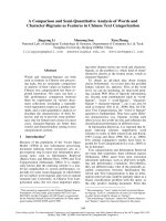
against PASKIN. Unexpectedly, although unphos-
phorylated PI showed only relatively low PASKIN
binding, this interaction was strongly increased by the
presence of a single phosphate group in PtdIns(4)P,
and reduced again when two or three phosphate
groups were present in PtdIns(4,5)P
2
and
PtdIns(3,4,5)P
3
, respectively (Fig. 4A). This finding
was corroborated by using dot blots with increasing
amounts of all possible PtdIns-phosphates: PASKIN
dose-dependently bound PtdIns-monophosphates bet-
ter than PtdIns-diphosphates, and nonphosphorylated
or tri-phosphorylated PtdIns bound PASKIN only
weakly (Fig. 4B, left). Similar results were obtained
with autophosphorylated PASKIN (Fig. 4B, right),
suggesting that PASKIN phosphorylation status does
not interfere with selective PtdIns-monophosphate
binding.
To localize the region responsible for PtdIns-mono-
phosphate binding, four different fragments of
PASKIN (Fig. 4C, left) were expressed and purified as
His
6
-tagged fusion proteins. However, only the kinase
domain of PASKIN bound PtdIns-monophosphates
(Fig. 4C, right), rather than the previously suggested
ligand-binding PAS domain (data not shown). We next
aimed to determine the effects of differently phosphor-
ylated PtdIns on PASKIN autophosphorylation. As
shown in Fig. 4D, autophosphorylation was dose-
dependently enhanced by all three PtdIns-monophos-
phates, whereas especially high concentrations of
PtdIns(4,5)P
2
and PtdIns(3,4,5)P
3
even inhibited auto-
phosphorylation, establishing a structure–function rela-
tionship between kinase domain–lipid interaction and
kinase activity.
PtdIns-monophosphate-dependent regulation of
PASKIN target phosphorylation
Because PtdIns-monophosphates stimulated PASKIN
autophosphorylation, we were interested in whether
they could also stimulate phosphorylation of the
PASKIN targets S6 and eEF1A1. Therefore, wild-type
and phosphoacceptor site mutant recombinant S6 and
eEF1A1 were used for PASKIN in vitro phosphoryla-
tion reactions in the presence of differently phosphory-
lated PtdIns-phosphates. As shown in Fig. 5, PASKIN
autophosphorylation was again stimulated by all three
PtdIns-monophosphates but inhibited by PtdIns(4,5)P
2
and PtdIns(3,4,5)P
3
. Although the phosphoacceptor
site mutant S6 and eEF1A1 GST fusion proteins
remained unphosphorylated, their wild-type counter-
parts were phosphorylated by PASKIN. Unexpectedly,
both S6 and eEF1A1 target phosphorylation was
inhibited by PtdIns-phosphates. The more phosphate
groups the inositol ring carries, the stronger the
PASKIN target protein phosphorylation was inhibited.
However, nonphosphorylated PtdIns did not signifi-
cantly change the target phosphorylation efficiency.
Discussion
In the present study, we identified various novel poten-
tial PASKIN substrates by peptide microarray phos-
phorylation, including glycogen synthase that was
known before to be phosphorylated by PASKIN [11].
Thus, the repetitive identification of this PASKIN
target confirms, at least partially, the validity of the
peptide array approach. Other peptides derived from
proteins involved in glycogen metabolism included
Targets and stimulation of PASKIN P. Schla
¨
fli et al.
1762 FEBS Journal 278 (2011) 1757–1768 ª 2011 The Authors Journal compilation ª 2011 FEBS
phosphorylase kinase, inhibitor of protein phosphatase
1 and yeast glycogen phosphorylase (Table S1). The
involvement of PASKIN in the regulation of glycogen
synthesis was demonstrated previously by showing that
both mammalian and yeast glycogen synthases, as well
as yeast UDP-glucose pyrophosphorylase, are known
phosphorylation targets of mammlian PASKIN and
yeast PSK1 and PSK2, respectively [8,11]. However,
although Ser640 was the main PASKIN kinase target
residue of mammalian glycogen synthase [11], the pep-
tides phosphorylated by PASKIN on the microarray
contained Ser3 and Ser7 but not Ser640. Of note, a
Ser640Ala mutation did not completely prevent phos-
phorylation [11]. Therefore, our data suggest that
PASKIN might phosphorylate Ser3 and ⁄ or Ser7 of
glycogen synthase in addition to Ser640.
Two peptides phosphorylated by PASKIN were
derived from enzymes involved in glycolysis: pyruvate
kinase and 6-phosphofructo-2-kinase ⁄ fructose 2,6-bis-
phosphatase 1 (Table S1). Obviously, the coordination
of glycolysis, gluconeogenesis and glycogen synthesis
appears to be physiologically meaningful, and hence it is
tempting to speculate that PASKIN is involved in the
regulation of all of these metabolic pathways. However,
pyruvate kinase could not be confirmed as a PASKIN
target using purified full-length pyruvate kinase GST
fusion proteins in in vitro assays (data not shown).
Interestingly, S6 was among the peptides phosphory-
lated by PASKIN and this phosphorylation could be
confirmed on the full-length protein level. Together
with the previously reported eEF1A1 phosphorylation
[18], this finding provides additional evidence that
PASKIN is involved in mammalian protein transla-
tion. The most important and best characterized S6
kinases are the mTOR-dependent p70 S6-kinases that
sequentially phosphorylate all five phosphorylatable
A
C
D
B
Fig. 4. Preferential PASKIN binding to (and
activation by) PtdIns-monophosphates.
(A) Recombinant His
6
-PASKIN protein was
allowed to bind to the indicated lipids
immobilized on a membrane, and subse-
quently detected using PASKIN antibodies.
(B) PASKIN dose-dependently bound
preferably PtdIns-monophosphates. PASKIN
was either detected by immunoblotting (left
panel) or by phosphorimaging after
autophosphorylation in the presence of
[c-
33
P]ATP (right panel). (C) Fragments of
PASKIN were expressed in E. coli and
purified as His
6
-tagged fusion proteins (left
panel). Subsequent to binding to the lipid
dot blots and detection using a His-tag anti-
body, only the kinase (KIN) domain of
PASKIN was found to interact with
PtdIns-monophosphates (right panel). (D)
His
6
-PASKIN autophosphorylation was
mainly stimulated by the presence of the
PtdIns-monophosphates. In vitro phosphory-
lation reactions in the presence of
[c-
33
P]ATP and the indicated synthetic diC8
PtdIns (3.16 l
M,10lM, 31.6 lM and
100 l
M) were separated by SDS ⁄ PAGE and
quantified by phosphorimaging. Values were
expressed relative to lipid-free control
reactions and are represented as the
mean ± SD of four independent
experiments (*P < 0.05; **P < 0.01;
***P < 0.001; t-test).
P. Schla
¨
fli et al. Targets and stimulation of PASKIN
FEBS Journal 278 (2011) 1757–1768 ª 2011 The Authors Journal compilation ª 2011 FEBS 1763
RRRRRR
Fig. 5. In vitro target phosphorylation of
His
6
-PASKIN is reduced in presence of
PtdIns. Recombinant His
6
-PASKIN purified
from Sf9 insect cells was used to in vitro
phosphorylate recombinant GST fusion
proteins with wild-type S6, with the
nonphosphorylatable double-mutant
S235 ⁄ 236A, with eEF1A1, or with its
nonphosphorylatable T432A mutant, in the
presence of [c-
33
P]ATP and PtdIns phos-
phates (100 l
M) as indicated. Subsequent to
separation by SDS ⁄ PAGE, protein phosphor-
ylation was viusalized (left panel, represen-
tative images) and quantified (right panel) by
phosphorimaging. His
6
-PASKIN autophos-
phorylation without lipid and target (first lane
from the left) was used for intra-assay nor-
malization of the values. Columns represent
the mean ± SD values of three independent
experiments (*P < 0.05; **P < 0.01;
***P < 0.001; t-test).
Targets and stimulation of PASKIN P. Schla
¨
fli et al.
1764 FEBS Journal 278 (2011) 1757–1768 ª 2011 The Authors Journal compilation ª 2011 FEBS
serines of S6, starting with S236 and S235 (i.e. the
same sites as shown in the present study for PASKIN)
followed by S240, S244 and S247 [25]. A second family
of S6 kinases are p90 ribosomal S6 kinases that phos-
phorylate S6 upon mitogenic stimulation at the same
sites as PASKIN [22]. Phosphorylation of S6 by
p70S6K has long been considered to increase protein
translation by selectively enhancing the translation of
5¢-terminal oligopyrimidine (TOP) mRNAs, a subset
of mRNAs containing an oligopyrimidine tract in their
5¢-UTRs. Of note, the 5¢-TOP mRNAs code for ribo-
somal proteins and translation factors, including
PASKIN targets S6 and eEF1A1 [26]. However, S6
phosphorylation and increased 5¢-TOP mRNA transla-
tion might be coincidental rather than causally related
[27] and, according to a newer hypothesis, might even
negatively influence translation if the phosphorylation
of S6 is considered as an inhibitory feedback signal
[28]. However, no significant difference in global [
35
S]-
Met incorporation could be observed in Paskin
) ⁄ )
MEFs (data not shown).
On the basis of the known functions of the
PASKIN-related FixL oxygen sensor in bacteria and
the PASKIN orthologues in yeast, and considering the
lack of any obvious phenotype in Paskin knockout
mice kept under normal housing conditions, it is tempt-
ing to assume that PASKIN has a ligand-mediated sen-
sor function that becomes apparent under currently ill-
defined stress situations [17]. However, only artificial
but no endogenous PASKIN ligands have been
reported to bind the PAS domain and lead to the de-
repression of the kinase domain-dependent autophos-
phorylation [7]. In the present study, we identified
phospholipids as the first biologically relevant PASKIN
ligands. Apparently, the presence of a charged phos-
phate moiety is required for stimulation of PASKIN
kinase activity, and PLD (but not PLC) can convert
phospholipids from low into high-affinity PASKIN
ligands. However, we currently do not know whether
PASKIN is a target of intracellular PLD cell signalling.
Unexpectedly, PtdIns-monophosphates were found
to be the best ligands of PASKIN, with clearly higher
affinities than PtdIns-diphosphates or PtdIns-triphos-
phate. PtdIns-binding domains have been reported to
display either well-defined 3D folds [29], or rather
unstructured regions with basic (for binding of the
phosphate groups) and hydrophobic residues, such as
in the noncanonical pleckstrin homology domain of
Tiam1 [30]. We identified a lysine rich region, spanning
from Lys1019 to Lys1034 of PASKIN, which shares
characteristic features with noncanonical pleckstrin
homology domains, including a double-lysine motif
(Lys1031 ⁄ 1032). However, mutation and deletion anal-
yses of this putative binding region did not affect lipid
binding by PASKIN (data not shown). Thus, it is diffi-
cult to predict the PtdIns-monophosphate binding site
within the PASKIN kinase domain and further work
will be necessary to identify the specific residues
involved in lipid binding.
Although PtdIns(4,5)P
2
and PtdIns(3,4,5)P
3
are
involved in signalling processes at the plasma mem-
brane, PtdIns-monophosphates are more abundant in
intracellular membrane structures such as the Golgi
apparatus and endosomes [31]. Within these structures,
PtdIns-monophosphates are involved in sorting and
signalling because the concentrations and localization
of differentially phosphorylated PtdIns can change
rapidly [29]. Therefore, it might be possible that
PtdIns-monophosphates not only regulate PASKIN
activity, but also its subcellular localization. This
hypothesis needs further investigation but is dependent
on the prior identification of the specific environmental
conditions that regulate PASKIN function.
As might be expected, we found a direct correlation
between ligand affinity and PASKIN autophosphoryla-
tion efficiency. However, the kinase domain rather
than the PAS domain was found to bind the PtdIns-
phosphates. This finding might explain why the activa-
tion of PASKIN-dependent S6 and eEF1A1 target
phosphorylation failed to comply with our initial
expectations: PASKIN autophosphorylation was not
directly related to target phosphorylation. However,
the results obtained in the present study are consistent
with a recent study reporting that PASKIN kinase
activity is independent of activation loop phosphoryla-
tion [19]. Thus, the original model of autophosphoryla-
tion-dependent kinase activity needs to be revised, and
the functional meaning of PASKIN autophosphoryla-
tion remains to be elucidated.
In conclusion, the in vitro data obtained in the
present study suggest the existence of downstream
effector functions of mammalian PASKIN similar to
those known from yeast: the coordination between
energy flux and translation. With the identification of
endogenous small molecule activators of PASKIN, we
have obtained the first indication of the upstream regu-
lators of PASKIN activity. It will be interesting to
examine how these regulators affect the downstream
processes mediated by PASKIN.
Experimental procedures
Plasmids
All cloning work was carried out using Gateway technology
(Invitrogen, Carlsbad, CA, USA). The human PASKIN
P. Schla
¨
fli et al. Targets and stimulation of PASKIN
FEBS Journal 278 (2011) 1757–1768 ª 2011 The Authors Journal compilation ª 2011 FEBS 1765
cDNA containing plasmids pENTR4-hPASK and pENTR4-
hKIN, as well as plasmids for recombinant expression of full
length His
6
-PASKIN, PASKIN truncations and eukaryotic
elongation factor 1A1, have been reported previously [18].
pENTR4-hPASK and pENTR4-hKIN were recombined
into pcDNA3.1 ⁄ c-myc-DEST [32] using LR recombinase
(Invitrogen) to generate pcDNA3.1 ⁄ c-myc-hPASK and
pcDNA3.1 ⁄ c-myc-hKIN for c-myc-tagged expression of
PASKIN or its kinase domain, respectively, in mammalian
cells. Human ribosomal protein S6 (IRAUp969B0849D6;
Deutsches Ressourcenzentrum fu
¨
r Genomforschung, Berlin,
Germany) was cloned into pENTR4 using primers 5¢-
TTATGTCGACATGAAGCTGAACAT-3¢ (forward) and
5¢-TACGTGCGGCCGCTTATTTCTGACTGGATTCAGA
CTTAG-3¢ (reverse), respectively, or 5¢-TACGTGGCGGC
CGCTTAAAGTCTGCGTCTCTTCGC-3¢ to introduce a
stop codon after residue L234. The PCR products were
ligated into the SalI and NotI restriction sites. The S6
S235 ⁄ 236A double mutant was produced with primers
5¢-GCGAAGAGACGCAGGCTAGCCGCTCTGCGAGC
TTCTAC-3¢ and 5¢-GTAGAAGCTCGCAGAGCGGCT
AGCCTGCGTCTCTTCGC-3¢ by Pfu polymerase-based
site-directed mutagenesis (Stratagene, La Jolla, CA, USA).
For expression as GST-tagged fusion proteins, pENTR4
based plasmids with the different S6 constructs were recom-
bined into pDEST15 using LR recombinase.
Purification of recombinant proteins
Recombinant proteins were purified as described previously
[18]. Briefly, full-length PASKIN was purified from Sf9 cells
using the Bac-to-Bac Baculovirus expression system (Invi-
trogen). GST-tagged fusion constructs and His
6
-tagged
PASKIN fragments were expressed in arabinose inducible
BL21 Escherichia coli. Recombinant proteins were purified
by FPLC (BioLogic DuoFlow; Bio-Rad, Hercules, CA,
USA) using HiTrap Chelating HP and GSTrap FF col-
umns (GE Healthcare, Milwaukee, WI, USA), respectively.
The kinase activity of purified recombinant PASKIN was
verified by autophosphorylation assays.
Kinase assays
His
6
-PASKIN or PKCd (Invitrogen) were incubated with
or without 2 lg of recombinant target proteins in kinase
buffer (25 mm Tris–HCl, pH 7.5, 10 mm MgCl
2
,1mm
dithiothreitol) for 20 min in the presence of 3 lCi
[c-
33
P]ATP (Hartmann Analytic, Brunswick, Germany).
Proteins were separated by SDS ⁄ PAGE and analyzed by
phosphorimaging of the dried gels (Molecular Imager FX;
Bio-Rad) using quantity one software (Bio-Rad). Lipids
(Sigma, St Louis, MO, USA or Fluka, Buchs, Switzerland)
were dissolved in CHCl
3
, aliquotted in test tubes and the
CHCl
3
evaporated under a stream of nitrogen. Lipids were
then resuspended in kinase assay master mixes by thorough
vortexing. PtdIns present in the phosphorylation reactions
were obtained from Echelon Biosciences (Salt Lake City,
UT, USA) as synthetic diC8-lipids and added to the reac-
tions from 1 mm aequous stock solutions to the final con-
centrations indicated.
Peptide microarrays
Peptide microarrays were phosphorylated with recombinant
PASKIN in accordance with the manufacturer’s instruc-
tions (Pepscan, Lelystad, The Netherlands). In brief, 50 lL
of a solution containing 500 ng recombinant PASKIN,
50 mm Hepes (pH 7.4), 20 mm MgCl
2
, 10% glycerol,
300 lCiÆmL
)1
[c-
33
P]ATP, 0.01% (v ⁄ v) Brij-35 and
0.01 mgÆmL
)1
BSA was added to the glass slide, covered
with a glass coverslip and incubated at 30 °C for 2 h in a
humidified incubator. After incubation, the coverslip was
removed with 1% Triton X-100 in NaCl ⁄ P
i
and the glass
slide was washed twice with 1% Triton X-100 in 2 m NaCl
and twice with water by over-head shaking, air-dried and
analyzed by phosphorimaging (Bio-Rad).
Phosphorylation of biotinylated peptides
PASKIN phosphorylation reactions were performed as
described above in the presence of N-terminally biotinylat-
ed 20-mer target peptides (JPT Peptide Technologies, Ber-
lin, Germany) at a final concentration of 200 lm. The
reactions were stopped by adding SDS to 0.5% final con-
centration and heating at 95 °C for 5 min. Streptavidin
sepharose beads (25 lL; GE Healthcare) and 500 lLof
100 mm Tris–HCl (pH 8.0) were added and incubated for
30 min at 4 °C. The beads were washed three times with
500 lL of a buffer containing 10 mm Tris–HCl (pH 8.0),
1mm EDTA, 400 mm NaCl, 0.1% Nonidet P-40 and once
with 500 lL of 100 mm Tris (pH 8.0). Phosphorylation of
the beads was quantified by liquid scintillation counting
(Packard Tri-Carb 2900TR; Perkin Elmer, Boston, MA,
USA).
Cell culture, transfections and immunoblotting
MEF cells were generated from Paskin
+ ⁄ +
and Pa-
skin
) ⁄ )
mice [14] at embryonic day 14. S6K1
) ⁄ )
⁄ S6K2
) ⁄ )
double-knockout MEFs were kindly provided by G. Tho-
mas and S. C. Kozma (Friedrich Miescher Institute for
Biomedical Research, Basel, Switzerland). MEF cells were
cultivated in DMEM (Sigma) supplemented with 10%
fetal bovine serum (Invitrogen) up to passage 12, suggest-
ing that they immortalized spontaneously. MEFs were
transiently transfected using Lipofectamine 2000 (Invitro-
gen) in accordance with the manufacturer’s instructions.
Thirty-six hours post-transfection, cells were harvested
and whole cell lysates were generated by heating the cells in
1% SDS for 5 min at 95 °C. After SDS ⁄ PAGE and
Targets and stimulation of PASKIN P. Schla
¨
fli et al.
1766 FEBS Journal 278 (2011) 1757–1768 ª 2011 The Authors Journal compilation ª 2011 FEBS
immunoblotting, the primary antibodies used were: human
PASKIN (Pierce, Rockford, IL, USA); mouse PASKIN,
phospho-S6 (S235 ⁄ 236), S6 kinase and phospho-S6 kinase
(T389) (Cell Signaling Technology, Beverly, MA, USA); S6
(Bethyl Laboratories, Montgomery, TX, USA); b-actin and
GST-tag (Sigma); and His-tag (Novagen, Madison, WI,
USA).
Lipid binding assays
Interactions between PASKIN and lipids were measured by
an ELISA-based assay, as described previously [33]. Briefly,
96-well plates (Sarstedt, Nu
¨
mbrecht, Germany) were coated
overnight with phospholipids dissolved in methanol, fol-
lowed by blocking with 3% BSA in NaCl ⁄ P
i
for 1 h. Puri-
fied His
6
-PASKIN (100 ng) was diluted in kinase buffer
and allowed to bind for 1 h at 30 °C. After three washing
steps (0.3% Tween-20 in NaCl ⁄ P
i
), bound PASKIN was
detected by anti-PASKIN mAb6, followed by secondary
goat anti-(mouse IgG) horseradish peroxidase-conjugated
sera (Pierce) using the 3,3 ¢,5,5¢-tetramethylbenzidine sub-
strate kit (Pierce). The peroxidase reaction was stopped by
adding H
2
SO
4
(final concentration of 1 m) and A
450
was
determined using a microplate reader (Digiscan; Asys
Hitech, Eugendorf, Austria). For PL experiments, phospho-
lipid-coated 96-well plates were treated with 0.2 units of
PLC or PLD (Sigma), diluted in reaction buffer (120 m m
CaCl
2
, 300 mm sodium acetate, pH 5.6) for 1 h at room
temperature.
Lipid binding arrays
Membranes spotted with phospholipids were obtained from
Echelon Biosciences (P-6002, P-6100) and used in accor-
dance with the manufacturer’s instructions. Generally, 1 lg
of protein diluted in 1% skimmed dry milk in NaCl ⁄ Tris
was allowed to bind to spotted phospholipids for 16–20 h
at 4 °C. Binding was detected using primary antibodies as
indicated and horseradish peroxidase-coupled secondary
antibodies for enhanced chemiluminescence detection
(Pierce).
Acknowledgements
The authors wish to thank J. Rutter, G. Thomas and
S. C. Kozma for the generous gifts of plasmids and
cell lines, as well as Gieri Camenisch and Daniel
P. Stiehl for helpful discussions. This work was
supported by by grants from the Wolfermann-Na
¨
geli
Stiftung, Stiftung fu
¨
r wissenschaftliche Forschung an
der Universita
¨
tZu
¨
rich ⁄ Baugarten Stiftung, the Univer-
sity Research Priority Program ‘Integrative Human
Physiology’ and the Swiss National Science Founda-
tion (Grant SNF 31003A_129962 ⁄ 1).
References
1 Ponting CP & Aravind L (1997) PAS: a multifunctional
domain family comes to light. Curr Biol 7, R674–R677.
2 Lowrey PL & Takahashi JS (2000) Genetics of the
mammalian circadian system: photic entrainment,
circadian pacemaker mechanisms, and posttranslational
regulation. Annu Rev Genet 34, 533–562.
3 Mimura J & Fujii-Kuriyama Y (2003) Functional role
of AhR in the expression of toxic effects by TCDD.
Biochim Biophys Acta 1619, 263–268.
4 Wenger RH (2002) Cellular adaptation to hypoxia:
O
2
-sensing protein hydroxylases, hypoxia-inducible
transcription factors, and O
2
-regulated gene expression.
FASEB J 16, 1151–1162.
5 Hofer T, Spielmann P, Stengel P, Stier B, Katschinski
DM, Desbaillets I, Gassmann M & Wenger RH (2001)
Mammalian PASKIN, a PAS-serine ⁄ threonine kinase
related to bacterial oxygen sensors. Biochem Biophys
Res Commun 288, 757–764.
6 Rutter J, Michnoff CH, Harper SM, Gardner KH &
McKnight SL (2001) PAS kinase: an evolutionarily con-
served PAS domain-regulated serine ⁄ threonine kinase.
Proc Natl Acad Sci USA 98, 8991–8996.
7 Amezcua CA, Harper SM, Rutter J & Gardner KH
(2002) Structure and interactions of PAS kinase
N-terminal PAS domain: model for intramolecular
kinase regulation. Structure 10, 1349–1361.
8 Rutter J, Probst BL & McKnight SL (2002) Coordinate
regulation of sugar flux and translation by PAS kinase.
Cell 111, 17–28.
9 Smith TL & Rutter J (2007) Regulation of glucose
partitioning by PAS kinase and Ugp1 phosphorylation.
Mol Cell 26, 491–499.
10 Grose JH, Smith TL, Sabic H & Rutter J (2007) Yeast
PAS kinase coordinates glucose partitioning in response
to metabolic and cell integrity signaling. EMBO J 26,
4824–4830.
11 Wilson WA, Skurat AV, Probst B, de Paoli-Roach A,
Roach PJ & Rutter J (2005) Control of mammalian gly-
cogen synthase by PAS kinase. Proc Natl Acad Sci
USA 102, 16596–16601.
12 da Silva Xavier G, Rutter J & Rutter GA (2004)
Involvement of Per-Arnt-Sim (PAS) kinase in the
stimulation of preproinsulin and pancreatic duodenum
homeobox 1 gene expression by glucose. Proc Natl
Acad Sci USA 101, 8319–8324.
13 An R, da Silva Xavier G, Hao HX, Semplici F, Rutter J
& Rutter GA (2006) Regulation by Per-Arnt-Sim (PAS)
kinase of pancreatic duodenal homeobox-1 nuclear
import in pancreatic beta-cells. Biochem Soc Trans 34,
791–793.
14 Katschinski DM, Marti HH, Wagner KF, Shibata J,
Eckhardt K, Martin F, Depping R, Paasch U, Gass-
mann M, Ledermann B et al. (2003) Targeted disrup-
P. Schla
¨
fli et al. Targets and stimulation of PASKIN
FEBS Journal 278 (2011) 1757–1768 ª 2011 The Authors Journal compilation ª 2011 FEBS 1767
tion of the mouse PAS domain serine ⁄ threonine kinase
PASKIN. Mol Cell Biol 23, 6780–6789.
15 Borter E, Niessen M, Zuellig R, Spinas GA, Spielmann
P, Camenisch G & Wenger RH (2007) Glucose-stimu-
lated insulin production in mice deficient for the PAS
kinase PASKIN. Diabetes 56, 113–117.
16 Hao HX, Cardon CM, Swiatek W, Cooksey RC,
Smith TL, Wilde J, Boudina S, Abel ED, McClain DA
& Rutter J (2007) PAS kinase is required for normal
cellular energy balance. Proc Natl Acad Sci USA 104,
15466–15471.
17 Schla
¨
fli P, Borter E, Spielmann P & Wenger RH (2009)
The PAS-domain kinase PASKIN: a new sensor in
energy homeostasis. Cell Mol Life Sci 66, 876–883.
18 Eckhardt K, Tro
¨
ger J, Reissmann J, Katschinski DM,
Wagner KF, Stengel P, Paasch U, Hunziker P, Borter
E, Barth S et al. (2007) Male germ cell expression of
the PAS domain kinase PASKIN and its novel target
eukaryotic translation elongation factor eEF1A1. Cell
Physiol Biochem 20, 227–240.
19 Kikani CK, Antonysamy SA, Bonanno JB, Romero R,
Zhang FF, Russell M, Gheyi T, Iizuka M, Emtage S,
Sauder JM et al. (2010) Structural bases of PAS
domain-regulated kinase (PASK) activation in the
absence of activation loop phosphorylation. J Biol
Chem 285, 41034–41043.
20 Ferrari S, Bandi HR, Hofsteenge J, Bussian BM &
Thomas G (1991) Mitogen-activated 70K S6 kinase.
Identification of in vitro 40 S ribosomal S6 phosphory-
lation sites. J Biol Chem 266, 22770–22775.
21 Pende M, Um SH, Mieulet V, Sticker M, Goss VL,
Mestan J, Mueller M, Fumagalli S, Kozma SC &
Thomas G (2004) S6K1() ⁄ )) ⁄ S6K2() ⁄ )) mice exhibit
perinatal lethality and rapamycin-sensitive 5¢-terminal
oligopyrimidine mRNA translation and reveal a
mitogen-activated protein kinase-dependent S6 kinase
pathway. Mol Cell Biol 24, 3112–3124.
22 Roux PP, Shahbazian D, Vu H, Holz MK, Cohen MS,
Taunton J, Sonenberg N & Blenis J (2007) RAS ⁄ ERK
signaling promotes site-specific ribosomal protein S6
phosphorylation via RSK and stimulates cap-dependent
translation. J Biol Chem 282, 14056–14064.
23 Kielbassa K, Mu
¨
ller HJ, Meyer HE, Marks F &
Gschwendt M (1995) Protein kinase Cd-specific
phosphorylation of the elongation factor eEF-a and an
eEF-1a peptide at threonine 431. J Biol Chem 270,
6156–6162.
24 Bell RM & Burns DJ (1991) Lipid activation of protein
kinase C. J Biol Chem
266, 4661–4664.
25 Krieg J, Hofsteenge J & Thomas G (1988) Identification
of the 40 S ribosomal protein S6 phosphorylation sites
induced by cycloheximide. J Biol Chem 263, 11473–
11477.
26 Meyuhas O (2000) Synthesis of the translational
apparatus is regulated at the translational level. Eur J
Biochem 267, 6321–6330.
27 Ruvinsky I, Sharon N, Lerer T, Cohen H, Stolovich-
Rain M, Nir T, Dor Y, Zisman P & Meyuhas O (2005)
Ribosomal protein S6 phosphorylation is a determinant
of cell size and glucose homeostasis. Genes Dev 19,
2199–2211.
28 Meyuhas O (2008) Physiological roles of ribosomal pro-
tein S6: one of its kind. Int Rev Cell Mol Biol 268, 1–37.
29 Lemmon MA (2008) Membrane recognition by
phospholipid-binding domains. Nat Rev Mol Cell Biol
9, 99–111.
30 Ceccarelli DF, Blasutig IM, Goudreault M, Li Z,
Ruston J, Pawson T & Sicheri F (2007) Non-canonical
interaction of phosphoinositides with pleckstrin homol-
ogy domains of Tiam1 and ArhGAP9. J Biol Chem
282, 13864–13874.
31 Di Paolo G & De Camilli P (2006) Phosphoinositides in
cell regulation and membrane dynamics. Nature 443,
651–657.
32 Barth S, Nesper J, Hasgall PA, Wirthner R, Nytko KJ,
Edlich F, Katschinski DM, Stiehl DP, Wenger RH &
Camenisch G (2007) The peptidyl prolyl cis ⁄ trans isom-
erase FKBP38 determines hypoxia-inducible transcrip-
tion factor prolyl-4-hydroxylase PHD2 protein stability.
Mol Cell Biol 27, 3758–3768.
33 Ghosh S, Strum JC, Sciorra VA, Daniel L & Bell RM
(1996) Raf-1 kinase possesses distinct binding domains
for phosphatidylserine and phosphatidic acid. Phospha-
tidic acid regulates the translocation of Raf-1 in 12-O-tet-
radecanoylphorbol-13-acetate-stimulated Madin–Darby
canine kidney cells. J Biol Chem 271, 8472–8480.
Supporting information
The following supplementary material is available:
Table S1. Rank order of the 75 most strongly phos-
phorylated PASKIN kinase targets on the peptide
microarray.
Table S2. Sequences of the eleven biotinylated peptides
tested for phosphorylation by recombinant PASKIN.
This supplementary material can be found in the
online version of this article.
Please note: As a service to our authors and readers,
this journal provides supporting information supplied
by the authors. Such materials are peer-reviewed and
may be re-organized for online delivery, but are not
copy-edited or typeset. Technical support issues arising
from supporting information (other than missing files)
should be addressed to the authors.
Targets and stimulation of PASKIN P. Schla
¨
fli et al.
1768 FEBS Journal 278 (2011) 1757–1768 ª 2011 The Authors Journal compilation ª 2011 FEBS