Báo cáo khoa học: Increased expression of c-Fos by extracellular signal-regulated kinase activation under sustained oxidative stress elicits BimEL upregulation and hepatocyte apoptosis pot
Bạn đang xem bản rút gọn của tài liệu. Xem và tải ngay bản đầy đủ của tài liệu tại đây (368.35 KB, 9 trang )
Increased expression of c-Fos by extracellular
signal-regulated kinase activation under sustained
oxidative stress elicits BimEL upregulation and hepatocyte
apoptosis
Yasuhiro Ishihara
1
, Fumiaki Ito
2
and Norio Shimamoto
1
1 Laboratory of Pharmacology, Faculty of Pharmaceutical Sciences at Kagawa, Tokushima Bunri University, Japan
2 Department of Biochemistry, Faculty of Pharmaceutical Sciences, Setsunan University, Osaka, Japan
Introduction
Apoptosis has several morphological features, includ-
ing cell shrinkage, nuclear condensation, and nucleoso-
mal DNA fragmentation. Extensive studies to uncover
the mechanisms underlying the induction of apoptosis
have yielded the generally accepted theory that mito-
chondria play a fundamental role in the process. Apop-
totic stimuli activate the mitochondrial permeability
transition pore and the release of apoptosis-promoting
molecules such as cytochrome c, apoptosis-inducing
factor, and endonuclease G [1]. The pathways
upstream of the mitochondria for apoptotic signal
transduction have recently been identified. Several
Keywords
apoptosis; Bim; c-Fos; extracellular signal-
regulated kinase (ERK); reactive oxygen
species
Correspondence
N. Shimamoto, Laboratory of Pharmacology,
Faculty of Pharmaceutical Sciences at
Kagawa, Tokushima Bunri University,
1314-1, Shido, Sanuki, Kagawa 769-2193,
Japan
Fax: +81 87 894 0181
Tel: +81 87 894 5111 ext. 6513
E-mail:
(Received 22 December 2010, revised 25
February 2011, accepted 22 March 2011)
doi:10.1111/j.1742-4658.2011.08105.x
We previously reported that the inhibition of catalase and glutathione per-
oxidase activities by treatment with 3-amino-1,2,4-triazole (ATZ) and mer-
captosuccinic acid evoked sustained increases in the levels of reactive
oxygen species and apoptosis in rat primary hepatocytes. Apoptosis was
accompanied by increased expression of BimEL, following activation of
extracellular signal-regulated kinase. The aim of this study was to charac-
terize the mechanism underlying hepatocyte apoptosis by identifying the
transcription factor that induces BimEL expression. The bim promoter
region was cloned into a promoterless-luc vector, and promoter activity
was monitored by a luciferase assay. The luciferase activity increased in the
presence of ATZ + mercaptosuccinic acid. Pretreatment with a MEK
inhibitor, U0126, or an antioxidant, vitamin C, suppressed the promoter
activity. Furthermore, ATZ + mercaptosuccinic acid-induced luciferase
activity was attenuated by mutation of the activator protein-1 binding site
in the bim promoter region. The amounts of total and phosphorylated
c-Fos increased over time in the presence of ATZ + mercaptosuccinic acid,
whereas the amounts of total and phosphorylated c-Jun remained
unchanged. Chromatin immunoprecipitation revealed that both c-Fos and
c-Jun localized to the activator protein-1-binding site in the bim promoter
region. BimEL expression and hepatocyte apoptosis were suppressed by
knockdown of c-Fos and c-Jun, respectively. These results indicate that
increases in c-Fos following extracellular signal-regulated kinase activation
are critical for BimEL upregulation and apoptosis.
Abbreviations
AP-1, activator protein-1; ATZ, 3-amino-1,2,4-triazole; ChIP, chromatin immunoprecipitation; ERK, extracellular signal-regulated kinase;
GAPDH, glyceraldehyde-3-phosphate dehydrogenase; ROS, reactive oxygen species; SE, standard error; siRNA, small interfering RNA.
FEBS Journal 278 (2011) 1873–1881 ª 2011 The Authors Journal compilation ª 2011 FEBS 1873
molecules that are known to be involved in prolifera-
tion and ⁄ or differentiation have been reported to
induce apoptosis [2,3].
Extracellular signal-regulated kinase (ERK) is a
classic mitogen-activated protein kinase that is acti-
vated by growth factors and induces cell cycle pro-
gression via cyclin transcription. However, increasing
evidence shows that ERK is activated by reactive
oxygen species (ROS), and that this is followed by
the induction of apoptosis [4–6]. ERK-dependent
apoptosis induced by ROS has been recognized in
several pathological conditions, such as alcoholic liver
injury [7,8], lung hyperoxia [9], and cisplatin-induced
renal toxicity [10]. However, little is known about the
mechanism responsible for apoptotic signaling elicited
by active ERK, and this process therefore needs to
be investigated.
The mechanism responsible for ERK activation by
ROS is well understood. The phosphorylation of ERK
or its upstream kinases is regulated by phosphatases
such as PTP1B [11], MKP3 [12], and LMW-PTP [13].
The cysteines in the active sites of these phosphatases
are easily inactivated by ROS, resulting in activation
of the ERK pathway [14]. However, factors that act
on the mitochondria downstream of ERK have been
rarely reported. Recently, we showed that ROS-acti-
vated ERK increased the transcriptional expression
of BimEL, a major isoform among the bim gene
products, leading to apoptosis in rat primary hepato-
cytes [15].
Bim is a member of the Bcl-2 family of proteins,
which play a fundamental role in the induction of mito-
chondria-driven apoptosis. Under normal conditions,
antiapoptotic Bcl-2 family members such as Bcl-2,
Bcl-xL and Mcl-1 interact with the proapoptotic Bcl-2
family members Bax ⁄ Bak, to inhibit the ability of
Bax ⁄ Bak to permeabilize the mitochondrial membrane.
Bim activates the mitochondrial permeability transition
mediated by Bax ⁄ Bak through two different mecha-
nisms [16]: (a) Bim binds to antiapoptotic Bcl-2 family
proteins to liberate Bax ⁄ Bak, leading to mitochondrial
permeability transition; and (ii) Bim directly activates
Bax ⁄ Bak (induces a conformational change), thus lead-
ing to pore formation.
The bim gene is a direct target of transcription
factors such as FOXO3A, Myb and c-Jun [17–21]. The
5¢-end of the bim gene contains binding sites for
FOXO, Myb, and activator protein-1 (AP-1) [18].
However, the mechanism underlying the transcrip-
tional activation of BimEL downstream of ERK acti-
vation is not known. The aim of this study was to
identify the ERK-responsive transcription factor that
regulates BimEL expression.
Results
We previously showed that treatment with 3-amino-
1,2,4-triazole (ATZ) and mercaptosuccinic acid
inhibited catalase and glutathione peroxidase, which
are antioxidative enzymes that eliminate hydrogen per-
oxide, and caused sustained increases in ROS levels
and apoptosis in rat primary hepatocytes [22,23]. In
addition, we recently reported that ROS-activated
ERK induces BimEL transactivation, followed by
hepatocyte apoptosis [15]. This study was designed to
examine the mechanism of hepatocyte apoptosis, with
a particular focus on identifying the transcription fac-
tor(s) that activate BimEL transcription downstream
of the ERK pathway.
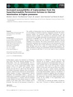
We cloned a 2.9-kb fragment of the rat bim pro-
moter region from rat primary hepatocytes. The bim
promoter region included an AP-1-binding site, a
FOXO-binding site, and three Myb-binding sites
(Fig. 1A). The bim promoter region was subcloned
into pGL4.24 (pGL4.24-BimProm). pGL4.24-BimProm
mutations were generated at each transcription factor-
binding site (mutated points are indicated in Fig. 1A),
and bim promoter activity in the presence of
ATZ + mercaptosuccinic acid was assessed with a
luciferase reporter assay. The mutations at the binding
sites used in this study reportedly attenuate the activity
of each transcription factor [19,24,25]. When rat pri-
mary hepatocytes were transfected with pGL4.24-Bim-
Prom and treated with ATZ + mercaptosuccinic acid
for 9 h, the luciferase activity increased 3.3 ± 0.3-fold
in comparison with untreated cells (Fig. 1B). However,
pretreatment with U0126, a potent inhibitor of MEK1,
or vitamin C, an antioxidant, largely suppressed
ATZ + mercaptosuccinic acid-induced luciferase activ-
ity (Fig. 1B). In addition, when rat primary hepato-
cytes were transfected with a mutated AP-1 (AP-1m)
promoter construct, the ATZ + mercaptosuccinic
acid-mediated increase in luciferase activity was greatly
attenuated (Fig. 1B). Transfection with a promoter
construct containing myb1m had no effect on the lucif-
erase activity, whereas transfection with myb2m,
myb3m or FOXOm promoters partially suppressed
ATZ + mercaptosuccinic acid-induced luciferase activ-
ity (Fig. 1B). These results suggest that AP-1 is
involved in increasing BimEL expression downstream
of ERK activation in response to treatment with
ATZ + mercaptosuccinic acid.
The AP-1 transcription factor consists of Fos and
Jun proteins [26]. Fos and Jun form a dimer, which in
turn binds to AP-1 regulatory elements and enhancer
regions of numerous mammalian genes. Jun forms
homodimers and heterodimers with Fos proteins,
Regulation of BimEL expression by c-Fos Y. Ishihara et al.
1874 FEBS Journal 278 (2011) 1873–1881 ª 2011 The Authors Journal compilation ª 2011 FEBS
whereas Fos proteins do not form homodimers, and
require heterodimerization to bind DNA [27,28].
Active ERK phosphorylates one of the major Fos fam-
ily proteins, c-Fos, and stabilizes it [29]; ERK also
phosphorylates c-Jun directly, leading to transactiva-
tion of AP-1. On the basis of these findings, we next
examined the expression and phosphorylation of c-Fos
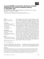
and c-Jun. The total amount of nuclear c-Fos
increased over time in the presence of ATZ + merca-
ptosuccinic acid (Fig. 2). Interestingly, phosphorylation
of c-Fos at Ser374 occurred in parallel with increases
in nuclear c-Fos levels (Fig. 2). Pretreatment with
U0126 or vitamin C largely suppressed the accumula-
tion of total and phosphorylated c-Fos in the presence
of ATZ + mercaptosuccinic acid (Fig. 2). In contrast,
there were no changes in the levels of total and phos-
phorylated nuclear c-Jun throughout the 9-h exposure
to ATZ + mercaptosuccinic acid (Fig. 2).
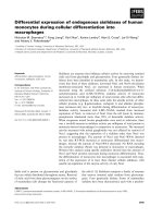
To show that AP-1 proteins directly bind to the con-
sensus AP-1 site in the bim promoter region (from
)2491 to )2497), a chromatin immunoprecipitation
(ChIP) assay was performed. A PCR analysis demon-
strated that c-Fos and c-Jun antibodies apparently pre-
cipitated the bim promoter region from rat primary
hepatocytes treated with ATZ + mercaptosuccinic
acid, whereas untreated hepatocytes and those pretreat-
ed with U0126 or vitamin C showed only slight DNA
binding (Fig. 3). Pretreatment with SP600125, an
inhibitor of c-Jun N-terminal kinase, showed no effect
on the DNA binding of c-Fos and c-Jun induced by
treatment with ATZ + mercaptosuccinic acid, indicat-
ing that JNK is not involved in the binding of AP-1 to
the bim promoter region. Nonspecific IgG also did not
exhibit DNA-binding activity (Fig. 3). These results
indicate that the AP-1 proteins bind specifically to the
AP-1 cis-regulatory region of the bim promoter in
hepatocytes treated with ATZ + mercaptosuccinic acid.
Next, we examined the effect of c-Fos and c-Jun on
BimEL transactivation and apoptosis. Transfection
with small interfering RNAs (siRNAs) targeted to
c-Fos and c-Jun clearly reduced the target protein lev-
els (Fig. 4A). Elevation of BimEL mRNA expression
by treatment with ATZ + mercaptosuccinic acid was
suppressed by transfection with siRNAs against c-Fos
and c-Jun (Fig. 4B). Increases in BimEL levels caused
by ATZ + mercaptosuccinic acid were also attenuated
by c-Fos or c-Jun knockdown (Fig. 4C). AT Z+ mer-
captosuccinic acid-induced cell death, chromatin
**
##
##
##
#
#
#
Fig. 1. AP-1-dependent Bim transcriptional
activation is induced by treatment with
ATZ + mercaptosuccinic acid. (A) A sche-
matic diagram of the rat bim promoter
(BimProm). The positions of the binding
sites for AP-1, Myb and FOXO are shown.
The mutation sequences of each transcrip-
tion factor-binding site are also presented.
(B) After cotransfection with pGL4.24-
BimProm or mutant pGL4.24-BimProm with
pRL-RSV into rat primary hepatocytes, cells
were cultured for 14 h. Cells were treated
with U0126 (40 l
M) or vitamin C (1 mM),
and then incubated for 9 h in the presence
or absence of ATZ (20 m
M) and merca-
ptosuccinic acid (7 m
M). Cell were collected
and lysed, and both firefly and Renilla
luciferase activities were measured. Values
for untreated cells carrying pGL4.24-
BimProm and pRL-RSV were set equal to 1.
The values are the means ± SE of six
separate experiments. Data were analyzed
with Student’s t-test or Dunnett’s test.
**P < 0.01 versus the untreated BimProm
group.
#
P < 0.05 and
##
P < 0.01 versus the
ATZ + mercaptosuccinic acid-treated
BimProm group.
Y. Ishihara et al. Regulation of BimEL expression by c-Fos
FEBS Journal 278 (2011) 1873–1881 ª 2011 The Authors Journal compilation ª 2011 FEBS 1875
condensation and DNA fragmentation were all abro-
gated by knockdown of c-Fos and c-Jun (Fig. 5A–C).
Transfection of scrambled siRNAs showed no effects
on the expression levels of c-Fos, c-Jun, or BimEL,
and did not affect hepatocyte apoptosis (Figs 4 and 5).
These results indicate that c-Fos and c-Hun are crucial
for BimEL expression and induction of hepatocyte
apoptosis.
Discussion
The bim promoter activity induced by treatment with
ATZ + mercaptosuccinic acid was largely attenuated
by mutating the AP-1-binding site in the bim promoter
region. Whereas the amounts of total and phosphory-
lated c-Fos increased in the presence of ATZ + mer-
captosuccinic acid, there was no change in the levels of
total and phosphorylated c-Jun throughout the experi-
mental period. Both c-Fos and c-Jun interacted with
the AP-1-binding site in the bim promoter region.
Knockdown of c-Fos or c-Jun suppressed not only
BimEL transactivation, but also hepatocyte apoptosis.
Pretreatment with U0126 or vitamin C largely abol-
ished ATZ + mercaptosuccinic acid-induced luciferase
activity, confirming that the ERK pathway elicited by
ROS is involved in Bim transcription in this experi-
mental system [15,23]. In addition, mutation of the
AP-1-binding site in the bim promoter region markedly
suppressed the luciferase activity induced by
ATZ + mercaptosuccinic acid, suggesting that AP-1 is
responsible for Bim transcription. Biswas et al.
reported that Bim expression was coregulated by three
transcription factors – c-Jun, FOXO, and Myb – when
PC12 cells were stimulated by nerve growth factor
deprivation, and insisted that the bim promoter acts as
a coincidence detector [18]. Interestingly, mutation of
the Myb-binding and FOXO-binding sites also slightly,
but significantly, reduced the luciferase activity in this
study. Therefore, the involvement of FOXO and Myb
in hepatocyte apoptosis should be examined further.
c-Fos is one of the main components of the AP-1
transcription factor complex [30]. Activated ERK
phosphorylates c-Fos at Ser-374, leading to its stabil-
ization [29,31]. Therefore, we examined the expression
and phosphorylation of c-Fos in this study. The total
and phosphorylated c-Fos levels increased over time in
the presence of ATZ + mercaptosuccinic acid, and
this increase was suppressed by pretreatment with
U0126. Therefore, c-Fos is stabilized by phosphoryla-
tion, which is mediated by ERK, allowing c-Fos to
accumulate. In contrast, c-Jun, another major compo-
nent of the AP-1 complex, is reportedly phosphory-
lated at Ser63 and Ser73 by active ERK, and this is
followed by increased c-Jun transcriptional activity
[32,33]. However, the total and phosphorylated c-Jun
levels in nuclei remained unaffected in the presence of
ATZ + mercaptosuccinic acid. Because c-Fos alone
cannot bind to DNA, c-Jun is required for transcrip-
tional activation [27,28]. Thus, BimEL expression is
dependent on both increased levels of c-Fos and basal
levels of c-Jun. This idea is supported by the results of
the ChIP assay, which indicated that both c-Fos and
Fig. 2. Increases in the expression of total and phosphorylated
c-Fos by treatment with ATZ + mercaptosuccinic acid. Primary rat
hepatocytes were treated with U0126 (40 l
M) or vitamin C (1 mM),
and then incubated for 9 h in the presence or absence of ATZ
(20 m
M) and mercaptosuccinic acid (7 mM). Nuclear proteins were
extracted, and the time courses of c-Fos, c-Fos phosphorylated at
Ser374, c-Jun, c-Jun phosphorylated at Ser63, c-Jun phosphory-
lated at Ser73 and histone H1 were evaluated by immunoblotting.
The results are representative of four independent experiments.
Fig. 3. Binding of c-Fos and c-Jun to the AP-1 site of the bim pro-
moter. Rat primary hepatocytes were treated with U0126 (40 l
M),
vitamin C (1 m
M), or SP600125 (40 lM), and then incubated for 9 h
in the presence or absence of ATZ (20 m
M) and mercaptosuccinic
acid (7 m
M). ChIP was used to assess the binding of c-Fos and
c-Jun to the AP-1-binding sites within the rat bim promoter. Rat
genomic DNA was used as a positive control, and immunoprecipita-
tion with a nonspecific antibody (IgG) was used as a negative
control. The results are representative of three independent experi-
ments.
Regulation of BimEL expression by c-Fos Y. Ishihara et al.
1876 FEBS Journal 278 (2011) 1873–1881 ª 2011 The Authors Journal compilation ª 2011 FEBS
A
B
C
(a)
(a)
(b)
(b)
(c)
Fig. 4. Suppression of BimEL expression by knockdown of c-Fos or c-Jun. After transfection of c-Fos or c-Jun siRNA or their scrambled siR-
NAs (Scr siRNA) into hepatocytes, cells were incubated for 14 h, and then further incubated in the presence or absence of ATZ (20 m
M) and
mercaptosuccinic acid (7 m
M) for 9 h. (A) The levels of c-Fos and c-Jun protein were determined by a western blot analysis (Aa), and bands
were then quantified and expressed as the fold change from the density of untreated hepatocytes as determined by densitometry (Ab,c).
The values are the means ± SE of five separate experiments. The data were analyzed with Dunnett’s test. **P < 0.01 versus the
ATZ + mercaptosuccinic acid-treated group. (B) The levels of BimEL mRNA were measured by real-time PCR. BimEL mRNA levels were nor-
malized using GAPDH mRNA. Values for untreated cells were set equal to 1. The values are the means ± SE of five separate experiments.
The data were analyzed with Dunnett’s test. **P < 0.01 versus the ATZ + mercaptosuccinic acid-treated group. (C) The expression of BimEL
proteins was evaluated by a western blot analysis (Ca). The bands were quantified and expressed as the fold change in their density as
compared with untreated hepatocytes (Cb). The values are the means ± SE of five separate experiments. The data were analyzed with
Dunnett’s test. **P < 0.01 versus the ATZ + mercaptosuccinic acid-treated group.
A
B
C
Fig. 5. Suppression of hepatocyte apoptosis by knockdown of c-Fos or c-Jun. After transfection of c-Fos or c-Jun siRNA into hepatocytes,
cells were incubated for 14 h, and then further incubated in the presence or absence of ATZ (20 m
M) and mercaptosuccinic acid (7 mM) for
24 h. Cell viability (A) and chromatin condensation (B) were assayed. The values are the means ± SE of five separate experiments. The data
were analyzed with Dunnett’s test. **P < 0.01 versus the ATZ + mercaptosuccinic acid-treated group. (C) Cellular DNA was extracted and
electrophoresed after a 24-h incubation. The results are representative of four independent experiments.
Y. Ishihara et al. Regulation of BimEL expression by c-Fos
FEBS Journal 278 (2011) 1873–1881 ª 2011 The Authors Journal compilation ª 2011 FEBS 1877
c-Jun localize to the AP-1-binding site in the bim pro-
moter region. Furthermore, knockdown of c-Fos or
c-Jun attenuated BimEL transactivation and apoptosis,
supporting the hypothesis that c-Fos and c-Jun act
coordinately to increase the expression of BimEL.
Increased c-Fos levels are therefore critical for BimEL
expression and apoptosis in this experimental system.
Active ERK is known to phosphorylate BimEL,
resulting in the ubiquitination and degradation of
BimEL [34,35]. Therefore, ERK activation was expected
to reduce the level of BimEL, leading to increased cell
survival as long as the proteasome maintains its normal
functions. We previously reported that BimEL degrada-
tion was suppressed in this experimental system, because
ROS generated by treatment with ATZ + merca-
ptosuccinic acid inhibited the activities of the protea-
some [15]. Namely, BimEL was upregulated by both
increased expression and decreased degradation in this
type of hepatocyte apoptosis. c-Fos was also reported to
be degraded by the ubiquitin–proteasome system [36].
In this study, pretreatment with U0126 did not com-
pletely abrogate the c-Fos expression induced by treat-
ment with ATZ + mercaptosuccinic acid. Therefore,
proteasome inhibition by ROS might be involved in the
increased expression of c-Fos in this experimental sys-
tem. The mechanism(s) underlying the upregulation of
c-Fos should be examined in greater detail.
The duration of the ERK signal is reported to be
important for c-Fos stability [37]. Transient activation
of ERK could increase c-Fos transcription but could
not lead to c-Fos phosphorylation, because the ERK
signal is inactivated when c-Fos protein is synthesized.
Nonphosphorylated c-Fos is rapidly degraded by the
ubiquitin–proteasome system [29,36]. In contrast, sus-
tained ERK activation increases c-Fos transcription
and phosphorylation, leading to phosphorylated c-Fos
accumulation. Therefore, under conditions where ERK
is persistently activated, c-Fos could transcriptionally
activate several genes, together with c-Jun. In this
experimental model, ERK was activated for 9 h after
the addition of ATZ + mercaptosuccinic acid, owing
to inactivation of protein tyrosine phosphatase caused
by sustained increases in intracellular ROS levels [15].
Therefore, we concluded that AP-1-dependent gene
expression occurred under the conditions of sustained
oxidative stress. This idea is supported by data show-
ing that transient oxidative stress for 3 or 6 h did not
induce apoptosis [38].
In conclusion, ERK activation resulting from sus-
tained oxidative stress increased the amounts of total
and phosphorylated nuclear c-Fos. Increased c-Fos
and basal c-Jun localized to the AP-1-binding site in
the bim promoter region and induced transcription of
BimEL mRNA, followed by hepatocyte apoptosis.
Therefore, the increase in c-Fos downstream of ERK
activation is critical for BimEL upregulation and apop-
tosis. The duration of exposure to oxidative stress
affects c-Fos stability and BimEL expression by chang-
ing the duration of the ERK signal. Therefore, the
duration of oxidative stress might be a fundamental
determinant of cellular fate.
Experimental procedures
Materials
ATZ and mercaptosuccinic acid were from Sigma–Aldrich
(St Louis, MO, USA). U0126 was from Promega (Madison,
WI, USA). SP600125 was from Bio Mol (Plymouth Meet-
ing, PA, USA). Vitamin C was from Wako Pure Industries
(Osaka, Japan). All other chemicals were obtained from
Sigma–Aldrich or Wako Pure Industries, and were of the
highest quality commercially available.
Preparation of rat primary hepatocytes
All procedures performed on animals were in accordance
with the Fundamental Guidelines for Proper Conduct of
Animal Experiment and Related Activities in Academic
Research Institutions under the jurisdiction of the Ministry
of Education, Culture, Sports, Science and Technology,
Japan, and the Animal Care and Use Committee of Toku-
shima Bunri University, Kagawa, Japan.
Rat primary hepatocytes were prepared from male Wistar
rats (body weight of 150–200 g) (Nippon CLEA, Osaka,
Japan) by collagenase perfusion, as described in our previous
report [39]. Cells were plated onto collagen type I-coated
dishes in hepatocyte culture medium (Williams’ medium E
containing 10% fetal bovine serum, 300 nm insulin, and
100 nm dexamethasone). After a 2-h attachment period, the
medium was exchanged and cells were used for experiments.
Cloning and site-directed mutagenesis of the rat
bim promoter region
Rat genomic DNA was extracted from rat primary hepato-
cytes with the DNeasy Blood & Tissue Kit (Qiagen, Valen-
cia, CA, USA). The bim promoter region, including the
transcriptional initiation site (2903 bp), was amplified with
Platinum Taq DNA polymerase (Invitrogen, Carlsbad, CA,
USA) (primers: Fw, 5¢-GCCAGGCGAGAAATTTAGT
GTC-3¢; and Rv, 5¢-CAACAAGCTGTTGACCCAGTG-3¢),
and ligated into pGL4.24 to create pGL4.24-BimProm,
which contains a BimProm-luc transcriptional fusion.
Mutation of the binding sites for AP-1, Myb and FOXO
in pGL4.24-BimProm was performed by site-directed
mutagenesis with the QuikChange kit (Stratagene, Santa
Regulation of BimEL expression by c-Fos Y. Ishihara et al.
1878 FEBS Journal 278 (2011) 1873–1881 ª 2011 The Authors Journal compilation ª 2011 FEBS
Clara, CA, USA) (primers: AP-1 Se, 5¢-CCGTCAGCGGT
GACTTGGATTCACAGAGAC-3¢; FOXO Se, 5¢-CAAGT
CACTAGGGTACCCACGCCGGGGTGG-3¢; Myb1 Se,
5¢-GACCAAGATGGTCCATC GGTGGGACGA CAG-3¢;
Myb2 Se, 5¢-CTCCCTGGTCTCTCATCTGTCCTTCCCA
CC-3¢; Myb3 Se, 5¢-CCTCCTGAGGCTTCCATCTGGCG
GCCGCGG-3¢). Mutations were confirmed by nucleotide
sequencing.
Transfection and luciferase activity assays
Cells were cotransfected with pGL4.24-BimProm or mutant
pGL4.24-BimProm and with pRL-RSV, using the Nucleo-
fection system (Amaxa, Koln, Germany), as described pre-
viously [40]. Luciferase reporter activity was measured with
the Dual-Glo Luciferase Assay System (Promega). Firefly
luciferase activity was normalized to Renilla luciferase activ-
ity and total protein levels.
Extraction of nuclear proteins and immunoblotting
Nuclear extracts were prepared according to our previous
report, with slight modifications [40]. Briefly, cells were sus-
pended in buffer A (10 mm Hepes, pH 7.8, 10 mm KCl,
2mm MgCl
2
, 0.1 mm EDTA, 0.5 mm dithiothreitol, and
protease inhibitor cocktail) and incubated on ice for
15 min. Nonidet-40 at a final concentration of 0.6% was
added to the cell suspension, which was immediately vor-
texed and centrifuged at 18 000 g for 30 sec. A white pellet
was washed with buffer A and used as a nuclear fraction.
Equal amounts of protein were loaded and separated by
SDS ⁄ PAGE with a 10% or 12% (w ⁄ v) polyacrylamide gel
and transferred onto a poly(vinylidene difluoride) mem-
brane. The blocked membranes were incubated with pri-
mary antibodies [anti-c-Fos; Rabbit IgG (Cell Signaling
Technology, Danvers, MA, USA); anti-c-Fos pSer374;
Mouse IgG
1
(Calbiochem, Darmstadt, Germany); anti-c-
Jun; Rabbit IgG (Cell Signaling Technology); anti-c-Jun
pSer63; Rabbit IgG (Cell Signaling Technology); anti-c-Jun
pSer73; Rabbit IgG (Cell Signaling Technology); anti-Bim;
Rabbit IgG (Cell Signaling Technology); anti-b-actin; Goat
IgG (Santa Cruz, CA, USA); anti-histone H1; Mouse IgG
2a
(Santa Cruz)]. The membranes were incubated with an
Alexa680-conjugated secondary antibody (Invitrogen) and
visualized.
ChIP assay
The cells were fixed in 1% formaldehyde for 10 min at room
temperature, and immunoprecipitation was performed with
antibodies against c-Fos and c-Jun (Santa Cruz), or control
IgG, with the ChIP-IT Express kit (Active Motif, Carlsbad,
CA, USA), according to the manufacturer’s instructions.
The immunoprecipitates including DNA were analyzed by
PCR (primers: Fw, 5¢-CCAGACAATCGTCTCGCCCA-3¢;
and Rv, 5¢-GGCTAGGTAACAGTTTAGCGAGGA-3¢).
Rat genomic DNA extracted from rat primary hepatocytes
was used as a positive control. PCR products were analyzed
by electrophoresis on 1.5% agarose gels.
Total RNA isolation and real-time PCR
Total RNA extraction from hepatocytes was performed with
an RNeasy Mini Kit (Qiagen). First-strand cDNA was
synthesized from total RNA with a ThermoScript RT-PCR
System (Invitrogen). The level of mRNA for BimEL was
measured by real-time quantitative RT-PCR with a 7500
Real-Time PCR System (Applied Biosystems, Foster City,
CA, USA), according to our previous report [15]. The
sequences of the forward and reverse primers were: Fw,
5¢-CCAGATCCCCACTTTTCATC-3¢; and Rv, 5¢-AAGAG
AAATACCCACTGGAGGA-3¢. The sequence of the Taq-
Man fluorogenic probe was 5¢-TGCTGTCC-3¢ (Universal
ProbeLibrary, Roche Diagnostics, Basel, Switzerland).
BimEL mRNA levels were corrected by glyceraldehyde-3-
phosphate dehydrogenase (GAPDH) mRNA.
Assays for cell death and apoptotic features
Chromatin condensation was assessed with the DNA-bind-
ing fluorochrome Hoechst 33342. Nuclei were visualized
with a BX51WI fluorescence microscope (Olympus, Tokyo,
Japan). To detect DNA fragmentation, an Apoptosis DNA
Ladder Kit (Wako) was used.
RNA interference
The siRNA targeted to rat c-Fos was synthesized by Sigma
Genosys (Ishikari, Japan) (Se: 5¢-CCGAGAUUGCCAAU
CUACUTT-3¢). The siRNAs targeted to rat c-Jun (siTrio,
Cat. No. SRF27A-2035) were purchased from B-Bridge
International (Mountain View, CA, USA). Scrambled
siRNAs against c-Fos and c-Jun siRNAs were synthesized by
Sigma Genosys (scrambled c-Fos siRNA Se, 5¢-GUACGCU
ACCACACUUGAUTT-3¢; scrambled c-Jun siRNA1 Se,
5¢-GGGAACAGAGCGGAUAGGATT-3¢; scrambled c-Jun
siRNA2 Se, 5¢-GAAAGAUGGCAGAAUAGAATT-3¢; and
scrambled c-Jun siRNA3 Se, 5¢-GAAAGCCUUAAGAA
UUGUATT-3¢). The transfection of rat primary hepatocytes
with siRNA(s) was carried out by electroporation with the
Nucleofection system (Amaxa), according to our previous
report [40].
Statistical analyses
Data for each variable are expressed as the means ± stan-
dard error (SE). The data obtained from two groups were
compared by the use of Student’s t-test, and data obtained
Y. Ishihara et al. Regulation of BimEL expression by c-Fos
FEBS Journal 278 (2011) 1873–1881 ª 2011 The Authors Journal compilation ª 2011 FEBS 1879
from three or more groups were compared by the use of Dun-
nett’s test. P-values < 0.05 were considered to be significant.
Acknowledgements
We thank T. Ohshima for helpful discussions, and
T. Shinohara for technical contributions.
References
1 van Gurp M, Festjens N, van Loo G, Saelens X & Van-
denabeele P (2003) Mitochondrial intermembrane pro-
teins in cell death. Biochem Biophys Res Commun 304,
487–497.
2 Song JY, Lee SW, Hong JP, Chang SE, Choe H &
Choi J (2009) Epidermal growth factor competes with
EGF receptor inhibitors to induce cell death in EGFR-
overexpressing tumor cells. Cancer Lett 283, 135–142.
3 Cagnol S, Van Obberghen-Schilling E & Chambard JC
(2006) Prolonged activation of ERK1,2 induces FADD-
independent caspase 8 activation and cell death. Apop-
tosis 11, 337–346.
4 Park BG, Yoo CI, Kim HT, Kwon CH & Kim YK
(2005) Role of mitogen-activated protein kinases in
hydrogen peroxide-induced cell death in osteoblastic
cells. Toxicology 215, 115–125.
5 Chen L, Liu L, Yin J, Luo Y & Huang S (2009) Hydro-
gen peroxide-induced neuronal apoptosis is associated
with inhibition of protein phosphatase 2A and 5, lead-
ing to activation of MAPK pathway. Int J Biochem Cell
Biol 41, 1284–1295.
6 Lee YJ, Cho HN, Soh JW, Jhon GJ, Cho CK, Chung
HY, Bae S, Lee SJ & Lee YS (2003) Oxidative stress-
induced apoptosis is mediated by ERK1 ⁄ 2 phosphoryla-
tion. Exp Cell Res 291, 251–266.
7 Higuchi H, Kurose I, Kato S, Miura S & Ishii H (1996)
Ethanol-induced apoptosis and oxidative stress in he-
patocytes. Alcohol Clin Exp Res 20, 340A–346A.
8 Valles SL, Blanco AM, Azorin I, Guasch R, Pascual
M, Gomez-Lechon MJ, Renau-Piqueras J & Guerri C
(2003) Chronic ethanol consumption enhances interleu-
kin-1-mediated signal transduction in rat liver and in
cultured hepatocytes. Alcohol Clin Exp Res 27, 1979–
1986.
9 Zhang X, Shan P, Sasidhar M, Chupp GL, Flavell RA,
Choi AM & Lee PJ (2003) Reactive oxygen species and
extracellular signal-regulated kinase 1 ⁄ 2 mitogen-acti-
vated protein kinase mediate hyperoxia-induced cell
death in lung epithelium. Am J Respir Cell Mol Biol 28,
305–315.
10 Jo SK, Cho WY, Sung SA, Kim HK & Won NH
(2005) MEK inhibitor, U0126, attenuates cisplatin-
induced renal injury by decreasing inflammation and
apoptosis. Kidney Int 67, 458–466.
11 Barrett WC, DeGnore JP, Keng YF, Zhang ZY, Yim
MB & Chock PB (1999) Roles of superoxide radical
anion in signal transduction mediated by reversible reg-
ulation of protein-tyrosine phosphatase 1B. J Biol Chem
274, 34543–34546.
12 Kim HS, Song MC, Kwak IH, Park TJ & Lim IK
(2003) Constitutive induction of p-Erk1 ⁄ 2 accompanied
by reduced activities of protein phosphatases 1 and 2A
and MKP3 due to reactive oxygen species during cellu-
lar senescence. J Biol Chem 278, 37497–37510.
13 Giannoni E, Raugei G, Chiarugi P & Ramponi G
(2006) A novel redox-based switch: LMW-PTP
oxidation enhances Grb2 binding and leads to ERK
activation. Biochem Biophys Res Commun 348, 367–373.
14 Chiarugi P & Buricchi F (2007) Protein tyrosine phos-
phorylation and reversible oxidation: two cross-talking
posttranslation modifications. Antioxid Redox Signal 9,
1–24.
15 Ishihara Y, Takeuchi K, Ito F & Shimamoto N (2011)
Dual regulation of hepatocyte apoptosis by reactive
oxygen species: increases in transcriptional expression
and decreases in proteasomal degradation of BimEL. J
Cell Physiol 226, 1007–1016.
16 Chipuk JE & Green DR (2008) How do BCL-2 proteins
induce mitochondrial outer membrane permeabilization?
Trends Cell Biol 18, 157–164.
17 Biswas SC, Liu DX & Greene LA (2005) Bim is a direct
target of a neuronal E2F-dependent apoptotic pathway.
J Neurosci 25 , 8349–8358.
18 Biswas SC, Shi Y, Sproul A & Greene LA (2007) Pro-
apoptotic Bim induction in response to nerve growth
factor deprivation requires simultaneous activation of
three different death signaling pathways. J Biol Chem
282, 29368–29374.
19 Gilley J, Coffer PJ & Ham J (2003) FOXO transcrip-
tion factors directly activate bim gene expression and
promote apoptosis in sympathetic neurons. J Cell Biol
162, 613–622.
20 Whitfield J, Neame SJ, Paquet L, Bernard O & Ham J
(2001) Dominant-negative c-Jun promotes neuronal sur-
vival by reducing BIM expression and inhibiting mito-
chondrial cytochrome c release. Neuron 29, 629–643.
21 Barreyro FJ, Kobayashi S, Bronk SF, Werneburg NW,
Malhi H & Gores GJ (2007) Transcriptional regulation
of Bim by FoxO3A mediates hepatocyte lipoapoptosis.
J Biol Chem 282, 27141–27154.
22 Shiba D & Shimamoto N (1999) Attenuation of endog-
enous oxidative stress-induced cell death by cyto-
chrome P450 inhibitors in primary cultures of rat
hepatocytes. Free Radic Biol Med 27, 1019–1026.
23 Ishihara Y, Shiba D & Shimamoto N (2005) Primary
hepatocyte apoptosis is unlikely to relate to caspase-3
activity under sustained endogenous oxidative stress.
Free Radic Res 39, 163–173.
Regulation of BimEL expression by c-Fos Y. Ishihara et al.
1880 FEBS Journal 278 (2011) 1873–1881 ª 2011 The Authors Journal compilation ª 2011 FEBS
24 Lee W, Mitchell P & Tjian R (1987) Purified transcrip-
tion factor AP-1 interacts with TPA-inducible enhancer
elements. Cell 49, 741–752.
25 Guehmann S, Vorbrueggen G, Kalkbrenner F & Moel-
ling K (1992) Reduction of a conserved Cys is essential
for Myb DNA-binding. Nucleic Acids Res 20, 2279–
2286.
26 Curran T and Franza BR Jr (1988) Fos and Jun: the
AP-1 connection. Cell 55, 395–397.
27 Rauscher FJ III, Voulalas PJ, Franza BR Jr & Curran T
(1988) Fos and Jun bind cooperatively to the AP-1 site:
reconstitution in vitro. Genes Dev 2, 1687–1699.
28 Nakabeppu Y & Nathans D (1989) The basic region of
Fos mediates specific DNA binding. EMBO J 8, 3833–
3841.
29 Ferrara P, Andermarcher E, Bossis G, Acquaviva C,
Brockly F, Jariel-Encontre I & Piechaczyk M (2003)
The structural determinants responsible for c-Fos pro-
tein proteasomal degradation differ according to the
conditions of expression. Oncogene 22, 1461–1474.
30 Shaulian E & Karin M (2001) AP-1 in cell proliferation
and survival. Oncogene 20, 2390–2400.
31 Chen RH, Abate C & Blenis J (1993) Phosphorylation
of the c-Fos transrepression domain by mitogen-acti-
vated protein kinase and 90-kDa ribosomal S6 kinase.
Proc Natl Acad Sci USA 90, 10952–10956.
32 Morton S, Davis RJ, McLaren A & Cohen P (2003) A
reinvestigation of the multisite phosphorylation of the
transcription factor c-Jun. EMBO J 22, 3876–3886.
33 Behrens A, Sibilia M & Wagner EF (1999) Amino-
terminal phosphorylation of c-Jun regulates
stress-induced apoptosis and cellular proliferation.
Nat Genet 21, 326–329.
34 Ley R, Balmanno K, Hadfield K, Weston C & Cook SJ
(2003) Activation of the ERK1 ⁄ 2 signaling pathway
promotes phosphorylation and proteasome-dependent
degradation of the BH3-only protein, Bim. J Biol Chem
278, 18811–18816.
35 Hubner A, Barrett T, Flavell RA & Davis RJ (2008)
Multisite phosphorylation regulates Bim stability and
apoptotic activity. Mol Cell 30, 415–425.
36 Acquaviva C, Bossis G, Ferrara P, Brockly F, Jariel-
Encontre I & Piechaczyk M (2002) Multiple degrada-
tion pathways for Fos family proteins. Ann N Y Acad
Sci 973, 426–434.
37 Murphy LO, Smith S, Chen RH, Fingar DC & Blenis J
(2002) Molecular interpretation of ERK signal duration
by immediate early gene products. Nat Cell Biol 4, 556–
564.
38 Ishihara Y & Shimamoto N (2007) Critical role of
exposure time to endogenous oxidative stress in hepato-
cyte apoptosis. Redox Rep 12, 275–281.
39 Ishihara Y, Shiba D & Shimamoto N (2006) Enhance-
ment of DMNQ-induced hepatocyte toxicity by cyto-
chrome P450 inhibition. Toxicol Appl Pharmacol 214,
109–117.
40 Ishihara Y & Shimamoto N (2006) Involvement of
endonuclease G in nucleosomal DNA fragmentation
under sustained endogenous oxidative stress. J Biol
Chem 281, 6726–6733.
Y. Ishihara et al. Regulation of BimEL expression by c-Fos
FEBS Journal 278 (2011) 1873–1881 ª 2011 The Authors Journal compilation ª 2011 FEBS 1881