Báo cáo khoa học: Sirt1 and mir-9 expression is regulated during glucose-stimulated insulin secretion in pancreatic b-islets ppt
Bạn đang xem bản rút gọn của tài liệu. Xem và tải ngay bản đầy đủ của tài liệu tại đây (1.11 MB, 8 trang )
Sirt1 and mir-9 expression is regulated during
glucose-stimulated insulin secretion in pancreatic b-islets
Deepti Ramachandran*, Upasana Roy*, Swati Garg, Sanchari Ghosh, Sulabha Pathak and
Ullas Kolthur-Seetharam
Department of Biological Sciences, Tata Institute of Fundamental Research, Colaba, Mumbai, India
Introduction
MicroRNAs (mirs) regulate protein expression due to
their ability to target the 3¢UTRs of mRNAs [1].
Although, in the recent past, there have been numer-
ous studies reporting mir targets and their physiologi-
cal implications, we still do not understand fully the
mechanisms that regulate their expression. This is cru-
cial as they are now known to play diverse roles and
are being considered as potential therapeutic targets.
Mirs have also been found to be important modulators
of changes in metabolic response, including endocrine
functions [2]. Several mirs involved in the control of
pancreatic development and insulin secretion have
been discovered recently [3,4]. Mir-375 was one of the
first mirs to be identified as a key factor affecting insu-
lin secretion by inhibiting glucose-stimulated insulin
secretion (GSIS) [4]. Another mir that has been impli-
cated in the control of insulin secretion is mir-9 [5].
Plaisance et al. [5] indicated a possible role for mir-9
in insulin secretion by showing that mir-9 targets
Onecut-2 (OC-2) mRNA and down-regulates its
expression in insulin-secreting cells. This decrease in
OC-2 consequently leads to an increase in the levels of
its target gene, granuphilin. Granuphilin has been well
characterized as a key player in insulin secretion and is
known to negatively regulate insulin exocytosis [6].
Therefore, on the basis of these results in INS-1E cells,
using exogenously expressed human growth hormone,
mir-9 has been proposed to negatively regulate insulin
exocytosis [5]. However, it is unclear whether altera-
tions in mir-9 levels and targeting are physiologically
Keywords
glucose-stimulated insulin secretion; mir-9;
Sirt1; b-islets
Correspondence
U. Kolthur-Seetharam, B-306, Department of
Biological Sciences, Tata Institute of
Fundamental Research, Homi Bhabha Road,
Colaba, Mumbai 400 005, India
Fax: +91 22 2280 4610
Tel: +91 22 2278 2721
E-mail:
*These authors contributed equally to this
work
(Received 17 November 2010, revised 5
January 2010, accepted 31 January 2011)
doi:10.1111/j.1742-4658.2011.08042.x
MicroRNA mir-9 is speculated to be involved in insulin secretion because
of its ability to regulate exocytosis. Sirt1 is an NAD-dependent protein
deacetylase and a critical factor in the modulation of cellular responses to
altered metabolic flux. It has also been shown recently to control insulin
secretion from pancreatic b-islets. However, little is known about the regu-
lation of Sirt1 and mir-9 levels in pancreatic b-cells, particularly during glu-
cose-dependent insulin secretion. In this article, we report that mir-9 and
Sirt1 protein levels are actively regulated in vivo in b-islets during glucose-
dependent insulin secretion. Our data also demonstrates that mir-9 targets
and regulates Sirt1 expression in insulin-secreting cells. This targeting is
relevant in pancreatic b-islets, where we show a reduction in Sirt1 protein
levels when mir-9 expression is high during glucose-dependent insulin secre-
tion. This functional interplay between insulin secretion, mir-9 and Sirt1
expression could be relevant in diabetes. It also highlights the crosstalk
between an NAD-dependent protein deacetylase and microRNA in pancre-
atic b-cells.
Abbreviations
ARBP, acidic ribosomal binding protein; GSIS, glucose-stimulated insulin secretion; LNA, locked nucleic acid; mir, microRNA; OC-2,
Onecut-2; pS, pSuper vector; pS9, pSuper mir-9 vector.
FEBS Journal 278 (2011) 1167–1174 ª 2011 The Authors Journal compilation ª 2011 FEBS 1167
relevant, particularly under conditions in which insulin
secretion is regulated in vivo.
The Sir2 family of proteins (sirtuins) are NAD-
dependent protein deacetylases that have been impli-
cated in several physiological processes [7]. Mamma-
lian Sirt1, one of the most well-studied members of the
family, is a nuclear protein. It is known to deacetylate
histones, transcription factors and co-regulators in an
NAD-dependent manner [8–10]. Interestingly, levels of
Sirt1 protein are known to fluctuate in tissues such as
the liver, white adipose tissue, brown adipose tissue
and muscle under different metabolic conditions (such
as calorie restriction and starvation) [11,12]. In addi-
tion to changes in its activity, as a result of fluctua-
tions in NAD levels [13–15], the modulation of Sirt1
protein levels is also important for its functions [11].
In the pancreatic islets, insulin secretion is linked to
glucose availability and is controlled by several factors,
including changes in ADP ⁄ ATP, mitochondrial mem-
brane potential and expression of proteins involved in
processes such as exocytosis [16]. Importantly, Sirt1
activity-dependent down-regulation of UCP2 levels has
been shown to affect insulin secretion [13]. Transgenic
mice that over-express Sirt1, specifically in the b-islets
(BESTO mice), have been used to show that Sirt1 is a
crucial player in GSIS [17]. Although, in these trans-
genic mice, there was no change in insulin secretion
under fed and starved conditions, there was a dramatic
effect on insulin secretion in response to glucose chal-
lenge (GSIS). These reports strongly suggest a role for
Sirt1 in the regulation of insulin secretion. In spite of
this, it is not known whether, in insulin-secreting cells,
Sirt1 protein levels are regulated in a manner similar
to that in other metabolically relevant tissues [11,12].
In this study, we have further investigated the role
of mirs in the control of insulin secretion. We report
that the 3¢UTR of Sirt1 mRNA is targeted by mir-9
and leads to a down-regulation of its protein levels.
Interestingly, we observe that this control mechanism
is relevant in insulin-secreting b-cells. In order to gain
more insight into the physiological role of mir-9 in
insulin secretion, we have specifically addressed the
link between GSIS and the regulation of mir-9 expres-
sion. We report, for the first time, that mir-9 levels are
regulated during GSIS in vivo in pancreatic b-islets.
We also show that this involves an increase in the
transcript levels of mir-9 derived from different chro-
mosomal loci. More importantly, Sirt1 protein levels
are modulated in the b-islets during GSIS, consistent
with mir-9 levels. In conclusion, our results indicate
that, in insulin-secreting cells, Sirt1 protein levels are
altered in response to changes in glucose availability,
and this is brought about by mir-9.
Results
Glucose-dependent changes in mir-9 expression
affect its levels in pancreatic b-islets
Mir-9 has been implicated in insulin secretion and has
been proposed to be regulated by glucose levels [18].
We therefore wished to determine whether mir-9
expression was regulated in vivo in pancreatic b-islets.
To this end, we isolated pancreatic b-islets from mice
that had been starved for 24 h and administered glu-
cose, intraperitoneally, to stimulate insulin secretion
(GSIS). Following glucose injections, sera and b-islets
were isolated at different time intervals. We assayed
for serum insulin levels in these mice (Fig. 1A). Mir-9
levels during GSIS were quantified from total RNA
isolated from b-islets. It was very interesting to see
that mir-9 levels showed no change at 30 min, but
increased significantly by 60 min post-glucose injection
(Fig. 1B). Importantly, this increase in mir-9 coincided
with the time point at which insulin levels started to
decline (Fig. 1A, B). Further, we also observed that
mir-9 levels remained high until 4 h post-glucose injec-
tion, when insulin secretion is expected to be low or
decreasing (Fig. 1A). To our knowledge, this is the
first report to clearly map the kinetics of mir-9 induc-
tion in vivo in b-islets during GSIS.
Given the implications of mir-9 function in the pan-
creas, it is important to understand the roles of both
cis- and trans-acting factors that control its transcrip-
tion. Mir-9 is encoded from three chromosomal loci,
in both humans and mice, and the molecular mecha-
nisms that regulate its expression from these loci are
not well understood. In mice, mir-9 is expressed from
chromosomes 3, 13 and 7, and its precursors are
denoted as pre-mir-9-1, pre-mir-9-2 and pre-mir-9-3,
respectively, to indicate the loci from which they origi-
nate. It should be noted that, although the mir-9
sequence is the same, those of the primary and precur-
sor mirs (pri-mir and pre-mir) derived from these loci
are different. Addressing the relative contributions of
these loci to mir-9 levels is an important aspect in
understanding the mechanisms and molecular factors
involved in its transcriptional induction. Therefore, in
order to determine whether mir-9 expression was dif-
ferentially regulated during GSIS in vivo, we examined
the levels of pri- ⁄ pre-mir-9 using primers designed to
distinguish the three pri- ⁄ pre-mirs (Table S1). It was
clear from our results that the levels of mir-9 tran-
scripts from chromosomes 3 and 13 started to increase
by 30 min post-glucose injection and peaked at 60 min
(Fig. 1C). Together, our data clearly demonstrated
that mir-9 levels were altered in vivo and this involved
Mir-9-dependent regulation of Sirt1 in b-cells D. Ramachandran et al.
1168 FEBS Journal 278 (2011) 1167–1174 ª 2011 The Authors Journal compilation ª 2011 FEBS
a differential contribution from chromosomes 3, 7 and
13. Further studies should help us to identify the
mechanism involved in this differential expression from
these loci, including the identification of transcription
factors.
Sequence analyses of mir-9 upstream regions indi-
cated that there were CpG islands at these loci in
humans and mice (Fig. S1). Indeed, mir-9 expression is
known to be altered by hypermethylation in cancers
[19,20]. Our results showed that pri- ⁄ pre-mir-9-3
expression was very low and barely detectable
(Fig. 1C). The analysis of CpG methylation at the mir-
9-3 locus using methylation-sensitive enzymes, followed
by PCR, showed that a low level of pri- ⁄ pre-mir-9-3 in
the islets was probably not caused by hypermethyla-
tion of this locus (Doc. S2 and Fig. S2).
Mir-9 negatively regulates Sirt1 protein
Mir-9 has been shown to target OC-2 in INS-1E cells
and to regulate the exocytosis of over-expressed human
growth hormone in these cells [5]. To further elucidate
the role of mir-9 in insulin secretion, we looked for
possible mir-9 targets using the online prediction tools
Pictar and Targetscan. We found Sirt1 mRNA to be
one of the candidate mRNAs for mir-9 targeting,
among several others, based on seed complementarity
and evolutionary conservation (Fig. 2A, B).
Taking into consideration the importance of both
Sirt1 and mir-9 in insulin secretion, we wished to
determine whether this targeting was true. In order to
assess this, Sirt1 3¢UTR from mouse cDNA was
cloned into pmir-Report plasmid that encodes a firefly
luciferase (Fig. S3). The pre-mir-9 sequence cloned
into pSuper vector (pS9) was used for the over-expres-
sion of mir-9, as described earlier by Plaisance et al.
[5] (Doc. S3 and S4). Empty pmir-Report vector that
lacked the 3¢UTR of Sirt1 did not show any changes
in luciferase activity in the presence or absence of
mir-9. Luciferase activity from pmir-Report carrying
the 3¢UTR sequence of Sirt1 decreased only in the
presence of mir-9 and in a dose-dependent manner.
To confirm this targeting, the mir-9 binding site in
Sirt1 3¢UTR was mutated and used in the luciferase
assay. Unlike the wild-type 3¢UTR, there was no
decrease in luciferase activity when mutant 3¢UTR
was transfected together with pS9 (Fig. 2C). These
results clearly show that mir-9 specifically targets the
3¢UTR of Sirt1.
Further, to determine whether mir-9 reduced the
endogenous Sirt1 protein levels, NIH3T3 cells were
transfected with control and mir-9 precursor (pre-mir-
9). The results indicated that exogenously added mir-9
was able to down-regulate endogenous Sirt1 protein
(Fig. 2D). Pre-mir-34a was used as a positive control
[21]. Hence, our data show that mir-9 is able to target
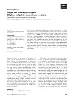
Fig. 1. Glucose-stimulated insulin secretion is accompanied by
changes in mir-9 levels in pancreatic b-islet cells. (A) Insulin levels
from the serum of mice were measured at 0, 15, 30, 60 and
240 min post-intraperitoneal glucose injections. (B) Mir-9 levels
determined by RT-qPCR analysis from total RNA isolated from pan-
creatic b-islets of the mice from (A). U6-snRNA levels were used for
normalization. (C) Real-time qRT-PCR analysis of total RNA from pan-
creatic b-islets of mice subjected to intraperitoneal glucose injec-
tions at the indicated time points to determine the levels of
transcripts pri ⁄ pre-mir-9-1, pri ⁄ pre-mir-9-2 and pri ⁄ pre-mir-9-3. ARBP
was used as the normalization control. (A–C) One-way ANOVA was
used for statistical analysis (n =3; *P < 0.05, **P < 0.01). In (B),
* and # indicate significance with respect to the 0- and 30-min time
points, respectively. In (C), # and * indicate significance with respect
to the 0-min time point for 9-1 and 9-2, respectively.
D. Ramachandran et al. Mir-9-dependent regulation of Sirt1 in b-cells
FEBS Journal 278 (2011) 1167–1174 ª 2011 The Authors Journal compilation ª 2011 FEBS 1169
Sirt1 mRNA and post-transcriptionally regulate its
expression.
Sirt1 is regulated in vivo in response to GSIS
Sirt1 is known to affect GSIS in b-islets [17]. However,
it is unclear whether its expression is regulated in vivo.
We found that mir-9 levels were regulated in response
to GSIS in vivo and that it targeted Sirt1 and down-
regulated its expression in cells in vitro. Hence, we
wished to determine whether Sirt1 levels were regu-
lated in vivo in pancreatic b-islets. If mir-9 targets Sirt1
in the b-islets, we would expect to see a decrease in
Sirt1 levels under conditions in which mir-9 levels are
high. Therefore, Sirt1 protein was assayed and, inter-
estingly, we found that Sirt1 levels were modulated in
pancreatic b-islets during GSIS (Fig. 3A). We observed
that there was a significant decrease in Sirt1 expression
240 min post-glucose injection. Importantly, the reduc-
tion in Sirt1 protein correlated well with increased
mir-9 levels in the b-islets when serum insulin secretion
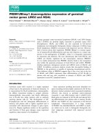
Fig. 2. Mir-9 targets the 3¢UTR of Sirt1 and down-regulates its
expression. (A) Alignment of mature mir-9 with the target sequence
on the 3¢UTR of mouse Sirt1 (using Target Scan). (B) Conservation
of the target site of mir-9 on the 3¢UTR of Sirt1 across vertebrates
(using Target Scan). (C) pmir-Report, pmir-Report Sirt1 3¢UTR (wild-
type or mutant) luciferase construct, pSuper-mir-9 and b-galactosi-
dase vector were co-transfected into HEK293T cells for 24 h, as
indicated. Luciferase activities were normalized to b-galactosidase
activities. Student’s t-test was used for statistical analysis (n =3;
*P < 0.05). (D) Western blot analysis of Sirt1 in NIH3T3 cells trans-
fected with 20 and 60 n
M of precursors of mir-control, mir-9 and
mir-34a. b-Actin was used as a loading control. The relative protein
levels were quantified using Adobe Photoshop and are indicated.
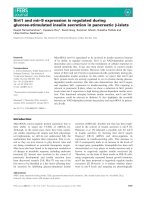
Fig. 3. Glucose-stimulated insulin secretion is accompanied by
changes in Sirt1 protein, but not mRNA levels, in pancreatic b-islet
cells. (A) Western blot analysis of endogenous Sirt1 protein levels
in pancreatic b-islets isolated at the indicated time points from mice
subjected to intraperitoneal glucose injections. b-Actin was used as
a loading control. (B) Relative Sirt1 protein levels in (A) were quanti-
fied using Adobe Photoshop and are plotted. One-way ANOVA was
used for statistical analysis (n =3;**P < 0.01). **indicates signifi-
cance with respect to the 0-min time point. (C) Sirt1 mRNA expres-
sion quantified by RT-qPCR from pancreatic b-islets isolated at the
indicated time points from mice subjected to intraperitoneal glu-
cose injections. ARBP was used as the normalization control.
Mir-9-dependent regulation of Sirt1 in b-cells D. Ramachandran et al.
1170 FEBS Journal 278 (2011) 1167–1174 ª 2011 The Authors Journal compilation ª 2011 FEBS
was decreasing (Fig. 1A, B). RT-qPCR analysis of
Sirt1 mRNA showed that, although the protein was
down-regulated (4 h post-glucose injection), this was
not a result of decreased mRNA levels (Fig. 3C). To
our knowledge, this is the first report to show that the
amount of Sirt1 protein is regulated (post-transcrip-
tionally) during GSIS in insulin-secreting b-islets
in vivo.
Sirt1 is down-regulated in insulin-secreting cells
in a mir-9-dependent manner
Although we have described the targeting in NIH3T3
cells, we wished to ascertain whether the post-tran-
scriptional regulation of Sirt1 expression in b-cells was
brought about by mir-9. We therefore used insulin-
secreting b-TC-6 cells to address this. b-TC-6 cells
were transfected with pre-mir-control ⁄ -9 and pS ⁄ pS9.
From Fig. 4A, B, it can be seen that Sirt1 protein is
reduced in cells transfected with pre-mir-9 and pS9,
respectively. Increased expression of mir-9 in pS9
transfected cells is shown in Fig. 4C. These findings
clearly demonstrate that mir-9 is indeed able to target
Sirt1 in b-TC-6 cells. In order to confirm this, b-TC-6
cells were transfected with control and anti-mir-9
locked nucleic acid (LNA). To mimic a declining insu-
lin secretion phase (Fig. 4D), the cells were subjected
to 8 h of glucose withdrawal, 16 h post-transfection.
The reduction in mir-9 after LNA transfection was
quantified by RT-qPCR (Fig. 4E). It was very interest-
ing to see that Sirt1 protein levels were significantly
higher in cells that had been transfected with anti-mir-
9 LNA (Fig. 4F). This result is consistent with reduced
Sirt1 expression in the pancreatic b-islets in vivo at
240 min post-glucose injection (when insulin secretion
is decreasing) (Fig. 3A, B). Importantly, this provides
mechanistic insight into the regulation of Sirt1 protein
in insulin-secreting b-cells brought about by mir-9.
Discussion
In this study, we have provided in vivo evidence for the
regulation of mir-9 expression in pancreatic b-islets.
The study by Plaisance et al. [5] clearly elucidated the
link between mir-9 and exocytosis in INS-1E cells.
However, whether mir-9 actually participated in GSIS
or whether it was itself regulated during this process
was not evident until now. Our in vivo data from
b-islets show, that mir-9 expression is temporally regu-
lated during GSIS. Specifically, we show that mir-9
levels increase during the fall phase of insulin secretion
(Fig. 1). We also show that, in b-islets, the increase in
mir-9 levels is not a result of an equal contribution
from the three chromosomal loci. Our results suggest
that mir-9 obtained from chromosomes 3 and 13 con-
tributes to an increase in its level in b-islets (Fig. 1C).
Further studies are required to dissect out molecular
players, such as transcription factors, involved in
the control of mir-9 expression. Our results suggest
that the increase in mir-9 levels in b-islets is an active
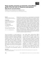
Fig. 4. Mir-9 targets and reduces Sirt1
expression in insulin-secreting cells. b-TC-6
cells were transfected with 60 n
M of precur-
sors of mir-control and mir-9 (A) and 1.5 lg
of pSuper and pSuper-mir-9 plasmids (B, C)
for 72 and 24 h, respectively. (D) b-TC-6
cells were cultured in high-glucose-contain-
ing medium and subjected to 8 h of glucose
withdrawal. Insulin levels in cell culture
supernatants were measured. (E, F) b-TC-6
cells were transfected with 100 n
M LNA
anti-mir-control and anti-mir-9 for 16 h and
subjected to glucose withdrawal for 8 h as
detailed in Experimental procedures. (C, E)
Mir-9 expression was quantified by
RT-qPCR normalized to U6 levels. (C–E)
Student’s t-test was used for statistical anal-
ysis (n =3;*P < 0.05, **P < 0.01). (A, B, F)
Western blot analysis for Sirt1 protein with
b-actin as the loading control.
D. Ramachandran et al. Mir-9-dependent regulation of Sirt1 in b-cells
FEBS Journal 278 (2011) 1167–1174 ª 2011 The Authors Journal compilation ª 2011 FEBS 1171
process. On the basis of our finding of the temporal
regulation of its expression and the earlier report on
exocytosis [5], we implicate mir-9 as a crucial factor in
the control of insulin secretion in response to glucose
stimulation in vivo.
Furthermore, we have also identified mir-9 to be a
key factor in the modulation of Sirt1 expression
in vivo. Recently, Saunders et al. [22] have shown that
mir-9 targets Sirt1 in mouse embryonic stem cells.
However, our results show that mir-9 targeting of Sirt1
is physiologically significant in insulin-secreting cells
(Figs 3 and 4).
Earlier studies have linked Sirt1 to GSIS. In Sirt1
transgenic (BESTO) mice, Sirt1-mediated control of
insulin secretion was found to be a result of the differ-
ential expression of genes, including those involved in
insulin secretion [17]. Banks et al. [23] used BAC trans-
genic mice over-expressing Sirt1 to show that it is a
crucial factor in insulin secretion under excess calorie
conditions. However, until now, it was unclear whether
Sirt1 itself was regulated in b-islets during insulin
secretion. Our results show, for the first time, that
Sirt1 expression is altered in insulin-secreting b-islets
during GSIS in vivo. Using b-TC-6 cells and conditions
that mimic the declining phase of insulin secretion, we
clearly demonstrate that mir-9 is involved in the regu-
lation of Sirt1 protein in these cells (Fig. 4). Our
results on the expression profile of Sirt1 during GSIS
fit well with the earlier findings on the ability of Sirt1
to positively regulate insulin secretion.
Recent reports have also examined the role of Sirt1 in
cytokine-dependent b-cell cytotoxicity [24]. Interest-
ingly, the data show that interferon-c and interleukin-1b
treatment of insulin-secreting cells leads to a down-
regulation of Sirt1. In another study by Chen et al. [25],
altered oxygen tension-mediated proliferation of insu-
lin-secreting cells was linked to changes in Sirt1
expression in INS-1E cells. These reports suggest that,
in addition to its significant role in insulin secretion,
Sirt1 is a crucial factor in b-cell proliferation and
survival. On the basis of our results that identify mir-9
as a negative regulator of Sirt1 in insulin-secreting cells,
it would be interesting to determine whether a similar
control is operating during cytokine-mediated changes
in Sirt1 levels in b-islets.
Given the known functions of Sirt1 in regulating
insulin secretion, our study adds a new facet by show-
ing, that Sirt1 protein levels are altered in insulin-secret-
ing cells during GSIS. To conclude, we have discovered
a functional interplay between glucose-dependent insu-
lin secretion, mir-9 levels and Sirt1 protein in b-cells. In
addition, we provide some evidence for the dynamic
(and differential) nature of mir-9 expression in pancre-
atic b-islets. Further insights into these mechanisms
may help in the understanding and tackling of diseases
such as diabetes.
Experimental procedures
Animal experiments
Adult Swiss male mice were maintained at the Tata Insti-
tute of Fundamental Research animal facility in accordance
with the institute’s animal ethics regulations. These mice
were used for GSIS. Briefly, the mice were starved over-
night and injected with glucose (3 gÆkg
)1
body weight)
intraperitoneally. Serum and tissue samples were collected
at 0, 15, 30, 60 and 240 min post-glucose injection. Three
animals per group were used and the experiment was
repeated at least twice.
Cell culture
NIH3T3 and HEK293T cells were grown in DMEM
(Sigma, St. Louis, MO, USA cat. no. D7777) supplemented
with 10% newborn calf serum (Gibco, USA cat. no. 16010-
159) and 10% fetal bovine serum (Gibco cat. no. 16000),
respectively. b-TC-6 cells were grown in DMEM supple-
mented with 15% heat-inactivated fetal bovine serum. For
glucose withdrawal, cells were washed with NaCl ⁄ P
i
and
then DMEM without glucose (Sigma cat. no. D5030), sup-
plemented with 5% heat-inactivated fetal bovine serum,
was added, as indicated in the figure legends.
Primers and constructs
The sequences of the primers used in this study are listed in
Table S1. Cloning of Sirt1 3¢UTR (wild-type and mutant)
into pmir-Report (Fig. S3) and the generation of pSuper-
mir-9 construct [5] are detailed in Doc. S3 and S4.
Transfections
Cells were transfected using Lipofectamine 2000 (Invitrogen,
Carlsbad, CA, USA cat. no. 11668-019) according to the
manufacturer’s instructions. Control pre-mir and pre-mir-9
were procured from Ambion (Austin, TX, USA). LNA con-
structs for control and anti-mir-9 were obtained from Exiqon
(Vedbaek, Denmark). Pre-mirs and anti-mirs were transfect-
ed into cells at concentrations of 20 ⁄ 60 and 100 nm, respec-
tively, and harvested after 24–48 h. Mir-9 was expressed in
cells by transfecting 1.5 lg of pSuper-mir-9.
RNA isolation and cDNA synthesis
Total RNA was isolated using Trizol (Invitrogen cat. no.
15596-026). SuperScript-III (Invitrogen cat. no. 18080-044)
Mir-9-dependent regulation of Sirt1 in b-cells D. Ramachandran et al.
1172 FEBS Journal 278 (2011) 1167–1174 ª 2011 The Authors Journal compilation ª 2011 FEBS
was used for cDNA synthesis with 1 lg of total RNA
employing random hexamers or with mir-specific RT-6
primers (Table S1).
Real-time PCR
qPCRs were performed in triplicate using SYBR green
(Qiagen, USA cat. no. 204056) according to the manufac-
turer’s instructions. qPCR for mir was performed as
described in ref. [26]. Briefly, short-mir-9 and MP-
fwd ⁄ MP-rev primers (Table S1) were used at concentra-
tions of 4 and 100 nm, respectively. U6 and ARBP were
used for the normalization of mir and mRNA expression,
respectively.
Pancreatic b-islet isolation
b-Islets were harvested by perfusing the pancreas as
described by Szot et al. [27] (Doc. S1).
Luciferase and b-galactosidase assays
HEK293T cells were transfected with pmir-Report-Sirt1
3¢UTR (wild-type or mutant), b-galactosidase vector and
pSuper or pSuper-mir-9 (pS ⁄ pS9) in 24-well plates. Cells
were harvested 24 h later and luciferase assay was carried
out using the Stratagene Luciferase Assay kit (Agilent
Technologies, Santa Clara, CA, USA cat. no. 219020)
according to the manufacturer’s instructions. Luciferase
activities were normalized to the b-galactosidase activity in
each case.
Western blots
Equal amounts of protein (estimated using the BCA kit,
Sigma-Aldrich, USA) were run on SDS ⁄ PAGE and trans-
ferred to poly(vinylidene difluoride) membranes (Roche,
Basel, Schweiz cat. no. 3 010 040 ⁄ ThermoFisher cat. no.
88518). Anti-Sirt1 (Millipore-Upsatate, MA, USA cat. no.
07-131) and anti-b-actin (Sigma cat. no. A1978) antibodies
were used for immunoblotting. Horseradish peroxidase-
conjugated secondary antibodies (Sigma cat. nos. A0545
and A2554) and Lumi-Light Western Blotting Substrate
(Roche cat. no. 12 015 196 001) were used to visualize the
bands.
Insulin ELISA
Sera and culture supernatants from b-TC-6 cells subjected
to control and glucose withdrawal conditions were assayed
for insulin. Insulin was quantified according to the manu-
facturer’s protocol using the Rat ⁄ Mouse Insulin 96-Well
Plate Assay Kit (Millipore, MA, USA cat. no. EZRMI-
13K).
Statistical analysis
All statistical analyses were carried out using GraphPad
InStat3 or SigmaPlot. Student’s t-test was performed when
two datasets were compared. One-way ANOVA was per-
formed for the datasets generated from the GSIS experi-
ments.
Acknowledgements
We thank Dr. Suryavanshi for helping with the animal
experimentation. We also thank A. Lazarus and K.
Banerjee for technical help. Funding from Department
of Biotechnology (Govt. of India) and Department of
Atomic Energy/TIFR (Govt. of India) is acknowl-
edged.
References
1 Valencia-Sanchez MA, Liu J, Hannon GJ & Parker R
(2006) Control of translation and mRNA degradation
by miRNAs and siRNAs. Genes Dev 20,
515–524.
2 Cuellar TL & McManus MT (2005) MicroRNAs and
endocrine biology. J Endocrinol 187, 327–332.
3 Walker MD (2008) Role of MicroRNA in pancreatic
beta-cells: where more is less. Diabetes 57, 2567–2568.
4 Poy MN, Eliasson L, Krutzfeldt J, Kuwajima S,
Ma X, Macdonald PE, Pfeffer S, Tuschl T, Rajewsky
N, Rorsman P et al. (2004) A pancreatic islet-specific
microRNA regulates insulin secretion. Nature 432,
226–230.
5 Plaisance V, Abderrahmani A, Perret-Menoud V,
Jacquemin P, Lemaigre F & Regazzi R (2006) Micro-
RNA-9 controls the expression of Granuphilin ⁄ Slp4
and the secretory response of insulin-producing cells.
J Biol Chem 281, 26932–26942.
6 Kato T, Shimano H, Yamamoto T, Yokoo T, Endo Y,
Ishikawa M, Matsuzaka T, Nakagawa Y, Kumadaki S,
Yahagi N et al. (2006) Granuphilin is activated by
SREBP-1c and involved in impaired insulin secretion in
diabetic mice. Cell Metab 4, 143–154.
7 Finkel T, Deng CX & Mostoslavsky R (2009) Recent
progress in the biology and physiology of sirtuins.
Nature 460, 587–591.
8 Langley E, Pearson M, Faretta M, Bauer UM, Frye
RA, Minucci S, Pelicci PG & Kouzarides T (2002)
Human SIR2 deacetylates p53 and antagonizes
PML ⁄ p53-induced cellular senescence. EMBO J 21,
2383–2396.
9 Vaquero A, Scher M, Lee D, Erdjument-Bromage H,
Tempst P & Reinberg D (2004) Human SirT1 interacts
with histone H1 and promotes formation of facultative
heterochromatin. Mol Cell 16, 93–105.
D. Ramachandran et al. Mir-9-dependent regulation of Sirt1 in b-cells
FEBS Journal 278 (2011) 1167–1174 ª 2011 The Authors Journal compilation ª 2011 FEBS 1173
10 Vaquero A, Scher M, Erdjument-Bromage H, Tempst
P, Serrano L & Reinberg D (2007) SIRT1 regulates the
histone methyl-transferase SUV39H1 during heterochro-
matin formation. Nature 450, 440–444.
11 Kwon HS & Ott M (2008) The ups and downs of
SIRT1. Trends Biochem Sci 33 , 517–525.
12 Kanfi Y, Peshti V, Gozlan YM, Rathaus M, Gil R &
Cohen HY (2008) Regulation of SIRT1 protein levels
by nutrient availability. FEBS Lett 582, 2417–2423.
13 Bordone L, Motta MC, Picard F, Robinson A, Jhala
US, Apfeld J, McDonagh T, Lemieux M, McBurney
M, Szilvasi A et al. (2006) Sirt1 regulates insulin secre-
tion by repressing UCP2 in pancreatic beta cells. PLoS
Biol 4, e31.
14 Revollo JR, Grimm AA & Imai S (2004) The NAD
biosynthesis pathway mediated by nicotinamide phos-
phoribosyltransferase regulates Sir2 activity in mamma-
lian cells. J Biol Chem 279, 50754–50763.
15 van der Veer E, Ho C, O’Neil C, Barbosa N, Scott R,
Cregan SP & Pickering JG (2007) Extension of human
cell lifespan by nicotinamide phosphoribosyltransferase.
J Biol Chem 282, 10841–10845.
16 Leibiger IB, Leibiger B & Berggren PO (2008) Insulin
signaling in the pancreatic beta-cell. Annu Rev Nutr 28,
233–251.
17 Moynihan KA, Grimm AA, Plueger MM, Bernal-Mizr-
achi E, Ford E, Cras-Meneur C, Permutt MA & Imai S
(2005) Increased dosage of mammalian Sir2 in
pancreatic beta cells enhances glucose-stimulated insulin
secretion in mice. Cell Metab 2, 105–117.
18 El Ouaamari A, Baroukh N, Martens GA, Lebrun P,
Pipeleers D & van Obberghen E (2008) miR-375 targets
3¢-phosphoinositide-dependent protein kinase-1 and reg-
ulates glucose-induced biological responses in pancreatic
beta-cells. Diabetes 57, 2708–2717.
19 Bandres E, Agirre X, Bitarte N, Ramirez N, Zarate R,
Roman-Gomez J, Prosper F & Garcia-Foncillas J
(2009) Epigenetic regulation of microRNA expression
in colorectal cancer. Int J Cancer 125, 2737–2743.
20 Lujambio A, Calin GA, Villanueva A, Ropero S,
Sanchez-Cespedes M, Blanco D, Montuenga LM, Rossi
S, Nicoloso MS, Faller WJ et al. (2008) A microRNA
DNA methylation signature for human cancer metasta-
sis. Proc Natl Acad Sci USA 105, 13556–13561.
21 Yamakuchi M, Ferlito M & Lowenstein CJ (2008)
miR-34a repression of SIRT1 regulates apoptosis. Proc
Natl Acad Sci USA 105, 13421–13426.
22 Saunders LR, Sharma AD, Tawney J, Nakagawa M,
Okita K, Yamanaka S, Willenbring H & Verdin E
(2010) miRNAs regulate SIRT1 expression during
mouse embryonic stem cell differentiation and in adult
mouse tissues. Aging (Albany NY) 2, 415–431.
23 Banks AS, Kon N, Knight C, Matsumoto M,
Gutierrez-Juarez R, Rossetti L, Gu W & Accili D
(2008) SirT1 gain of function increases energy effi-
ciency and prevents diabetes in mice. Cell Metab 8,
333–341.
24 Lee JH, Song MY, Song EK, Kim EK, Moon WS,
Han MK, Park JW, Kwon KB & Park BH (2009)
Overexpression of SIRT1 protects pancreatic beta-cells
against cytokine toxicity by suppressing the
nuclear factor-kappaB signaling pathway. Diabetes 58,
344–351.
25 Chen JH, Jones RH, Tarry-Adkins J, Smith NH &
Ozanne SE (2008) Adverse effects of reduced
oxygen tension on the proliferative capacity of rat
kidney and insulin-secreting cell lines involve DNA
damage and stress responses. Exp Cell Res 314, 3075–
3080.
26 Sharbati-Tehrani S, Kutz-Lohroff B, Bergbauer R,
Scholven J & Einspanier R (2008) miR-Q: a novel
quantitative RT-PCR approach for the expression
profiling of small RNA molecules such as miRNAs
in a complex sample. BMC Mol Biol 9, 34.
27 Szot GL, Koudria P & Bluestone JA (2007) Murine
pancreatic islet isolation. J Vis Exp 7, 255.
Supporting information
The following supplementary material is available:
Fig. S1. Genomic loci encoding Mus. musculus (mmu)-
mir-9-1 and 9-3.
Fig. S2. Analysis of the methylation status of the mir-
9-3 promoter region.
Fig. S3. Generation of Sirt1 3¢UTR luciferase con-
struct.
Doc. S1. Pancreatic b-islet isolation [27].
Doc. S2. Analysis of the methylation status of the mir-
9-3 promoter region.
Doc. S3. pSuper-mir-9 construct.
Doc. S4. Sirt1 3¢UTR wild-type and mutant luciferase
construct.
Table S1. Primer sequences.
This supplementary material can be found in the
online version of this article.
Please note: As a service to our authors and readers,
this journal provides supporting information supplied
by the authors. Such materials are peer-reviewed and
may be re-organized for online delivery, but are not
copy-edited or typeset. Technical support issues arising
from supporting information (other than missing files)
should be addressed to the authors.
Mir-9-dependent regulation of Sirt1 in b-cells D. Ramachandran et al.
1174 FEBS Journal 278 (2011) 1167–1174 ª 2011 The Authors Journal compilation ª 2011 FEBS