Impact of Gene Expression Profiling Tests on Breast Cancer Outcomes potx
Bạn đang xem bản rút gọn của tài liệu. Xem và tải ngay bản đầy đủ của tài liệu tại đây (2.85 MB, 230 trang )
Evidence Report/Technology Assessment
Number 160
Impact of Gene Expression Profiling Tests on Breast
Cancer Outcomes
Prepared for:
Agency for Healthcare Research and Quality
U.S. Department of Health and Human Services
540 Gaither Road
Rockville, MD 20850
www.ahrq.gov
Contract No. 290-02-0018
Prepared by:
The Johns Hopkins University Evidence-based Practice Center, Baltimore, MD
Investigators
Luigi Marchionni, M.D., Ph.D.
Renee F. Wilson, M.Sc.
Spyridon S. Marinopoulos, M.D., M.B.A.
Antonio C. Wolff, M.D.
Giovanni Parmigiani, M.D.
Eric B. Bass, M.D., M.P.H.
Steven N. Goodman, M.D., M.H.S., Ph.D.
AHRQ Publication No. 08-E002
January 2008
This report is based on research conducted by the Johns Hopkins University Evidence-based
Practice Center (EPC) under contract to the Agency for Healthcare Research and Quality (AHRQ),
Rockville, MD (Contract No. 290-02-0018). The findings and conclusions in this document are
those of the author(s), who are responsible for its content, and do not necessarily represent the
views of AHRQ. No statement in this report should be construed as an official position of AHRQ
or of the U.S. Department of Health and Human Services.
The information in this report is intended to help clinicians, employers, policymakers, and others
make informed decisions about the provision of health care services. This report is intended as a
reference and not as a substitute for clinical judgment.
This report may be used, in whole or in part, as the basis for the development of clinical practice
guidelines and other quality enhancement tools, or as a basis for reimbursement and coverage
policies. AHRQ or U.S. Department of Health and Human Services endorsement of such
derivative products may not be stated or implied.
ii
This document is in the public domain and may be used and reprinted without permission except
those copyrighted materials noted for which further reproduction is prohibited without the
specific permission of copyright holders.
Suggested Citation:
Marchionni L, Wilson RF, Marinopoulos SS, Wolff AC, Parmigiani G, Bass EB, Goodman SN.
Impact of Gene Expression Profiling Tests on Breast Cancer Outcomes. Evidence
Report/Technology Assessment No. 160. (Prepared by The Johns Hopkins University Evidence-
based Practice Center under contract No. 290-02-0018). AHRQ Publication No. 08-E002.
Rockville, MD: Agency for Healthcare Research and Quality. January 2008.
The investigators have no relevant financial interests in the report. The investigators
have no employment, consultancies, honoraria, or stock ownership or options, or
royalties from any organization or entity with a financial interest or financial conflict
with the subject matter discussed in the report.
iii
Preface
The Agency for Healthcare Research and Quality (AHRQ), through its Evidence-Based
Practice Centers (EPCs), sponsors the development of evidence reports and technology
assessments to assist public- and private-sector organizations in their efforts to improve the
quality of health care in the United States. The Centers for Disease Control and Prevention
(CDC) requested and provided funding for this report. The reports and assessments provide
organizations with comprehensive, science-based information on common, costly medical
conditions and new health care technologies. The EPCs systematically review the relevant
scientific literature on topics assigned to them by AHRQ and conduct additional analyses when
appropriate prior to developing their reports and assessments.
To bring the broadest range of experts into the development of evidence reports and health
technology assessments, AHRQ encourages the EPCs to form partnerships and enter into
collaborations with other medical and research organizations. The EPCs work with these partner
organizations to ensure that the evidence reports and technology assessments they produce will
become building blocks for health care quality improvement projects throughout the Nation. The
reports undergo peer review prior to their release.
AHRQ expects that the EPC evidence reports and technology assessments will inform
individual health plans, providers, and purchasers as well as the health care system as a whole by
providing important information to help improve health care quality.
We welcome comments on this evidence report. They may be sent by mail to the Task Order
Officer named below at: Agency for Healthcare Research and Quality, 540 Gaither Road,
Rockville, MD 20850, or by e-mail to
Carolyn M. Clancy, M.D.
Director
Agency for Healthcare Research and Quality
Jean Slutsky, P.A., M.S.P.H.
Director, Center for Outcomes and Evidence
Agency for Healthcare Research and Quality
Julie Louise Gerberding, M.D., M.P.H.
Director
Centers for Disease Control and Prevention
Gurvaneet Randhawa, M.D., M.P.H.
EPC Program Task Order Officer
Agency for Healthcare Research and Quality
Beth Collins Sharp, Ph.D., R.N.
Director, EPC Program
Agency for Healthcare Research and Quality
iv
Acknowledgments
The Evidence-based Practice Center thanks Michael Oladubu, D.D.S. and Allison Jonas, for their
assistance with literature searching and database management, and project organization; Aly
Shogan for her assistance in completing the sections on economics; Brenda Zacharko for her
assistance with budget matters, and for her assistance with final preparations of the report. The
Center also wishes to thank Gurvaneet Randhawa, M.D., M.P.H., AHRQ Task Order Officer, for
his efforts in guiding this project and coordination with the CDC EGAPP group.
v
Structured Abstract
Objective: To assess the evidence that three marketed gene expression-based assays improve
prognostic accuracy, treatment choice, and health outcomes in women diagnosed with early stage
breast cancer.
Data Sources: MEDLINE
®
, EMBASE, the Cochrane databases, test manufacturer Web sites,
and information provided by manufacturers.
Review Methods: We evaluated the evidence for three gene expression assays on the market;
Oncotype DX™, MammaPrint® and the Breast Cancer Profiling (BCP or H/I ratio) test, and for
gene expression signatures underlying the assays. We sought evidence on: (a) analytic
performance of tests; (b) clinical validity (i.e., prognostic accuracy and discrimination); (c)
clinical utility (i.e., prediction of treatment benefit); (d) harms; and (e) impact on clinical
decision making and health care costs.
Results: Few papers were found on the analytic validity of the Oncotype DX and MammaPrint
tests, but these showed reasonable within-laboratory replicability. Pre-analytic issues related to
sample storage and preparation may play a larger role than within-laboratory variation. For
clinical validity, studies differed according to whether they examined the actual test that is
currently being offered to patients or the underlying gene signature. Almost all of the Oncotype
DX evidence was for the marketed test, the strongest validation study being from one arm of a
randomized controlled trial (NSABP-14) with a clinically homogeneous population. This study
showed that the test, added in a clinically meaningful manner to standard prognostic indices. The
MammaPrint signature and test itself was examined in studies with clinically heterogeneous
populations (e.g., mix of ER positivity and tamoxifen treatment) and showed a clinically relevant
separation of patients into risk categories, but it was not clear exactly how many predictions
would be shifted across decision thresholds if this were used in combination with traditional
indices. The BCP test itself was examined in one study, and the signature was tested in a variety
of formulations in several studies. One randomized controlled trial provided high quality
retrospective evidence of the clinical utility of Oncotype DX to predict chemotherapy treatment
benefit, but evidence for clinical utility was not found for MammaPrint or the H/I ratio. Three
decision analyses examined the cost-effectiveness of breast cancer gene expression assays, and
overall were inconclusive.
Conclusions: Oncotype DX is furthest along the validation pathway, with strong retrospective
evidence that it predicts distant spread and chemotherapy benefit to a clinically relevant extent
over standard predictors, in a well-defined clinical subgroup with clear treatment implications.
The evidence for clinical implications of using MammaPrint was not as clear as with Oncotype
DX, and the ability to predict chemotherapy benefit does not yet exist. The H/I ratio test requires
further validation. For all tests, the relationship of predicted to observed risk in different
populations still needs further study, as does their incremental contribution, optimal
implementation, and relevance to patients on current therapies.
vi
Contents
Executive Summary 1
Evidence Report………………………………………………………………………………….9
Chapter 1. Introduction 11
Breast Cancer 11
Gene expression profiling 12
Breast Cancer Assays on the Market 13
RT-PCR 14
Microarrays 15
Sources of Variability in Gene Expression Analysis 16
Objectives of the Evidence Report 17
Structured Approach to Assessment of the Questions 18
Chapter 2. Methods 21
Recruitment of Technical Experts and Peer Reviewers 21
Key Questions 21
Literature Search Methods 21
Sources 22
Search terms and strategies 22
Organization and tracking of literature search 23
Title Review 23
Abstract Review 23
Inclusion and exclusion criteria 23
Article Inclusion/Exclusion 24
Data Abstraction 26
Quality Assessment 26
Data Synthesis 27
Data Entry and Quality Control 27
Grading of the Evidence 27
Peer Review 27
Chapter 3. Results 29
Key Question 1. What is the direct evidence that gene expression profiling tests in women
diagnosed with breast cancer, or any specific subset of this population, lead to
improvement in outcomes? 29
Key Question 2. What are the sources of and contributions to analytic validity in
these gene expression-based prognostic estimators for women diagnosed with
breast cancer? 29
Oncotype DX™ 30
MammaPrint® 34
H/I Ratio 36
Key Question 3. What is the clinical validity of gene expression profiling tests in women
diagnosed with breast cancer? 38
vii
Oncotype DX 38
MammaPrint 39
H/I Ratio 41
Key Question 4. What is the clinical utility of these tests? 45
Oncotype DX 46
MammaPrint 52
H/I Ratio 54
Ongoing Studies 55
TAILORx 55
MINDACT 55
Other Relevant Studies 55
Studies Excluded Upon Complete Review 57
Chapter 4. Discussion 87
Oncotype DX 88
Analytic validity 88
Clinical validity 89
Clinical utility 90
Questions regarding the clinical validity and utility of the Oncotype DX assay 93
MammaPrint 93
Analytic validity 94
Clinical validity 94
Clinical utility 95
H/I Ratio Signature and Breast Cancer Profiling (BCP) 96
General Comments on Analytic Validity and Laboratory Quality Control 96
Overall implications and recommendations 97
Assay validation 97
Potential for scale problems 97
Genetic variability and gene expression 98
The need for databases, reproducibility, and standards 98
Where is the field going? 98
“Comparative effectiveness” studies 99
Conclusion 99
References and Included Studies 101
Tables
Table 1. Description of the three gene expression profile assays 59
Table 2. Successful assays, Oncotype DX 62
Table 3. Variability and reproducibility, Oncotype DX 63
Table 4. Analytic validity, Oncotype DX 64
Table 5. RT-PCR vs. IHC comparison assays, Oncotype DX 65
Table 6. Successful assays, MammaPrint 67
Table 7. Reproducibility, MammaPrint 68
Table 8. Analytic validity, MammaPrint 69
viii
Table 9. Successful assays, two-gene signature and H/I ratio assays 70
Table 10. Reproducibility, two-gene signature and H/I ratio assay 71
Table 11. RT-PCR vs. IHC comparison assays, two-gene signature and H/I ratio assay 72
Table 12. Clinical validity, Oncotype DX 73
Table 13. Risk classification of Oncotype DX against the St. Gallen criteria 75
Table 14. Risk classification of Oncotype DX against the 2004 NCCN guidelines 75
Table 15. Risk classification of Oncotype DX against the Adjuvant! Guidelines 75
Table 16. Clinical Validity, MammaPrint and 70-gene signature 76
Table 17. MammaPrint compared with traditional composite risk markers 79
Table 18. Clinical Validity, two-gene signature and H/I ratio assays 80
Table 19. Clinical Utility, Oncotype DX 83
Table 20. Comparison of economic studies 85
Table 21. Clinical Utility, two-gene signature and H/I ratio 86
Figures
Figure 1. Increasing complexity of information from genome to trascriptome and proteome:
gene expression analysis focuses on the analysis of the transcriptome……………… 12
Figure 2. Quantitative RT-PCR 15
Figure 3. Schematic model for microarray hybridizations… 16
Figure 4. Summary of literature search and review process (number of articles) 25
Appendixes
Appendix A: List of Acronyms
Appendix B: Glossary
Appendix C: Description of Genes
Appendix D: Technologies
Appendix E: Technical Experts and Peer Reviewers
Appendix F: Detailed Electronic Database Search Strategies
Appendix G: Review Forms
Appendix H: Excluded Articles
Appendix I: Evidence Tables
Appendixes and Evidence Tables for this report are provided electronically at
1
Executive Summary
Introduction
Breast cancer is the most commonly diagnosed cancer in women. This tumor is the second
leading cause of cancer-related deaths in women in the United States, with approximately
178,000 new cases and 40,000 deaths expected among U.S. women in 2007. Treatment for
breast cancer usually involves surgery to remove the tumor and involved lymph nodes.
Frequently, surgery is followed by radiation therapy (in case of breast conservation or in women
with large tumors or many involved lymph nodes), endocrine therapy (for essentially all women
with tumors that express the estrogen receptor (ER-positive)), and/or chemotherapy (for women
having a high risk for a poor outcome such as those with large tumors, involved lymph nodes,
advanced disease, or inflammatory breast cancer). More than three-quarters of patients are
expected to survive with this multi-modality approach.
Gene expression profiling has been proposed as an approach to address this issue in clinical
settings, and three breast cancer gene expression assays are now available in the U.S. The
Oncotype DX™ Breast Cancer Assay, the MammaPrint
®
Test, and the Breast Cancer Profiling
test (BCP or H/I ratio). MammaPrint is based on the use of microarray technology, while the
other two assays are based on the reverse transcriptase polymerase chain reaction (RT-PCR). All
of these tests combine the measurements of gene expression levels within the tumor to produce a
number associated with the risk of distant disease recurrence. These tests aim to improve on risk
stratification schemes based on clinical and pathologic factors currently used in clinical practice.
As therapeutic decisions are based on risk estimates, tests that improve such estimates have the
potential to affect clinical outcome in breast cancer patients by either avoiding unnecessary
chemotherapy and its attendant morbidity or by employing it where it might not otherwise have
been used, thereby reducing recurrence risk.
The literature was searched for evidence about the use of gene expression profiling in breast
cancer. Our analytical framework for reporting the results distinguishes between the assays, as
they are offered to patients, and the underlying signatures, which comprise the genes whose
expression is measured. This measurement of expression can be done in a number of ways that
may not be identical to the procedures used for the marketed test, producing an unknown number
of different predictions. We also distinguish between developmental and validation studies.
Methods
Working with the Agency for Healthcare Research and Quality (AHRQ), the Centers for
Disease Prevention and Control (CDC), the Evaluation of Genomic Applications in Practice and
Prevention (EGAPP) working group, and members of a technical expert panel, we formulated
four key questions, and addressed them on the basis of the evidence available about the specific
assays and the underlying gene expression signatures. The original set of key questions was
refined to focus primarily on two gene expression profiling tests: Oncotype DX (Genomic
Health, Inc.) and MammaPrint (Agendia). During the course of the evaluation, a third gene
expression profiling test came to our attention, the H/I ratio test based on the two-gene signature
(AviaraDX/Quest Diagnostics, Inc.), and was thus investigated. We searched and retrieved
2
studies in MEDLINE
®
, EMBASE, and the Cochrane databases (1990-2006). We supplemented
this search with recent publications that appeared after the time period initially considered in the
systematic search, and about the two-gene test (H/I ratio). We also searched for relevant
documents on the Food and Drug Administration’s web site, and solicited additional
documentation from the companies offering the tests. The systematic searches yielded a total of
12983 citations. Specific inclusion and exclusion criteria were developed and pairs of readers
reviewed each title; the same procedure was used to review selected abstracts. We identified 63
studies for full text review. We developed tables to summarize each article. Initial data were
abstracted by investigators and entered directly into evidence tables. Quality and consistency of
the abstracted data was then evaluated by a second reviewer, and a senior investigator examined
all reviews to identify potential problems with data abstraction. These were discussed at
meetings of group members. A system of random data checks was applied to ensure data
abstraction accuracy.
Results
Literature on Key Questions
Key Question 1. What is the direct evidence that gene expression profiling tests in women
diagnosed with breast cancer (or any specific subset of this population) lead to improvement in
outcomes?
Direct evidence was defined as a study where the primary intervention is the use of a
prognostic test (with therapeutic decisionmaking directed by the result) and the outcomes are
patient morbidity, mortality and/or quality of life. No direct evidence was found in the published
data on improvement of patients’ outcomes due to such testing in women diagnosed with breast
cancer, nor were there any randomized studies using the tests’ predictions to manage patients.
However, as described under Key Questions 3 and 4, some of the tests’ supporting evidence was
derived from past randomized controlled trials (RCTs) with prospectively gathered patient
samples, giving them strong evidential value. Two ongoing RCTs, TAILORx and MINDACT
(using Oncotype DX, and MammaPrint respectively), will provide further evidence allowing
almost direct inference about the impact on patient outcomes.
Key Question 2. What are the sources of and contributions to analytic validity in these two
gene expression-based prognostic estimators for women diagnosed with breast cancer?
In the field of gene expression there are no “gold standards” outside the technologies used in
the tests under study, i.e., microarrays and RT-PCR. Consequently, a definitive evaluation of the
analytic validity of expression-based tests is difficult. Evidence about operational characteristics
was partial and limited to a few publications. A 2007 paper by Cronin and colleagues, on the
analytic validity of Oncotype DX was the most detailed study for any of these tests so far,
showing good performance for a number of analytic components of the assay. Data about the
sources and contributions to variability of the tests and about their reproducibility was generally
limited to analyses of few samples, and thus a complete evaluation of the impact of such
variability on risk assessment was not available. Partial evidence about analytic validity was
provided in the percentage of subjects whose samples were successfully analyzed with these
tests, and those numbers were fairly good. Continuous monitoring of laboratory procedures and
3
careful evaluation of the quality of the submitted specimens are major factors affecting test
reliability.
Key Question 3. What is the clinical validity of these tests in women diagnosed with breast
cancer?
a. How well does this testing predict recurrence rates for breast cancer compared to
standard prognostic approaches? Specifically, how much do these tests add to currently
known factors or combination indices that predict the probability of breast cancer
recurrence, (e.g., tumor type or stage, age, ER, and human epidermal growth factor
receptor 2 (HER-2) status)?
b. Are there any other factors, which may not be components of standard predictors of
recurrence (e.g., race/ethnicity or adjuvant therapy), that affect the clinical validity of
these tests, and thereby generalizability of results to different populations?
Clinical validity is defined as the degree to which a test accurately predicts the risk of an
outcome (i.e., calibration), as well as its ability to separate patients with different outcomes into
separate risk classes (discrimination). Clinical validity was documented to some degree for all
three gene expression signatures. Oncotype DX was validated on a homogenous population of
lymph node negative, ER positive patients all treated with tamoxifen, derived from an arm of an
RCT, the National Surgical Adjuvant Breast and Bowel Project (NSABP-14). MammaPrint, on
the other hand, was validated on samples from a clinical series with a wide range of clinical and
treatment characteristics, and sometimes it was the signature and not the MammaPrint test itself
that was validated. Data that made clear the incremental value of the test over standardized risk
predictors using classical clinical factors, in the form of risk reclassification tables, was limited
to Oncotype DX in one population, and for one of those predictors (Adjuvant! Online for
MammaPrint). The evidence behind the two-gene test is quite heterogeneous, in that the specific
manner in which the index was calculated differed in each, and only one examines the index that
is to be used as part of the BCP (or H/I ratio) test in a study that was still using statistical
methods to find optimal cut points, i.e., a training study. So the Oncotype DX test, which has
been validated in exactly the form given to patients on clinically homogeneous samples with
clear treatment implications, is regarded as the index with the strongest claim to clinical validity.
It is not yet as clear to which populations MammaPrint best applies, and how much incremental
value it would have within those clinically homogeneous populations above various standard
predictors. Since the number of validation studies for any of the tests is still relatively small,
more remains to be learned about stability between different populations of the relationship
between expression-based score and the absolute observed risk. Essentially nothing is known
about how specific characteristics of these populations might affect test performance.
While the H/I ratio test shows some promise, it must be regarded as still being in a
developmental phase; it cannot yet be considered fully validated. It was not clear whether
samples were processed by Quest Diagnostics, which hold the current license. There are a
number of intriguing biological insights and plausible mechanisms to support the rationale for
the test, but its consistent value in well-defined clinical settings has not yet been firmly
established.
Key Question 4. What is the clinical utility of these tests?
a. To what degree do the results of these tests predict the response to chemotherapy, and
what factors affect the generalizability of that prediction?
4
b. What are the effects of using these two tests and the subsequent management options on
the following outcomes: testing or treatment related psychological harms, testing or
treatment related physical harms, disease recurrence, mortality, utilization of adjuvant
therapy, and medical costs.
c. What is known about the utilization of gene expression profiling in women diagnosed
with breast cancer in the United States?
d. What projections have been made in published analyses about the cost-effectiveness of
using gene expression profiling in women diagnosed with breast cancer?
Few studies addressed the clinical utility of Oncotype DX recurrence score (RS) in predicting
the benefits of adjuvant chemotherapy, although the probability of recurrence represents an
upper bound on the degree of absolute benefit. One fairly strong retrospective study produced
preliminary evidence that the RS has predictive power in assessing the benefit of chemotherapy
usage in ER-positive, lymph node negative breast cancer patients. This study was embedded
within a large, well conducted RCT (National Surgical Adjuvant Breast and Bowel Project
(NSABP B-20)). Some patients from the tamoxifen-only arm of the trial were in the training data
sets for the Oncotype DX assay development, and this could potentially translate into a
somewhat enhanced estimate of the discriminatory effect of Oncotype DX, although it is unlikely
to eliminate entirely the effect seen here. Other studies produced preliminary evidence that the
RS from the Oncotype DX assay has predictive power in assessing the likelihood of pathologic
complete response after pre-operative chemotherapy with various drugs and regimens, although
very limited sets of patients have been used. One study produced preliminary evidence that the
RS cannot predict pathologic complete response after primary chemotherapy in advanced breast
cancer patients.
One study produced preliminary evidence that the knowledge of the RS from the Oncotype
DX assay can have an impact on the clinical management of patients diagnosed with ER
positive, lymph node negative, and early breast cancer. However, it did not report specifically
what the patients (or doctors) were told or understood about their absolute risk of recurrence, and
therefore was minimally informative as to the actual risk thresholds used by women and their
treating physicians, or whether absolute risks even entered into the decision.
There were no studies that addressed the clinical utility of the MammaPrint or H/I ratio tests.
Three published studies have addressed economic outcomes associated with use of the breast
cancer gene expression tests. One study reported that using the 21-gene RT-PCR assay to
reclassify patients who were defined by 2005 National Comprehensive Cancer Network (NCCN)
criteria as low risk (to intermediate or high risk) would lead to an average gain in survival per
reclassified patient of 1.86 years. The associated cost-utility of using recurrence score testing for
this cohort was $31,452 per quality-adjusted life-year (QALY) gained. The analysis also reported
that using the 21-gene RT-PCA assay to reclassify patients who were defined by 2005 NCCN
criteria as high risk (to low risk) was cost saving. In a hypothetical population of 100 patients
with characteristics similar to those of the NSABP B-14 participants, more than 90 percent of
whom were NCCN-defined as high risk, using the 21-gene RT-PCR assay was expected to
improve quality-adjusted survival by a mean of 8.6 years and reduce overall costs by about
$203,000. However, the EPC team had only moderate confidence in the results of this analysis
because the study was sponsored in part by the manufacturer of the 21-gene RT-PCR assay and
the authors did not provide sufficient information about methodological and structural
uncertainties as well as other potential sources of bias such as the derivation of the utility
5
estimates. Furthermore, the 2007 NCCN guideline indicates that the use of chemotherapy in
these patients is now considered optional, further diminishing the usefulness of these projections.
The second study reported that use of the 21-gene RT-PCR assay was associated with a gain
of 0.97 QALYs and a cost-utility ratio of $4432 per QALY compared with use of tamoxifen
alone, and a gain of 1.71 QALYs with net cost savings when compared with the chemotherapy
and tamoxifen combination. However, the EPC team had little confidence in the results of this
analysis, which was supported in part by the manufacturer, because the study did not meet many
of the standards that the team used for appraising the quality of the analysis.
The third study compared the cost-effectiveness of the Netherlands Cancer Institute gene
expression profiling (GEP) assay (MammaPrint) to the U.S. National Institutes of Health (NIH)
guidelines for identification of early breast cancer patients who would benefit from adjuvant
chemotherapy. The GEP assay was projected to yield a poorer quality-adjusted survival than the
NIH guidelines (9.68 vs. 10.08 QALYs) and lower total costs ($29,754 vs. $32,636). To improve
quality-adjusted survival, the GEP assay would need to have a sensitivity of at least 95 percent
for detecting high risk patients while also having a specificity of at least 51 percent. The EPC
team had confidence in the results of this analysis because it met most of the standards for
appraising the quality of an economic analysis.
Based on the appraisal of these three studies, the overall body of evidence on economic
outcomes was inconclusive.
Limitations of the Report
The report included only English publications and was restricted to three gene expression
tests.
Limitations of the Literature and Implications for
Future Research
There are several issues that concern all of these tests.
1. While all of the tests exhibit a fair bit of risk discrimination (i.e., separating patients into
different risk groups), the calibration of the estimates (i.e., how close the predicted risk is
to the observed risk) in varying settings is still not as well established. Of greatest interest
is the observed risk in the lowest risk groups, since the absolute level of this risk is
critical for informed decisionmaking, and patients may forego chemotherapy on the basis
of this information.
2. The manner in which the tests are best used–in combination with other prediction scores,
as continuous scores, or as categorical predictors–has not been established. In addition,
the current cut-points for designation of Low and High risks (with or without an
intermediate category) are not clearly derived from decision-analytic criteria.
3. The incremental value of these tests is best assessed from cross-classification tables that
show how many subjects are placed in different risk categories (corresponding to
different clinical decisions) by the addition of the information from the test in comparison
6
or in addition to standard predictors. Such tables have been developed for Oncotype DX,
but for only one set of risk thresholds, and some of the conventional guidelines used for
those comparisons have since been updated.
4. In practice, pre-analytic issues related to sample preparation, transport and processing
could cause the tests to perform differently in practice than in investigational contexts;
continued monitoring of test procedures and performance will be important as they are
used more widely.
5. The relevance of validation studies in past tamoxifen-treated populations for current
populations treated with aromatase inhibitors needs further research.
6. Studies examining the use of the tests should provide women and physicians with
quantitative risk information and report how this alters clinical decisionmaking. The
manner in which this risk information is presented should also be studied.
Oncotype DX
1. The role of the RS in guiding treatment of HER-2 positive patients is unclear, as most of
these patients were classified in the high RS group in the initial trials.
2. While awaiting the TAILORx results, the findings of the Paik 2006 study predicting
treatment benefit need independent confirmation.
MammaPrint
1. The prognostic value of the 70-gene signature has been assessed in different populations
facing different therapeutic choices. In the analysis by van de Vijver and colleagues, 130
of the 295 patients received adjuvant therapy in a non-randomized fashion. Patients in the
original development cohort were not treated, and Buyse validated the marketed assay in
untreated patients. It is not yet clear which are the optimal patient populations for the use
of this test, exactly what its performance is in those populations, and how many of its
predictions would result in different therapeutic decisions. Larger independent validation
studies in therapeutically homogeneous groups would be very valuable.
2. There is no evidence for the degree to which this test predicts the benefit of adjuvant
chemotherapy.
Breast Cancer Profile (H/I ratio) Test
1. The BCP test is not yet as well validated as either of the other tests, with most of the
supporting studies examining slightly different ways of either performing (e.g., different
reference standards) or calculating the index. More work needs to be done documenting
the risk discrimination and risk calibration of the marketed test in clinically homogeneous
populations, as well as its incremental value.
2. There is no evidence for the degree to which this test predicts the benefit of adjuvant
chemotherapy.
7
In addition to the conclusions above, a series of other observations were made on the basis of
what was learned in this investigation.
Assay Validation
In general, it is clear that validation studies need to deal with populations for whom the
decision-making implications of various risk groupings are clear. For all tests except Oncotype
DX, both validation and development studies have been on mixed populations, without sufficient
sample sizes to stratify into large enough homogeneous groups to guide clinical decisionmaking.
In addition, validation samples are often re-used by other investigators; the pool of such samples
in the public domain needs to be greatly expanded.
Potential for Scale Problems
One problem that may be faced in the future is that of the consequences of an increase in
demand for these tests. Whether the degree of accuracy seen in investigational settings can be
maintained with increasing demands should be monitored by scientific or regulatory bodies.
Genetic Variability and Gene Expression
It is unknown whether gene expression profiles are more or less likely than more traditional
biomarkers to be generalizable beyond the populations in which they were initially developed.
Gene expression may reflect fundamental biological tumor features, and thus be relatively stable
across ethnic groups. This speaks to the importance of validating these tests in populations with
varying genetic background. Of particular interest will be the variation of the observed absolute
risk in those populations, and its correlates.
The Need for Databases, Reproducibility, and Standards
Consideration should be given to the development of databases with complete data on each
patient tested with these and future tests (absent identifiers). The data should include all the
analyses performed, laboratory logs, the raw and processed data, and all the information about
procedures and analyses that have been performed to produce a risk estimate from a tumor
sample.
Where is the Field Going?
We can expect many new tests, as well as new uses for the assays that already exist. More
genes might be added to the signatures, and in the particular case of MammaPrint this will be
possible without changing the experimental procedures, since the array contains more genes than
the ones that are incorporated in the 70-gene signature. In this regard, we might also expect other
modifications: subsets of the current signatures might be proposed as alternatives to current
clinical risk factors, or be proposed in different populations or for different purposes. For
Oncotype DX, a natural evolution could be related to its use as an alternative to
immunohistochemistry and/or pathology to evaluate tumor Grade, S-phase index, ER,
8
progesterone receptor, and HER2 expression, since such genes are part of the set included in the
assay. Reporting of individual gene expression results may also prove useful.
“Comparative Effectiveness” Studies
As these tests mature and proliferate, an important question will be how they compare to
each other, and whether there is value in their combination. In the therapeutic domain, this has
been called “comparative effectiveness” research. Such research has traditionally been difficult
to fund by government or by industry, because it may not hold out as much therapeutic promise
as new discoveries, and because industry understandably is not anxious to fund head-to-head
comparisons with competitive products. This same dynamic could easily take hold in the risk
prediction arena, with a proliferation of licensed prediction indices without any clear notion of
what new ones are contributing over previous tests. In this perspective, development of future
expression-based predictors should account for direct contrasts with “established” methods.
Conclusion
The introduction of these gene-expression tests has ushered in a new era in which many
conventional clinical markers and predictors may be seen merely as surrogates for more
fundamental genetic and physiologic processes. The multidimensional nature of these predictors
demands both large numbers of clinically homogeneous patients to be used in the validation
process, and exceptional rigor and discipline in the validation process, all with an eye toward
how the test will be used in a clinical decisionmaking context. Every study provides an
opportunity to tweak a genetic signature, but we must find the right balance between speed of
innovation and development of scientifically and clinically reliable tools. Going forward, it will
be important to harness, if possible, as much genetic and clinical information on patients who
undergo these tests to facilitate achieving each goal without unduly sacrificing the other.
Evidence Report
11
Chapter 1. Introduction
Breast Cancer
Breast cancer is the most commonly diagnosed cancer in women.
1
This tumor is currently the
second leading cause of cancer-related deaths in women in the U.S., with approximately 178,000
new cases and 40,000 deaths expected among U.S. women in 2007.
1
Treatment for breast cancer
usually involves surgery to remove the tumor and involved lymph nodes. Frequently, surgery is
followed by radiation therapy (in case of breast conservation or in women with large tumors or
many involved lymph nodes), endocrine therapy (for essentially all women with tumors that are
estrogen receptor (ER)-positive (see Appendix A
a
for a list of acronyms), and/or chemotherapy
(for women having a high risk for a poor outcome, such as those with large tumors, involved
lymph nodes, advanced disease, or inflammatory breast cancer). Chemotherapy administered in
addition to surgery is called “adjuvant” chemotherapy. More than three-quarters of all patients
are expected to survive with this multi-modality approach.
One major challenge in breast cancer treatment relates to the decision about whether or not to
use adjuvant chemotherapy. Although adjuvant chemotherapy can reduce the annual odds of
recurrence and death for many women with breast cancer, especially those with ER-negative
tumors,
2
it has considerable adverse effects. Even though most women with early-stage breast
cancer are advised to undergo chemotherapy, not all will benefit from it and some may remain
free of disease recurrence at 10 years without it, especially those with small tumors and ER-
positive disease. Decisionmaking protocols have been proposed with the intent of guiding
clinicians involved in breast cancer treatment. Examples include the National Institutes of Health
(NIH) Consensus Development criteria,
3,4
the St. Gallen expert opinion criteria,
5
the National
Comprehensive Cancer Network (NCCN) guideline,
6
and the computer-based algorithm
Adjuvant! Online,
7,8
which produces risk assessment and recommendations based on patient
information, clinical data, tumor staging, and tumor characteristics (including age, menopausal
status, comorbidity, tumor size, number of positive axillary nodes, and ER status). In addition,
measurement of the human epidermal growth factor receptor 2 (HER-2) is now established as
another predictive marker and has been incorporated into some of these indices,
9
as it serves to
identify candidates for adjuvant therapy with the monoclonal antibody trastuzumab (Herceptin
®
;
Genentec, Inc., San Francisco, CA). Such patients may also be candidates for adjuvant treatment
with other new agents such as the tyrosine kinase anti-HER-2 inhibitor lapatinib (Tykerb
®
, GSK,
PA) and the anti-vascular epithelial growth factor (VEGF) receptor antibody bevacizumab
(Avastin
®
; Genentech), which are being studied in trials now in progress. With the proliferation
of treatment advances in breast cancer, treatment decisions have become more complex, thereby
increasing the demand for tests and predictive models that could help identify those patients most
likely to benefit from specific therapies.
Breast cancer is increasingly understood as a broad umbrella label, with various tumor
subtypes exhibiting different prognoses and different responses to the various treatment options
available for use in the adjuvant setting. Evidence from large randomized trials, and systematic
reviews, forms the basis of the various treatment algorithms and nomograms described above.
These tools help caregivers determine the risk of recurrence and death and the chances of
a
Appendixes cited in this report are provided electronically at:
12
benefiting from a specific therapy within a tumor subtype (e.g., anti-estrogens alone for ER-
positive disease, trastuzumab for HER-2-positive disease). Unfortunately, the predictive utility of
these tools for an individual patient within a specific tumor subset is quite limited, and a large
number of patients with ER-positive disease or HER-2-positive disease still experience tumor
recurrence and die from their disease despite having received adjuvant anti-estrogen therapy or
trastuzumab, respectively. Therefore, there is great interest in developing, testing, and validating
strong predictive markers that can be used in daily clinical practice to accurately identify those
patients most likely to benefit from specific therapy options such as chemotherapy, endocrine
therapy, and anti-HER-2 therapy, alone or in combination.
Gene Expression Profiling
Gene expression profiling (see Glossary, Appendix B) is an emerging technology for
identifying genes whose activity may be helpful in assessing disease prognosis and guiding
therapy. Gene expression profiling examines the composition of cellular messenger ribonucleic
acid (RNA) populations. The identity of the RNA transcripts (see Glossary, Appendix B) that
make up these populations and the number of these transcripts in the cell provide information
about the global activity of genes that give rise to them. The number of mRNA transcripts
derived from a given gene is a measure of the “expression” of that gene. Given that messenger
RNA (mRNA) molecules are translated into proteins, changes in mRNA levels are ultimately
related to changes in the protein composition of the cells, and consequently to changes in the
properties and functions of tissues and cells in the body. However, only 2 percent of the genome
(see Glossary, Appendix B) is translated into proteins, and little is known about how the
expression of this 2 percent is controlled. The key intermediate is the transcriptome (see
Glossary, Appendix B), which is made up of all the individual transcripts produced by the cell
(see Figure 1).
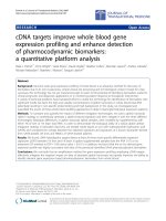
Figure 1: Increasing complexity of information from genome to transcriptome and proteome: gene
expression profiling focuses on the analysis of the transcriptome.
13
Investigators have developed approaches to gene expression analysis that have led to
substantial advances in our understanding of basic biology. Gene expression profiling has been
applied to numerous mammalian tissues, as well as plants, yeast, and bacteria.
10-14
These studies
have examined the effects of treating cells with chemicals and the consequences of
overexpression of regulatory factors in transected cells. Studies also have compared mutant
strains with parental strains to delineate functional pathways. In cancer research, such
investigation has been used to find gene expression changes in transformed cells and metastases,
to identify diagnostic markers, and to classify tumors based on their gene expression profiles (see
Glossary, Appendix B).
15-18
The use of this approach for specific clinical problems, however, is
relatively recent and poses several challenges related to the validity, reproducibility, and
reliability required for use in diagnostic or predictive testing.
In recent years, gene expression profiling has been successfully used in breast cancer
research. For instance, distinct subtypes of breast tumors (such as tumors expressing HER-2)
have been identified as having distinctive gene expression profiles, representing diverse biologic
entities associated with differences in clinical outcome.
19-23
Other investigators
24
have found
gene expression signatures (see Glossary, Appendix B) associated with the ER and lymph node
status of patients, thus identifying subgroups of patients with different clinical outcomes after
therapy. From such studies, investigators have proposed a number of gene expression profiles
that could be used to classify prognosis. In a case-control study from the Netherlands Cancer
Institute (Amsterdam, the Netherlands), one such gene profile, consisting of 70 genes, was
developed using archived frozen tissue from 78 young, node-negative women with breast
cancer.
21
In this study, tumors from patients who suffered rapid relapses after primary therapy
had gene expression profiles that were quite distinct from those who remained disease-free.
These gene expression profiles were then applied to a second validation set of 295 frozen tissue
specimens collected from young women (including 61 patients from the previous cohort),
yielding very similar results.
25
Indeed, it appeared that this 70-gene profile more accurately
predicted outcomes than did the traditional clinical criteria. Results from these preliminary
studies further suggested that gene expression profiling may provide a powerful tool for
estimating prognosis and the likelihood of benefit from selected therapeutic agents.
Breast Cancer Assays on the Market
Three breast cancer gene expression profiling-based assays are now available in the U.S.
These assays investigate the expression of specific panels of genes by measuring their RNA
levels in breast cancer specimens using different techniques, real-time reverse transcription-
polymerase chain reaction (RT-PCR)
26
(Glossary) and DNA microarrays
27
(see Glossary,
Appendix B):
1. The Oncotype DX™ Breast Cancer Assay (Genomic Health, Redwood City, CA)
quantifies gene expression for 21 genes in breast cancer tissue by RT-PCR.
28
This test is
intended to predict the likelihood of recurrence in women of all ages with newly
diagnosed Stage I or II breast cancer, lymph node-negative and ER-positive, who will be
treated with tamoxifen, an anti-estrogen agent.
2. The MammaPrint
®
Test is based on microarray technology, uses the 70-gene expression
profile developed by van’t Veer and colleagues,
21,25
and is marketed by Agendia
(Amsterdam, the Netherlands). This is a prognostic test for women 61 years of age or
14
younger with primary invasive breast cancer who are lymph node-negative and ER-
positive or negative. The company voluntarily submitted this test to the U.S. Food and
Drug Administration for approval under proposed new guidelines for such tests, and
received such approval in February 2007. These guidelines were finalized in July 2007.
3. The Breast Cancer Profiling Test is based on the expression ratio of the two genes
HOXB13 and IL17RB, and for this reason is also known as the H/I ratio test. The assay
was developed by AviaraDX and licensed to Quest Diagnostics, Inc. (Lyndhurst, NJ).
This assay is based on RT-PCR and is offered to treatment-naïve women with ER-
positive, lymph node-negative breast cancer.
All three tests have defined protocols for evaluating the tumor content of the specimens to be
analyzed, preparing the RNA samples, normalizing the raw expression measurements, and
computing summary indices which are related to patient prognosis. The characteristics of the
assays, the gene panels used, and the procedures involved in the analysis are summarized in
Table 1. Detailed descriptions of the genes can be found in Appendix C. These differences
between tests must be taken into account in the evaluation of the available evidence about such
tests. In the following section, we provide a brief description of the technologies that are used. A
more detailed description is presented in Appendix D.
RT-PCR
RT-PCR is a molecular biology technique that combines reverse transcription with real-time
PCR (see Glossary, Appendix B). This methodology allows the quantification of a defined RNA
molecule. It is accomplished by reverse transcription of the specific RNA into its complementary
DNA, followed by amplification of the resulting DNA using PCR. The quantification of the
DNA produced after each round of amplification is accomplished by the use of fluorescent dyes
that intercalate with double-stranded DNA, or by modified DNA oligonucleotide probes (see
Glossary, Appendix B) that fluoresce when hybridized with complementary DNA.
In a PCR template, relative ratios of the product and reagent vary. At the beginning of the
reaction, reagents are in excess, and template and product are present in low concentrations and
do not compete with primer binding, so that the amplification proceeds at a constant, exponential
rate. After this initial phase, the process enters a linear phase of amplification, and then in the
late reaction cycles, the amplification reaches a plateau phase and no more product accumulates
To achieve accuracy and precision, it is necessary to collect quantitative data during the
exponential phase of amplification, since in this phase the reaction is extremely reproducible. In
RT-PCR, this process is automated, and measurements are made at each cycle. Finally, several
implementations of this technique allow multiple DNA species to be measured in the same
sample (multiplex PCR), since fluorescent dyes with different emission spectra may be attached
to the different probes. Multiplex PCR allows internal controls to be co-amplified with the target
transcripts (see Glossary, Appendix B) and permits allele discrimination in single-tube,
homogeneous assays (Figure 2).
15
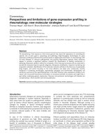
Figure 2: Quantitative RT-PCR. Panel A: PCR reaction using sets of quenched primers and probes. Panel B:
binding of fluorescent probe molecules to double-stranded DNA. Panel C: fluorescence intensity curves for
different dyes and samples: on the x-axis, the number of PCR cycle is shown, and on the y-axis, the
corresponding fluorescence detected is indicated; the dashed line is used to calculate the cycle threshold
for each sample. Panel D: computation of the relative levels of expression.
This technique is extremely sensitive. The development of novel chemistries and
instrumentation platforms has led to widespread adoption of real-time RT-PCR as the method of
choice for quantifying absolute changes in gene expression. Moreover, this technique has
become the preferred method for validating results obtained from microarray analyses and other
techniques that evaluate gene expression changes on a global scale.
Microarrays
The analysis of gene expression by microarray technology is based on the Watson-Crick
pairing of complementary nucleic acid molecules. In this technique, a collection of DNA
sequences, called probes (see Glossary, Appendix B), are “arrayed” on a miniaturized solid
support (microarray) and used to detect the concentration of the corresponding complementary
RNA sequences, called targets (see Glossary, Appendix B), present in a sample of interest. The
advancements made in attaching or synthesizing nucleic acid sequences to solid supports and
robotics have allowed investigators to miniaturize the scale of the reactions, and it is now
possible to assess the expression of thousands of different genes in a single reaction.
29-31
In the basic microarray experiment, RNA harvested from the sample of interest is labeled
with a fluorescent dye and hybridized to the microarray, then incubated in the presence of RNA
from a different sample labeled with a different fluorescent dye. In this two-color experimental
design, samples can be directly compared to one another or to a common reference RNA, and
their relative expression levels can be quantified. After hybridization, gray-scale images
corresponding to fluorescent signals are obtained by scanning the microarray with dedicated
instruments, and the fluorescence intensity corresponding to each gene investigated is quantified
by specific software. After normalization, the intensity of the hybridization signals can be
compared to detect differential expression by using sophisticated computational and statistical
techniques (Figure 3).
16
Figure 3: Schematic model for microarray hybridizations. Panel A: two-color scheme design. Panel B: single-
color design.
Sources of Variability in Gene Expression Analysis
Gene expression analysis poses several general challenges that can affect the reproducibility
and reliability of the measurements obtained. The control of such sources of variability is clearly
a concern when such technologies are used to make decisions about the clinical management of
patients. Given the complexity of the procedures used in this type of investigation, the sources of
uncertainty are multiple, from the preparation of tissue specimens to the computational analysis
used to quantify expression levels.
The first source of variability relates to the various types of specimens that can be used to
prepare the RNA to be used in gene expression analysis, including tissue specimens obtained in
vivo. In this case, the resulting RNA template will be a mixture of the RNA content of all the
cells contained in the specimen, and the relative content of the different cell populations
(malignant vs. normal) present in the specimen processed is a major source of variability in gene
expression. For this reason, special care must be taken when tumors are sampled for gene
expression analysis. In general, macro- or micro-dissection of the samples is performed to ensure
that the specimens contain a sufficient percentage of cancer cells.
A second major source of variability is related to the protocols used to prepare the specimens,
since several alternatives have been used in the field, including the use of formalin-fixed,
paraffin-embedded (FFPE) tumor specimens or laser-captured, micro-dissected (see Glossary,
Appendix B) specimens and fresh or snap-frozen samples. Other factors likely to affect RNA
quality include storage time and the reagents, and particular batches used. Unlike DNA, RNA is
very unstable. The degradation of RNA can be triggered by pH changes as well as by specific
enzymes called ribonucleases (see Glossary, Appendix B) that are present in cells and that can
remain active in the RNA preparation if the RNA isolation is not properly carried out.
Watson-Crick hybridization of complementary nucleic acid moieties is the fundamental
principle that forms the basis of any gene expression analysis. For this reason, sequence selection
and gene annotation (see Glossary, Appendix B) are among the most relevant factors that can
contribute to variability in the analysis of gene expression.