Báo cáo khoa học: Ibuprofen binding to secondary sites allosterically modulates the spectroscopic and catalytic properties of human serum heme–albumin doc
Bạn đang xem bản rút gọn của tài liệu. Xem và tải ngay bản đầy đủ của tài liệu tại đây (243.71 KB, 9 trang )
Ibuprofen binding to secondary sites allosterically
modulates the spectroscopic and catalytic properties
of human serum heme–albumin
Alessandra di Masi
1
, Francesca Gullotta
2,3
, Alessandro Bolli
1
, Gabriella Fanali
4
, Mauro Fasano
4
and
Paolo Ascenzi
1
1 Department of Biology, and Interdepartmental Laboratory of Electron Microscopy, University Roma Tre, Italy
2 Department of Experimental Medicine and Biochemical Sciences, University of Rome ‘Tor Vergata’, Italy
3 Interuniversity Consortium for the Research on the Chemistry of Metals in Biological Systems, Bari, Italy
4 Department of Structural and Functional Biology, and Center of Neuroscience, University of Insubria, Busto Arsizio (VA), Italy
Introduction
Human serum albumin (HSA), the most abundant
protein in plasma, provides a depot and carrier for
many endogenous and exogenous compounds. Among
other roles, HSA affects the pharmacokinetics of many
drugs, holds some ligands in a strained orientation that
results in their metabolic modification, renders poten-
tial toxins harmless by transporting them to disposal
sites, accounts for most of the antioxidant capacity of
human serum, and displays (pseudo)enzymatic proper-
ties [1–8].
HSA is a single nonglycosylated all-a-chain protein,
composed of 585 amino acids, and containing three
homologous domains (labeled I, II, and III). Each
domain is composed of two separate subdo-
mains (named A and B) connected by random coils.
Interdomain helical regions link subdomain IB to
Keywords
allostery; human serum heme–albumin;
ibuprofen binding; modulation of reactivity
and spectroscopic properties; recombinant
truncated human serum heme–albumin
(Asp1–Glu382)
Correspondence
P. Ascenzi, Department of Biology, and
Interdepartmental Laboratory of Electron
Microscopy, University Roma Tre, Viale
Guglielmo Marconi 446, I-00146 Rome, Italy
Fax: +39 06 5733 6321
Tel: +39 06 5733 3200(2)
E-mail:
(Received 14 July 2010, revised 24
November 2010, accepted 6 December
2010)
doi:10.1111/j.1742-4658.2010.07986.x
The ibuprofen primary binding site FA3–FA4 is located in domain III of
human serum albumin (HSA), the secondary clefts FA2 and FA6 being
sited in domains I and II. Here, the thermodynamics of ibuprofen binding
to recombinant Asp1–Glu382 truncated HSA (tHSA)–heme-Fe(III) and
nitrosylated tHSA–heme-Fe(II), encompassing domains I and II only, is
reported. Moreover, the allosteric effect of ibuprofen on the kinetics of
tHSA–heme-Fe(III)-mediated peroxynitrite isomerization and nitrosylated
tHSA–heme-Fe(II) denitrosylation has been investigated. The present data
indicate, for the first time, that the allosteric modulation of tHSA–heme
and HSA–heme reactivity by ibuprofen depends mainly on drug binding to
the FA2 and FA6 secondary sites rather than drug association with the
FA3–FA4 primary cleft. Thus, tHSA is a valuable model with which to
investigate the allosteric linkage between the heme cleft FA1 and the
ligand-binding pockets FA2 and FA6, all located in domains I and II of
(t)HSA.
Abbreviations
FA, fatty acid; HSA, human serum albumin; HSA–heme-Fe, human serum heme-albumin; HSA–heme-Fe(III), ferric HSA–heme-Fe;
HSA–heme-Fe(II), ferrous HSA–heme-Fe; HSA–heme-Fe(II)-NO, nitrosylated HSA–heme-Fe(II); tHSA, truncated HSA; tHSA–heme-Fe(III),
ferric heme tHSA; tHSA–heme-Fe(II), ferrous tHSA–heme-Fe; tHSA–heme-Fe(II)-NO, nitrosylated tHSA–heme-Fe(II).
654 FEBS Journal 278 (2011) 654–662 ª 2011 The Authors Journal compilation ª 2011 FEBS
subdomain IIA, and subdomain IIB to subdo-
main IIIA (Fig. 1) [8,9].
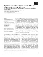
The structural organization of HSA provides several
ligand-binding sites (Fig. 1). HSA has seven binding
clefts hosting chemically diverse ligands, including
fatty acids (FAs), that are labeled FA1–FA7 (Fig. 1).
In particular, FA1 (located in subdomain IB) has
evolved to specifically bind heme, FA3 and FA4 make
up the so-called Sudlow site II (located in subdo-
main IIIA), which preferentially recognizes aromatic
carboxylates with an extended conformation, and FA7
is the so-called Sudlow site I (located in subdo-
main IIA), which binds, in particular, bulky heterocy-
clic anions. Remarkably, warfarin, a coumarinic
anticoagulant drug, and ibuprofen, a nonsteroidal
anti-inflammatory drug, are considered to be stereotyp-
ical ligands for Sudlow sites I and II, respectively. In
contrast to warfarin, ibuprofen has been reported to
also bind to secondary sites in HSA domains I and II
that have been characterized by X-ray crystallographic
and solution spectroscopic studies [1,2,6,8–20].
Recently, the recombinant truncated form of HSA
(tHSA), encompassing residues Asp1–Glu382 (corre-
sponding to domains I and II, containing only the ibu-
profen secondary binding sites), has been preliminarily
characterized [19]. Here, ibuprofen binding to tHSA–
heme-Fe(III) and tHSA–heme-Fe(II) is reported.
Moreover, the allosteric effect of ibuprofen on tHSA–
heme-Fe(III)-mediated peroxynitrite isomerization and
denitrosylation of tHSA–heme-Fe(II)-NO has been
investigated. The present data indicate, for the first
time, that the allosteric modulation of tHSA–heme
and HSA–heme reactivity by ibuprofen depends
mainly on drug binding to the secondary sites FA2
and FA6 rather than drug association with the FA3–
FA4 primary cleft.
Results
Ibuprofen binding to tHSA–heme-Fe(III) and
HSA–heme-Fe(III)]
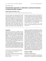
Figure 2A shows the binding isotherm for ibuprofen
binding to tHSA–heme-Fe(III) and HSA–heme-Fe(III).
FA1
FA2
FA7
FA6
Glu382
FA3-FA4
FA5
Fig. 1. HSA structure. The six subdomains of HSA are colored as
follows: IA, blue; IB, cyan; IIA, dark green; IIB, light green; IIIA,
orange; IIIB, red. The heme (red) fits the primary cleft in subdo-
main IB, corresponding to FA1. Sudlow site I (in subdomain IIA,
corresponding to FA7) is occupied by warfarin (purple). Glu382 is
highlighted. Sudlow site II (in subdomain IIIA, corresponding to
FA3–FA4) and FA6 (in subdomain IIB) are occupied by ibuprofen
(magenta). Sites FA2 (at the subdomain I–IIA interface) and FA5 (in
subdomain IIIB) are occupied by myristate (orange). Atomic coordi-
nates were taken from Protein Data Bank entries 1O9X [40], 2BXD,
and 2BXG [6]. For details, see text.
Fig. 2. Ibuprofen binding to tHSA–heme-Fe(III) and HSA–heme
Fe(III). (A) Thermodynamics of ibuprofen binding to tHSA–heme-
Fe(III). The continuous line was calculated according to Eqn (1) by
non-linear regression curve fitting with the following parameters:
K
2
= (1.7 ± 0.2) · 10
)5
M; K
3
= (8.9 ± 0.9) · 10
)4
M; a = 0.18 ±
0.03; and 1 – a = 0.82 ± 0.04. [tHSA–heme-Fe(III)] = 1.9 · 10
)6
M.
(B) Thermodynamics of ibuprofen binding to HSA–heme-Fe(III). The
continuous line was calculated according to Eqn (1) by non-linear
regression curve fitting with the following parameters: K
2
=
(6.9 ± 0.7) · 10
)6
M; K
3
= (8.1 ± 0.7) · 10
)4
M; a = 0.14 ± 0.03; and
1 ) a = 0.86 ± 0.04. [HSA–heme-Fe(III)] = 2.2 · 10
)6
M. The ibupro-
fen concentration corresponds to that of the free ligand. Where not
shown, the standard deviation is smaller than the symbol. All data
were obtained at pH 7.0 and 20.0 °C. For further details, see text.
A. di Masi et al. Ibuprofen binding to truncated HSA secondary sites
FEBS Journal 278 (2011) 654–662 ª 2011 The Authors Journal compilation ª 2011 FEBS 655
The analysis of the data given in Fig. 2A,B, according
to Eqn (1), allowed the determination of K
2
and K
3
values for ibuprofen binding to tHSA–heme-Fe(III)
(1.7 · 10
)5
and 8.9 · 10
)4
m, respectively) and to
HSA–heme-Fe(III) (6.9 · 10
)6
and 8.1 · 10
)4
m,
respectively). The spectroscopic contributions of ibu-
profen binding to the high-affinity and low-affinity
sites of tHSA–heme-Fe(III) and HSA–heme-Fe(III)
[represented by a and (1 – a), respectively, in Eqn (1)]
are 0.18 and 0.82, and 0.14 and 0.86, respectively
(Table 1).
Effect of ibuprofen on peroxynitrite isomerization
by tHSA–heme-Fe(III)
In the absence and presence of tHSA–heme-Fe(III),
the kinetic data of peroxynitrite isomerization were fit-
ted to a single-exponential decay for more than 95%
of their course (Eqn 2). According to the literature
[21], this indicates that the formation of the transient
tHSA–heme-Fe(III)-OONO species represents the
rate-limiting step in catalysis, the conversion of the
tHSA–heme-Fe(III)-OONO complex to tHSA–heme-
Fe(III) and NO
À
3
being faster by at least one order of
magnitude.
In the absence and presence of ibuprofen, the
observed rate constants for tHSA–heme-Fe(III)-cata-
lyzed isomerization of peroxynitrite (l
obs
) increased
linearly with the tHSA–heme-Fe(III) concentration
(Fig. 3A). The analysis of data reported in Fig. 3A,
according to Eqn (3), allowed the determination of val-
ues of the second-order rate constant for peroxynitrite
isomerization by tHSA–heme-Fe(III) (l
on
= 4.3 · 10
5
m
)1
Æs
)1
, corresponding to the slope of the linear plots)
and of the first-order rate constant for peroxynitrite
isomerization in the absence of tHSA–heme-Fe(III)
(l
0
= 2.4 · 10
)1
s
)1
, corresponding to the y-intercept
of the linear plots).
Ibuprofen dose-dependently impairs tHSA–heme-
Fe(III)-mediated isomerization of peroxynitrite
(Fig. 3A,B). Indeed, values of l
on
for tHSA–heme-
Fe(III)-catalyzed isomerization of peroxynitrite
decreased from 4.3 · 10
5
m
)1
Æs
)1
in the absence of ibu-
profen [l
on(top)
] to 5.8 · 10
4
m
)1
Æs
)1
at [ibupro-
fen] = 1.0 · 10
)2
m (Fig. 3A,B). On the other hand,
values of l
0
were unaffected by ibuprofen, being depen-
dent only on CO
2
(Fig. 3A,C). The values of l
on
and
l
0
for tHSA–heme-Fe(III)-catalyzed isomerization of
peroxynitrite determined here are very similar to those
previously reported for HSA–heme-Fe(III)-mediated
peroxynitrite isomerization [21].
The analysis of the dependence of l
on
on ibuprofen
concentration for tHSA–heme-Fe(III)-catalyzed isom-
erization of peroxynitrite (Fig. 3B), according to
Eqn (4), allowed determination of the value of the
dissociation equilibrium constant for ibuprofen binding
to tHSA–heme-Fe(III) (K
3
= 9.3 · 10
)4
m). Under
conditions where [ibuprofen] >> K
3
, tHSA–heme-
Fe(III) did not catalyze the isomerization of peroxy-
nitrite. Indeed, the value of l
obs
(2.6 · 10
)1
s
)1
) was
independent of the tHSA–heme-Fe(III) concentration,
and corresponded to that obtained in the absence of
tHSA–heme-Fe(III) (l
0
= 2.4 · 10
)1
s
)1
).
Ibuprofen binding to tHSA–heme-Fe(II)-NO
Figure 4 shows the binding isotherm for ibuprofen
binding to tHSA–heme-Fe(II)-NO. Analysis of the
dependence of the molar fraction of the ibuprofen-
bound tHSA–heme-Fe(II)-NO (Y) on the ibuprofen
concentration (Fig. 4), according to Eqn (5), allowed
to determine the value of the dissociation equilibrium
Table 1. Values of the dissociation equilibrium constants for ibuprofen binding to tHSA–heme and HSA–heme derivatives obtained by differ-
ent experimental methods. For consistency with previous studies [20,21], equilibrium dissociation constants and Hill coefficients are indi-
cated as K
1
, K
2
, and K
3
, and as n
1
, n
2
, and n
3
, respectively.
Protein Experimental method K
1
(M) n
1
K
2
(M) n
2
K
3
(M) n
3
tHSA–heme-Fe(III)
a
Absorbance spectroscopy –
b
–
b
1.7 · 10
–5
0.98 8.9 · 10
–4
1.01
tHSA–heme-Fe(III)
a
Peroxynitrite isomerization –
b
–
b
–
b
–
b
9.3 · 10
–4
0.99
tHSA–heme-Fe(II)-NO
a
Absorbance spectroscopy –
b
–
b
–
b
–
b
2.1 · 10
–3
0.99
tHSA–heme-Fe(II)-NO
a
Denitrosylation kinetics –
b
–
b
1.3 · 10
–4
0.99 2.5 · 10
–3
1.01
HSA–heme-Fe(III)
a
Absorbance spectroscopy –
b
–
b
6.9 · 10
–6
1.00 8.1 · 10
–4
0.99
HSA–heme-Fe(III)
c
Peroxynitrite isomerization –
b
–
b
–
b
–
b
9.7 · 10
–4
–
d
HSA–heme-Fe(II)-NO
e
Absorbance spectroscopy –
b
–
b
–
b
–
b
2.6 · 10
–3
–
d
HSA–heme-Fe(II)-NO
e
Denitrosylation kinetics 3.1 · 10
–7
–
d
1.7 · 10
–4
–
d
2.2 · 10
–3
–
d
a
pH 7.0 and 20.0 °C. Present study.
b
Not applicable; see text.
c
pH 7.2 and 22.0 °C. From [21].
d
Although values of n
1
, n
2
and n
3
have not
been reported [20,21], the data fit to simple equilibria, implying n
1
=1,n
2
= 1, and n
3
=1.
e
pH 7.0 and 10.0 °C. From [20].
Ibuprofen binding to truncated HSA secondary sites A. di Masi et al.
656 FEBS Journal 278 (2011) 654–662 ª 2011 The Authors Journal compilation ª 2011 FEBS
constant for drug binding to tHSA–heme-Fe(II)-NO
(K
3
= 2.1 · 10
)3
m) (Fig. 4 and Table 1).
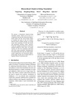
Effect of ibuprofen on tHSA–heme-Fe(II)-NO
denitrosylation
In the absence and presence of ibuprofen, the time
course for NO dissociation from tHSA–heme-Fe(II)-
NO conformed to a single-exponential decay for more
than 95% of its course (Eqn 6). According to the
literature [20], this indicates that the formation of
the transient tHSA–heme-Fe(II) species represents the
rate-limiting step in tHSA–heme-Fe(II)-CO formation.
Values of the first-order rate constant for NO dissocia-
tion from tHSA–heme-Fe(II)-NO (k
off
) were indepen-
dent of wavelength and [CO] in the presence of excess
dithionite.
Values of k
off
for tHSA–heme-Fe(II)-NO denitrosy-
lation increased from 1.5 · 10
)4
s
)1
in the absence of
ibuprofen (k
þ
off
in Eqn 7) to 8.5 · 10
)3
s
)1
in the pres-
ence of 1.0 · 10
)2
m ibuprofen (Fig. 5). The k
off
value
for tHSA–heme-Fe(II)-NO denitrosylation determined
here in the absence of ibuprofen is very similar to that
previously obtained for HSA–heme-Fe(II)-NO denitro-
sylation [20,22].
Analysis of the dependence of k
off
for tHSA–heme-
Fe(II)-NO denitrosylation on the ibuprofen concentra-
tion (Fig. 5), according to Eqn (7), allowed us to
determine the values of the dissociation equilibrium
constants for drug binding to tHSA–heme-Fe(II)-NO
(K
2
= 1.3 · 10
)4
m and K
3
= 2.5 · 10
)3
m) (Fig. 5).
The values of k
off(2)
and k
off(3)
for tHSA–heme-Fe(II)-
NO denitrosylation determined here (1.1 · 10
)3
and
9.1 · 10
)3
s
)1
, respectively) are very similar to those
previously obtained for HSA–heme-Fe(II)-NO denitro-
sylation [20].
Fig. 4. Ibuprofen binding to tHSA–heme-Fe(II)-NO. The continuous
line was calculated according to Eqn (5) with K
3
= (2.1 ± 0.3) ·
10
)3
M). The tHSA–heme-Fe(II)-NO concentration was 4.8 · 10
)6
M.
Where not shown, the standard deviation is smaller than the
symbol. All data were obtained at pH 7.0 (1.0 · 10
)1
M phosphate
buffer) and 20.0 °C. For further details, see text.
Fig. 3. Ibuprofen inhibits peroxynitrite isomerization by tHSA–
heme-Fe(III). (A) Dependence of the pseudo-first-order rate
constant for peroxynitrite isomerization (l
obs
) on the tHSA–heme-
Fe(III) concentration. The peroxynitrite concentration was
2.5 · 10
)4
M. The ibuprofen concentrations were 0.0 M (trace a),
1.5 · 10
)3
M (trace b), and 7.5 · 10
)3
M (trace c). The continuous
lines in (A) were calculated according to Eqn (3) with the following
parameters: trace a, l
on
= (4.3 ± 0.4) · 10
5
M
)1
Æs
)1
and
l
0
= (2.4 ± 0.3) · 10
)1
s
)1
; trace b, l
on
= (1.8 ± 0.2) · 10
5
M
)1
Æs
)1
and
l
0
= (2.8 ± 0.3) · 10
)1
s
)1
; and trace c, l
on
= (6.8 ± 0.4) · 10
4
M
)1
Æs
)1
and l
0
= (2.7 ± 0.3) · 10
)1
s
)1
. (B) Effect of ibuprofen concentration
on the second-order rate constant for tHSA–heme-Fe(III)-catalyzed
isomerization of peroxynitrite (l
on
). The filled symbol on the ordinate
indicates the l
on
value obtained in the absence of ibuprofen [l
on(-
top)
= (4.3 ± 0.4) · 10
5
M
)1
Æs
)1
]. The continuous line was calculated
according to Eqn (4) with l
on(top)
= (4.3 ± 0.4) · 10
5
M
)1
Æs
)1
and
K
3
= (9.3 ± 1.0) · 10
)4
M. (C) Effect of ibuprofen concentration on
the first-order rate constant for tHSA–heme-Fe(III)-catalyzed isomer-
ization of peroxynitrite (l
0
). The filled symbol on the ordinate indi-
cates the l
0
value obtained in the absence of ibuprofen
[(2.4 ± 0.3) · 10
)1
s
)1
]. Values of l
0
are independent of ibuprofen
concentration; the average l
0
value is (2.6 ± 0.3) · 10
)1
s
)1
. Where
not shown, the standard deviation is smaller than the symbol. All
data were obtained at pH 7.0 (1.0 · 10
)1
M phosphate buffer) and
20.0 °C. For further details, see text.
A. di Masi et al. Ibuprofen binding to truncated HSA secondary sites
FEBS Journal 278 (2011) 654–662 ª 2011 The Authors Journal compilation ª 2011 FEBS 657
Discussion
The present results highlight the rol e of ibuprofen second-
ary sites in modulating HSA–heme spectroscopic p roper-
ties and reactivity. Indeed, values of the diss ociation
equilibrium constants for ibuprofen b inding to secon dary
sites present in tHSA–heme (present study) are in excel-
lent agreement with those for drug binding to full-
length HSA–heme (Table 1). However, the abse nce of
domain III in tH SA–heme precludes the binding of
ibuprofen to its primary clef t [19–21] (Table 1).
The analysis of thermodynamic parameters reported
in Table 1 allows the following conclusions to be drawn.
l
Ibuprofen binds to two secondary sites of tHSA–
heme-Fe(III) and HSA–heme-Fe(III) (present study),
affecting the absorption spectra. However, only ibupro-
fen binding to the (t)HSA–heme-Fe(III) secondary site
showing the lowest drug affinity allosterically modulates
peroxynitrite isomerization (present study and [21]).
l
Ibuprofen affects the absorption spectra of tHSA–
heme-Fe(II)-NO (present study) and HSA–heme-
Fe(II)-NO [20] by binding to only one secondary site.
However, the allosteric modulation of tHSA–heme-
Fe(II)-NO denitrosylation (present study) reflects ibu-
profen binding to two secondary sites. By contrast,
HSA–heme-Fe(II)-NO denitrosylation is allosterically
modulated by ibuprofen binding not only to both
secondary sites located in domains I and II, but also
to the primary cleft sited in domain III [20].
l
Values of K
2
and K
3
for ibuprofen binding to
tHSA–heme-Fe(III) and HSA–heme-Fe(III) (present
study) are lower than those for drug binding to tHSA–
heme-Fe(II)-NO (present study) and HSA–heme-
Fe(II)-NO [20]. This indicates that the redox and the
(un)ligated state of the heme Fe atom allosterically
affects ibuprofen binding to (t) HSA–heme.
l
Values of K
2
and K
3
for ibuprofen binding to
tHSA–heme-Fe (present study) are in excellent agree-
ment with those reported for drug binding to HSA–
heme species (present study and [20,21]). Also, the
kinetic parameters for ibuprofen-mediated peroxyni-
trite isomerization by (t)HSA–heme-Fe(III) and for
(t)HSA–heme-Fe(II)-NO denitrosylation are very simi-
lar (present study and [20,21]). This indicates that the
removal of domain I from HSA does not significantly
affect the functional and structural properties of
domains I and II [i.e. of the 1–382 region of (t)HSA].
l
The values of the Hill coefficients (n
1
, n
2
, and n
3
) for
ibuprofen binding to tHSA–heme and HSA–heme
derivatives range between 0.98 and 1.01 (present study
Table 1), indicating that drug association with (t)HSA–
heme is a non-cooperative event, under all the experi-
mental conditions.
l
The ibuprofen-dependent (t)HSA–heme-Fe(III)-
mediated peroxynitrite isomerization and (t)HSA–
heme-Fe(II)-NO denitrosylation reflect drug-dependent
structural changes occurring at the heme-binding
pocket (FA1). Indeed, ibuprofen binding to HSA–heme
has been reported to induce the hexa-coordination of
the heme Fe atom [18,21,23]. Thus, peroxynitrite can-
not bind to the heme Fe(III) atom of hexa-coordinated
HSA–heme-Fe(III), and therefore cannot undergo facil-
itated isomerization [21]. Moreover, the increase in k
off
for NO dissociation from HSA–heme-Fe(II)-NO upon
stabilization of the hexa-coordinated heme-Fe(II)-NO
atom is reminiscent of what has been reported
for abacavir-induced and warfarin-induced hexa-
coordination of HSA–heme-Fe(II)-NO [22], and for
1-methyl-imidazole-mediated hexa-coordination of the
heme-Fe(II)-NO model compound [24].
The ibuprofen primary binding cleft (i.e. Sudlow
site II, formed by the FA3 and FA4 sites) and the
drug secondary pocket (i.e. the FA6 region) have been
substantiated by X-ray crystallography [6]. The third
low-affinity ibuprofen-binding site has been tentatively
identified with the FA2 site. Indeed, it acts as
the modulatory site that controls the FA-induced
conformational switch [18,21,25,26]. Ibuprofen binding
to the third site induces the stabilization of the
hexa-coordinate derivative of the HSA–heme-Fe(III)
and HSA–heme-Fe(II)-NO species, which are instead
predominantly penta-coordinated in the absence of
allosteric effectors. Indeed, high (> 1.0 · 10
)3
m) ibu-
profen concentration clearly induces the coordination
of a histidine nitrogen as the sixth coordination posi-
Fig. 5. Effect of ibuprofen on tHSA–heme-Fe(II)-NO denitrosylation.
The continuous line was calculated according to Eqn (7) with
k
off(2)
= (1.1 ± 0.1) · 10
)3
s
)1
, K
2
= (1.3 ± 0.2) · 10
)4
M, k
off(3)
=
(9.1 ± 1.0) · 10
)3
s
)1
, K
3
= (2.5 ± 0.3) · 10
)3
M), and k
þ
off
=
(1.5 ± 0.2) · 10
)4
s
)1
. The tHSA–heme-Fe(II)-NO concentration was
2.9 · 10
)6
M. Where not shown, the standard deviation is smaller
than the symbol. All data were obtained at pH 7.0 (1.0 · 10
)1
M
phosphate buffer) and 20.0 °C. For further details, see text.
Ibuprofen binding to truncated HSA secondary sites A. di Masi et al.
658 FEBS Journal 278 (2011) 654–662 ª 2011 The Authors Journal compilation ª 2011 FEBS
tion of the heme Fe, as the result of the conforma-
tional transition following ligand binding to FA2 [18].
The present considerations appear to apply only to
ibuprofen, as other Sudlow site II ligands, such as FAs,
diazepam, and diflunisal, display different binding
properties. FAs bind to multiple sites with different
affinities and functional effects. In particular, the FA6
(corresponding to the highest-affinity site for ibuprofen
in tHSA) and the FA7 (also named Sudlow site I and
corresponding to the warfarin-binding pocket) sites
show low affinity for FAs, whereas the FA1 (also named
the heme site) and the FA2 (corresponding to the
regulatory site of the neutral-to-basic conformational
transition) pockets display high affinity for FAs [17].
Therefore, the effect of FAs is opposite to that of
ibuprofen [8]. Moreover, FA binding to the FA1 site
induces heme dissociation from HSA–heme, thus
impairing HSA–heme reactivity [25]. Diazepam has
been reported to bind only to Sudlow site II [17], so it
does not bind to tHSA, which is deprived of domain III
(Fig. 1). Finally, the FA6 site is also the secondary bind-
ing cleft for diflunisal, another Sudlow site II ligand.
However, diflunisal binds not only to the FA6 site
(corresponding to the highest-affinity site for ibuprofen
in tHSA), but also to the FA7 cleft (corresponding to
the warfarin pocket) [17], thus inducing mixed effects,
combining the actions of ibuprofen and warfarin [8].
It is worth noting that the modular architecture of
HSA allows the removal of domain III without this
affecting the conformational stability of the remaining
protein scaffold. Moreover, lost contacts between par-
alogous HSA domains do not impair the correct fold-
ing of single domains, as shown by the agreement
between thermodynamic and kinetic parameters in
HSA and tHSA derivatives. As a whole, the present
data: (a) demonstrate unequivocally, for the first time,
that ibuprofen allosterically modulates the spectro-
scopic and reactivity properties of tHSA–heme and
HSA–heme by binding to the low-affinity secondary
sites FA2 and FA6 rather than associating with the
primary-high affinity cleft FA3–FA4; and (b) reinforce
the idea that HSA could be taken as the prototype of
monomeric allosteric proteins [8,19,27,28]. Finally,
tHSA is a valuable model with which to investigate the
allosteric linkage between ligand-binding pockets
located in domains I and II of HSA.
Experimental procedures
Chemicals
HSA (‡ 96%, essentially FA-free), hemin [Fe(III)-proto-
porphyrin IX] chloride and ibuprofen were purchased
from Sigma-Aldrich (St Louis, MO, USA). Recombinant
tHSA was expressed and purified as previously reported
[19]. NO (Aldrich Chemical Co., Milwaukee, WI,
USA) was purified by flowing it through an NaOH
column in order to remove acidic nitrogen oxides. CO
was purchased from Linde AG (Ho
¨
llriegelskreuth,
Germany).
tHSA–heme-Fe(III) and HSA–heme-Fe(III) were pre-
pared by adding a 0.8 mol heme-Fe(III) per mol tHSA and
HSA (1.0 · 10
)1
m sodium phosphate buffer, pH 7.2) at
20.0 °C [19–21]. The final tHSA–heme-Fe(III) and HSA–
heme-Fe(III) concentrations ranged between 1.9 · 10
)6
and
5.0 · 10
)5
m, and between 2.2 · 10
)6
and 5.0 · 10
)5
m,
respectively. tHSA–heme-Fe(II)-NO (final concentration,
4.8 · 10
)6
m) was obtained, under anaerobic conditions, by
blowing purified NO over the ferrous heme–protein solu-
tion (1.0 · 10
)1
m sodium phosphate buffer, pH 7.0) at
10.0 °C [20].
The ibuprofen stock solution (1.0 · 10
)1
m) was prepared
by dissolving the drug in 1.0 · 10
)1
m phosphate buffer
(pH 7.0) at 20.0 °C [23]. The final ibuprofen concentration
ranged between 1.0 · 10
)6
and 1.0 · 10
)2
m.
Peroxynitrite was synthesized from KO
2
and NO or from
HNO
2
and H
2
O
2
, and stored in small aliquots at )80.0 °C
[29,30]. The peroxynitrite stock solution (2.0 · 10
)3
m) was
diluted immediately before use with degassed 5.0 · 10
)2
m
NaOH to achieve the desired concentration [21,31–35].
Nitrate and nitrite contamination levels were in the ranges
of 0–7% and 8–19% of the peroxynitrite concentration,
respectively [21]. The concentration of peroxynitrite was
determined spectrophotometrically prior to each experiment
by measuring the absorbance at 302 nm (e
302 nm
= 1.705 ·
10
3
m
)1
Æcm
)1
) [29,30].
The CO solution was prepared by keeping the
1.0 · 10
)1
m phosphate buffer solution (pH 7.0) in a
closed vessel with CO at a pressure of 760.0 mmHg, under
anaerobic conditions, at 20.0 °C [20,36].
All of the other chemicals were obtained from Sigma-
Aldrich and Merck AG (Darmstadt, Germany). All prod-
ucts were of analytical or reagent grade, and were used
without further purification.
Ibuprofen binding to tHSA–heme-Fe(III) and
HSA–heme-Fe(III)
Values of the dissociation equilibrium constants for ibupro-
fen binding to tHSA–heme-Fe(III) and HSA–heme-Fe(III)
(i.e. K
2
and K
3
) were obtained spectrophotometrically,
at pH 7.0 (1.0 · 10
)1
m phosphate buffer) and 20.0 °C.
Ibuprofen-dependent absorbance changes were recorded
between 350 and 450 nm. Small aliquots of the ibuprofen
(1.0 · 10
)1
m) stock solution were added to the tHSA–
heme-Fe(III) (1.9 · 10
)6
m) and HSA–heme-Fe(III)
(2.2 · 10
)6
m) solutions, and the ibuprofen-dependent
absorbance changes of tHSA–heme-Fe(III) and HSA–
A. di Masi et al. Ibuprofen binding to truncated HSA secondary sites
FEBS Journal 278 (2011) 654–662 ª 2011 The Authors Journal compilation ª 2011 FEBS 659
heme-Fe(III) were recorded after incubation for 10 min,
after each addition [19,21]. Test measurements performed
after 2 h excluded slow kinetic events.
Ibuprofen binding to tHSA–heme-Fe(III) and HSA–
heme-Fe(III) was analyzed by plotting the molar fraction of
drug–tHSA–heme-Fe(III) and drug–HSA–heme-Fe(III)
complexes (Y) as a function of the free ibuprofen concen-
tration (ranging between 1.0 · 10
)6
m and 1.0 · 10
)2
m).
Data were analyzed according to Eqn (1) [36]:
Y ¼½a Âf½ibuprofen=ðK
2
þ½ibuprofenÞg
þ½ð1 À aÞÂf½ibuprofen=ðK
3
þ½ibuprofenÞg
ð1Þ
where a and 1 ) a are the relative spectroscopic contri-
butions to the total electronic absorbance change of ibu-
profen binding to the high-affinity and low-affinity sites,
respectively.
Effect of ibuprofen on peroxynitrite isomerization
by tHSA–heme-Fe(III)
Kinetic data for peroxynitrite isomerization in the absence
and presence of tHSA–heme-Fe(III) and ibuprofen were
recorded with the SMF-100 rapid-mixing stopped-flow
apparatus (Bio-Logic SAS, Claix, France). The light path
of the observation cuvette was 10 mm, and the dead time
was 1.4 ms. The kinetics were monitored at 302 nm, the
characteristic absorbance maximum of peroxynitrite
(e
302 nm
= 1.705 · 10
3
m
)1
Æcm
)1
) [29,30,35,37]. Kinetic data
were obtained in the absence and presence of tHSA–heme-
Fe(III) (final concentration, 5.0 · 10
)6
to 5.0 · 10
)5
m) and
ibuprofen (final concentration, 1.0 · 10
)6
to 1.0 · 10
)2
m),
by rapid mixing of the protein-buffered solution with the
peroxynitrite solution (final concentration, 2.5 · 10
)4
m).
Kinetic data were obtained at pH 7.0 (1.0 · 10
)1
m
phosphate buffer) and 20.0 °C; no gaseous phase was
present [21].
The kinetics of peroxynitrite isomerization by tHSA–
heme-Fe(III), in the absence and presence of ibuprofen,
were analyzed in the framework of the following minimum
reaction scheme [21]:
tHSAÀhemeÀFeðIIIÞþHOONO
!
l
on
tHSAÀhemeÀFeðIIIÞÀ OONO þ H
þ
tHSAÀhemeÀFeðIIIÞÀOONO
!
fast
tHSAÀhemeÀFeðIIIÞþNO
3
À
Values of the pseudo-first-order rate constant for tHSA–
heme-Fe(III)-mediated peroxynitrite isomerization (l
obs
)
were determined, in the absence and presence of ibuprofen,
at pH 7.0 (1.0 · 10
)1
m phosphate buffer) and 20.0 °C,
from the analysis of the time-dependent absorbance
decrease at 302 nm, according to Eqn (2) [21]:
½peroxynitrite
t
¼½peroxynitrite
i
 e
Àl
obs
Ât
ð2Þ
Values of the second-order rate constant for tHSA–
heme-Fe(III)-mediated peroxynitrite isomerization (l
on
) and
of the first-order rate constant for peroxynitrite isomeriza-
tion in the absence of tHSA–heme-Fe(III) (l
0
) were deter-
mined, in the absence and presence of ibuprofen, at pH 7.0
and 20.0 °C, from the linear dependence of l
obs
on the
tHSA–heme-Fe(III) concentration, according to Eqn (3)
[21,32,38,39]:
l
obs
¼ l
on
½tHSAÀhemeÀFeðIIIÞ þ l
0
ð3Þ
The value of the dissociation equilibrium constant for
ibuprofen binding to tHSA–heme-Fe(III) (K
3
) was deter-
mined, at pH 7.0 (1.0 · 10
)1
m phosphate buffer) and
20.0 °C, from the dependence of l
on
on the free drug con-
centration (ranging between 1.0 · 10
)6
and 1.0 · 10
)2
m).
The effect of the drug concentration on l
on
was analyzed
according to Eqn (4) [21,32,38,39]:
l
on
¼ l
onðtopÞ
Àfðl
onðtopÞ
½ibuprofenÞ = ðK
3
þ½ibuprofenÞg ð4Þ
where l
on(top)
represents the asymptotic value of l
on
under
conditions where [ibuprofen] = 0 (i.e. l
on(top)
= l
on
).
Ibuprofen binding to tHSA–heme-Fe(II)-NO
The value of the dissociation equilibrium constant for ibu-
profen binding to tHSA–heme-Fe(II)-NO (K
3
) was deter-
mined spectrophotometrically, at pH 7.0 (1.0 · 10
)1
m
phosphate buffer) and 20.0 °C. Ibuprofen-dependent absor-
bance changes were recorded between 350 and 450 nm. Small
aliquots of the ibuprofen (1.0 · 10
)1
m) stock solution were
added to the tHSA–heme-Fe(II)-NO (4.8 · 10
)6
m) solution,
and the ibuprofen-dependent absorbance changes of tHSA–
heme-Fe(II)-NO were recorded after incubation for 10 min,
after each addition [20]. Test measurements performed after
2 h excluded slow kinetic events.
Ibuprofen binding to tHSA–heme-Fe(III) was analyzed by
plotting the molar fraction of the drug–tHSA–heme-Fe(II)-
NO complex (Y) as a function of the free ibuprofen concen-
tration (ranging between 1.0 · 10
)6
and 1.0 · 10
)2
m). Data
were analyzed according to Eqn (5) [20]:
Y ¼½ibuprofen=ðK
3
þ½ibuprofenÞ ð5Þ
Effect of ibuprofen on tHSA–heme-Fe(II)-NO
denitrosylation
Values of the first-order rate constant for NO dissociation
from tHSA–heme-Fe(II)-NO (i.e. for NO replacement by
CO; k
off
) were obtained by mixing the tHSA–heme-Fe(II)-
NO (final concentration, 2.9 · 10
)6
m) solution with the CO
(final concentration, 1.0 · 10
)4
to 5.0 · 10
)4
m) solution in
the presence of dithionite (final concentration, 1.0 · 10
)2
m),
under anaerobic conditions, at pH 7.0 (1.0 · 10
)1
m sodium
Ibuprofen binding to truncated HSA secondary sites A. di Masi et al.
660 FEBS Journal 278 (2011) 654–662 ª 2011 The Authors Journal compilation ª 2011 FEBS
phosphate buffer) and 20.0 °C [20], in the absence and pres-
ence of ibuprofen (final concentration, 1.0 · 10
)7
to
1.0 · 10
)2
m). The excess of NO was pumped off gently
before recording of ligand dissociation kinetics [20].
The kinetics were monitored between 350 and 460 nm.
Absorbance electronic spectra were collected every 30 s.
The time course for tHSA–heme-Fe(II)-NO denitrosylation
was fitted to a single-exponential process according to the
minimum reaction mechanism represented by the following
scheme [20]:
tHSAÀhemeÀFeðIIÞÀNO þ CO
!
k
off
tHSAÀhemeÀFeðIIÞÀCO þ NO
Values of k
off
were determined from data analysis
according to Eqn (6) [20]:
½HSAÀhemeÀFe ðIIÞÀNO
t
¼½HSAÀhemeÀFeðIIÞÀNO
i
 e
Àk
off
Ât
ð6Þ
Values of the dissociation equilibrium constants for ibu-
profen binding to tHSA–heme-Fe(II)-NO (K
2
and K
3
) were
obtained from the dependence of k
off
on the free ibuprofen
concentration. Values of K
2
and K
3
were determined from
data analysis, according to Eqn (7) [20]:
k
off
¼ðk
offð2Þ
Âð½ibuprofen=ðK
2
½ibuprofenÞ
þðk
offð3Þ
Âð½ibuprofen=ðK
3
½ibuprofenÞÞ þ k
þ
off
ð7Þ
where k
off(2)
and k
off(3)
indicate values of k
off
occurring at
K
2
< [ibuprofen] < K
3
, and at K
2
< K
3
< [ibuprofen],
respectively, and k
þ
off
is the k
off
value obtained in the
absence of ibuprofen.
Data analysis
Kinetic and thermodynamic data were analyzed with the
matlab program (The Math Works, Natick, MA, USA).
The results are given as mean values of at least four experi-
ments plus or minus the corresponding standard deviation.
Acknowledgements
This work was partially supported by grants from
the Ministero dell’Istruzione, dell’Universita
`
e della
Ricerca of Italy (PRIN 2007ECX29E_002 and Univer-
sity Roma Tre, CLAR 2009, to P. Ascenzi).
References
1 Sudlow G, Birkett DJ & Wade DN (1975) The charac-
terization of two specific drug binding sites on human
serum albumin. Mol Pharmacol 11, 824–832.
2 Peters T Jr, ed. (1996) All about Albumin: Biochemistry,
Genetics and Medical Applications. Academic Press,
San Diego and London.
3 Curry S (2002) Beyond expansion: structural studies on
the transport roles of human serum albumin. Vox Sang
83(Suppl), 1.
4 Kragh-Hansen U, Chuang VT & Otagiri M (2002)
Practical aspects of the ligand-binding and enzymatic
properties of human serum albumin. Biol Pharm Bull
25, 695–704.
5 Fasano M, Curry S, Terreno E, Galliano M, Fanali G,
Narciso P, Notari S & Ascenzi P (2005) The extraordi-
nary ligand binding properties of human serum
albumin. IUBMB Life 57, 787–796.
6 Ghuman J, Zunszain PA, Petitpas I, Bhattacharya AA,
Otagiri M & Curry S (2005) Structural basis of the
drug-binding specificity of human serum albumin.
J Mol Biol 353, 38–52.
7 Ascenzi P, Bocedi A, Notari S, Fanali G, Fesce R &
Fasano M (2006) Allosteric modulation of drug binding
to human serum albumin. Mini Rev Med Chem 6, 483–
489.
8 Ascenzi P & Fasano M (2010) Allostery in a monomeric
protein: the case of human serum heme-albumin.
Biophys Chem 148, 16–22.
9 Curry S (2009) Lessons from the crystallographic
analysis of small molecule binding to human serum
albumin. Drug Metab Pharmacokinet 24, 342–
357.
10 Curry S, Mandelkov H, Brick P & Franks N (1998)
Crystal structure of human serum albumin complexed
with fatty acid reveals an asymmetric distribution of
binding sites. Nat Struct Biol 5, 827–835.
11 Bhattacharya AA, Curry S & Franks NP (2000)
Binding of the general anesthetics propofol and
halothane to human serum albumin: high resolution
crystal structures. J Biol Chem 275, 38731–38738.
12 Bhattacharya AA, Gru
¨
ne T & Curry S (2000) Crystallo-
graphic analysis reveals common modes of binding of
medium and long-chain fatty acids to human serum
albumin. J Mol Biol 303, 721–732.
13 Dockal M, Chang M, Carter DC & Ru
¨
ker F (2000)
Five recombinant fragments of human serum albumin:
tools for the characterization of the warfarin binding
site. Protein Sci 9, 1455–1465.
14 Petitpas I, Bhattacharya AA, Twine S, East M & Curry
S (2001) Crystal structure analysis of warfarin binding
to human serum albumin: anatomy of drug site I.
J Biol Chem 276, 22804–22809.
15 Chuang VTG & Otagiri M (2002) How do fatty acids
cause allosteric binding of drugs to human serum albu-
min? Pharm Res 19, 1458–1464.
16 Hamilton JA (2004) Fatty acid interactions with pro-
teins: what X-ray crystal and NMR solution structures
tell us. Prog Lipid Res 43, 177–199.
17 Simard JR, Zunszain PA, Hamilton JA & Curry S
(2006) Location of high and low affinity fatty acid
binding sites on human serum albumin revealed by
A. di Masi et al. Ibuprofen binding to truncated HSA secondary sites
FEBS Journal 278 (2011) 654–662 ª 2011 The Authors Journal compilation ª 2011 FEBS 661
NMR drug-competition analysis. J Mol Biol 361,
336–351.
18 Nicoletti FP, Howes BD, Fittipaldi M, Fanali G,
Fasano M, Ascenzi P & Smulevich G (2008) Ibuprofen
induces an allosteric conformational transition in the
heme complex of human serum albumin with significant
effects on heme ligation. J Am Chem Soc 130,
11677–11688.
19 Fanali G, Pariani G, Ascenzi P & Fasano M (2009)
Allosteric and binding properties of Asp1–Glu382 trun-
cated recombinant human serum albumin: an optical
and NMR spectroscopic investigation. FEBS J 276,
2241–2250.
20 Ascenzi P, di Masi A, de Sanctis G, Coletta M &
Fasano M (2009) Ibuprofen modulates allosterically
NO dissociation from ferrous nitrosylated human serum
heme–albumin by binding to three sites. Biochem
Biophys Res Commun 387, 83–86.
21 Ascenzi P, di Masi A, Coletta M, Ciaccio C, Fanali G,
Nicoletti FP, Smulevich G & Fasano M (2009) Ibupro-
fen impairs allosterically peroxynitrite isomerization by
ferric human serum heme–albumin. J Biol Chem 284,
31006–31017.
22 Ascenzi P, Imperi F, Coletta M & Fasano M (2008)
Abacavir and warfarin modulate allosterically kinetics
of NO dissociation from ferrous nitrosylated human
serum heme–albumin. Biochem Biophys Res Commun
369, 686–691.
23 Baroni S, Mattu M, Vannini A, Cipollone R, Aime S,
Ascenzi P & Fasano M (2001) Effect of ibuprofen and
warfarin on the allosteric properties of haem–human
serum albumin: a spectroscopic study. Eur J Biochem
268, 6214–6220.
24 Kharitonov VG, Sharma VS, Magde D & Koesling D
(1997) Kinetics of nitric oxide dissociation from five-
and six-coordinate nitrosyl hemes and heme proteins,
including soluble guanylate cyclase. Biochemistry 36,
6814–6818.
25 Fanali G, Fesce R, Agrati C, Ascenzi P & Fasano M
(2005) Allosteric modulation of myristate and Mn(III)-
heme binding to human serum albumin: optical and
NMR spectroscopy characterization. FEBS J 272,
4672–4683.
26 Fanali G, Bocedi A, Ascenzi P & Fasano M (2007)
Modulation of heme and myristate binding to human
serum albumin by anti-HIV drugs. An optical and
NMR spectroscopic study. FEBS J 274, 4491–4502.
27 Fanali G, Ascenzi P & Fasano M (2007) Effect of
prototypic drugs ibuprofen and warfarin on global
chaotropic unfolding of human serum heme–albumin: a
fast-field-cycling 1H-NMR relaxometric study. Biophys
Chem 129, 29–35.
28 Fanali G, De Sanctis G, Gioia M, Coletta M, Ascenzi
P & Fasano M (2009) Reversible two-step unfolding of
heme–human serum albumin: a
1
H-NMR relaxometric
and circular dichroism study. J Biol Inorg Chem 14,
209–217.
29 Bohle DS, Glassbrenner PA & Hansert B (1996)
Syntheses of pure tetramethylammonium peroxynitrite.
Methods Enzymol 269, 302–311.
30 Koppenol WH, Kissner R & Beckman JS (1996)
Syntheses of peroxynitrite: to go with the flow or on
solid grounds? Methods Enzymol 269, 296–302.
31 Herold S, Exner M & Boccini F (2003) The mechanism
of the peroxynitrite mediated oxidation of myoglobin in
the absence and presence of carbon dioxide. Chem Res
Toxicol 16, 390–402.
32 Herold S, Kalinga S, Matsui T & Watanabe Y (2004)
Mechanistic studies of the isomerization of peroxynitrite
to nitrate catalyzed by distal histidine metmyoglobin
mutants. J Am Chem Soc 126, 6945–6955.
33 Ascenzi P & Fasano M (2007) Abacavir modulates
peroxynitrite-mediated oxidation of ferrous nitrosylated
human serum heme–albumin. Biochem Biophys Res
Commun 353, 469–474.
34 Ascenzi P & Visca P (2008) Scavenging of reactive
nitrogen species by mycobacterial truncated hemoglo-
bins. Methods Enzymol 436, 317–337.
35 Goldstein S & Mere
´
nyi G (2008) The chemistry of
peroxynitrite: implications for biological activity.
Methods Enzymol 436, 49–61.
36 Antonini E & Brunori M (1971) Hemoglobin and Myo-
globin in their Reactions with Ligands. North Holland
Publishing Co., Amsterdam and London.
37 Goldstein S, Lind J & Mere
´
nyi G (2005) Chemistry
of peroxynitrites and peroxynitrates. Chem Rev 105,
2457–2470.
38 Herold S, Matsui T & Watanabe Y (2001) Peroxynitrite
isomerization catalyzed by His64 myoglobin mutants.
J Am Chem Soc 123, 4085–4086.
39 Herold S & Kalinga S (2003) Metmyoglobin and methe-
moglobin catalyze the isomerization of peroxynitrite to
nitrate. Biochemistry 42, 14036–14046.
40 Zunszain PA, Ghuman J, Komatsu T, Tsuchida E &
Curry S (2003) Crystal structural analysis of human
serum albumin complexed with hemin and fatty acid.
BMC Struct Biol 3,6.
Ibuprofen binding to truncated HSA secondary sites A. di Masi et al.
662 FEBS Journal 278 (2011) 654–662 ª 2011 The Authors Journal compilation ª 2011 FEBS