TUBERCULOSIS PNEUMONIA AS A PRIMARY CAUSE OF RESPIRATORY FAILURE-REPORT OF TWO CASES pdf
Bạn đang xem bản rút gọn của tài liệu. Xem và tải ngay bản đầy đủ của tài liệu tại đây (57.3 KB, 7 trang )
Indian Journal of Tuberculosis
TUBERCULOSIS PNEUMONIA AS A PRIMARY CAUSE OF RESPIRATORY
FAILURE-REPORT OF TWO CASES
Case Report
M.M. Puri
1
, Subodh Kumar
2
, Brahma Prakash
3
, K. Lokender
4
, A . Jaiswal
1
and D. Behera
5
(Received on 20.10.2009; Accepted on 29.10.2009)
Summary: Tuberculosis (TB) is one of the treatable diseases rarely causing Acute Respiratory Failure (ARF). Hypoxic
respiratory failure is often fatal in miliary tuberculosis and acute tuberculous bronchopneumonia. We describe two
patients of tuberculous pneumonia with ARF who were successfully treated with early appropriate anti-tuberculosis
therapy.
Key words: Tuberculosis, Pneumonia, Acute Respiratory Failure, Miliary Tuberculosis
[Indian J Tuberc 2010; 57: 41-47]
INTRODUCTION
Tuberculosis as a primary cause of
respiratory failure is an uncommon occurrence
1
with
an incidence of 1.5% in patients hospitalized with
pulmonary TB
2
. Patients with miliary or disseminated
disease are especially prone to develop respiratory
failure. Tuberculous Pneumonia has rarely been
identified as a cause of ARF
3-4
. Acute tuberculous
pneumonia presents as parenchymal consolidation
with or without endobronchial spread mimicking
bacterial pneumonia. It probably represents an
exudative hypersensitivity reaction to
tuberculoprotein, rather than actual inflammation
caused by the Mycobacterium tuberculosis organism
per se. These infiltrates can appear within a matter
of days and can clinically simulate acute bacterial
pneumonia. Anti-tubercular treatment has been
considered to be an important factor affecting
patient’s outcome. In this report, we describe two
patients with tuberculosis who developed ARF and
were successfully treated with early appropriate anti-
tuberculosis therapy. The experience with these
cases serves to re-emphasize the importance of
quality sputum examination
routinely for AFB in
patients at risk of TB with
respiratory failure and
pneumonic infiltrates, particularly in endemic areas
since specific and effective therapy for tuberculosis
is available in contrast to most other conditions
associated with respiratory failure.
Case-1. Mr. “S” 18 years’ old, young male, non-
smoker, unmarried, student, resident of Delhi was
admitted on 17 May 2008 with complaints of
haemoptysis, fever and shortness of breath for one
week’s duration. A year ago, he had haemoptysis
and for which he had taken 6 month Category-I
anti-tuberculosis treatment from a DOTS centre,
as a case of smear positive pulmonary tuberculosis.
He improved with the treatment except for some
residual early morning cough with expectoration and
was declared cured after sputum examination for
AFB. He remained well for two months, when in
May, 2008 he developed cough, expectoration, fever
and haemoptysis. Fever was insidious in onset, high
grade, and more in the evening. Cough was
productive with yellow colour sputum and
sometimes mixed with blood. There were 2-3
episodes of haemoptysis in one week with 150-200
ml of blood loss in each episode. He was admitted
at a peripheral hospital and received two units of
whole blood transfusion. There was no history of
1. Chest Physician 2. Senior Resident 3. Junior Resident ( Specialist Grade I) 4. Chest Physician (Specialist Grade II)
5. Director
Department of Tuberculosis and Respiratory Diseases , LRS Institute of Tuberculosis and Respiratory Diseases, New Delhi.
Corresspondence: Dr. M.M. Puri, Chest Physician (Specialist Grade I), LRS Institute of Tuberculosis and Respiratory
Diseases, Sri Aurbindo Marg, New Delhi-110030.E-mail :
Indian Journal of Tuberculosis
alcohol abuse or smoking. During his hospitalization,
his breathlessness progressively increased and he
was referred to our institute on 17
th
May, 2008. On
admission, examination revealed a lethargic young
man in respiratory distress; blood pressure was 100/
70 mm Hg, pulse rate was 136 beats per minute,
temperature was 102
O
F and respiratory rate was
42 per minute. Abnormal findings were limited to
coarse crackles all over the chest. There was no
pallor, cyanosis, lymphadenopathy, or pedal edema.
Laboratory data revealed the following values:
hemoglobin 13.0g%; total leucocytes count 10,800/
cu.mm (90 per cent polymorphonuclear leukocytes,
9 per cent lymphocytes and 1 percent monocytes);
serum protein, 5.8 g/dl; serum albumin 3.0 g/dl
total bilirubin levels, 0.51mg/dl; SGOT levels 52U/
L; SGPT levels 50U/L; and alkaline phosphatase
level, 261U/L. Serum electrolytes were: Na
+
140
mmol/L; K
+
4.2mmol/L; Cl
–
106 m mol/L and the
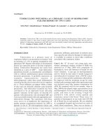
result of urine analysis were normal. A chest X-ray
film (Fig. 1A) showed multiple ill-defined confluent
nodular opacities widely distributed throughout both
the lungs. The nodules were larger than those of
miliary shadows. Multiple small cavities were present
in left upper zone. Initial therapy with ceftriaxone 1
gm intravenously 12 hourly, Hydrocortisone 100 mg
intravenously 8 hourly was begun. Gram stain of
sputum revealed scanty leukocytes and no
pathogens. A culture of sputum grew normal oral
flora. Sputum smear examination was positive for
acid-fast bacilli. Anti-tuberculosis treatment (Cat II)
thrice a week with injection streptomycin 0.75 gram
intramuscular, capsule rifampicin 450 mg., tablet
isoniazid 600 mg., tablet pyrazinamide 1500 mg. and
tablet ethambutol 1200 mg was started. On
admission oxygen saturation (SaO
2
) at room air
was 74%. The SaO
2
rose to 87.5% with oxygen
delivered by venturi mask (FIO
2
=32%). Arterial
blood gas analysis at FIO
2
of 32 % showed the
following values: pH, 7.409; PaCO
2
: 50.8mm Hg;
and PaO
2
:53.3 mm Hg. PaO
2
/ FiO
2
ratio was 167.
With FiO
2
of 50 percent he was able to maintain SaO
2
above 90 per cent. His breathlessness gradually
improved and on 3
rd
day respiratory rate settled to
28 per minute with pulse rate of 100 beats per
minute. Repeat chest X-ray on 5
th
day did not reveal
any marked change, however patient was able to
maintain SaO
2
above 90 % at room air and his fever
also responded. Within two weeks, he was doing
his routine activity and oxygen therapy was stopped.
Anti-tuberculosis therapy was continued and
Corticosteroids were tapered and stopped. At three
weeks he was maintaining oxygen saturation (SaO
2
)
of 96% at room air. After a week, he had high
grade fever and found to have urinary tract infection
and cholelithiasis. He was treated for urinary tract
Fig. 1a: CXR-PA view on admission revealing
poorly defined nodules in upper and lower
lung fields of both lungs. The nodules
are larger than those of miliary shadows.
A cavity is seen in right upper lung field.
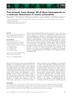
Fig. 1b: After 8 months, chest X-ray PA view
revealing healing of cavity and fibrotic
lesions in upper and middle lung fields of
both lungs with complete resolution of
nodular densities.
M.M. PURI ET AL42
Indian Journal of Tuberculosis
infection. He was discharged and referred to DOTS
centre for completion of Anti tuberculosis treatment.
His hospital stay was 57 days. Chest X-ray after
completion of eight months of Cat II anti-
tuberculosis treatment revealed significant resolution
of opacities (Fig. 1 B).
Case- 2. Mr. “M S” a 25-year-young male,
rickshaw-puller, non smoker attended Chest OPD
of LRS Institute of Tuberculosis and respiratory
Diseases on 14 February, 2009 with symptoms of
cough, expectoration, fever, breathlessness on
exertion, loss of weight and appetite for three weeks.
Ten days ago he had haemoptysis with loss of 10-
15 ml of blood followed by blood mixed in sputum
for three days. In the past, ten years ago he had
inadequate unsupervised daily anti-tuberculosis
treatment for three months. In last three years he
had history of abuse of 250 ml alcohol per day. His
sputum smear examination was found to be positive
for AFB. He was referred to DOTS centre for
Category-II anti-tuberculosis treatment. After four
days, before the initiation of ATT, he was
hospitalized on 23 February 2009 with high grade
fever and respiratory distress. Examination revealed
a cachectic man with BMI of 14.7 Kg/ m
2
in
respiratory distress, with blood pressure of 130/76
mm Hg, pulse rate of 116 per minute, temperature
of 101
o
F, and respiratory rate of 36 per minute.
Pertinent findings included coarse crackles all over
the chest and hepatomegaly. Laboratory data revealed
the following values: hemoglobin 10.7g%; total
leucocytes count 22,900/cu mm (80 per cent
polymorphonuclear leucocytes and 20 per cent
lymphocytes); blood urea nitrogen (BUN) level, 23.8
mg/100 ml; bilirubin level, 0.77 mg/100 ml; alkaline
phosphatase level, 1134 international units (IU)/L;
and serum glutamic-oxaloacetic transaminase
(SGOT) level, 964 IU/L and serum glutamic-Pyruvic
transaminase (SGPT) level, 737 IU/L. The findings
from urine analysis were normal. Six weeks into
treatment sputum culture grew Mycobacterium
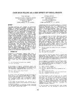
tuberculosis. The chest X-ray film taken on
admission (Fig. 2a) showed widespread poorly
defined opacities in upper and lower lung fields of
both lungs with air bronchogram. Analysis of arterial
blood gases while the patient breathing oxygen 4
liter per minute by nasal canulae revealed a pH of
7.406, an arterial oxygen pressure (PaO
2
) of 45.3
mm Hg; and arterial carbon dioxide tension (PaCO
2
),
56.6 mm Hg. PaO
2
/ FiO
2
ratio of 142. Gradually,
he was able to maintain oxygen saturation(SaO
2
)
above 90% with 0.50 FiO
2
with venturi mask and
arterial blood gas levels revealed : pH, 7.421; PaO
2
of 85.1 mm Hg; PaCO
2
of 58.0 mm Hg. Initial
therapy included Injection Ceftriaxone 2 gram
Fig. 2a: Chest X-ray P.A. view on admission
revealing widespread poorly defined
opacities in upper and lower lung fields
of both lungs. Note the air bronchogram.
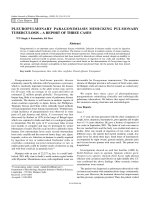
Fig. 2b: Chest X-ray PA view after one week
revealing partial resolution of opacities.
Note air bronchogram is more prominent.
TUBERCULOUS PNEUMONIA WITH ACUTE RESPIRATORY FAILURE 43
Indian Journal of Tuberculosis
intravenously 12 hourly, Hydrocortisone 100 mg
intravenously 12 hourly along with anti-tuberculosis
treatment (ATT) . Gram stain of sputum revealed
scanty leukocytes and no pathogens. A culture of
sputum grew normal oral flora. Therapy with
ceftriaxone was stopped. In view of deranged liver
functions, modified daily ATT with injection
streptomycin 0.75 gram intramuscular, tablet
ethambuol 1000 mg and levofloxacin 750 mg was
started. Repeat X-ray chest after a week showed
radiological improvement with partial resolution of
opacities (Fig. 2b). Corticosteroids were tapered
and stopped in two weeks’ time. With the
improvement of liver functions thrice a week,
Category-II ATT was initiated on 16th March 2009
with injection streptomycin 0.75 gram
intramuscular, capsule rifampicin 450 mg., tablet
isoniazid 600 mg., tablet pyrazinamide 1500 mg. and
tablet ethambutol 1200 mg. Gradually in 8 weeks
he was able to maintain 90% oxygen saturation
(SaO
2
) at room air. Anti-tuberculosis therapy was
continued and at 12 weeks he was maintaining
oxygen saturation (SaO
2
) of 94% at room air. He
was discharged and referred to DOTS centre for
completion of Anti tuberculosis treatment. On
discharge, arterial blood gas levels revealed: pH,
7.471; PaO
2
of 67.5 mm Hg; PaCO
2
of 37.1 mm
Hg. His hospital stay was 111 days.
DISCUSSION
Identification of the primary cause of
respiratory distress is vital for the initiation of
appropriate therapy. Active pulmonary TB is a rare
primary cause of ARF and is associated with very
high mortality
1
. Important factors contributing to
ARF in TB patients included Gram-negative
pneumonia and/or sepsis, chronic obstructive
pulmonary disease, prior TB with anti-TB medication
non-compliance, and malignancy
5
. Tuberculosis
occurring initially as an acute, rapidly progressive
pneumonia is unusual because tubercle bacilli
multiply only once every 18 to 24 hours as opposed
to most pathogenic bacteria, which can multiply
every 20 to 30 minutes. It is suggested that for this
to occur, either a massive number of tubercle bacilli
or, more likely tuberculoprotein must be aspirated
causing an acute exudative hypersensitivity reaction
into new areas of the lung
6
. This is usually due
to liquefaction of a caseous lesion and its erosion
into a bronchus. Perforation of a lymph node into
a bronchus may be a factor in this reaction
7
. Acute
exudative consolidation was experimentally
induced by intratracheal injection of acid-fast
organisms into rabbits
8
and the importance of a
hypersensitivity reaction associated with
tuberculoprotein was confirmed by intratracheal
injections of tuberculin into normal and
tuberculous guinea pigs
9
. In human tuberculosis,
Rich
6
found areas of fresh pneumonic exudates
surrounding caseous foci in which few or no acid-
fast bacilli were seen and attributed this peripheral
reaction to a hypersensitivity response to
tuberculoprotein. The pathogenesis of ARDS in
both pulmonary and miliary tuberculosis is not
well understood. It has been speculated that
lipoarabinomannan, a component of mycobacterial
cell wall has been shown to induce the production
of tumor necrosis factor in human macrophages,
which might contribute to the development of
ARDS.
Acute tuberculous pneumonia is
characterized by fever, productive cough, and high
temperature with signs of severe toxicity and of
consolidation, presence of large confluent dense
shadows on the chest x-ray film involving at least
one lobe; and tubercle bacilli in the sputum
7
. The
rapidly progressive course of acute tuberculous
pneumonia can mimic bacterial pneumonia. The
longer duration of symptoms before admission is
the most important factor differentiating TB from
other infectious
causes
3
. In acute tuberculous
pneumonia symptoms are usually less than one
month
10
. The reported mean duration of symptoms
before
admission was 29 ± 28 days in various
studies
3, 12 - 13
. Patients with acute massive tuberculous
pneumonia are subjectively better than those with a
bacterial pneumonia of equal extent with less pleuritic
pain, toxemia, and dyspnea. It is difficult to
differentiate radiologically
between TBP and severe
bacterial pneumonia as causes of ARF,
meaning
accurate diagnosis can be delayed. The white blood
cell count is rarely greater than 15,000/cu mm, and
the temperature is usually between 37.8
0
C and
38.9
0
C (100
0
F and 102
0
F)
11
.
M.M. PURI ET AL44
Indian Journal of Tuberculosis
The hospital mortality for tuberculosis patients
mechanically ventilated compared with that for non-
tuberculous pneumonia with similar APACHE II
scores was significantly worse (69% VS 36%, p <
0.025 )
14
. In tuberculous pneumonia patients (TBP)
advanced age, longer duration of symptoms before
hospital admission, the presence of shock unrelated
to sepsis and non-use of steroids influence patient
survival
12
. Advanced age and presence of shock
unrelated to sepsis were independently associated
with poor outcomes; however, the use of
corticosteroids was a favourable prognostic factor
for patients with TBP
12
. Acute respiratory distress
syndrome (ARDS) is the most common reasons for
ICU admission of patients with TB
13, 15
. ARDS is
characterized by
16-17
: (a) acute onset , (b) bilateral
infiltrates on chest radiograph ,(c) pulmonary artery
wedge pressure < 18 mmHg (obtained by pulmonary
artery catheterization), if this information is available;
if unavailable, then lack of clinical evidence of left
ventricular failure suffices (d) if PaO
2
:FiO
2
< 300
mmHg acute ling injury (ALI) is considered to be
present (e) if PaO
2
:FiO
2
< 200 mmHg acute
respiratory distress syndrome (ARDS) is considered
to be present. Sharma et al reported ARDS in
1.06% hospitalized adult patients with active TB
18
.
Presence of duration of illness beyond 30 days at
presentation, absolute lymphocyte count < 1625/
mm
3
and serum ALT > 100 IU were independent
predictors of ARDS development. Patients with
APACHE II score >18; those with APACHE II score
<18 in the presence of hyponatraemia and PaO
2
/
FIO
2
ratio <108.5 were likely to have more mortality
18
.
ARF is more common in miliary tuberculosis
than in tuberculous bronchopneumonia and also has
a worse prognosis
19
. ARDS caused by miliary TB
is associated with a high fatality rate
20
. The mortality
rate in the patients with pulmonary tuberculosis
requiring mechanical ventilation is very high, with
multiple
organ failure and consolidation on chest
radiographs
21
. Concomitant extra pulmonary TB,
ARDS or DIC were more common in the MTB group
than in the TBP group (p<0.05). However, there
were no significant differences in hospital mortality
rates between the two groups (68.2 vs 58.3%, p =
0.385)
12
. Treatment has been considered to be an
important factor affecting patient’s outcome
14, 22-23
.
Higher mortality is present in patients who did not
receive an optimal treatment with a triple combination
including INH and RMP. Impaired liver function
being a major reason to withdraw the INH and RMP;
however, other causes have been also described
24
.
With anti tuberculosis treatment, diffusing capacities
may improve rapidly. Usually it returns to normal in
three weeks, however sometimes defect persists for
months. In three weeks, our patient with tuberculous
bronchopneumonia, was able to maintain oxygen
saturation (SaO
2
) of 96% at room air, while patient
with tuberculous pneumonia in case 2 was able to
maintain SaO
2
of 90% at room air at six weeks.
Organ dysfunction in critically ill patients
is another cause for changes in the treatment
regimen. Although the duration from exhibition of
first symptoms to treatment onset was outlined as a
crucial factor to mortality
25
. HIV status and longer
history of symptoms such as fever or haemoptysis
did not show a significantly worse outcome in study
reported by Kim et al
12
. Nosocomial infection during
ICU stay has significant impact on the mortality of
critically ill TB patients
26
. Interestingly, some of the
predictive factors for mortality, such as nosocomial
infections, were actually related to the intensive care
procedures.
The beneficial effects of corticosteroids in the
management of TBP with ARF are suggested by
several reports. Mycobacterial antigen can induce
release of pyrogens from monocytes, lymphokines
from specifically sensitised lymphocytes and
cytokines, such as tumor necrosis factor, from
macrophages and peripheral blood mononuclear
cells, which may be responsible for constitutional
symptoms and tissue damage
27
. Corticosteroids can
inhibit the release and activities of lymphokines and
cytokines. The granulomatous host response to TB
may paradoxically protect sequestered M.
tuberculosis from anti-TB therapy. The adjuvant
corticosteroids may be beneficial in permitting anti-
TB drugs to penetrate into granulomas, by disrupting
granuloma formation
28
. Tuberculous pneumonia
patients with ARF receiving corticosteroid therapy
showed a lower mortality rate than those not
receiving corticosteroid therapy (56.7% vs 77.8%;
p = 0.046)
12
. The use of systemic corticosteroid
TUBERCULOUS PNEUMONIA WITH ACUTE RESPIRATORY FAILURE 45
Indian Journal of Tuberculosis
was based entirely on the attending physician’s
decision and/or the patient’s underlying condition;
and the corticosteroids did not affect either the
duration of mechanical ventilation (p = 0.603) or
arterial oxygenation i.e. arterial oxygen tension/
inspiratory oxygen fraction (p = 0.182)
12
. Further
randomised controlled
trials are necessary to clarify
the role of corticosteroids
in the management of
tuberculous pneumonia with ARF.
Any benefit of
adjuvant corticosteroids in patients with miliary
Tuberculosis with ARF is not clear, since only limited
evidence with conflicting results are available. A
beneficial response was observed in one study
29
,
but such benefit was not documented in another
30
.
CONCLUSION
Identification of the primary cause of
respiratory distress is vital for the initiation of
appropriate therapy. Active pulmonary TB is a
rare primary cause of ARF and is associated with
very high mortality. Acute pneumonia probably
represents an exudative hypersensitivity
reaction to tuberculoprotein, rather than actual
inflammation caused by the Mycobacterium
tuberculosis organism per se. These infiltrates
can appear within a matter of days and can
clinically simulate acute bacterial pneumonia.
Tuberculosis should be considered in the
differential diagnosis of acute pneumonic
infiltrates with respiratory failure.
REFERENCES
1. Keim LW, Schuldt S, Bedell GN. Tuberculosis in the
intensive care unit. Heart Lung 1977; 6: 624–34.
2. Levy H, Kallenbach JM, Feldman C, Thorburn JR,
Abramowitz JA. Acute respiratory failure in active
tuberculosis. Crit Care Med 1987; 15: 221–25
3. Septimus EJ, Awe RJ, Greenberg SD, Raleigh JW. Acute
tuberculous pneumonia. Chest 1977; 71: 774–75.
4. Erbes R, Oettel K, Raffenberg M, et al. Characteristics
and outcome of patients with active pulmonary
tuberculosis requiring intensive care. Eur Respir J 2006;
27: 1223–28.
5. Frame RN, Johnson MC, Eichenhorn MS, Bower GC,
Popovich J Jr. Active tuberculosis in the medical
intensive care unit: a 15-year retrospective analysis.
Crit Care Med 1987; 15:1012–14.
6. Rich AR: The Pathogenesis of Tuberculosis. Springfield,
Ill, Charles C Thomas, 1944, pp828-30.
7. Schwartz WS, Moyer EE. The management of massive
tuberculous pneumonia. Am Rev Tuberc 1951; 64: 41-9.
8. Austrian CH, Willis HS: The pulmonary effects of
intratracheal injections of tubercie bacilli and blood in
rabbits. Am Rev Tuberc. 1926; 14: 306.
9. Larson A, Long ER: Experimental tuberculin
pneumonia. Am Rev Tuberc 1931; 23: 41-4.
10. Calix AA, Ziskind MM, Leonard AJ, et al. Acute
tuberculous pneumonia in the Negro. Am Rev Tuberc
1953; 68: 382-92.
11. Pinner M: Pulmonary Tuberculosis in the Adult.
Springfield,Ill, Charles C Thomas, 1946, p 241.
12. Kim Y. J., Pack K. M., E., et.al. Pulmonary tuberculosis
with acute respiratory failure. Eur Respir J 2008; 32:
1625-30.
13. Zahar JR, Azoulay E, Klement E, et al. Delayed
treatment contributes to mortality in ICU patients with
severe active pulmonary tuberculosis and acute
respiratory failure. Intensive Care Med 2001; 27: 513–
20.
14. Penner C, Roberts D Kunimoto D, Manfreda J, Long R.
Tuberculosis as a primary cause of respiratory failure
requiring mechanical ventilation. Am J Respir Crit Care
Med.1995 Mar; 151(3 Pt 1):867-72.
15. Sydow M, Schauer A, Crozier TA, Burchardi H. Multiple
organ failure in generalized disseminated tuberculosis.
Respir Med 1992; 86: 517–19.
16. Irwin RS, Rippe JM (2003). Irwin and Rippe’s Intensive
Care Medicine (5th ed. ed.). Lippincott Williams &
Wilkins.
17. Bernard G, Artigas A, Brigham K, Carlet J, Falke K,
Hudson L, Lamy M, Legall J, Morris A, Spragg R (1994).
“The American-European Consensus Conference on
ARDS. Definitions, mechanisms, relevant outcomes,
and clinical trial coordination”. Am J Respir Crit Care
Med 149 (3 Pt 1): 818–24.
18. Sharma S K, Mohan A, Banga A, Saha P K, Guntupalli K
K. Predictors of development and outcome in patients
with acute respiratory distress syndrome due to
tuberculosis. Int J Tuberc Lung Dis 2006 Apr; 10(4):
429-35.
19. Shneerson J M. Respiratory failure in tuberculosis: a
modern perspective. Clin Med 2004 Jan-Feb; 4(1):72-
6.
20. Kim JY. Park YB, Kim Y S, Kang S B, Shin JW, Park I
W,Choi B W. Miliary tuberculosis and acute respiratory
distress syndrome. Int J Tuberc Lung Dis 2003 Apr;
7(4): 359-64.
21. P.L. Lee, J.S. Jerng, Y.L. Chang, C.F. Chen, P.R. Hsueh,
C.J. Yu, P.C. Yang and K.T. Luh. Patient mortality of
active pulmonary tuberculosis requiring mechanical
ventilation. Eur Respir J 2003; 22: 141-47.
22. Pablos-Me´ndez A, Sterling TR, Frieden TR. The
relationship between delayed or incomplete treatment
and all cause mortality in patients with tuberculosis.
JAMA 1996; 276: 1223–28.
23. Sacks LV, Pendle S. Factors related to in-hospital deaths
in patients with tuberculosis. Arch Intern Med 1998;
158: 1916–22.
M.M. PURI ET AL46
Indian Journal of Tuberculosis
24. Schaberg T, Rebhan K, Lode H. Risk factors for side-
effects of isoniazid, rifampin and pyrazinamide in
patients hospitalized for pulmonary tuberculosis. Eur
Respir J 1996; 9: 2026–30.
25. Mathur P, Sacks L, Auten G, Sall R, Levy C, Gordin
F.Delayed diagnosis of pulmonary tuberculosis in city
hospitals. Arch Intern Med 1994; 154: 306–10.
26. Dahmash NS, Arora SC, Fayed DF, Chowdhury
MN.Infections in critically ill patients: experience in
MICU at a major teaching hospital. Infection 1994; 22:
264–70.
27. Muthuswamy P, Hu TC, Carasso B, et al. Prednisolone
as adjunctive therapy in the management of pulmonary
tuberculosis. Report of 12 cases and review of the
literature. Chest 1995; 107: 1621–30.
28. Wallis RS. Reconsidering adjuvant immunotherapy for
tuberculosis. Clin Infect Dis 2005; 41 :201–08.
29. Sun TN, Yang JY, Zheng LY, et al. Chemotherapy and
its combination with corticosteroids in acute miliary
tuberculosis in adolescents and adults: analysis of 55
cases. Chin Med J 1981; 94: 309–14.
30. Massaro D, Katz S, Sachs M. Choroidal tubercles. A clue
to haemotogenous tuberculosis. Ann Intern Med 1964;
60: 231-41.
TUBERCULOUS PNEUMONIA WITH ACUTE RESPIRATORY FAILURE 47