NUTRITION AND IMMUNE FUNCTION potx
Bạn đang xem bản rút gọn của tài liệu. Xem và tải ngay bản đầy đủ của tài liệu tại đây (3.46 MB, 440 trang )
Nutrition00
4/9/02
4:03 PM
Page i
FRONTIERS IN NUTRITIONAL SCIENCE
This series of books addresses a wide range of topics in nutritional science. The
books are aimed at advanced undergraduate and graduate students,
researchers, university teachers, policy makers and nutrition and health professionals. They offer original syntheses of knowledge, providing a fresh perspective on key topics in nutritional science. Each title is written by a single author
or by groups of authors who are acknowledged experts in their field. Titles
include aspects of molecular, cellular and whole body nutrition and cover
humans and wild, captive and domesticated animals. Basic nutritional science,
clinical nutrition and public health nutrition are each addressed by titles in the
series.
Editor in Chief
P Calder, University of Southampton, UK
.C.
Editorial Board
A. Bell, Cornell University, Ithaca, New York, USA
F. Kok, Wageningen University, The Netherlands
A. Lichtenstein, Tufts University, Massachusetts, USA
I. Ortigues-Marty, INRA, Thiex, France
P Yaqoob, University of Reading, UK
.
K. Younger, Dublin Institute of Technology, Ireland
Titles available
1. Nutrition and Immune Function
Edited by P Calder, C.J. Field and H.S. Gill
.C.
Nutrition00
4/9/02
4:03 PM
Page ii
Nutrition00
4/9/02
4:03 PM
Page iii
NUTRITION AND IMMUNE
FUNCTION
Edited by
Philip C. Calder
University of Southampton, UK
Catherine J. Field
University of Alberta, Canada
and
Harsharnjit S. Gill
Massey University, New Zealand
CABI Publishing
in association with
The Nutrition Society
Nutrition00
4/9/02
4:03 PM
Page iv
CABI Publishing is a division of CAB International
CABI Publishing
CAB International
Wallingford
Oxon OX10 8DE
UK
Tel: +44 (0)1491 832111
Fax: +44 (0)1491 833508
E-mail:
Web site: www.cabi-publishing.org
CABI Publishing
10 E 40th Street
Suite 3203
New York, NY 10016
USA
Tel: +1 212 481 7018
Fax: +1 212 686 7993
E-mail:
© CAB International 2002. All rights reserved. No part of this publication may
be reproduced in any form or by any means, electronically, mechanically, by
photocopying, recording or otherwise, without the prior permission of the copyright owners.
A catalogue record for this book is available from the British Library, London,
UK.
Library of Congress Cataloging-in-Publication Data
Nutrition and immune function / edited by Philip C. Calder.
p. cm. -- (Frontiers in nutritional science ; no. 1)
Includes bibliographical references and index.
ISBN 0-85199-583-7
1. Immune system. 2. Nutrition. 3. Natural immunity. 4. Dietary
supplements. I. Calder, Philip C. II. Series.
QR182 .N88 2002
616.07Ј9--dc21
2002004470
ISBN 0 85199 583 7
Typeset in Souvenir Light by Columns Design Ltd, Reading
Printed and bound in the UK by Biddles Ltd, Guildford and King’s Lynn
Nutrition00
4/9/02
4:03 PM
Page v
Contents
Contributors
vii
Preface
ix
Part 1: The Immune System
1. The Immune System: an Overview
G. Devereux
2. Evaluation of the Effects of Nutrients on Immune Function
S. Cunningham-Rundles
1
21
Part 2: Individual Nutrients, Infection and Immune Function
3. Effect of Post-natal Protein Malnutrition and Intrauterine
Growth Retardation on Immunity and Risk of Infection
R.K. Chandra
41
4. Fatty Acids, Inflammation and Immunity
P.C. Calder and C.J. Field
57
5. Arginine and Immune Function
M.D. Duff and J.M. Daly
93
6. Glutamine and the Immune System
P.C. Calder and P. Newsholme
109
7. Sulphur Amino Acids, Glutathione and Immune Function
R.F. Grimble
133
v
Nutrition00
4/9/02
4:03 PM
Page vi
vi
Contents
8. Vitamin A, Infection and Immune Function
R.D. Semba
151
9. Antioxidant Vitamins and Immune Function
D.A. Hughes
171
10. Zinc, Infection and Immune Function
A.S. Prasad
193
11. Role of Iron in Immunity and Infection
S. Kuvibidila and B.S. Baliga
209
12. Selenium and the Immune System
229
R.C. McKenzie, J.R. Arthur, S.M. Miller, T.S. Rafferty and G.J. Beckett
13. Probiotics and Immune Function
H.S. Gill and M.L. Cross
251
Part 3: Nutrition and Immunity through the Life Cycle
14. Role of Local Immunity and Breast-feeding in Mucosal
Homoeostasis and Defence against Infections
P. Brandtzaeg
273
15. Food Allergy
E. Opara
321
16. Exercise and Immune Function – Effect of Nutrition
E.W. Petersen and B.K. Pedersen
347
17. Nutrition and Ageing of the Immune System
B. Lesourd, A. Raynaud-Simon and L. Mazari
357
18. Nutrition, Infection and Immunity:
Public Health Implications
A. Tomkins
375
Index
413
Nutrition00
4/9/02
4:03 PM
Page vii
Contributors
J.R. Arthur, Division of Cell Integrity, Rowett Research Institute, Bucksburn,
Aberdeen AB21 9SB, UK.
B.S. Baliga, Department of Pediatrics, College of Medicine, University of South
Alabama, 2451 Fillingim Street, Mobile, AL 36617, USA.
G.J. Beckett, Department of Clinical Biochemistry, University of Edinburgh,
Lauriston Building, Royal Infirmary of Edinburgh, Edinburgh EH3 9YW, UK.
P Brandtzaeg, Laboratory for Immunohistochemistry and Immunopathology
.
(LIIPAT), Institute of Pathology, University of Oslo, Rikshospitalet, N-0027
Oslo, Norway.
P Calder, Institute of Human Nutrition, School of Medicine, University of
.C.
Southampton, Bassett Crescent East, Southampton SO16 7PX, UK.
R.K. Chandra, Janeway Child Health Centre, Room 2J740, 300 Prince Philip
Drive, St John’s, Newfoundland, Canada A1B 3V6.
M.L. Cross, Institute of Food, Nutrition and Human Health, Massey University,
Palmerston North, New Zealand.
S. Cunningham-Rundles, Immunology Research Laboratory, Division of
Hematology and Oncology, Department of Pediatrics, New York
Presbyterian Hospital, Cornell University Weill Medical College, 1300
York Avenue, New York, NY 10021, USA.
J.M. Daly, Department of Surgery, New York Presbyterian Hospital, Weill
Medical College of Cornell University and 525 East 68th Street, New
York, NY 10021, USA.
G. Devereux, Aberdeen Royal Infirmary, Foresterhill, Aberdeen AB25 2ZD, UK.
M.D. Duff, Department of Surgery, New York Presbyterian Hospital, Weill
Medical College of Cornell University and 525 East 68th Street, New
York, NY 10021, USA.
C.J. Field, Nutrition and Metabolism Research Group, Department of
Agricultural, Food and Nutritional Science, 4–10 Agriculture Forestry
Centre, University of Alberta, Edmonton, Canada T6G 2P5.
vii
Nutrition00
viii
4/9/02
4:03 PM
Page viii
Contributors
H.S. Gill, Institute of Food, Nutrition and Human Health, Massey University,
Palmerston North, New Zealand.
R.F. Grimble, Institute of Human Nutrition, School of Medicine, University of
Southampton, Bassett Crescent East, Southampton SO16 7PX, UK.
D.A. Hughes, Nutrition and Consumer Science Division, Institute of Food
Research, Norwich Research Park, Norwich NR4 7UA, UK.
S. Kuvibidila, Division of Hematology/Oncology, Department of Pediatrics,
Louisiana State University Health Sciences Center, Box T8-1, 1542
Tulane Avenue, New Orleans, LA 70112, USA.
B. Lesourd, Département de Gérontologie Clinique, Hôpital Nord du CHU de
Clermont-Ferrand, BP 56, 63118 Cébazat, France.
R.C. McKenzie, Department of Medical and Radiological Sciences, University of
Edinburgh, Lauriston Building, Royal Infirmary of Edinburgh, Edinburgh
EH3 9YW, UK. Corresponding address: Section of Dermatology, Lauriston
Building, Royal Infirmary of Edinburgh, Edinburgh EH3 9YW, UK.
L. Mazari, Département de Gérontologie Clinique, Hôpital Nord du CHU de
Clermont-Ferrand, BP 56, 63118 Cébazat, France.
S.M. Miller, Department of Clinical Biochemistry, University of Edinburgh,
Lauriston Building, Royal Infirmary of Edinburgh, Edinburgh EH3 9YW, UK.
P Newsholme, Department of Biochemistry, Conway Institute of Biomolecular
.
and Biomedical Research, University College Dublin, Belfield, Dublin 4,
Republic of Ireland.
E. Opara, School of Life Sciences, Kingston University and Faculty of Health
and Social Care Sciences, St George’s Hospital Medical School, Penrhyn
Road, Kingston upon Thames, Surrey KT1 2EE, UK.
B.K. Pedersen, Copenhagen Muscle Research Centre and Department of
Infectious Diseases, Rigshospitalet, University of Copenhagen, Tagensvej
20, 2200 Copenhagen N, Denmark.
E.W. Petersen, Copenhagen Muscle Research Centre and Department of
Infectious Diseases, Rigshospitalet, University of Copenhagen, Tagensvej
20, 2200 Copenhagen N, Denmark.
A.S. Prasad, Division of Hematology and Oncology, Department of Internal
Medicine, Wayne State University School of Medicine, 4201 St Antoine,
Detroit, MI 48201, USA.
T.S. Rafferty, Department of Medical and Radiological Sciences, University of
Edinburgh, Lauriston Building, Royal Infirmary of Edinburgh, Edinburgh
EH3 9YW, UK.
A. Raynaud-Simon, Département de Gérontologie Clinique, Hôpital Nord du
CHU de Clermont-Ferrand, BP 56, 63118 Cébazat, France.
R.D. Semba, Department of Opthalmology, Johns Hopkins University School
of Medicine, Baltimore, MD 21205, USA. Correspondence address:
550 North Broadway, Suite 700, Baltimore, MD 21205, USA.
A. Tomkins, Centre for International Child Health, Institute of Child Health, 30
Guilford Street, London WC1N 1EH, UK.
Nutrition00
4/9/02
4:03 PM
Page ix
Preface
‘This fortress built by Nature for herself
Against infection and hand of war’
(The Tragedy of King Richard II, Act II, Scene I, lines 43 and 44,
William Shakespeare)
It has been recognized for many years that states of nutrient deficiency are associated with an impaired immune response and with increased susceptibility to infectious disease. In turn, infection can affect the status of several nutrients, thus setting
up a vicious circle of under nutrition, compromised immune function and infection. Thus, the focus of much of the research into nutrition, infection and immunity has been related to identifying the effects of nutrient deficiencies upon
components of the immune response (often using animal models) and, importantly, upon attempts to reduce the occurrence and severity of infectious diseases
(often in human settings). Although it is often considered that the problems of
under nutrition relate mainly to the developing world, they exist in developed
countries, especially among the elderly, individuals with eating disorders, alcoholics, patients with certain diseases and premature and small-for-gestational-age
babies. Thus, immunological problems in these groups probably relate, at least in
part, to nutrient status. In addition, many diseases that exist among the apparently
well nourished have a strong immunological component and it is now recognized
that at least some of these diseases relate to diet and that their course may be
modified by specific changes in nutrient supply. Examples of these diseases
include rheumatoid arthritis, Crohn’s disease and atopic diseases. Furthermore, it
is now recognized that atherosclerosis, a disease strongly influenced by diet, has
an immunological component. Thus, understanding the interaction between nutrition and immune function is fundamental to understanding the development of a
multitude of communicable and non-communicable diseases and will offer preventive and therapeutic opportunities to control the incidence and severity of
those diseases. It is also now recognized that immune dysfunction plays a role in
ix
Nutrition00
x
4/9/02
4:03 PM
Page x
Preface
the events that follow trauma, burns or major surgery, and which, in some
patients, can lead to organ failure and death. Thus, understanding the interaction
between nutrition and immune function is fundamental in designing therapies to
control the severity of these aberrant responses and to improve patient outcome.
The aim of this book is to provide a state of the art description of the interaction between nutrition and immunity, with an emphasis on the mechanism(s)
of action of the nutrients concerned and the impact on human health. The
book is divided into three parts.
Part 1 contains two chapters. The first is an overview of the immune system, its components and the way in which it functions and regulates its activities. The second is a description, using examples from the recent literature, of
the methodological approaches that can be used to investigate the impact of
altered nutrient supply on immune outcomes.
Part 2 contains 11 chapters. The first of these is devoted to the immunological effects of protein–energy malnutrition and of intrauterine growth retardation. Each of a further nine chapters is devoted to a specific nutrient or a family
of nutrients: fatty acids, arginine, glutamine, sulphur amino acids, vitamin A,
antioxidant vitamins (vitamins C and E and -carotene), zinc, iron and selenium are all featured. The final chapter in this section deals with probiotics, an
emerging area of great interest.
Part 3 contains five chapters. Rather than taking a nutrient-led approach
these deal with changes in immune competence through the life cycle and with
how nutrition affects these. The development of immunity in early life and the
role of breast-feeding are covered in one chapter. A later chapter describes the
current understanding of the impact of ageing on immune competence and
how nutrient status plays a role in accelerating or delaying this ageing process.
In between these two chapters are chapters on food allergy and on the influence of exercise on immune function. The final chapter tackles the public
health implications of our increased understanding of the interaction between
nutrition and immune function and poses important questions about how we
can harness our knowledge for greater benefit.
Each chapter of this book includes an extensive reference list, which will
guide the reader who wishes to seek more detailed information.
The true remedy for all diseases is Nature’s remedy. Nature and Science
are at one … Nature has provided, in the white corpuscles as you call them
– in the phagocytes as we call them – a natural means of devouring and
destroying all disease germs. There is at bottom only one genuinely
scientific treatment for all diseases, and that is to stimulate the phagocytes.
Stimulate the phagocytes… The phagocytes are stimulated; they devour
the disease; and the patient recovers.
The Doctor’s Dilemma, Bernard Shaw
P Calder, C.J. Field and H.S. Gill
.C.
Editors
December 2001
Nutrition00
4/9/02
4:03 PM
Page vii
Contributors
J.R. Arthur, Division of Cell Integrity, Rowett Research Institute, Bucksburn,
Aberdeen AB21 9SB, UK.
B.S. Baliga, Department of Pediatrics, College of Medicine, University of South
Alabama, 2451 Fillingim Street, Mobile, AL 36617, USA.
G.J. Beckett, Department of Clinical Biochemistry, University of Edinburgh,
Lauriston Building, Royal Infirmary of Edinburgh, Edinburgh EH3 9YW, UK.
P Brandtzaeg, Laboratory for Immunohistochemistry and Immunopathology
.
(LIIPAT), Institute of Pathology, University of Oslo, Rikshospitalet, N-0027
Oslo, Norway.
P Calder, Institute of Human Nutrition, School of Medicine, University of
.C.
Southampton, Bassett Crescent East, Southampton SO16 7PX, UK.
R.K. Chandra, Janeway Child Health Centre, Room 2J740, 300 Prince Philip
Drive, St John’s, Newfoundland, Canada A1B 3V6.
M.L. Cross, Institute of Food, Nutrition and Human Health, Massey University,
Palmerston North, New Zealand.
S. Cunningham-Rundles, Immunology Research Laboratory, Division of
Hematology and Oncology, Department of Pediatrics, New York
Presbyterian Hospital, Cornell University Weill Medical College, 1300
York Avenue, New York, NY 10021, USA.
J.M. Daly, Department of Surgery, New York Presbyterian Hospital, Weill
Medical College of Cornell University and 525 East 68th Street, New
York, NY 10021, USA.
G. Devereux, Aberdeen Royal Infirmary, Foresterhill, Aberdeen AB25 2ZD, UK.
M.D. Duff, Department of Surgery, New York Presbyterian Hospital, Weill
Medical College of Cornell University and 525 East 68th Street, New
York, NY 10021, USA.
C.J. Field, Nutrition and Metabolism Research Group, Department of
Agricultural, Food and Nutritional Science, 4–10 Agriculture Forestry
Centre, University of Alberta, Edmonton, Canada T6G 2P5.
vii
Nutrition00
viii
4/9/02
4:03 PM
Page viii
Contributors
H.S. Gill, Institute of Food, Nutrition and Human Health, Massey University,
Palmerston North, New Zealand.
R.F. Grimble, Institute of Human Nutrition, School of Medicine, University of
Southampton, Bassett Crescent East, Southampton SO16 7PX, UK.
D.A. Hughes, Nutrition and Consumer Science Division, Institute of Food
Research, Norwich Research Park, Norwich NR4 7UA, UK.
S. Kuvibidila, Division of Hematology/Oncology, Department of Pediatrics,
Louisiana State University Health Sciences Center, Box T8-1, 1542
Tulane Avenue, New Orleans, LA 70112, USA.
B. Lesourd, Département de Gérontologie Clinique, Hôpital Nord du CHU de
Clermont-Ferrand, BP 56, 63118 Cébazat, France.
R.C. McKenzie, Department of Medical and Radiological Sciences, University of
Edinburgh, Lauriston Building, Royal Infirmary of Edinburgh, Edinburgh
EH3 9YW, UK. Corresponding address: Section of Dermatology, Lauriston
Building, Royal Infirmary of Edinburgh, Edinburgh EH3 9YW, UK.
L. Mazari, Département de Gérontologie Clinique, Hôpital Nord du CHU de
Clermont-Ferrand, BP 56, 63118 Cébazat, France.
S.M. Miller, Department of Clinical Biochemistry, University of Edinburgh,
Lauriston Building, Royal Infirmary of Edinburgh, Edinburgh EH3 9YW, UK.
P Newsholme, Department of Biochemistry, Conway Institute of Biomolecular
.
and Biomedical Research, University College Dublin, Belfield, Dublin 4,
Republic of Ireland.
E. Opara, School of Life Sciences, Kingston University and Faculty of Health
and Social Care Sciences, St George’s Hospital Medical School, Penrhyn
Road, Kingston upon Thames, Surrey KT1 2EE, UK.
B.K. Pedersen, Copenhagen Muscle Research Centre and Department of
Infectious Diseases, Rigshospitalet, University of Copenhagen, Tagensvej
20, 2200 Copenhagen N, Denmark.
E.W. Petersen, Copenhagen Muscle Research Centre and Department of
Infectious Diseases, Rigshospitalet, University of Copenhagen, Tagensvej
20, 2200 Copenhagen N, Denmark.
A.S. Prasad, Division of Hematology and Oncology, Department of Internal
Medicine, Wayne State University School of Medicine, 4201 St Antoine,
Detroit, MI 48201, USA.
T.S. Rafferty, Department of Medical and Radiological Sciences, University of
Edinburgh, Lauriston Building, Royal Infirmary of Edinburgh, Edinburgh
EH3 9YW, UK.
A. Raynaud-Simon, Département de Gérontologie Clinique, Hôpital Nord du
CHU de Clermont-Ferrand, BP 56, 63118 Cébazat, France.
R.D. Semba, Department of Opthalmology, Johns Hopkins University School
of Medicine, Baltimore, MD 21205, USA. Correspondence address:
550 North Broadway, Suite 700, Baltimore, MD 21205, USA.
A. Tomkins, Centre for International Child Health, Institute of Child Health, 30
Guilford Street, London WC1N 1EH, UK.
Nutrition Chapter 01
4/9/02
1
4:03 PM
Page 1
The Immune System:
an Overview
GRAHAM DEVEREUX
Aberdeen Royal Infirmary, Foresterhill, Aberdeen AB25 2ZD, UK
Introduction
To parasitic microorganisms, the human body represents an extremely attractive environment and source of nutrients. Consequently, we live under the constant threat of overwhelming attack by viruses, bacteria and parasites.
Microorganisms evolve more rapidly than humans, so that the nature of the
microbiological threat to humans is changing as exposure to new or variant
organisms occurs. To combat this potentially devastating threat, evolution has
provided humans with a highly sophisticated, flexible and potent immune
system, which is able to protect humans against rapidly evolving microorganisms. The critical protective function of the immune system becomes apparent
when it fails. The inherited and acquired immunodeficiency states are characterized by increased susceptibility to all infections, including those organisms
not normally considered to be pathogenic.
The immune system is a two-edged sword: the extremely potent and toxic
biological effector mechanisms of the immune system can destroy not only
threatening microorganisms but also body tissues. Usually the tissue destruction
and inflammation associated with the eradication of a microbiological threat
are acceptable and functionally insignificant. However, in several human
diseases, the immunologically associated tissue destruction and inflammation
are harmful, e.g. tuberculosis, fulminant hepatitis and meningitis, and, although
this may be advantageous to the species as a whole, the effect on the individual may be devastating. It is because of their potential to destroy tissues that
the effector mechanisms of the immune system are very tightly regulated.
Failure of these regulatory mechanisms results in the full might of the immune
system being inappropriately directed against body tissues and the development of autoimmune diseases, such as rheumatoid arthritis, systemic lupus
erythematosus (SLE), myasthenia gravis and multiple sclerosis. If immune
responses are directed against innocuous targets, such as allergens or transplanted
© CAB International 2002. Nutrition and Immune Function
(eds P.C. Calder, C.J. Field and H.S. Gill)
1
Nutrition Chapter 01
4/9/02
4:03 PM
Page 2
2
G. Devereux
organs, the resulting immunologically mediated tissue damage and inflammation are the basis of allergy and transplant rejection. The immune response to
microorganisms is divided into two general systems: innate (natural) immunity
and adaptive (specific, acquired) immunity.
Innate Immunity (Medzhitov and Janeway, 1997)
Innate immunity comprises physical barriers, soluble factors and phagocytic
cells, which can be considered to provide an immediate first line of defence
against invading microorganisms. Innate immunity is encoded in the germline, it
is very similar among normal individuals and there is no memory effect, with reexposure to the same pathogen eliciting the same response. Innate immunity is
directed against molecular structures of microorganisms that are essential for
microbial survival, present in many types of microorganisms and unique to
pathogenic microorganisms, e.g. bacterial lipopolysaccharides and teichoic
acids. The major cells of innate immunity are phagocytic macrophages and neutrophils, which possess surface receptors specific for common bacterial surface
molecules. Engagement of these receptors triggers phagocytosis and destruction
of the microorganism. Although pathogenic microorganisms have evolved
mechanisms to evade the innate immune response, e.g. bacterial capsules, they
are usually eliminated by the adaptive immune response, which is able to mount
an appropriate neutralizing response directed specifically against the invading
microorganism. Although innate immunity is inflexible, it provides a very rapid
first line of defence until the more powerful and flexible adaptive immune
response takes effect. The innate and adaptive immune systems are not independent; the innate immune response probably influences the character of the
adaptive response and the effector arm of the adaptive response harnesses
innate effector mechanisms, such as phagocytes (Fearon and Locksley, 1996).
Adaptive Immunity (Huston, 1997)
Cells and tissues involved
It is the functional properties of B lymphocytes (B-cells) and T lymphocytes
(T-cells) that enable the adaptive immune response to be extremely powerful
and yet, at the same time, regulated and flexible. Lymphocytes originate in the
bone marrow from a common lymphoid stem cell. Further development and
maturation of B- and T-cells occur in the bone marrow and thymus, respectively. Mature T- and B-cells enter the bloodstream; specific receptors enable
adherence to capillary endothelial cells and migration into peripheral lymphoid
organs. These comprise the lymph nodes, spleen, bronchial-associated lymphoid tissue, mucosa-associated lymphoid tissue and gut-associated lymphoid
tissues (tonsils, adenoids, appendix and the Peyer’s patches of the small intestine). Peripheral lymphoid organs are highly anatomically and functionally
organized to facilitate interactions between migrating lymphocytes and antigens
Nutrition Chapter 01
4/9/02
4:03 PM
Page 3
The Immune System
3
transported actively (by antigen-presenting cells) or passively (in lymph) to the
peripheral lymphoid organs from the tissues. Lymphocytes that do not
encounter antigen re-enter the bloodstream by way of efferent lymphatics and
the thoracic duct. The functional consequence of this T- and B-cell circulation is
that all of the body tissues are under continuous immunological surveillance for
invading pathogens.
Clonal expansion of lymphocytes
Each T- and B-cell bears surface receptors with a single antigenic specificity, but
the specificity of each individual lymphocyte is different. The population of
T- and B-cells in a human is able to recognize an estimated 1011 different antigens. This huge receptor repertoire is generated during lymphocyte development by the random rearrangement of a limited number of receptor genes
(Fanning et al., 1996). Although the immune system is able to recognize a huge
number of antigens, any single antigen is recognized by relatively few lymphocytes, typically 1 in 1,000,000; consequently, there are not enough lymphocytes
to immediately eliminate an invading microorganism. When a lymphocyte antigen receptor engages its complementary antigen, the lymphocyte ceases migration, enlarges and rapidly proliferates so that, within 3–5 days, there are a large
number of effector cells, each specific for the initiating antigen. This antigen-driven clonal expansion accounts for the characteristic delay of several days before
adaptive immune responses become effective. Some of the effector cells generated by clonal expansion are very long-living and are the basis of the immunological memory that is characteristic of adaptive immunity. Functionally,
immunological memory enables a more rapid and effective immune response
upon re-exposure to microorganisms. In contrast to innate immunity, the antigen
specificities of adaptive immunity reflect the individual’s lifetime exposure to
infectious agents and will consequently differ between individuals.
B-cells, immunoglobulins and humoral immunity
Protection against certain infections can be transferred by serum. This is called
humoral immunity and is mediated by circulating antibodies, also known as
immunoglobulins (Ig). The cell surface of B-cells incorporates the membranebound form of immunoglobulin, which functions as an antigen-specific receptor. Engagement of surface Ig by complementary antigen initiates B-cell
proliferation, with the majority of the resulting cells transforming into plasma
cells secreting large amounts of antibody with the same specificity as the progenitor B-cell surface Ig receptor.
Structure of immunoglobulins (Huston, 1997)
The general structural features of antibodies can be demonstrated by
immunoglobulin G (IgG) (molecular weight 150 kDa), which comprises two
Nutrition Chapter 01
4/9/02
4:03 PM
Page 4
4
G. Devereux
identical heavy chains (50 kDa each) and two identical light chains (25 kDa
each). Each of the two heavy chains is linked to the other and to a light chain
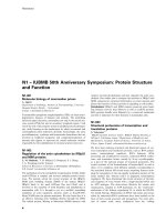
by disulphide bonds, giving a roughly Y-shaped molecule (Fig. 1.1). Each
immunoglobulin molecule possesses two antigen-binding (Fab) sites, each with
the same specificity situated at the amino ends of the light and heavy chains.
The Fab segments are divided into a variable (V) and a constant (C) region and
the structural diversity of the V regions produces the diversity of antibody specificity. There are five main types of heavy chain, , ␦, ␥, ␣ and ⑀, which confer
differing functional properties between the five major classes (isotypes) of
immunoglobulin, namely IgM, IgD, IgG, IgA and IgE, respectively. The functional activity of antibodies resides at the carboxyl-terminal (Fc) region of the
heavy chains.
Immunoglobulin isotypes
The antigen specificity of antibodies is mediated by the two antigen-binding
sites, while the differing Fc regions of the various immunoglobulin isotypes
engage differing effector mechanisms. Monomeric IgM and IgD act as B-cell
surface antigen-specific receptors. The affinity of each IgM antigen-binding site
tends to be low; however, IgM in serum usually polymerizes into a pentamer
with ten antigen-binding sites, which give the antibody high binding strength.
IgM dominates the initial humoral immune response; however, IgG and
IgA predominate later, although IgE is prominent during an allergic response.
This process is known as isotype switching and is the consequence of DNA
Antigen-binding site
Antigen-binding site
Variable region
Fab
Light chain
Constant region
Fc
Heavy chain
Fig. 1.1. Schematic representation of an IgG molecule. The two domains of each of the two
light chains are termed VL and CL. The four domains of each of the two heavy chains are
termed VH, CH1, CH2 and CH3. The amino terminal (dark) domain of each chain is the
variable region and it is the tips of these regions that form the two antigen-binding sites of the
molecule.
Nutrition Chapter 01
17/9/02
12:21 PM
Page 5
The Immune System
5
rearrangements in the genes encoding for the C (but not the V) regions of the
heavy chains (Stavnezer, 1996). Isotype switching results in differing classes of
antibodies with differing functional properties, although antigen specificity
remains constant. Isotype switching is dependent on T-cells and their secretion
of cytokines, with interleukin-4 (IL-4) inducing B-cell switching to IgE; this is
antagonized by interferon-␥ (IFN-␥) (Pene et al., 1988). Switching to IgA is promoted by transforming growth factor- (TGF-), in combination with IL-10
(Defrance et al., 1992). In addition to isotype switching, as the humoral
immune response matures, point mutations in the immunoglobulin V-region
genes occur. A T-cell-dependent process, known as affinity maturation, selects
those B-cells with point mutations producing antibodies with an increased affinity for the stimulating antigen. Consequently, as the humoral immune response
progresses, the affinity and specificity of the antibodies increase and the resulting memory cells provide highly effective protection against reinfection by the
same microorganism (Neuberger and Milstein, 1995).
IgG antibodies are monomeric and are further subdivided into IgG1, IgG2,
IgG3 and IgG4, with IgG1 being found in the greatest quantities in serum. IgG1
and IgG3 are transferred across the placenta to the fetus. IgA circulates in the
bloodstream but, of more functional importance, IgA is secreted across mucous
membranes and is found in intestinal and bronchial secretions, tears and breast
milk. Circulating IgA is monomeric, while secreted IgA polymerizes into dimers;
polymerization is required for transport across epithelia. IgA is subdivided into
IgA1 and IgA2. IgE is the principal antibody isotype involved in the immune
response to parasites and in allergic reactions. The ⑀ heavy chains possess an
extra constant heavy-chain (CH) domain and the Fc component binds with
high affinity to the Fc⑀R1 receptor found on the surface membranes of mast
cells, basophils and activated eosinophils.
Effector functions of immunoglobulins
The humoral arm of the adaptive immune responses is particularly effective
against extracellular microorganisms and their toxins. Antibodies bind to functionally critical antigenic sites on soluble toxins and to the surface antigens of
extracellular microorganisms. Such binding effectively neutralizes toxins and
microorganisms by preventing binding to host-cell surface molecules.
Antibodies bound to bacteria are also able to activate a series of plasma proteins, known as complement, to produce molecules that are chemotactic for
phagocytes, promote phagocytosis and can also directly destroy bacteria
(Lambris et al., 1999).
Antibodies bind to bacteria by the amino-terminal antigen-binding sites,
leaving the Fc component of the antibody exposed. Engagement of these
exposed Fc fragments by surface Fc receptors on phagocytic cells induces
phagocytosis and destruction of the coated bacterium; this process is known as
opsonization. Macrophages and neutrophils possess IgM- and IgG-specific Fc
receptors, while eosinophils possess IgE-specific Fc receptors. Phagocytes form
part of the innate immune system and possess very limited antigen-specific
receptors. Opsonizing antibodies enable phagocytes to recognize a wide range
Nutrition Chapter 01
4/9/02
4:03 PM
Page 6
6
G. Devereux
of antigens by effectively converting an antigen to an Fc segment that is easily
recognized by phagocytes that are otherwise unable to engage and destroy the
bacteria.
Antibodies are mainly directed against extracellular pathogens; however,
they can be effective against virally infected cells that express viral antigens on
their surfaces. These exposed viral antigens are bound by antigen-specific antibodies and the infected cell is destroyed by natural killer (NK) cells. NK cells are
large granular lymphocytes, defined by the absence of surface immunoglobulin
or T-cell receptors and the presence of Fc␥ receptors. NK cells do not undergo
clonal expansion; instead, they provide innate cytotoxic immune responses
directed against virally infected cells, although they can interact with the adaptive immune response as outlined above (Fearon and Locksley, 1996).
T-cells and cell-mediated immunity
Antibodies are highly effective against extracellular pathogens, but they have
very limited potency against intracellular pathogens, such as viruses and certain
bacteria. T-cells, however, are particularly effective against intracellular
pathogens, because of their ability to identify infected cells and then mount and
coordinate an effective cell mediated immune response.
The T-cell receptor
Each T-cell possesses approximately 30,000 antigen-specific T-cell receptor
(TCR) molecules on its surface, each with the same antigen specificity. Unlike
B-cell immunuoglobulin molecules, TCR is always surface-bound, is not
secreted and does not undergo any form of isotype switching or somatic hypermutation. The TCR (Fig. 1.2) comprises two transmembrane glycoprotein
chains, linked by a disulphide bond. A single ␣ and a single  chain associate
to form the majority (90%) of TCRs. However, 10% of T-cell TCRs are composed of a single ␥ chain and a single ␦ chain. The true functional significance
of ␣ and ␥␦ T-cells is unknown. Each TCR traverses the T-cell membrane, and
the external part of each TCR chain consists of a V and a C domain, with the V
region being highly polymorphic, and the single antigen-binding site is formed
by the apposition of the two amino-terminal V regions. TCR antigen-specificity
diversity is generated during T-cell maturation by random rearrangement of
gene segments encoding the TCR V␣ and V regions. Rearrangement of the
genes encoding the ␣ TCR produces an estimated 1015 variants, each with a
different antigen specificity; ␥␦ chain diversity is even greater, with an estimated
1018 specificities. In contrast to B-cells, T-cells are only able to recognize antigens displayed on cell surfaces. Infection of a cell by an intracellular pathogen
is signalled by the surface expression of pathogen-derived peptide fragments,
expressed in conjunction with glycoproteins encoded by the major histocompatibility complex (MHC). It is the combination of pathogen peptide fragment
bound to MHC molecule that is recognized by T-cells (Fremont et al., 1996).
Nutrition Chapter 01
4/9/02
4:03 PM
Page 7
The Immune System
7
Antigen-binding site
␣ chain
 chain
V␣
V
Variable region
C␣
C
Constant region
T-cell membrane
Fig. 1.2. Schematic representation of a T-cell-receptor molecule. Each of the constituent ␣
and  chains comprises a V and a C domain. The apposition of the two V (dark) domains
forms the antigen-binding site of the molecule. The two chains are linked by a disulphide
bond and anchored in the T-cell surface membrane.
The MHC (Germain, 1994; Huston, 1997)
The MHC is a large complex of genes that encode the major histocompatibility
glycoproteins. These large cell-surface glycoproteins are present in some form
on every nucleated cell and there are two structural variants (MHC class I and
MHC class II). The MHC was originally identified and characterized by its profound influence on the rejection or acceptance of transplanted organs. The
MHC is the molecular basis by which T-cells recognize intracellular pathogens
in order to initiate or effect an immune response.
An MHC class I molecule (Fig. 1.3) consists of a highly polymorphic
44 kDa ␣ chain that is non-covalently associated with a smaller non-polymorphic 12 kDa 2-microglobulin chain. The ␣ chain spans the cell membrane and
forms a cleft into which the pathogen-derived peptide fragment is inserted during assembly of the MHC molecule. An MHC class II molecule comprises a
34 kDa ␣ chain and a 29 kDa  chain; both span the cell membrane (Fig. 1.4).
Each chain is divided into two domains, with association of the ␣1 and 1
domains forming an open-ended peptide-binding cleft into which a processed
antigen peptide fragment is incorporated. MHC class I molecules bind peptides
of eight to ten amino acids that originate from pathogen proteins synthesized
within the cell cytosol, typically from viruses and certain bacteria. MHC class II
molecules bind peptides derived from pathogens that have been phagocytosed
by macrophages or endocytosed by antigen-presenting cells’ such as
macrophages, B-cells and professional antigen-presenting cells. MHC–
pathogen–peptide complexes are very stable and are expressed on the cell surface, ready for recognition by a T-cell with TCRs specific for the peptide–MHC
complex; this is known as MHC restriction.
Nutrition Chapter 01
4/9/02
4:03 PM
Page 8
8
G. Devereux
Peptide-binding cleft
␣1
␣2
2-microglobulin
␣3
Cell membrane
Fig. 1.3. Schematic representation of an MHC class I molecule. A single ␣ chain is
composed of three domains, ␣1, ␣2 and ␣3, and the apposition of the ␣1 and ␣2 domains
forms the peptide-binding cleft. The ␣ chain is non-covalently associated with a smaller nonpolymorphic protein 2-microglobulin.
Peptide-binding cleft
␣ chain
 chain
␣1
1
␣2
2
Cell membrane
Fig. 1.4. Schematic representation of an MHC class II molecule. Each of the constituent ␣
and  chains comprises two domains. Apposition of the ␣1 and 1 domains forms the
peptide-binding cleft.
T-cells expressing the CD8 antigen recognize peptides complexed with
MHC class I molecules, which are expressed by all nucleated cells. The CD8
antigen is a surface molecule that acts as a co-receptor by simultaneously binding to the TCR and the MHC class I ␣3 domain. MHC class II–peptide complexes are recognized by T-cells expressing the CD4 antigen, which acts as a
co-receptor (like CD8) by binding to the 2 domain of the MHC class II molecules already bound by TCR. In humans, approximately one-third of peripheral
blood T-cells are CD8, two-thirds are CD4 and approximately 5–10% are
CD4Ϫ CD8Ϫ, the functions of which are unclear.
Nutrition Chapter 01
4/9/02
4:03 PM
Page 9
The Immune System
9
The structure of the peptide-binding cleft determines the peptide-binding
specificity of an MHC molecule, such that it binds to peptides with a broadly
similar structure. There are several genetic organizational features of the MHC
that result in nucleated cells expressing a highly polymorphic set of MHC molecules, each with differing peptide-binding specificities. The polymorphic nature
of the MHC is the consequence of the MHC being formed by three major class
I genes designated human leucocyte antigen (HLA)-A, HLA-B and HLA-C, and
three main class II genes, HLA-DP HLA-DQ and HLA-DR; each of these loci is
,
highly polymorphic. Furthermore, most individuals are heterozygous for MHC
genes and there is co-dominant expression of the antigens coded by the maternally and paternally derived loci. Consequently, nearly all individuals express
six class I and ten class II molecules, each with differing specificities. During an
infection, it is highly likely that the proteins of a pathogen include peptide
sequences that are recognized and presented to T-cells by at least one MHC
molecule. The general explanation for MHC polymorphism is that it is an evolutionary response to pathogenic diversity, enabling the immune systems of
individuals to respond to a wide range of existing and evolving pathogens.
MHC polymorphism results in individuals with differing immunological capabilities to combat an individual pathogen, but on a population scale it is highly
unlikely that any individual pathogen will be able to evade the immune system
of every individual.
The generation of effector T-cells (Janeway and Bottomly, 1994)
Activation of a T-cell is a complex, tightly regulated process. This is necessary in
order to ensure that T-cell activation is directed only against pathogens and not
against body tissues. Furthermore, increased complexity decreases the likelihood that a microorganism can evolve mechanisms to subvert T-cell activation.
T-cell activation takes place in the peripheral lymphoid organs. However,
before this can occur, antigen is processed and presented in association with
MHC molecules, and the antigen is then transported from the site of infection
to the peripheral lymphoid organs and presented to T-cells. The processing,
transportation and presentation of antigen are performed by antigen-presenting
cells, the most important and efficient of which are dendritic cells. Dendritic
cells are mandatory for the initiation of a primary immune response against a
new pathogen, although both dendritic cells and non-professional antigenpresenting cells, such as macrophages and B-cells, are able to initiate secondary (memory) responses against reinfecting organisms.
Dendritic cells (Banchereau and Steinman, 1998)
These are generated in the bone marrow but are subsequently widely distributed throughout the tissues, typically in association with epithelial surfaces.
When viewed by phase-contrast microscopy, dendritic cells extend long, delicate, motile processes in all directions. In peripheral tissues, so-called ‘immature’ dendritic cells have poor T-cell stimulatory activity. Instead, they act as
Nutrition Chapter 01
4/9/02
4:03 PM
Page 10
10
G. Devereux
sentinels, constantly sampling the surrounding tissues for pathogens. Immature
dendritic cells accumulate foreign antigens in their surroundings by
macropinocytosis of soluble antigens and phagocytosis of particulate antigens
and microorganisms. These processes are so efficient that dendritic cells can initiate immune responses with pico- and nanomolar concentrations of antigens,
compared with the micromolar concentrations required by non-professional
antigen-presenting cells, such as B-cells and macrophages.
After a dendritic cell captures a pathogen-associated antigen, its sampling
function declines and, instead, it starts to process pathogenic antigens and present them in association with MHC molecules on its cell surface. Endocytosed
antigens are presented in association with MHC class II molecules, while endogenously produced antigen, e.g. from a virus infecting the dendritic cell, is presented
in association with MHC class I molecules. Dendritic cells are able to process and
present, in a class I-restricted manner, antigens that do not enter the cytosolic
compartment, e.g. viruses unable to infect dendritic cells. However, the mechanism for this is unclear. As antigens are processed and expressed, dendritic cells
up-regulate surface expression of T-cell co-stimulatory molecules, such as CD40
and B7. Dendritic-cell maturation is also associated with secretion of cytokines
and chemotactic cytokines (chemokines), which recruit macrophages, granulocytes, NK cells and more dendritic cells to counter the invading pathogen.
After processing and presenting antigen, dendritic cells bearing processed
antigen migrate from the site of infection to the T-cell areas of local lymph
nodes. There migration stops and they interact with T- and B-cells to initiate an
immune response. Mature dendritic cells are extremely potent activators of Tcells, with a single dendritic cell being able to activate 100–3000 T-cells. This is
because of the high density of MHC, co-stimulatory and adhesion molecules
expressed by dendritic cells and the secretion of cytokines that profoundly influence T-cells, e.g. IL-12.
Dendritic–T-cell interactions
As T-cells circulate around the body, they pass through the peripheral lymphoid
organs, where they transiently adhere to antigen-presenting cells. Contact is
made with many thousands of dendritic cells every day. This enables T-cells to
‘sample’ the many MHC–peptide complexes on the surface of the antigenpresenting cells. Rarely, a circulating T-cell will possess TCRs that conform to
the peptide–MHC complex. Binding of the TCR and peptide–MHC complex
induces conformational changes in adhesion molecules that increase the interaction between the antigen-presenting cell and the T-cell and keep the T-cell
and its progeny in close proximity to the source of their stimulation. T-cell activation is not induced solely by ligation of a TCR, CD4 or CD8 co-receptor with
a specific MHC–peptide complex. T-cell proliferation requires a further stimulus
from the antigen-presenting cell and this is provided by the antigen-presenting
cell surface glycoproteins B7.1 (CD80) and B7.2 (CD86) binding to their receptor (CD28) present on the T-cell. Typically, a TCR binding to an MHC–peptide
complex in the absence of co-stimulation leads to T-cell anergy (unresponsiveness) or apoptosis.
Nutrition Chapter 01
4/9/02
4:03 PM
Page 11
The Immune System
11
Clonal expansion and differentiation of T-cells into effector cells
Antigen-specific and co-stimulatory interaction between T-cell and antigenpresenting cell triggers T-cell proliferation. After a few days, thousands of T-cell
progeny emerge from the peripheral lymphoid organs and localize to the areas
of infection or inflammation. Each of these effector T-cells possesses the same
antigen specificity as the parent T-cell and they are now available to counteract
the stimulating pathogen. These effector T-cells differ from the parent T-cell,
because they do not require the co-stimulation provided by antigen-presenting
cells; therefore, further encounters by effector T-cells with their specific antigen
results in immunological attack. The nature of immunological attack depends
on the effector T-cell CD4/CD8 status.
Effector CD8 T-cells
Effector CD8 T-cells (also known as cytotoxic T-cells) play a vital role in counteracting viral infections (Fig. 1.5), which are intracellular and almost completely hidden from the humoral immune response. Effector CD8 T-cells are
Virus infects cell
Surface expression of
viral peptide + MHC
class I molecule
CD8+ T-cell binds to
viral peptide + MHC
class I molecule
CD8+ T-cell destroys
virally infected cell
Virally infected cell
destroyed
Fig. 1.5. Schematic representation of virally infected cell by destruction CD8+ effector T-cell.