First Aid for the Basic Sciences: General Principles pot
Bạn đang xem bản rút gọn của tài liệu. Xem và tải ngay bản đầy đủ của tài liệu tại đây (18.37 MB, 577 trang )
General Principles
New York Chicago San Francisco Lisbon London Madrid Mexico City
Milan New Delhi San Juan Seoul Singapore Sydney Toronto
SENIOR EDITORS
TAO LE, MD, MHS
Assistant Clinical Professor
Chief, Section of Allergy and Clinical Immunology
Department of Medicine
University of Louisville
KENDALL KRAUSE, MD
Resident
Department of Emergency Medicine
Harvard Affi liated Emergency Medicine Residency
EDITORS
ELIZABETH EBY
Vanderbilt University School of Medicine
Class of 2009
JUSTIN P. FOX, MD
Resident
Department of Surgery
Wright State University
DANIELLE GUEZ
Yale University School of Medicine
Class of 2011
M. KENNEDY HALL
Albert Einstein College of Medicine
Class of 2009
SANDY MONG
Harvard Medical School
Class of 2009
KONSTANTINA M. VANEVSKI, MD
Senior Fellow
Department of Obstetrics and Gynecology
F. Edward Hébert School of Medicine Unifomed
Services University of the Health Sciences
Copyright © 2009 by The McGraw-Hill Companies, Inc. All rights reserved. Except as permitted under the United States Copyright Act of 1976, no part of this publica-
tion may be reproduced or distributed in any form or by any means, or stored in a database or retrieval system, without the prior written permission of the publisher.
ISBN: 978-0-07-154546-4
MHID: 0-07-154546-8
The material in this eBook also appears in the print version of this title: ISBN: 978-0-07-154545-7, MHID: 0-07-154545-X.
All trademarks are trademarks of their respective owners. Rather than put a trademark symbol after every occurrence of a trademarked name, we use names in an
editorial fashion only, and to the benefit of the trademark owner, with no intention of infringement of the trademark. Where such designations appear in this book, they
have been printed with initial caps.
McGraw-Hill eBooks are available at special quantity discounts to use as premiums and sales promotions, or for use in corporate training programs. To contact a
representative please visit the Contact Us page at www.mhprofessional.com.
Disclosure: The opinions or assertions contained herein are the private views of the authors and are not to be construed as offcial or as reflecting the views of the
Department of Defense, the United States Military, or the Department of Health and Human Services.
Medicine is an ever-changing science. As new research and clinical experience broaden our knowledge, changes in treatment and drug therapy are required. The authors
and the publisher of this work have checked with sources believed to be reliable in their efforts to provide information that is complete and generally in accord with the
standards accepted at the time of publication. However, in view of the possibility of human error or changes in medical sciences, neither the authors nor the publisher nor
any other party who has been involved in the preparation or publication of this work warrants that the information contained herein is in every respect accurate or
complete, and they disclaim all responsibility for any errors or omissions or for the results obtained from use of the information contained in this work. Readers are
encouraged to confirm the information contained herein with other sources. For example and in particular, readers are advised to check the product information sheet
included in the package of each drug they plan to administer to be certain that the information contained in this work is accurate and that changes have not been made
in the recommended dose or in the contraindications for administration. This recommendation is of particular importance in connection with new or infrequently used
drugs.
The following material was reproduced, with permission, from Le T, et al. First Aid for the USMLE Step 1: 2008, New York: McGraw-Hill, 2008: Figures 1-3, 1-6, 1-7,
1-8, 1-19, 1-20, 1-22, 1-24, 1-25, 1-26, 1-27, 1-28, 1-30, 1-32, 1-33, 3-5, 3-7, 3-8, 3-9, 3-14, 3-15, 3-16, 3-17, 3-25, 3-27, 3-32, 3-34, 3-40, 3-42, 3-44, 3-45, 3-48, 3-49,
3-50, 3-52, 3-59, 3-70, 3-73, 3-101, 3-119, 3-120, 3-121, 3-122, 3-123, 4-3, 4-5, 4-6, 4-11, 4-25, 5-6, 5-7, 5-10, 5-37, 5-38, 5-40, 6-4, 6-5, 6-10, 6-11, 8-1, and 8-2; Tables
1-7, 3-20, 3-28, 4-7, 5-51, 6-6, 6-7, 6-10, and 6-12.
Cover images are modified and reproduced, with permission, from the following sources: Chandrasoma P, Taylor CR. Concise Pathology, 3rd ed. Originally published by
Appleton & Lange. Copyright © 1998 by The McGraw-Hill Companies, Inc.; and Wolff K, Johnson RA, Suurmond D. Fitzpatrick’s Color Atlas & Synopsis of Clinical
Dermatology, 5th ed. New York: McGraw-Hill, 2005.
TERMS OF USE
This is a copyrighted work and The McGraw-Hill Companies, Inc. (“McGraw-Hill”) and its licensors reserve all rights in and to the work. Use of this work is subject to
these terms. Except as permitted under the Copyright Act of 1976 and the right to store and retrieve one copy of the work, you may not decompile, disassemble, reverse
engineer, reproduce, modify, create derivative works based upon, transmit, distribute, disseminate, sell, publish or sublicense the work or any part of it without
McGraw-Hill’s prior consent. You may use the work for your own noncommercial and personal use; any other use of the work is strictly prohibited. Your right to use the
work may be terminated if you fail to comply with these terms.
THE WORK IS PROVIDED “AS IS.” McGRAW-HILL AND ITS LICENSORS MAKE NO GUARANTEES OR WARRANTIES AS TO THE ACCURACY, ADEQUA-
CY OR COMPLETENESS OF OR RESULTS TO BE OBTAINED FROM USING THE WORK, INCLUDING ANY INFORMATION THAT CAN BE ACCESSED
THROUGH THE WORK VIA HYPERLINK OR OTHERWISE, AND EXPRESSLY DISCLAIM ANY WARRANTY, EXPRESS OR IMPLIED, INCLUDING BUT
NOT LIMITED TO IMPLIED WARRANTIES OF MERCHANTABILITY OR FITNESS FOR A PARTICULAR PURPOSE. McGraw-Hill and its licensors do not war-
rant or guarantee that the functions contained in the work will meet your requirements or that its operation will be uninterrupted or error free. Neither McGraw-Hill nor
its licensors shall be liable to you or anyone else for any inaccuracy, error or omission, regardless of cause, in the work or for any damages resulting therefrom.
McGraw-Hill has no responsibility for the content of any information accessed through the work. Under no circumstances shall McGraw-Hill and/or its licensors be liable
for any indirect, incidental, special, punitive, consequential or similar damages that result from the use of or inability to use the work, even if any of them has been advised
of the possibility of such damages. This limitation of liability shall apply to any claim or cause whatsoever whether such claim or cause arises in contract, tort or
otherwise.
DEDICATION
To the contributors to this and future editions, who took time to share their
knowledge, insight, and humor for the benefi t of students.
and
To our families, friends, and loved ones, who supported us
in the task of assembling this guide.
This page intentionally left blank
v
Contributing Authors . . . . . . . . . . . . . . . . . . . . . . . . . vi
Faculty Reviewers . . . . . . . . . . . . . . . . . . . . . . . . . . . vii
Preface . . . . . . . . . . . . . . . . . . . . . . . . . . . . . . . . . . . . ix
Acknowledgments . . . . . . . . . . . . . . . . . . . . . . . . . . . xi
How to Contribute. . . . . . . . . . . . . . . . . . . . . . . . . . .xiii
CHAPTER 1. Anatomy and Histology. . . . . . . . . .1
Cellular Anatomy and Histology . . . . . . . . . . . . . . . . .
2
Gross Anatomy and Histology. . . . . . . . . . . . . . . . . 18
CHAPTER 2. Behavioral Science . . . . . . . . . . . . 39
Epidemiology
. . . . . . . . . . . . . . . . . . . . . . . . . . . . . .
40
Disease Prevention
. . . . . . . . . . . . . . . . . . . . . . . . . 49
Ethics . . . . . . . . . . . . . . . . . . . . . . . . . . . . . . . . . . . . 55
Statistics . . . . . . . . . . . . . . . . . . . . . . . . . . . . . . . . . . 62
Life Cycle . . . . . . . . . . . . . . . . . . . . . . . . . . . . . . . . . 66
Psychology . . . . . . . . . . . . . . . . . . . . . . . . . . . . . . . . 70
CHAPTER 3. Biochemistry
. . . . . . . . . . . . . . . . . 75
Molecular Biology . . . . . . . . . . . . . . . . . . . . . . . . . .
76
Mutations and DNA Repair . . . . . . . . . . . . . . . . . . . 95
Enzymes. . . . . . . . . . . . . . . . . . . . . . . . . . . . . . . . . 109
The Cell . . . . . . . . . . . . . . . . . . . . . . . . . . . . . . . . . 116
Connective Tissue . . . . . . . . . . . . . . . . . . . . . . . . . 122
Homeostasis and Metabolism . . . . . . . . . . . . . . . 130
Amino Acids. . . . . . . . . . . . . . . . . . . . . . . . . . . . . . 144
Nutrition . . . . . . . . . . . . . . . . . . . . . . . . . . . . . . . . . 164
Fed versus Unfed State . . . . . . . . . . . . . . . . . . . . . 175
Laboratory Tests and Techniques. . . . . . . . . . . . . 214
Genetics . . . . . . . . . . . . . . . . . . . . . . . . . . . . . . . . . 225
CHAPTER 4. Embyology
. . . . . . . . . . . . . . . . . . 233
CHAPTER 5. MIcrobiology . . . . . . . . . . . . . . . . 261
Bacteriology . . . . . . . . . . . . . . . . . . . . . . . . . . . . . . 262
Spirochetes . . . . . . . . . . . . . . . . . . . . . . . . . . . . . . 326
Mycology . . . . . . . . . . . . . . . . . . . . . . . . . . . . . . . . 332
Helminths. . . . . . . . . . . . . . . . . . . . . . . . . . . . . . . . 344
Zoonotic Bacteria . . . . . . . . . . . . . . . . . . . . . . . . . . 355
Virology. . . . . . . . . . . . . . . . . . . . . . . . . . . . . . . . . . 358
Microbiology: Systems. . . . . . . . . . . . . . . . . . . . . . 408
Antimicrobials. . . . . . . . . . . . . . . . . . . . . . . . . . . . . 424
CHAPTER 6. Immunology . . . . . . . . . . . . . . . . . 457
Principles of Immunology
. . . . . . . . . . . . . . . . . . . 458
Pathology
. . . . . . . . . . . . . . . . . . . . . . . . . . . . . . . . 478
CHAPTER 7. Pathology
. . . . . . . . . . . . . . . . . . . 503
CHAPTER 8. General Pharmacology . . . . . . . 527
Pharmacodynamics . . . . . . . . . . . . . . . . . . . . . . . . 528
Pharmacokinetics. . . . . . . . . . . . . . . . . . . . . . . . . . 531
Toxicology. . . . . . . . . . . . . . . . . . . . . . . . . . . . . . . . 534
Index . . . . . . . . . . . . . . . . . . . . . . . . . . . . . . . . . . . 543
About the Editors . . . . . . . . . . . . . . . . . . . . . . . . . 560
Contents
vi
CONTRIBUTING AUTHORS
Kevan M. Akrami
Albert Einstein College of Medicine
Class of 2009
Ryan Patrick Bayley
Vanderbilt University School of Medicine
Class of 2008
Johnathan A. Bernard
Yale University School of Medicine
Harvard School of Public Health
Class of 2009
Melissa A Buryk, MD
Resident
Department of Pediatrics
Portsmouth Naval Medical Center
Trevor A. Crowell
Keck School of Medicine
University of Southern California
Class of 2008
Beverly (Ye) Du
Harvard Medical School
Class of 2009
Cicely Anne Dye
F. Edward Hérbert School of Medicine
Uniformed Services University of the Health Sciences
Class of 2009
Elizabeth Eby
Vanderbilt University School of Medicine
Class of 2009
Matthew C. Egalka, MD
Resident
Department of Pediatrics
Yale-New Haven Children’s Hospital
Mary Evans, MD
Resident
Department of Internal Medicine
Boston Medical Center
Rolf Graning, MD
Uniformed Services University of the Health Sciences
Class of 2007
Britney L. Grayson
Vanderbilt University School of Medicine
Class of 2012
Alison Hanson
Vanderbilt University School of Medicine
Class of 2012
Nicole M. Hsu
F. Edward Hébert School of Medicine
Uniformed Services University of the Health Sciences
Class of 2009
Sean L. Jersey, MD
Flight Surgeon
99th Reconnaissance Squadron Air Force Base, California
Serge Kobsa
Yale University School of Medicine
Class of 2011
Pamela Landsteiner, MD
Transitional Resident
David Grant USAF Medical Center
Travis Air Force Base, California
Peter Liang
Harvard Medical School
Class of 2009
Joel Musee
Vanderbilt University School of Medicine
Class of 2013
Gregory Nelson, MD, MHS
Resident
Department of Orthopaedic Surgery
Barnes-Jewish Hospital
Washington University in St. Louis
Scott E. Potenta
Harvard Medical School
Class of 2011
Sofi e Rahman
Vanderbilt University School of Medicine
Class of 2009
Rachel Reinert
Vanderbilt University School of Medicine
Class of 2013
Jakub Sroubek
Albert Einstein College of Medicine
Class of 2011
Brendon G. Tillman
F. Edward Hébert School of Medicine
Uniformed Services University of the Health Sciences
Class of 2009
Yolanda D. Tseng
Harvard Medical School
Class of 2009
Joshua Tyler, MD
Resident
Department of General Surgery
Brooke Army Medical Center
Eric Vaillant
Resident
University of California Davis Medical Center
Joshua J. Weaver, MD
Resident
Department of Neurology
New York Presbyterian /Weill Cornell
Cicely Williams
Yale University School of Medicine
Class of 2011
David Young
Vanderbilt University School of Medicine
Class of 2008
vii
FACULTY REVIEWERS
Ronald Arky, MD
Charles S. Davidson Distinguished Professor of Medicine,
Brigham and Women’s Hospital
Harvard Medical School
James B. Atkinson, MD, PhD
Professor
Department of Pathology
Vanderbilt University Medical Center
Susan J. Baserga, MD, PhD
Professor, Molecular Biophysics and Biochemistry, Genetics,
Therapeutic Radiology
Yale University School of Medicine
Stephen C. Baum, MD
Professor of Medicine, Microbiology and Immunology
Albert Einstein College of Medicine
Douglas A. Berv, MD
Lecturer
Department of Psychiatry
Yale University School of Medicine
Jonathan Bogan, MD
Assistant Professor of Medicine and Cell Biology
Yale University School of Medicine
Florence M. Brown, MD
Assistant Professor of Medicine
Beth Israel-Deaconess Medical Center
Harvard Medical School
Yung-Chi “Tommy” Cheng, PhD
Henry Bronson Professor of Pharmacology
Yale University School of Medicine
Yoon Andrew Cho-Park, MD
Clinical Fellow in Neurology
Massachusetts General Hospital/Brigham and Women’s Hospital
Harvard Medical School
Christoper Cimino, MD
Professor of Clinical Neurology
Albert Einstein College of Medicine
Celena Dancourt, MD
Chief Resident
Department of Psychiatry
Beth Israel Medical Center
Albert Einstein College of Medicine
Sachin Desai, MD
Clinical Fellow
Department of Pediatrics
Yale University School of Medicine
Jorg Dietrich, MD, PhD
Chief Resident
Department of Neurology
Massachusetts General Hospital/Brigham and Women’s Hospital
Harvard Medical School
Eric Elster, MD
Assistant Professor of Surgery
Uniformed Services University of the Health Sciences
Susan Farrell, MD
Assistant Professor of Medicine
Brigham and Women’s Hospital
Harvard Medical School
Dennis Finkielstein, MD
Assistant Professor of Medicine
Albert Einstein College of Medicine
David E. Golan, MD, PhD
Professor
Department of Biological Chemistry & Molecular Pharmacology
Harvard Medical School
Attilio V. Granata, MD, MBA
Associate Professor
Yale School of Medicine and Yale School of Public Health
Yvonne Grimm-Jorgensen, PhD
Assistant Professor
Department of Cell Biology
University of Connecticut Health Center
Mehrnaz Hojjati, MD
Assistant Professor
Department of Medicine
Medical School at the University of Minnesota
Paul D. Holtom, MD
Associate Professor of Medicine and Orthopedics
Keck School of Medicine of University of Southern California
Edmund G. Howe III, MD, JD
Professor of Psychiatry
Uniformed Services University of the Health Sciences
Anand D Jeyasekharan, MD, MBBS
Department of Oncology and Hutchison/MRC Research Centre
University of Cambridge
Shanta Kapadia, MD, MBBS
Lecturer
Department of Surgery
Yale School of Medicine
Joel Katz, MD
Assistant Professor of Medicine
Brigham and Women’s Hospital
Harvard Medical School
George L. King, MD
Professor of Medicine
Brigham and Women’s Hospital
Harvard Medical School
Philip Kingsley, MS
Assistant in Biochemistry
Vanderbilt University Medical Center
Michael Klompus, MD, PhD
Instructor
Department of Ambulatory Care and Prevention
Harvard Medical School
Fotios Koumpouras, MD
Assistant Professor of Medicine
University of Pittsburg School of Medicine
viii
Patricia Kritek, MD
Instructor in Medicine
Brigham and Women’s Hospital
Harvard Medical School
Jayde Kurland, MD
Staff Gastroenterologist
National Naval Medical Center
Joseph D. LaBarbera, PhD
Associate Professor
Department of Psychiatry
Vanderbilt University School of Medicine
John McArdle, MD
Assistant Professor of Medicine
Yale University School of Medicine
Cynthia Macri, MD
Assistant Professor, Obstetrics and Gynecology
Uniformed Services University of the Health Sciences
Jason B. Martin, MD
Division of Allergy, Pulmonary, and Critical Care
Department of Medicine
Vanderbilt University School of Medicine
Andrew Miller, DO
Senior Rheumatology Fellow
Vanderbilt University Medical Center
Tracey A. Milligan, MD, MS
Instructor in Neurology
Brigham and Women’s Hospital
Harvard Medical School
Kenneth L. Muldrew, MD, MPH
Winchester Fellow in Clinical Microbiology
Department of Laboratory Medicine
Yale School of Medicine
Michael Parker, MD
Assistant Professor of Medicine
Beth Israel-Deaconess Hospital
Harvard Medical School
Jonathan P. Pearl, MD
Assistant Professor
Department of Surgery
Uniformed Services University of the Health Sciences
Cathleen C. Pettepher, PhD
Professor, Cancer Biology and Cell & Developmental Biology
Vanderbilt University School of Medicine
Staci E. Pollack, MD
Assistant Professor of Reproductive Endocrinology & Infertility
Department of Obstetrics, Gynecology & Women’s Health
Albert Einstein College of Medicine
E. Matthew Ritter, MD
Assistant Professor of Surgery
Norman M. Rich Department of Surgery
Uniformed Services University of the Health Sciences
David H. Roberts, MD
Assistant Professor of Medicine
Beth Israel-Deaconess Medical Center
Harvard Medical School
Jeffrey J. Schwartz, MD
Associate Professor
Department of Anesthesiology
Yale University School of Medicine
Ravi Shah, MD
Resident in Internal Medicine
Massachusetts General Hospital
Keegan Smith, MD
Clinical Fellow
Division of Allergy, Pulmonary, and Critical Care Medicine
Vanderbilt University Medical Center
Darko Stefanovski, MSc
PhD Graduate Student; PIBBS (Physiology & Biophysics)
Keck School of Medicine of University of Southern California
Richard S. Stein, MD
Professor of Medicine
Vanderbilt University Medical Center
Howard M. Steinman, MD
Professor of Biochemistry
Albert Einstein College of Medicine
Srinivas Susarla, DMD
Clinical Fellow in Oral and Maxillofacial Surgery
Massachusetts General Hospital
Louise D. Teel, PhD
Research Assistant Professor
Department of Microbiology and Immunology
Uniformed Services University of the Health Sciences
Mark Thomas, MD
Associate Professor
Physical Medicine and Rehabilitation
Albert Einstein College of Medicine
Joseph I. Wolfsdorf, MB, BCh
Professor of Pediatrics
Children’s Hospital
Harvard Medical School
ix
Preface
With this fi rst edition of First Aid for the Basic Sciences: General Principles, we
continue our commitment to providing students with the most useful and up-
to-date preparation guides for the USMLE Step 1. Both this text and its com-
panion, First Aid for the Basic Sciences: Organ Systems, are designed to fi ll
the need for a high-quality, in-depth, conceptually driven study guide for Step
1 of the USMLE. They are designed to be used either alone, or in conjunc-
tion with the original First Aid for the USMLE Step 1. In this way, students
can tailor their own studying experience, calling on either book, according to
their mastery of each subject.
These books would not have been possible without the help of the hundreds
of students and faculty members who contributed their feedback and sugges-
tions. We invite students and faculty to please share their thoughts and ideas
to help us improve First Aid for the Basic Sciences: General Principles. (See
How to Contribute, p. xiii.)
Tao Le
Louisville
Kendall Krause
Boston
This page intentionally left blank
xi
Acknowledgments
This has been a collaborative project from the start. We gratefully acknowl-
edge the thoughtful comments and advice of the residents, international med-
ical graduates, and faculty who have supported the editors and authors in the
development of First Aid for the Basic Sciences: General Principles.
We wish to extend a sincere and heartfelt thanks to our amazing editor, Isabel
Nogueira, who truly was the backbone of this project. Without her enthusi-
asm and commitment, the publication of this project would not have been
possible. For support and encouragement throughout the process, we are
grateful to Thao Pham, Louise Petersen, and Selina Franklin.
Furthermore, we wish to give credit to our wonderful editors and authors, who
worked tirelessly on the manuscript. We never cease to be amazed by their
dedication, thoughtfulness, and creativity.
Thanks to Catherine Johnson and our publisher, McGraw-Hill, for their
assistance and guidance. For outstanding editorial work, we thank Karla
Schroeder, Michael Shelton, and Alison Kelley. A special thanks to Rainbow
Graphics for remarkable production work.
Thanks to Peter Anderson of the Department of Pathology, University of Al-
abama at Birmingham, for use of images from the Pathology Education In-
structional Resource Digital Library (). We also thank faculty at
Uniformed Services University of the Health Sciences (USUHS) for use of
their images.
Tao Le
Louisville
Kendall Krause
Boston
This page intentionally left blank
xiii
How to Contribute
To continue to produce a high-yield review source for the USMLE Step 1,
you are invited to submit any suggestions or corrections. We also offer paid
internships in medical education and publishing ranging from three months
to one year (see below for details). Please send us your suggestions for:
• New facts, mnemonics, diagrams, and illustrations.
• High-yield topics that may reappear on future Step 1 exams
• Corrections and other suggestions
For each entry incorporated into the next edition, you will receive a $10 gift
certifi cate, as well as personal acknowledgment in the next edition. Diagrams,
tables, partial entries, updates, corrections, and study hints are also appreci-
ated, and signifi cant contributions will be compensated at the discretion of
the authors. Also let us know about material in this edition that you feel is low
yield and should be deleted.
The preferred way to submit entries, suggestions, or corrections is via our
blog:
www.fi rstaidteam.com.
Otherwise, please send entries, neatly written or typed, or on disk (Microsoft
Word), to:
First Aid Team
914 N. Dixie Avenue, Suite 100
Elizabethtown, KY 42701
Attention: First Aid General Principles
NOTE TO CONTRIBUTORS
All entries become property of the authors and are subject to editing and re-
viewing. Please verify all data and spellings carefully. In the event that similar
or duplicate entries are received, only the fi rst entry received will be used.
Include a reference to a standard textbook to facilitate verifi cation of the fact.
Please follow the style, punctuation, and format of this edition if possible.
AUTHOR OPPORTUNITIES
The author team is pleased to offer opportunities in medical education and
publishing to motivated medical students and physicians. Projects may range
from three months (e.g., a summer) up to a full year. Participants will have
an opportunity to author, edit, and earn academic credit on a wide variety of
projects, including the popular First Aid series. English writing/editing experi-
ence, familiarity with Microsoft Word, and Internet access are required. Go
to our blog www.fi rstaidteam.com to apply for an internship. A sample of
your work or a proposal of a specifi c project is helpful.
This page intentionally left blank
1
CHAPTER 1
Anatomy and Histology
Cellular Anatomy and Histology 2
THE CELL 2
HEMATOPOIESIS 9
Gross Anatomy and Histology 18
ABDOMINAL WALL ANATOMY 18
SPLENIC ANATOMY 22
T
HE LYMPHATIC SYSTEM 23
PERIPHERAL NERVOUS SYSTEM 25
THE INTEGUMENTARY SYSTEM 29
THE RESPIRATORY SYSTEM 32
THE GASTROINTESTINAL SYSTEM 35
THE ADRENAL SYSTEM 36
2
CHAPTER 1 ANATOMY AND HISTOLOGY
Cellular Anatomy and Histology
THE CELL
All living things, with the exception of viruses, are composed of cells. Cells,
therefore, are considered the most basic unit of life. Each cell is a collection
of diverse components; each component contributes to the integral biochemi-
cal processes that sustain the life of the organism. The most important eukary-
otic cellular components will be covered in the following sections.
Plasma Membrane
Every eukaryotic cell is enveloped by an asymmetrical bilayer lipid mem-
brane. This membrane consists primarily of two sheets of phospholipids,
each one molecule thick. Phospholipids are amphiphilic (also referred to
as amphipathic) molecules, containing both hydrophilic and hydrophobic
regions (see Figure 1-1).
Q
The hydrophilic portions (i.e., phosphate groups) of the outer layer
face the extracellular environment, and those of the inner layer face the
cytoplasm.
Q
The hydrophobic portions of each layer (i.e., fatty acid chains) intermin-
gle within the center of the membrane.
This bilayer membrane also contains steroid molecules (derived from cho-
lesterol), glycolipids (fatty acids with sugar moieties), sphingolipids, proteins,
and glycoproteins (proteins with sugar moieties). The cholesterol and glyco-
lipid molecules alter the physical properties of the membrane (e.g., increase
the melting point), in relative proportion to their presence. The proteins serve
important specifi c roles in the transport and traffi cking of nutrients across
the membrane, signal transduction, and interactions between the cell and its
environment.
The cell membrane performs the following functions:
Q
Enhancing cellular structural stability.
Q
Protecting internal organelles from the external environment.
Q
Regulating the internal environment (chemical and electrical po-
tential).
Q
Enabling interactions with the external environment (e.g., signal transduc-
tion and cellular adhesion).
Nucleus and Nucleolus
The nucleus is the control center of the cell. The nucleus contains genetically
encoded information, DNA, which directs the life processes of the cell. It is
surrounded by two lipid bilayers: The inner membrane defi nes the boundar-
ies of the nucleus, while the outer membrane is continuous with the rough
endoplasmic reticulum (RER) (see Figure 1-2). In addition to DNA, the
nucleus houses a number of important proteins that enable the maintenance
(protection, repair, and replication), expression (transcription), and transpor-
tation of genetic material (DNA, RNA).
Most of the cell’s ribosomal RNA (rRNA) is produced within the nucleus by
the nucleolus. The rRNA then passes through the nuclear pores (transmem-
brane protein complexes that regulate traffi cking across the nuclear mem-
brane), to the cytosol, where it associates with the RER.
CLINICAL
CORRELATION
Proteins comprise transmembrane
transporters, ligand-receptor
complexes, and ion channels;
protein dysfunction underlies many
diseases.
FLASH
FORWARD
Genetic mutations may cause
dysfunction of regulatory proteins,
especially repair mechanisms, often
leading to debilitating diseases, such
as xeroderma pigmentosum.
3
CHAPTER 1ANATOMY AND HISTOLOGY
Rough Endoplasmic Reticulum and Ribosomes
As previously described, the RER is home to the majority of the cell’s ribo-
somes (the many ribosomes studding the surface of the RER membrane give
rise to its name). These rRNA doublets associate with transfer RNA (tRNA) to
translate messenger RNA (mRNA) into amino acid sequences, and, eventu-
ally, proteins (see Figure 1-3). The RER functions primarily as the location for
membrane and secretory protein production as well as protein modifi cation
(see Figure 1-2). The RER is most well developed in cell types that produce
proteins for secretion (pancreatic acinar cells or plasma cells).
Smooth Endoplasmic Reticulum (SER)
The SER is the site of fatty acid and phospholipid production. Most eukary-
otic cells have a relatively small SER, with some exceptions. For example,
hepatocytes, constantly engaged in detoxifying hydrophobic compounds
through conjugation and excretion, have well-developed SER.
FIGURE 1-1. Amphipathic lipids. Formation of lipid membranes, micelles, emulsions, and liposomes from amphipathic lipids (e.g.,
phospholipids). (Modifi ed, with permission, from Murray RK, Granner DK, Rodwell VW. Harper’s Illustrated Biochemistry, 27th ed., New
York: McGraw-Hill, 2006: 130.)
Aqueous phase
Aqueous phase
Aqueous phase
Aqueous phase
Aqueous
phase
Nonpolar
phase
“Oil” or
nonpolar phase
“Oil” or nonpolar phase
C. MicelleB. Lipid bilayer D. Oil in water emulsion
E. Liposome (unilamellar) F. Liposome (multilamellar)
Nonpolar
phase
Aqueous
phase
Aqueous
compartments
Lipid
bilayer
Lipid
bilayers
Nonpolar or
hydrophobic groups
Polar or
hydrophilic groups
A. Amphipathic Lipid
4
CHAPTER 1 ANATOMY AND HISTOLOGY
FIGURE 1-2. Diagrammatic representation of the RER branch of protein sorting. Newly
synthesized proteins are inserted into the endoplasmic reticulum membrane or lumen from
membrane-bound polyribosomes (small black circles studding the cytosolic face of the endo-
plasmic reticulum). Those proteins that are transported out of the endoplasmic reticulum (solid
black arrows) do so from ribosome-free transitional elements. Such proteins may then pass
through the various subcompartments of the Golgi until they reach the trans-Golgi network
(TGN), the exit side of the Golgi. In the TGN, proteins are segregated and sorted. Secretory
proteins accumulate in secretory storage granules from which they may be expelled, as shown
in the upper right side of the fi gure. Proteins destined for the plasma membrane or those that
are secreted in a constitutive manner are carried out to the cell surface in transport vesicles, as
indicated in the upper middle area of the fi gure. Some proteins enter prelysosomes (late endo-
somes) and fuse with endosomes to form lysosomes, as depicted in the upper left corner of the
fi gure. Retrieval from the Golgi apparatus to the endoplasmic reticulum is not considered in
this scheme. CGN = cis-Golgi network; RER = rough endoplasmic reticulum. (Modifi ed, with
permission, from Murray RK, Granner DK, Rodwell VW. Harper’s Illustrated Biochemistry,
27th ed. New York: McGraw-Hill, 2006: 508.)
Prelysosome
(or late endosome)
Cytosol
Golgi
apparatus
Lysosome
Endoplasmic
reticulum
Nuclear
envelope
TGN
trans
medial
cis
CGN
Plasma membrane
Secretory
storage
granule
Constitutive
(excretory)
transport
vesicle
Early
endosome
FIGURE 1-3. Schematic representation of ribosomal RNA (rRNA). Here, the 40S and 60S
subunits of rRNA are shown, translating a portion of mRNA in the 5′ to 3′ direction. Many of
these ribosomes are located within the membrane of the rough endoplasmic reticulum so that
their initial protein product ends up within the lumen of the rough endoplasmic reticulum,
where it undergoes further modifi cation. E site = holds Empty tRNA as it Exits; P site =
accommodates growing Peptide; A site = incoming Aminoacyl tRNA.
3'
5'
Ribosome
40S
E
P
A
60S
5
CHAPTER 1ANATOMY AND HISTOLOGY
FIGURE 1-4. Tunneling electron microscopy of Golgi apparatus. The cis and trans faces
of the Golgi apparatus are shown in relation to other important organelles, including the rough
endoplasmic reticulum, smooth endoplasmic reticulum, and cell membrane. (SER = smooth
endoplasmic reticulum; RER = rouch endoplasmic reticulum.) (Reproduced, with permission,
from Junqueira LC, Carneiro J. Basic Histology: Text and Atlas, 11th ed. New York: McGraw-
Hill, 2005: 36.)
1
2
3
REIR
SER
Cell
membrane
Cell
membrane
trans
face
cis
face
Golgi Apparatus
Shortly after being synthesized, proteins from the RER are packaged into
transport vesicles and secreted from the RER. These vesicles travel to and fuse
with the Golgi vesicles. Within the lumen of this organelle, secretory and
membrane-bound proteins undergo modifi cation. Depending on their fi nal
destination, these proteins may be modifi ed in one of the three major regions
or Golgi networks: Cis (CGN), medial (MGN), or trans (TGN). These pro-
teins are then packaged in a second set of transport vesicles, which bud from
the trans side, and are delivered to their target locations (e.g., organelle mem-
branes, plasma membrane, and lysosomes; see Figures 1-2 and 1-4).
FUNCTIONS OF THE GOLGI APPARATUS
Q
Distributing proteins and lipids from the ER to the plasma membrane,
ly sosomes, and secretory vesicles.
Q
Modifying N-oligosaccharides on asparagines.
Q
Adding O-oligosaccharides to serine and threonine residues.
Q
Assembling proteoglycans from core proteins.
Q
Sulfating sugars in proteoglycans and tyrosine residues on proteins.
Q
Adding mannose-6-phosphate to specifi c proteins (targets the proteins to
the lysosome).
Lysosomes
The lysosome is the trash collector of the cell. Bound by a single lipid bilayer,
the lysosome is responsible for hydrolytic degradation of obsolete cellular
components. Extracellular materials, ingested via endocytosis or phagocytosis,
are enveloped in an endosome (temporary vesicle), which fuses with the lyso-
CLINICAL
CORRELATION
I-cell disease, also known as
mucolipidosis type II, results from
the failed modifi cation of lysosomal
proteins. Rather than being targeted
for the lysosome through the
addition of mannose-6-phosphate,
enzymes are secreted from the
cell, thus hindering the disposal
of intracellular waste. Coarse
facial features and restricted joint
movements result.
6
CHAPTER 1 ANATOMY AND HISTOLOGY
some, leading to enzymatic degradation of endosomal contents. Lysosomal
enzymes (nucleases, proteases, and phosphatases) are activated at a pH below
4.8. To maintain this pH, the membrane of the lysosome contains a hydrogen
ion pump, which hydrolyzes ATP to move protons against the concentration
gradient.
Mitochondria
This is the primary site of ATP production in aerobic respiration. The proteins
of the outer membrane enable the transport of large molecules (molecular
weight ~ 10,000) for oxidative respiration. The inner membrane, separated
from the outer by the intermembranous space, is highly selectively perme-
able (see Figure 1-5). While the inner membrane’s surface area is greatly
increased by numerous folds, known as cristae, its selectivity is maintained
by transmembrane proteins. These proteins, comprising the electron transport
chain, maintain a proton gradient between the intermembrane space and the
lumen of the inner membrane. The role of the electron transport chain is to
generate energy for storage in the bonds of ATP.
Microtubules and Cilia
These aggregate intracellular protein structures are important for cellular
support, rigidity, and locomotion. Microtubules consist of α- and β-tubulin
di mers, each bound to two guanosine triphosphate molecules. They com-
bine to form cylindrical polymers of 24-nm diameter and variable lengths (see
CLINICAL
CORRELATION
A number of lysosomal storage
diseases, such as Tay-Sachs disease,
result from lysosomal dysfunction.
CLINICAL
CORRELATION
Chédiak-Higashi disease, resulting
from abnormal microtubular
assembly, leads to decreased
PMN phagocytosis and frequent
infections.
FIGURE 1-5. Structure of the mitochondrial membranes. In reality, the inner membrane
contains many folds, or cristae. (Modifi ed, with permission, from Murray RK, Granner DK,
Rodwell VW. Harper’s Illustrated Biochemistry, 27th ed. New York: McGraw-Hill, 2006: 101.)
INNER
MEMBRANE
Cristae
MATRIX
Enzymes of inner membrane including:
Electron carriers (complexes I-IV)
ATP synthase
Membrane transporters
Enzymes of the mitochondrial matrix
include:
Citric acid cycle enzymes
β-oxidation enzymes
Pyruvate dehydrogenase
OUTER MEMBRANE
Enzymes in the outer membrane
include:
Acyl CoA synthetase
Glycerolphosphate acyl transferase
INTER-
MEMBRANE
SPACE
7
CHAPTER 1ANATOMY AND HISTOLOGY
FIGURE 1-6. Microtubules. (A) Structure. The cylindrical structure of a microtubule is
depicted as a circumferential array of 13 dimers of α- and β-tubulin. Each dimer binds two
guanosine triphosphate molecules. (B) Ciliary structure. Nine microtubule doublets, circum-
ferentially arranged, create motion via coordinated dynein ATP cleavage.
24 nm
Microtubule
doublets
Dynein ATPase
AB
Figure 1-6). Polymerization occurs slowly from the end of the microtubule,
but depolymerization occurs rapidly.
Microtubules are incorporated into both fl agella and cilia. Within cilia, the
microtubules occur in pairs, known as doublets. A single cilium contains nine
doublets around its circumference, each linked by an ATPase, dynein (Figure
1-6). These motile proteins, anchored to one doublet, move along the length
of a neighboring doublet in a coordinated fashion, resulting in ciliary motion.
Epithelial Cell Junctions
These transmembrane proteins mediate intercellular interaction by providing
cellular adhesion and cell signaling. Cellular adhesion and communication is
vitally important to both the integrity and the function of an organ.
Organs and tissues exposed to the external environment are the most resil-
ient. These tissues are referred to as epithelial, primarily due to their embryo-
logic origin. The epithelial cells of these external tissues contain an array of
cell junctions that mediate cellular adhesion and communication processes.
There are fi ve principal types of cell junctions: Zona occludens (tight junc-
tions), zona adherens (intermediate junctions), macula adherens (desmo-
somes), gap junctions (communicating junctions), and hemidesmosomes
(see Figure 1-7).
ZONA OCCLUDENS
Tight junctions, also referred to as occluding junctions have the following
two primary functions:
Q
Determine epithelial cell polarity, separating the apical pole from the
basolateral pole.
Q
Regulate passage of substances across the epithelial barrier (paracellular
transport).
In a typical epithelial tissue, the membranes of adjacent cells meet at regu-
lar intervals to seal the inter- or paracellular space, thus surrounding the cell
like a belt. These connections occur at the interaction of the junctional pro-
tein complex of neighboring cells. This complex is composed of the proteins
occludin, a four-span transmembrane protein, and claudin.
ZONA ADHERENS
Intermediate junctions are located just below tight junctions, near the api-
cal surface of an epithelial layer. Like the zona occludens, the zona adherens
KEY FACT
Drugs that act on microtubules:
Drug Disease
Mebendazole/
thiabendazole
Parasitic
infections
Taxol Breast cancer
Griseofulvin Fungal
infections
Vincristine/
vinblastine
Cancers
Colchicine Gout
CLINICAL
CORRELATION
A number of diseases arise from
ineffective or insuffi cient ciliary
motion.
Kartagener syndrome = dynein arm
defect.
Cystic fi brosis = respiratory
secretions that are too thick to be
cleared by ciliary motion.
CLINICAL
CORRELATION
Often, cells of adenocarcinomas
lose their usual epithelial cell
junctions, allowing them to infi ltrate
surrounding tissues and metastasize.
8
CHAPTER 1 ANATOMY AND HISTOLOGY
occurs periodically along the circumference of the cell, in a belt-like distri-
bution. Inside the cell, these transmembrane protein complexes are associ-
ated with actin microfi laments. Outside the cell, cadherins (see adjacent
mnemonic) from adjacent cells use a calcium-dependent mechanism to span
wider intercellular spaces than can the zona occludens.
MACULA ADHERENS
As opposed to the belt-like distribution of the zona occludens and adherens,
desmosomes resemble spot welds; single rivets erratically spaced below the
apical surface of the epithelium. Intracellularly, they are associated with kera-
tin intermediate fi laments, providing strength and rigidity to the epithelial sur-
face. This intercellular adhesion is also mediated by calcium-dependent cad-
herin interactions.
GAP JUNCTIONS
These intercellular junctions allow for rapid transmission of biochemical
information from one cell to the next (via chemical or electrical potential).
A connexon is formed from a complex of six connexin proteins. Each single
connexon exists as a hollow cylindrical structure spanning the plasma mem-
brane. When a connexon of one cell is bound to a connexon of an adjacent
cell, a gap junction is formed, creating an open channel for fl uid and electro-
lyte transport across cell membranes.
HEMIDESMOSOMES
These asymmetrical anchors provide epithelial adhesion to the underlying
connective tissue layer, the basal lamina. The hemidesmosome contains
laminin 5 (instead of cadherins), an anchoring protein fi lament that binds the
cell to the basal lamina. Although the intracellular portion structurally resem-
bles that of the desmosome, none of the protein components are conserved,
except for the cytoplasmic association with intermediate fi laments.
MNEMONIC
Cadherins are Calcium-dependent
ADHEsion proteins.
FLASH
FORWARD
Gap junctions allow for “coupling”
of cardiac myocytes, enabling the
rapid transmission of electrical
depolarization and coordinating
contraction during the cardiac cycle.
FIGURE 1-7. Epithelial cell junctions. Five types of epithelial cell junctions are depicted
along with their supporting and component proteins.
Actin
filaments
Desmoplakin
Connection
with central
channel
Keratin
Integrin––maintains
integrity of basement
membrane
Hemidesmosome––connects cells to underlying extracellular matrix
E-cadherin
Zona occludens (tight junction)—
prevents diffusion across intracellular space
Zona adherens (intermediate junction)—
surrounds perimeter just below
zona occludens
Macula adherens (desmosome)—
small, discrete sites of attachment
Gap junction—
allows adjacent cells to communicate for electric
and metabolic functions
9
CHAPTER 1ANATOMY AND HISTOLOGY
HEMATOPOIESIS
Hematopoietic cells are primarily individual cells engaged in processes of cel-
lular interaction, physiologic transport, and immune surveillance.
Blood
Blood is a connective tissue composed of cells suspended in a liquid phase.
This liquid phase, which consists of water, proteins, and electrolytes is known
as plasma or serum. The O
2
-carrying red blood cells, known as erythrocytes,
make up about 45% of blood by volume (this percentage is known as the
hematocrit). Erythrocytes can be separated from white blood cells, or leuko-
cytes, and platelets by centrifugation. The erythrocytes form the lowest layer,
while the leukocytes and platelets form the next layer, also known as the buffy
coat.
The Pluripotent Stem Cell
The hematopoietic stem cell is the grandfather of all major blood cells. These
cells reside within the bone marrow, where hematopoiesis (blood cell differ-
entiation) occurs. They are capable of asymmetric reproduction: Simultane-
ous self-renewal and differentiation.
Q
Self-renewal, integral to the maintenance of future hematopoietic poten-
tial, preserves the pool of stem cells.
Q
Differentiation leads to the production of specialized mature cells, neces-
sary for carrying out the major functions of blood.
Two differentiated cell lines derive from the pluripotent stem cell: Myeloid
and lymphoid (see Figure 1-8). These cells are considered committed; they
have begun the process of differentiation, and no longer have the potential to
become any blood cell. The myeloid lineage produces fi ve colony-forming
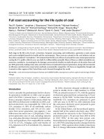
FIGURE 1-8. Blood cell differentiation. A chart of the pluripotent hematopoietic stem cell’s differentiation potential. Each lineage is
depicted as a separate column.
Pluripotent hematopoietic stem cell
Proerythroblast MegakaryoblastMonoblastMyeloblast
BasophilEosinophilNeutrophil
Reticulocyte Megakaryocyte
Erythrocyte
PlateletsMonocyteMyelocyte
Metamyelocyte
Stab cell
BasophilEosinophilNeutrophil
Promyelocyte
Lymphoblast
T cellB cell
Plasma
cell
Active
T cell
10
CHAPTER 1 ANATOMY AND HISTOLOGY
units (CFUs), each ending in a distinct mature cell: erythroid (producing
erythrocytes), megakaryocyte (producing platelets), basophil, eosinophil, and
granulocyte-macrophage (producing monocytes and neutrophils). The lym-
phoid lineage produces two cell lines: T cells and B cells.
Erythrocytes
Erythrocytes are nonnucleated biconcave disks designed for gas exchange.
These cells measure 7.8 μm in diameter, and their biconcave shape increases
their surface area for gas exchange. These cells lack organelles, which are jet-
tisoned shortly after they enter the bloodstream. Instead, they contain only a
plasma membrane, a cytoskeleton, hemoglobin, and glycolytic enzymes that
help them survive via anaerobic respiration (90%) and the hexose monophos-
phate shunt (10%). This limits the red blood cell life span to approximately
120 days, after which they are typically removed via macrophages in the
spleen. Mature erythrocytes are replaced by immature reticulocytes, which
mature 1–2 days after entering the circulation. These precursors are active in
protein metabolism because the mature red blood cells have expelled their
nucleus and ribosomes. Reticulocytes are thereby distinguished from mature
erythrocytes by their retained nucleus and slightly larger diameter.
Erythrocyte metabolism begins with the transport of glucose across the red
cell membrane via the GLUT1 transporter. At this point, glycolytic enzymes
produce ATP and lactic acid via anaerobic metabolism. Important aspects of
erythrocyte metabolism are listed in Table 1-1.
Leukocytes
Leukopoiesis is the process by which white blood cells develop from
hematopoietic stem cells. Neutrophils and monocytes develop through the
granulocyte-macrophage CFU precursor. Basophils and eosinophils each
have a lineage-specifi c CFU. Lymphocytes, although separate from myeloid
cells, are also considered leukocytes, and arise from the lymphoid stem cell.
All leukocytes are involved in some aspect of the immune response:
Q
Neutrophils affect nonspecifi c innate immunity in the acute infl amma-
tory response.
Q
Basophils mediate allergic responses.
Q
Eosinophils fi ght off parasitic infections.
Q
Lymphocytes are integral to both innate and humoral immunity.
NEUTROPHILS
These products of the myeloid lineage act as acute-phase granulocytes. They
begin in the bone marrow as myeloid stem cells (see Figure 1-9) and mature
over a period of 10–14 days, producing both primary and secondary granules
(promyelocyte stage; see Figures 1-9, 1-10, and 1-11). Once mature, these leu-
kocytes are vital to the success of the innate immune system and are especially
prominent in the acute infl ammatory response.
Histologically, these cells are distinguished by their large spherical size, mul-
tilobed nuclei, and azurophilic primary granules (lysosomes). These cells
have earned the alternate name polymorphonuclear cells (PMNs) due to
their multilobed nucleus. The key to their immune function, however, lies
not in the nucleus, but in the ability of PMNs to phagocytose microbes and
destroy them via reactive oxygen species (superoxide, hydrogen peroxide, per-
oxyl radicals, and hydroxyl radicals). Their azurophilic granules contain sev-
CLINICAL
CORRELATION
Red cell cytoskeletal
abnormalities (e.g., hereditary
spherocytosis, elliptocytosis)
and hemoglobinopathies (e.g.,
thalassemias, sickle cell anemia)
cause signifi cant morbidity and
mortality.
CLINICAL
CORRELATION
Reticulocyte counts increase
when the bone marrow increases
production to replenish red cell
levels in the blood in response to a
bleed or hemolytic process.
KEY FACT
Leukos = Greek for white.
Cytos = Greek for cell.